Page 1
Purdue UniversityPurdue e-Pubs
Open Access Theses Theses and Dissertations
2013
Development of a Starch-Based Mussel-MimeticAdhesive PolymerJeffrey Kazimir de KozlowskiPurdue University, [email protected]
Follow this and additional works at: https://docs.lib.purdue.edu/open_access_theses
Part of the Biochemistry Commons
This document has been made available through Purdue e-Pubs, a service of the Purdue University Libraries. Please contact [email protected] foradditional information.
Recommended Citationde Kozlowski, Jeffrey Kazimir, "Development of a Starch-Based Mussel-Mimetic Adhesive Polymer" (2013). Open Access Theses. 7.https://docs.lib.purdue.edu/open_access_theses/7
Page 2
Graduate School ETD Form 9
(Revised 12/07)
PURDUE UNIVERSITY GRADUATE SCHOOL
Thesis/Dissertation Acceptance
This is to certify that the thesis/dissertation prepared
By
Entitled
For the degree of
Is approved by the final examining committee:
Chair
To the best of my knowledge and as understood by the student in the Research Integrity and
Copyright Disclaimer (Graduate School Form 20), this thesis/dissertation adheres to the provisions of
Purdue University�s �Policy on Integrity in Research� and the use of copyrighted material.
Approved by Major Professor(s): ____________________________________
____________________________________
Approved by: Head of the Graduate Program Date
Jeffrey de Kozlowski
Development of a Starch-Based Mussel-Mimetic Adhesive Polymer
Master of Science
Bernard Tao
Jonathan Wilker
Nathan Mosier
Bernard Tao
Bernie Engel 10/11/2013
Page 3
i
i
DEVELOPMENT OF A STARCH-BASED MUSSEL-MIMETIC ADHESIVE
POLYMER
A Thesis
Submitted to the Faculty
of
Purdue University
by
Jeffrey Kazimir de Kozlowski
In Partial Fulfillment of the
Requirements for the Degree
of
Master of Science
December 2013
Purdue University
West Lafayette, Indiana
Page 4
ii
ii
To Amanda,
I know it was not easy for you to move with me to Indiana without the guarantee of a job.
The past two years have brought us closer than I imagined and in the meantime you
discovered your true professional aspirations. I am so happy that things worked out for
the best and I thank you for putting up with the frustrations of my life as a graduate
student. This thesis represents the time and commitment you deserve every day. I love
you.
Page 5
iii
iii
ACKNOWLEDGEMENTS
I would like to first thank my advisor Dr. Bernie Tao for his support throughout my
graduate career. I have learned immensely from him educationally, professionally, and
personally through conversations and advice. I am extremely grateful for his guidance
throughout my project and towards my professional goals. I am glad to have worked
with him.
Next, I would like to thank the Indiana and Iowa Corn Growers Associations for making
this project possible. Thank you for your patience and support throughout this project.
I want to thank my committee member Dr. John Wilker for his insights into this project
and for allowing me to use his organic chemistry lab. Additionally, I owe extreme
gratitude to the Wilker lab group, especially Jess Roman, Cori Jenkins, Heather Meredith,
and Michael North for their unwavering assistance in the lab and for helpful discussions.
I would also like to thank Lisa Mauer, Brad Reuhs, Anton Terekov, and the entire
Whistler Center for their assistance with NMR and IR spectroscopy and for allowing me
to use their labs. A special thanks to Anton Terekov for training me on NMR and other
equipment.
I owe thanks to many members of the Hamaker lab including Madhuvanti Kale and
Byung-Hoo Lee for their patience and knowledge in HPLC analysis of polysaccharides.
Page 6
iv
iv
Thank you to Karl Wood for his timely assistance with mass spectrometry and general
knowledge in analytical chemistry.
Thank you to my committee member Dr. Nate Mosier and to Dr. Michael Ladisch for
their mentorship and support in my professional and academic goals.
To the faculty and staff of ABE and Food Science, thank you for everything you have
done for me and other graduate students. Without your commitment, graduate school
would be a nightmare.
Thanks to Dr. Bruce Applegate for my cat and his vaccinations, and interesting
conversations.
Thank you to Dr. Yuan Yao and Dr. Susan Nielson for allowing me to attend the food
science tour of China. It was a once-in-a-lifetime experience that I will never forget. I
want to especially thank Dr. Yao for his guidance on the trip, for helpful discussions
regarding starch and carbohydrate chemistry, and his friendship.
Finally, I want to thank my family and friends for their support. You are my motivation
and backbone.
Page 7
v
v
TABLE OF CONTENTS
Page
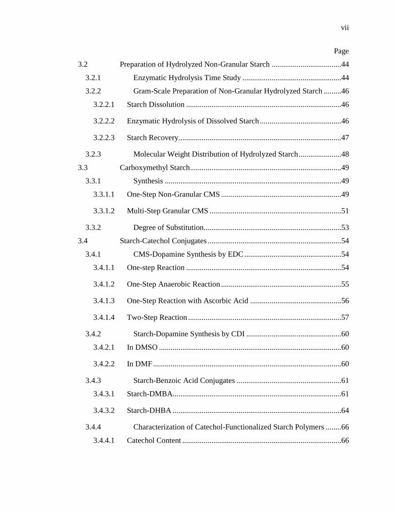
LIST OF TABLES .............................................................................................................. x
LIST OF FIGURES ........................................................................................................... xi
LIST OF ABBREVIATIONS .......................................................................................... xiv
ABSTRACT ............................................................................................................ xvi
CHAPTER 1. INTRODUCTION ................................................................................. 1
1.1 Objectives ...................................................................................................1
1.2 Organization ...............................................................................................1
CHAPTER 2. LITERATURE REVIEW ...................................................................... 3
2.1 Adhesives ...................................................................................................3
2.1.1 Introduction .........................................................................................3
2.1.2 Motivation for Green Adhesives .........................................................4
2.1.3 Concepts ..............................................................................................5
2.1.4 Mechanisms of Adhesion ....................................................................7
2.1.4.1 Mechanical Adhesion .............................................................................7
2.1.4.2 Chemical Adhesion ................................................................................7
2.1.5 Adhesion in Nature – Mussel Adhesion..............................................8
2.2 DOPA Chemistry .....................................................................................11
2.2.2 DOPA in Adhesion ...........................................................................13
2.2.3 DOPA in Cohesion ............................................................................14
2.3 Mussel-Mimetic Adhesive Polymers .......................................................16
2.3.1. Catechol-Functionalized Polymers ..........................................................17
2.3.1.1 Benzotriazoles ......................................................................................17
Page 8
vi
vi
Page
2.3.1.2 Carbodiimide ........................................................................................18
2.3.1.3 Schiff Base............................................................................................21
2.3.2 Polymerization of Catechol-Functionalized Monomers ...................22
2.3.2.1 Condensation ........................................................................................22
2.3.2.2 Ring-Opening Addition of N-Carboxyanhydrides ...............................23
2.3.2.3 Reactive Anhydride or Acid Chloride ..................................................24
2.3.2.4 Vinyl Polymerization............................................................................25
2.3.3 Characterizing DOPA-polymer Conjugates ......................................26
2.3.3.1 DOPA Content......................................................................................26
2.3.3.2 Verification of Conjugation ..................................................................27
2.3.4 Performance of Mussel-Inspired Adhesives .....................................28
2.3.5 DOPA for Adhesive Polymer Crosslinking ......................................30
2.3.5.1 Chemical Oxidants ...............................................................................30
2.3.5.2 Metals ...................................................................................................31
2.3.5.3 Enzymes ...............................................................................................32
2.4 Opportunity for Catechol-Functionalized Biopolymers...........................32
2.5 Starch and Starch Adhesives ....................................................................34
2.5.1 Introduction .......................................................................................34
2.5.2 History of Starch Adhesives..............................................................34
2.5.3 Structure ............................................................................................35
2.5.4 Modified Starch .................................................................................37
2.5.5 Carboxymethyl Starch .......................................................................38
2.5.6 CMS Bioconjugates ..........................................................................39
2.6 Future Studies in Mussel-Inspired Biopolymer Adhesives ......................40
CHAPTER 3. MATERIALS AND METHODS ........................................................ 42
3.1 Materials ...................................................................................................42
Page 9
vii
vii
Page
3.2 Preparation of Hydrolyzed Non-Granular Starch ....................................44
3.2.1 Enzymatic Hydrolysis Time Study ...................................................44
3.2.2 Gram-Scale Preparation of Non-Granular Hydrolyzed Starch .........46
3.2.2.1 Starch Dissolution ................................................................................46
3.2.2.2 Enzymatic Hydrolysis of Dissolved Starch ..........................................46
3.2.2.3 Starch Recovery....................................................................................47
3.2.3 Molecular Weight Distribution of Hydrolyzed Starch ......................48
3.3 Carboxymethyl Starch ..............................................................................49
3.3.1 Synthesis ...........................................................................................49
3.3.1.1 One-Step Non-Granular CMS ..............................................................49
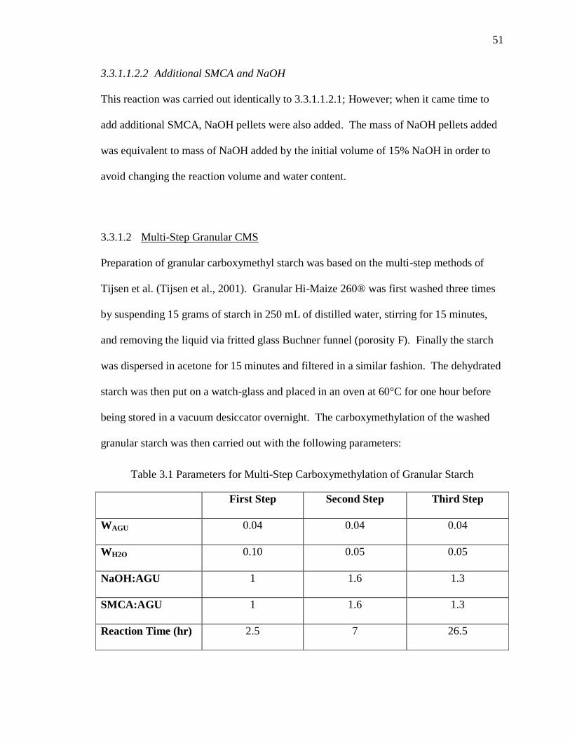
3.3.1.2 Multi-Step Granular CMS ....................................................................51
3.3.2 Degree of Substitution.......................................................................53
3.4 Starch-Catechol Conjugates .....................................................................54
3.4.1 CMS-Dopamine Synthesis by EDC ..................................................54
3.4.1.1 One-step Reaction ................................................................................54
3.4.1.2 One-Step Anaerobic Reaction ..............................................................55
3.4.1.3 One-Step Reaction with Ascorbic Acid ...............................................56
3.4.1.4 Two-Step Reaction ...............................................................................57
3.4.2 Starch-Dopamine Synthesis by CDI .................................................60
3.4.2.1 In DMSO ..............................................................................................60
3.4.2.2 In DMF .................................................................................................60
3.4.3 Starch-Benzoic Acid Conjugates ......................................................61
3.4.3.1 Starch-DMBA.......................................................................................61
3.4.3.2 Starch-DHBA .......................................................................................64
3.4.4 Characterization of Catechol-Functionalized Starch Polymers ........66
3.4.4.1 Catechol Content ..................................................................................66
Page 10
viii
viii
Page
3.4.4.2 Verification of Conjugation Bond-Type by FTIR ................................70
3.4.4.3 Characterization of Dihydroxybenzoic acid-Phenylboronic acid Esters .
..............................................................................................................70
3.4.5 Lap-Shear Adhesive Test ..................................................................71
CHAPTER 4. RESULTS AND DISCUSSION .......................................................... 72
4.1 Enzymatic Hydrolysis of Hi-Maize® 260 ...............................................72
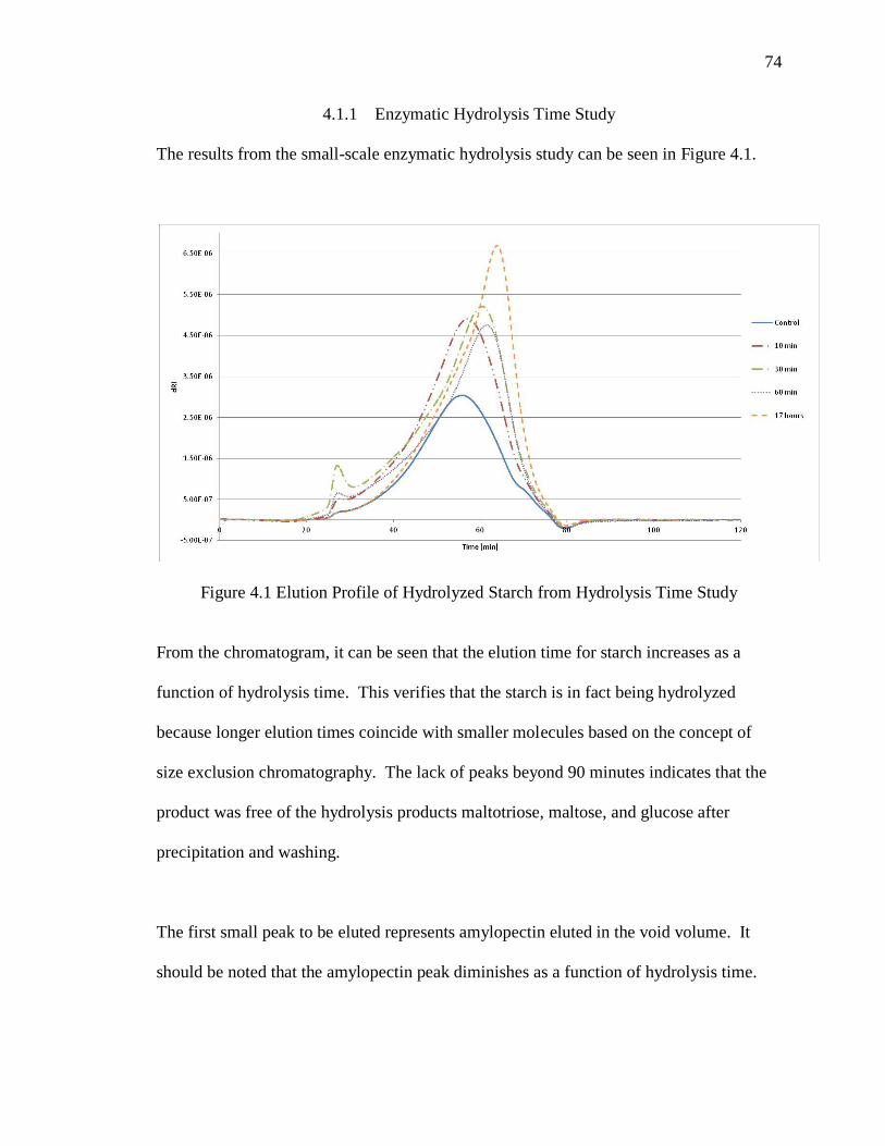
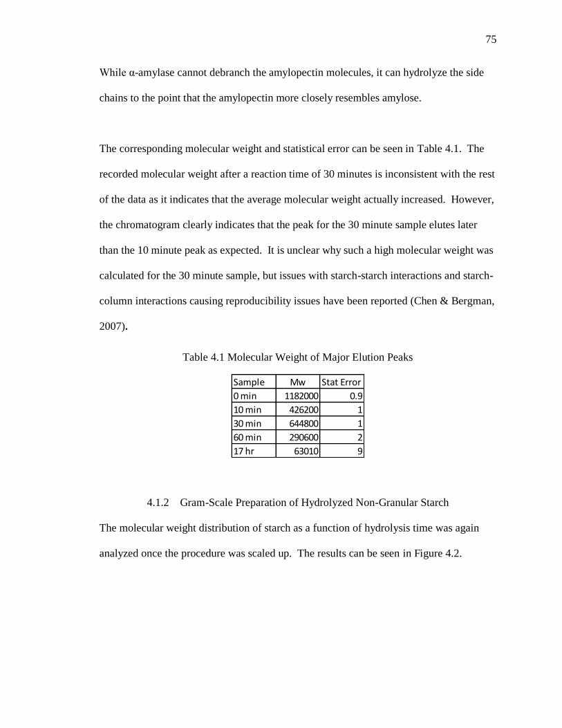
4.1.1 Enzymatic Hydrolysis Time Study ...................................................74
4.1.2 Gram-Scale Preparation of Hydrolyzed Non-Granular Starch .........75
4.2 Carboxymethylation of Hi-Maize® 260 ..................................................77
4.2.1 One-Step Non-Granular CMS ...........................................................77
4.2.2 Fed-Batch Non-Granular CMS .........................................................78
4.2.2.1 Additional SMCA .................................................................................78
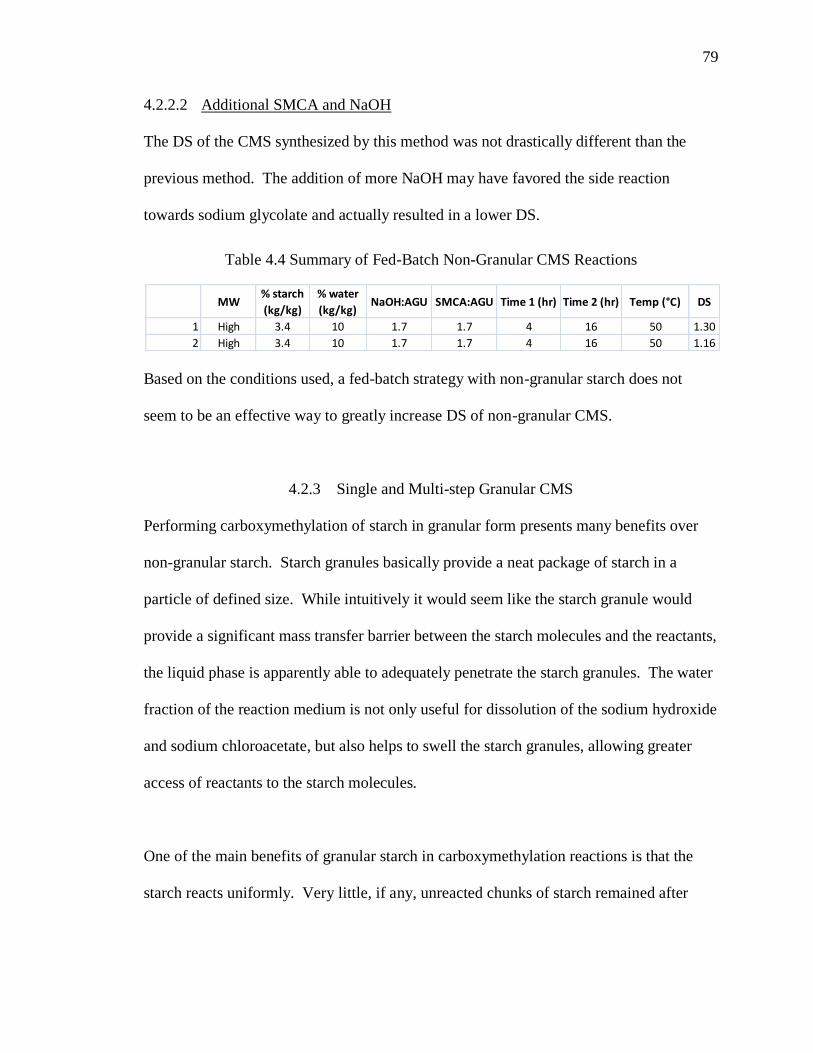
4.2.2.2 Additional SMCA and NaOH ..............................................................79
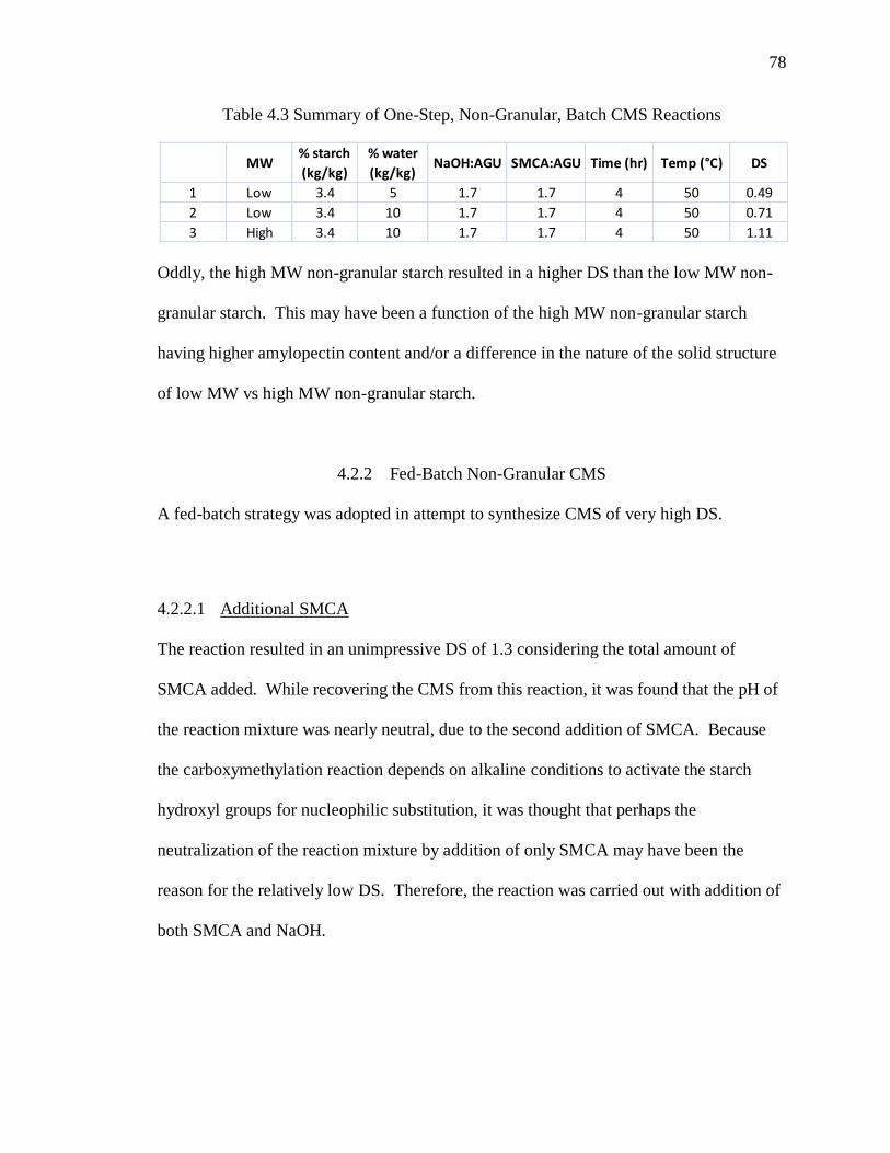
4.2.3 Single and Multi-step Granular CMS ...............................................79
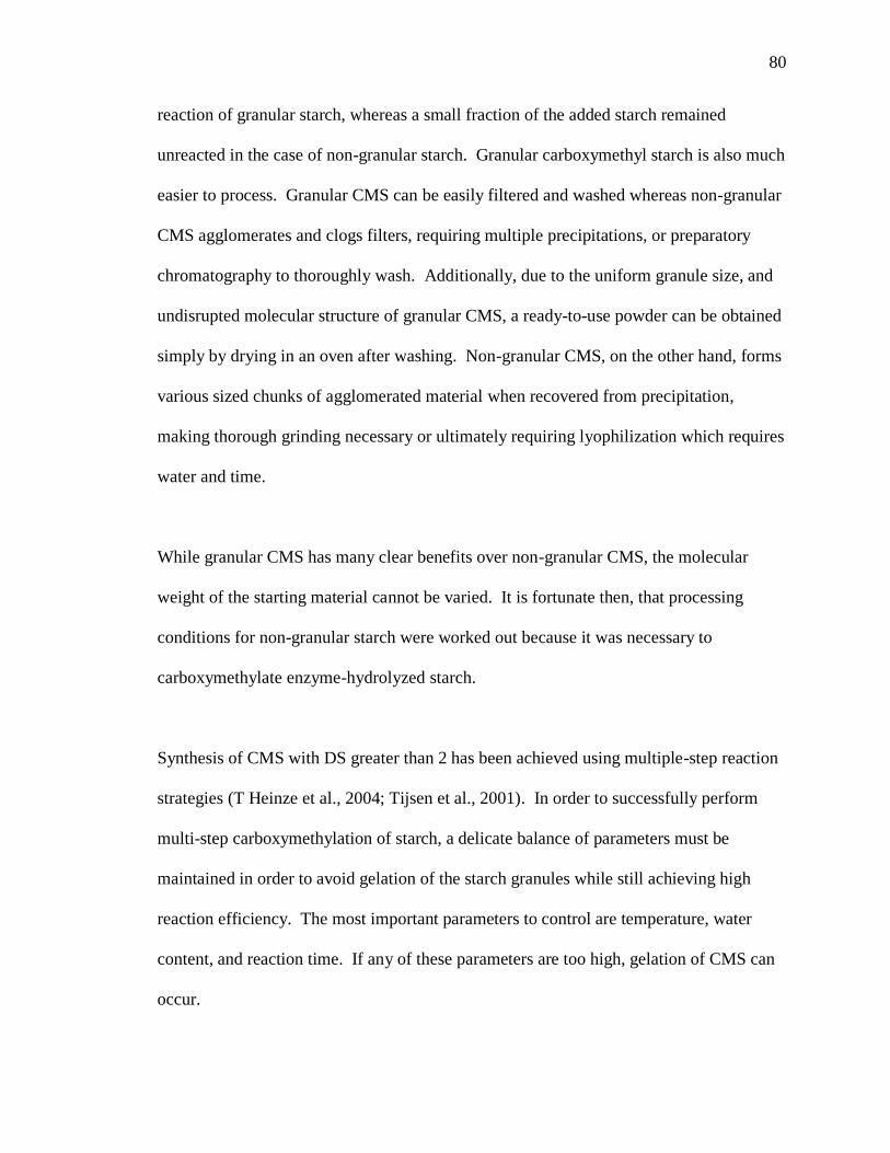
4.2.3.1 Method 1 ...............................................................................................81
4.2.3.2 Method 2 ...............................................................................................81
4.3 Starch-Catechol Conjugates .....................................................................82
4.3.1 Synthesis of CMS-dopamine by EDC...............................................82
4.3.1.1 One-step Reaction ................................................................................83
4.3.1.2 Two-Step Reaction ...............................................................................90
4.3.1.3 Summary of EDC Reactions ................................................................93
4.3.1.4 FTIR Characterization of CMS-Dopamine ..........................................94
4.3.1.5 Adhesive Strength of CMS-Dopamine .................................................97
4.3.2 Starch-Catechol Conjugates ..............................................................98
4.3.2.1 Starch-DMBA.....................................................................................102
4.3.2.2 Starch-DHBA .....................................................................................109
Page 11
ix
ix
Page
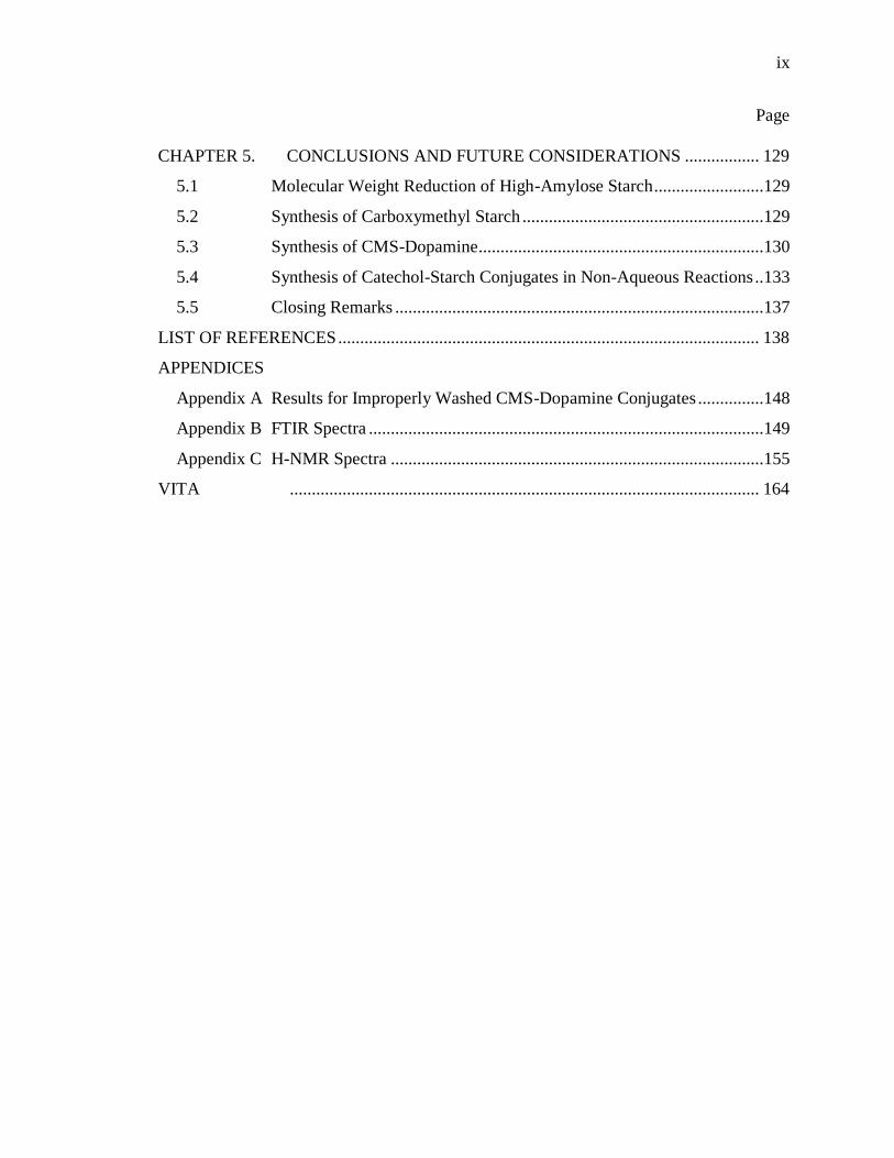
CHAPTER 5. CONCLUSIONS AND FUTURE CONSIDERATIONS ................. 129
5.1 Molecular Weight Reduction of High-Amylose Starch .........................129
5.2 Synthesis of Carboxymethyl Starch .......................................................129
5.3 Synthesis of CMS-Dopamine .................................................................130
5.4 Synthesis of Catechol-Starch Conjugates in Non-Aqueous Reactions ..133
5.5 Closing Remarks ....................................................................................137
LIST OF REFERENCES ................................................................................................ 138
APPENDICES
Appendix A Results for Improperly Washed CMS-Dopamine Conjugates ...............148
Appendix B FTIR Spectra ..........................................................................................149
Appendix C H-NMR Spectra .....................................................................................155
VITA ........................................................................................................... 164
Page 12
x
x
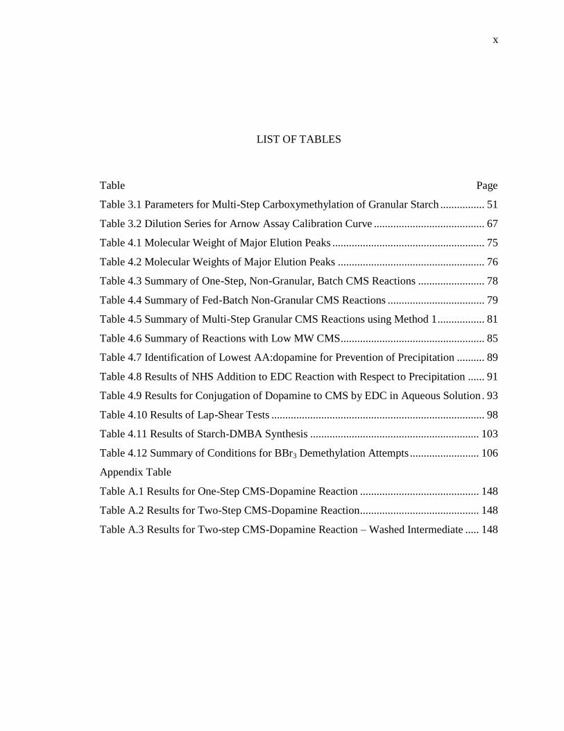
LIST OF TABLES
Table .............................................................................................................................. Page
Table 3.1 Parameters for Multi-Step Carboxymethylation of Granular Starch ................ 51
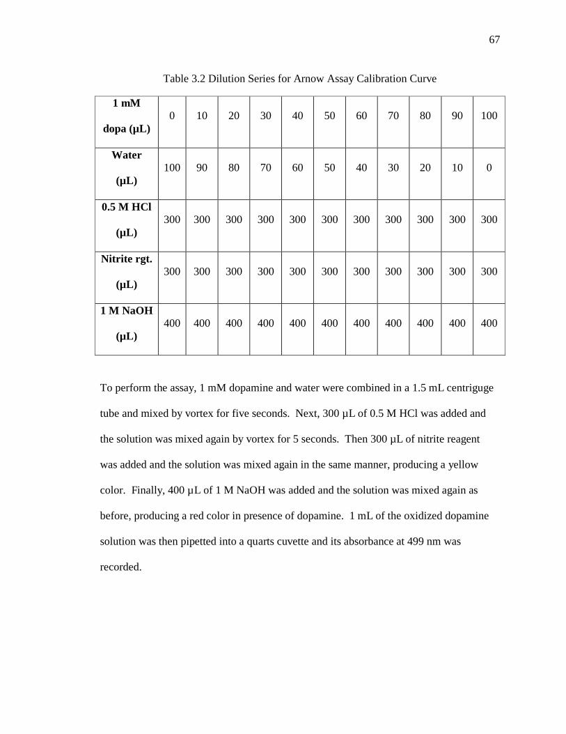
Table 3.2 Dilution Series for Arnow Assay Calibration Curve ........................................ 67
Table 4.1 Molecular Weight of Major Elution Peaks ....................................................... 75
Table 4.2 Molecular Weights of Major Elution Peaks ..................................................... 76
Table 4.3 Summary of One-Step, Non-Granular, Batch CMS Reactions ........................ 78
Table 4.4 Summary of Fed-Batch Non-Granular CMS Reactions ................................... 79
Table 4.5 Summary of Multi-Step Granular CMS Reactions using Method 1 ................. 81
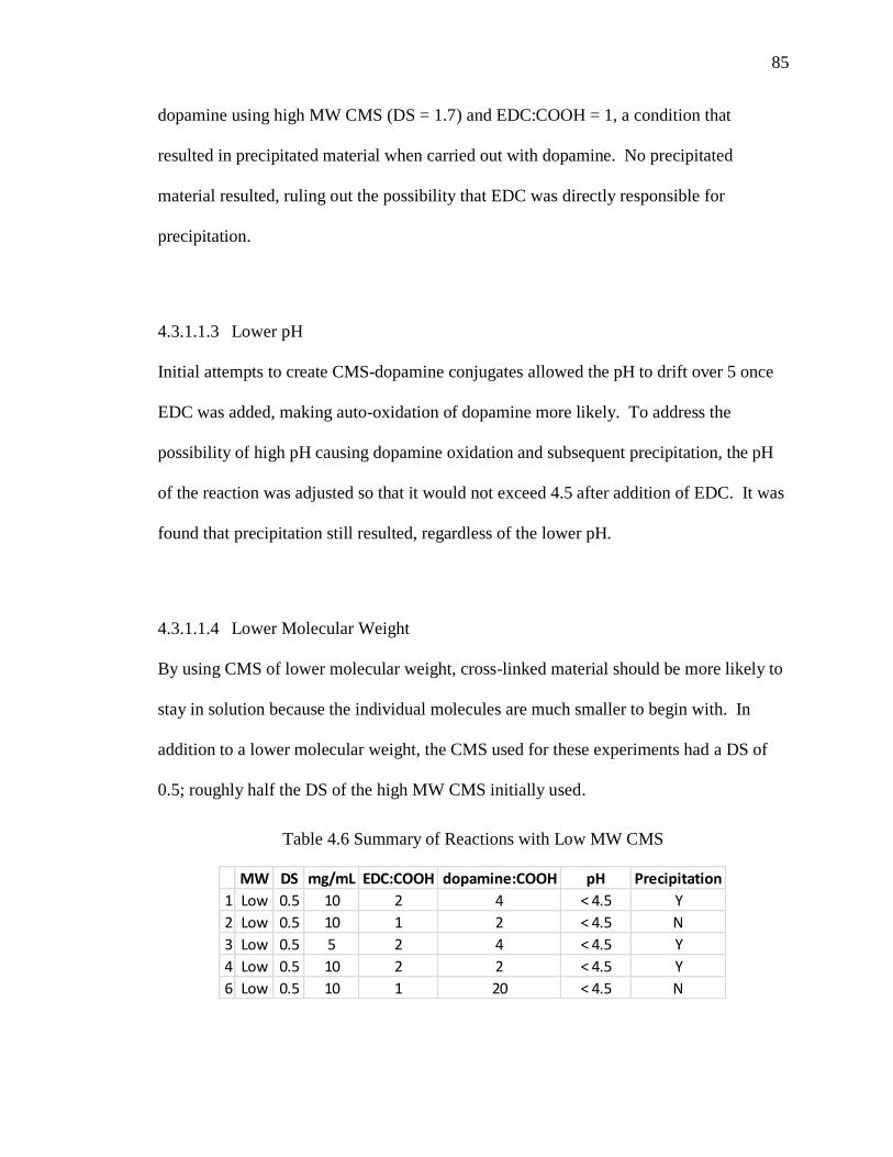
Table 4.6 Summary of Reactions with Low MW CMS.................................................... 85
Table 4.7 Identification of Lowest AA:dopamine for Prevention of Precipitation .......... 89
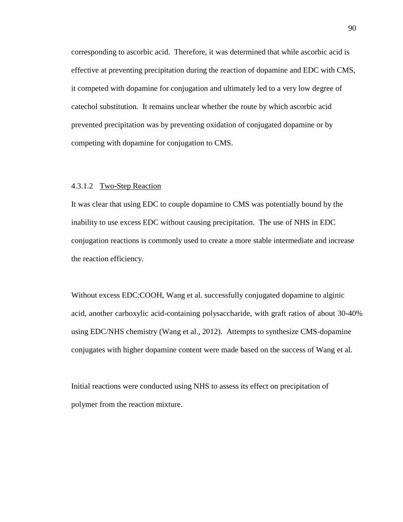
Table 4.8 Results of NHS Addition to EDC Reaction with Respect to Precipitation ...... 91
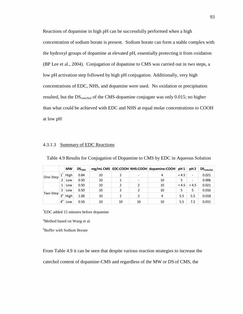
Table 4.9 Results for Conjugation of Dopamine to CMS by EDC in Aqueous Solution . 93
Table 4.10 Results of Lap-Shear Tests ............................................................................. 98
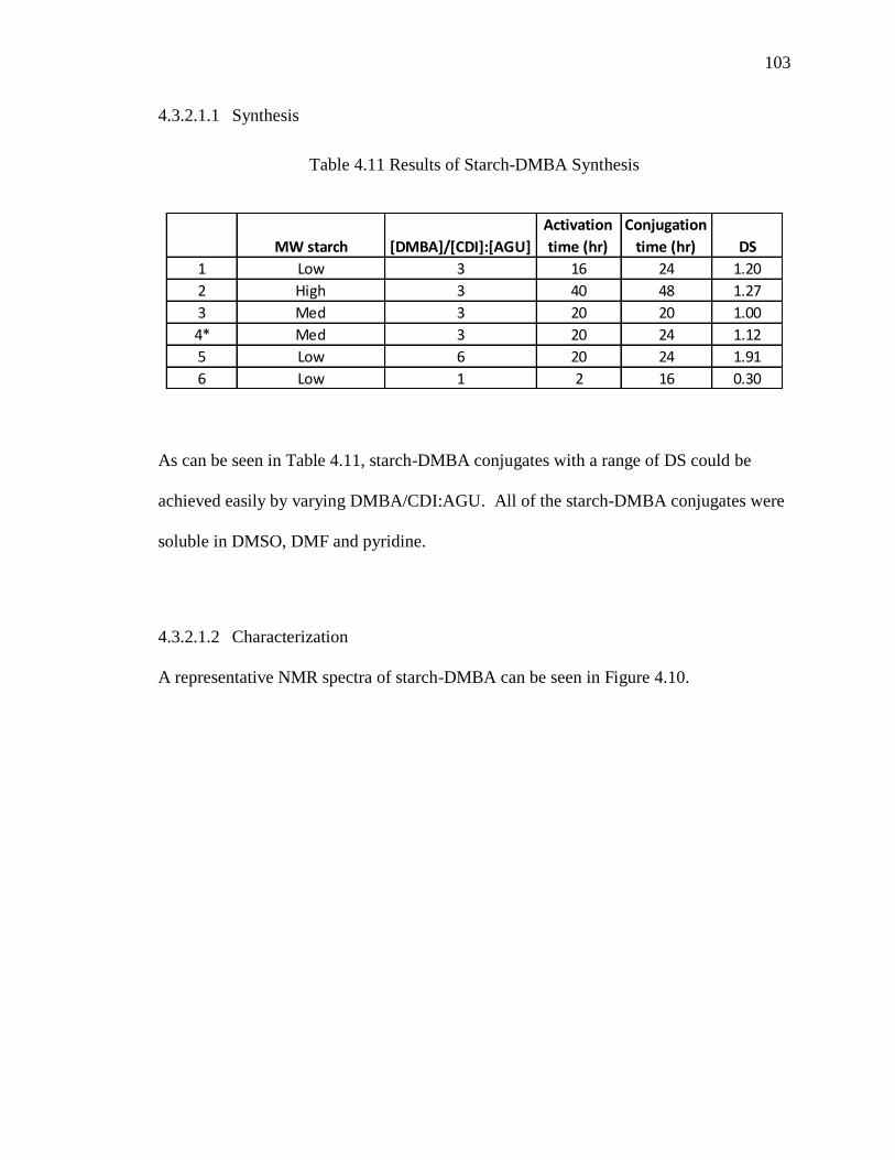
Table 4.11 Results of Starch-DMBA Synthesis ............................................................. 103
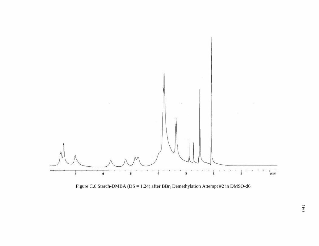
Table 4.12 Summary of Conditions for BBr3 Demethylation Attempts ......................... 106
Appendix Table
Table A.1 Results for One-Step CMS-Dopamine Reaction ........................................... 148
Table A.2 Results for Two-Step CMS-Dopamine Reaction ........................................... 148
Table A.3 Results for Two-step CMS-Dopamine Reaction – Washed Intermediate ..... 148
Page 13
xi
xi
LIST OF FIGURES
Figure ............................................................................................................................. Page
Figure 2.1 Share of Adhesives and Sealants Market Value by Industry Application (Bosik,
2012) ................................................................................................................................... 3
Figure 2.2 Basic diagram of a mussel byssal thread extending from the stem (within
mussel) to attachment surface (Waite et al., 2005). .......................................................... 10
Figure 2.3 Adhesive and cohesive interactions of DOPA (Wiegemann, 2005) ............... 15
Figure 2.4 Mechanism for amine-to-carboxyl conjugation by EDC (Hermanson, 2008) 19
Figure 2.5 Simplified Mechanism of Amide Bond Formation via Schiff Base................ 21
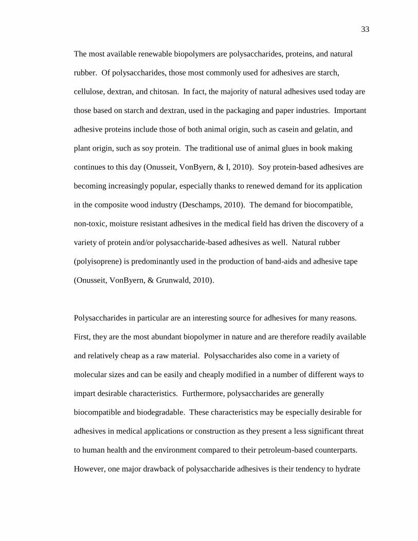
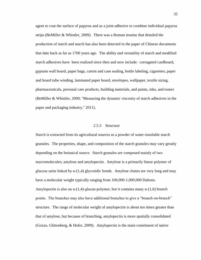
Figure 2.6 Basic structures of amylose and amylopectin (Nuffield Foundation) ............. 36
Figure 2.7 "Branch-on-branch" structure of amylopectin (Thompson, 2000) .................. 36
Figure 3.1 Standard Curve for Colorimetric Dopamine Assay ......................................... 68
Figure 4.1 Elution Profile of Hydrolyzed Starch from Hydrolysis Time Study ............... 74
Figure 4.2 Elution Profile for Hydrolyzed Starch from Gram-Scale Preparation ............ 76
Figure 4.3 UV-Vis Spectrum of CMS-dopamine-AA ...................................................... 88
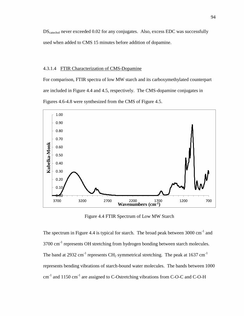
Figure 4.4 FTIR Spectrum of Low MW Starch ................................................................ 94
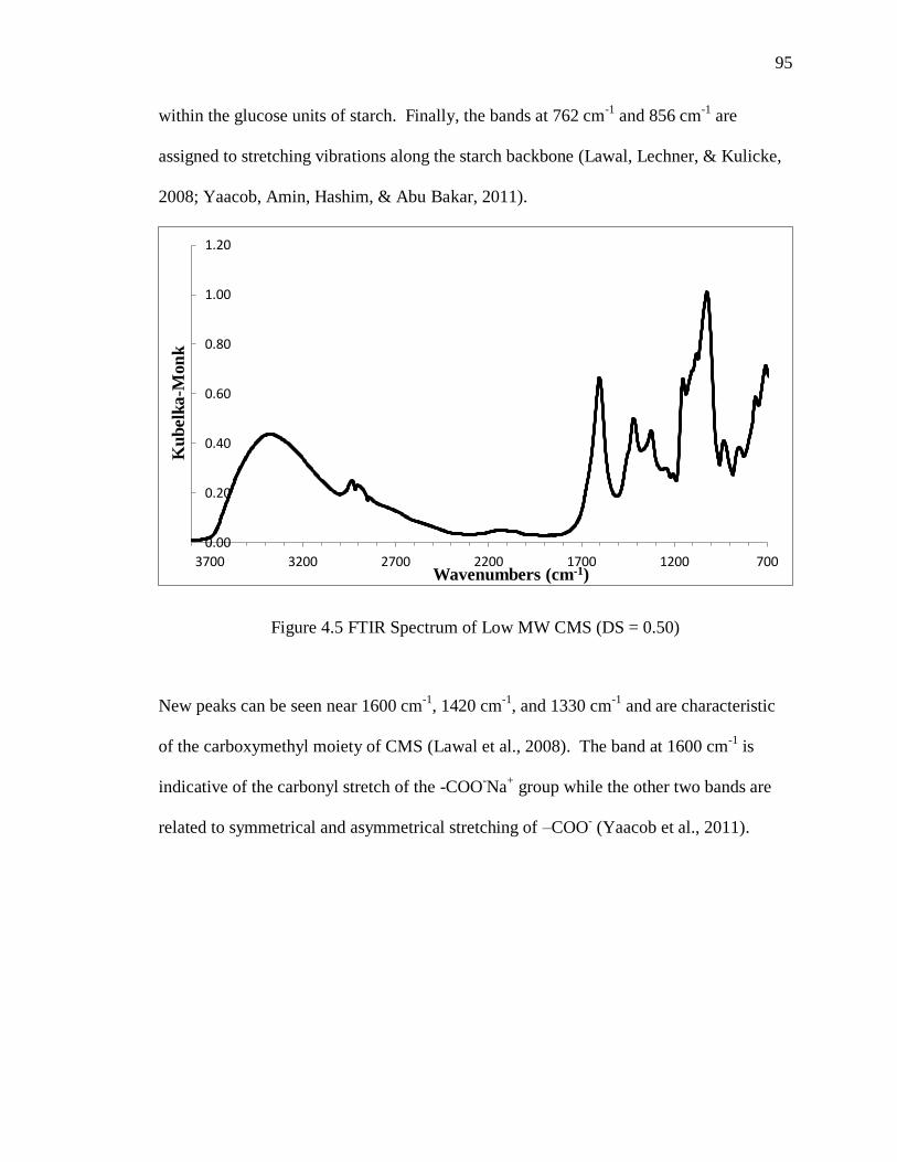
Figure 4.5 FTIR Spectrum of Low MW CMS (DS = 0.50) .............................................. 95
Figure 4.6 Representative FTIR Spectrum of CMS-Dopamine by One-Step Reaction with
EDC................................................................................................................................... 96
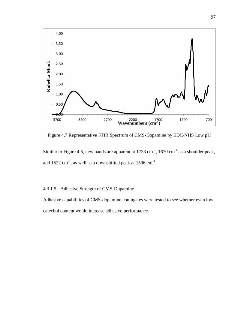
Figure 4.7 Representative FTIR Spectrum of CMS-Dopamine by EDC/NHS Low pH .. 97
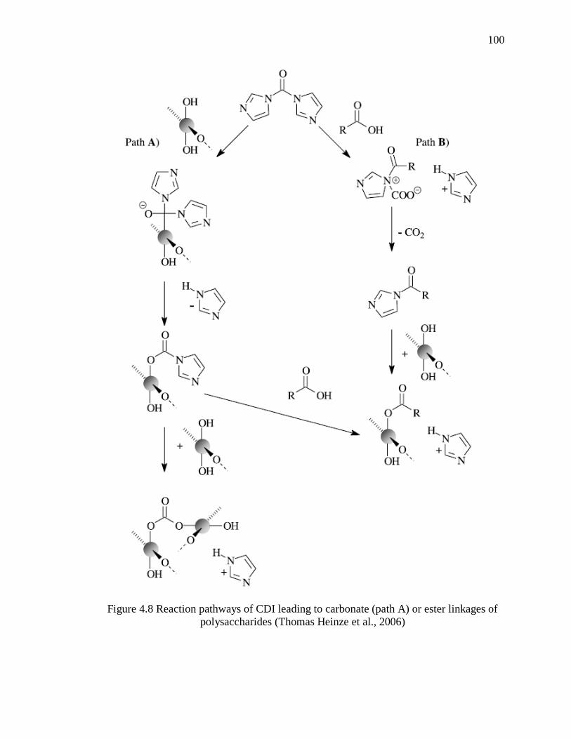
Figure 4.8 Reaction pathways of CDI leading to carbonate (path A) or ester linkages of
polysaccharides (Thomas Heinze et al., 2006) ............................................................... 100
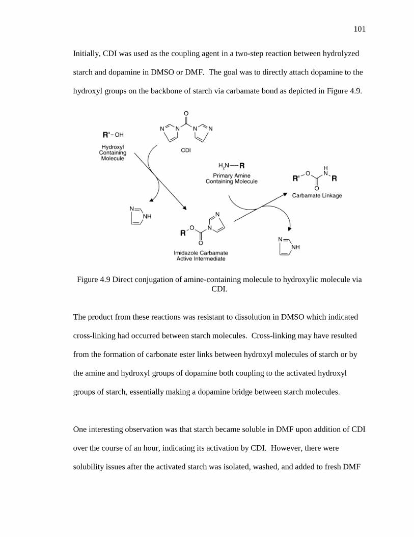
Figure 4.9 Direct conjugation of amine-containing molecule to hydroxylic molecule via
CDI. ................................................................................................................................. 101
Figure 4.10 Representative H-NMR Spectrum of Starch-DMBA Conjugate ................ 104
Page 14
xii
xii
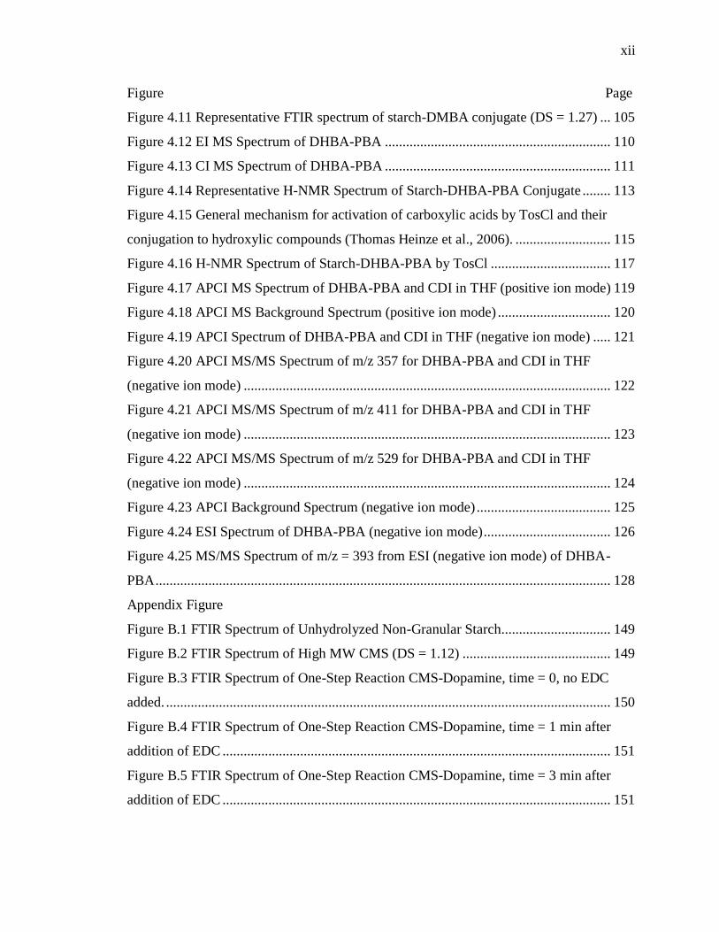
Figure Page
Figure 4.11 Representative FTIR spectrum of starch-DMBA conjugate (DS = 1.27) ... 105
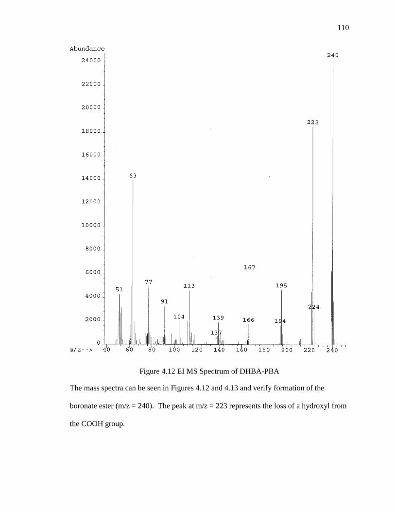
Figure 4.12 EI MS Spectrum of DHBA-PBA ................................................................ 110
Figure 4.13 CI MS Spectrum of DHBA-PBA ................................................................ 111
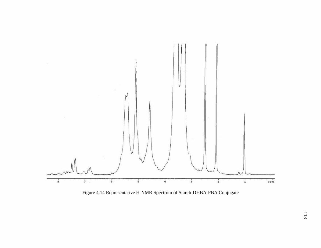
Figure 4.14 Representative H-NMR Spectrum of Starch-DHBA-PBA Conjugate ........ 113
Figure 4.15 General mechanism for activation of carboxylic acids by TosCl and their
conjugation to hydroxylic compounds (Thomas Heinze et al., 2006). ........................... 115
Figure 4.16 H-NMR Spectrum of Starch-DHBA-PBA by TosCl .................................. 117
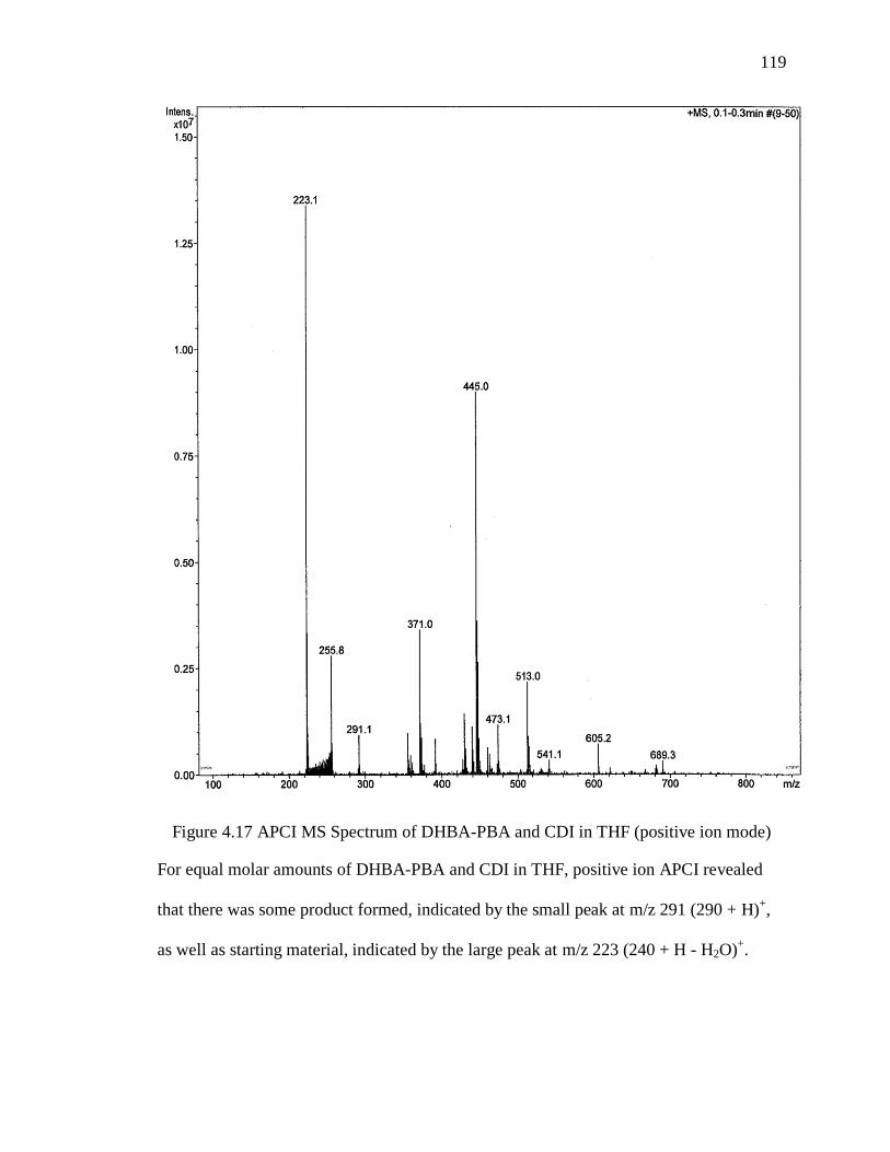
Figure 4.17 APCI MS Spectrum of DHBA-PBA and CDI in THF (positive ion mode) 119
Figure 4.18 APCI MS Background Spectrum (positive ion mode) ................................ 120
Figure 4.19 APCI Spectrum of DHBA-PBA and CDI in THF (negative ion mode) ..... 121
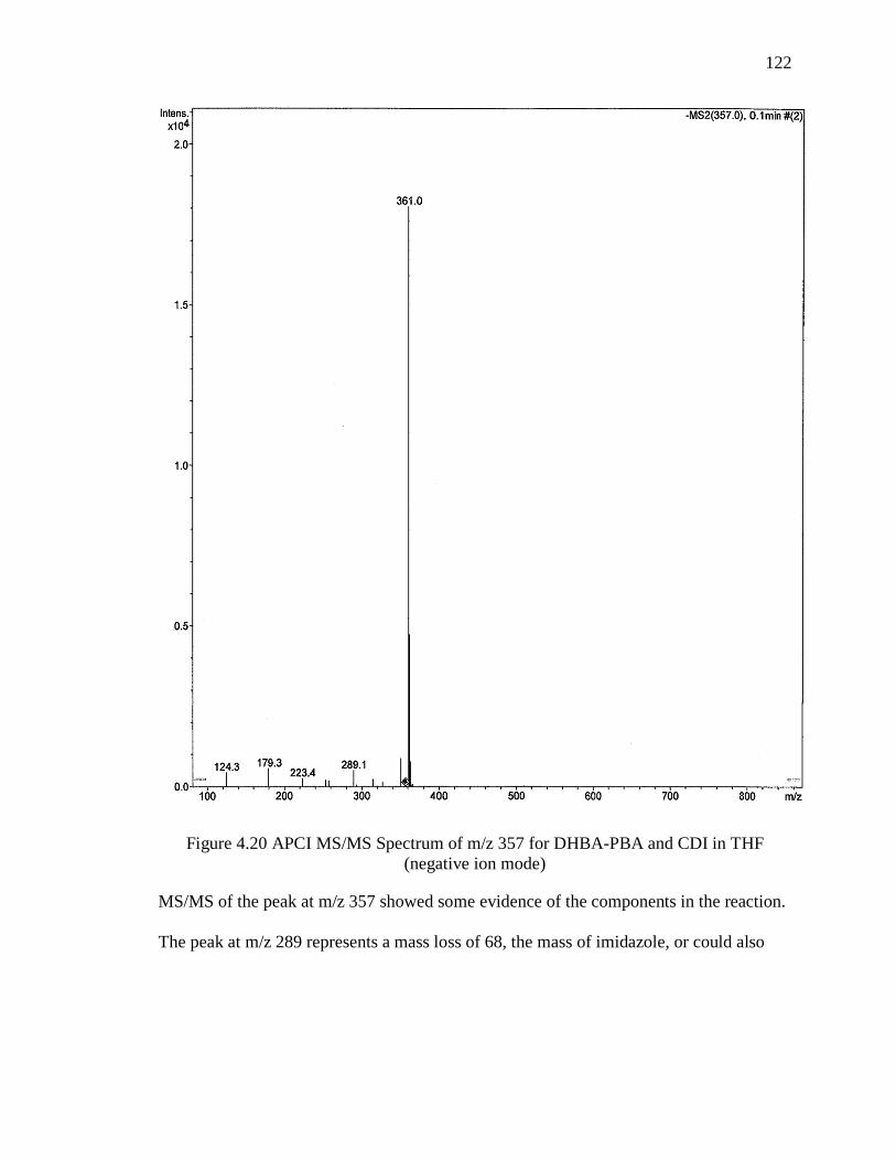
Figure 4.20 APCI MS/MS Spectrum of m/z 357 for DHBA-PBA and CDI in THF
(negative ion mode) ........................................................................................................ 122
Figure 4.21 APCI MS/MS Spectrum of m/z 411 for DHBA-PBA and CDI in THF
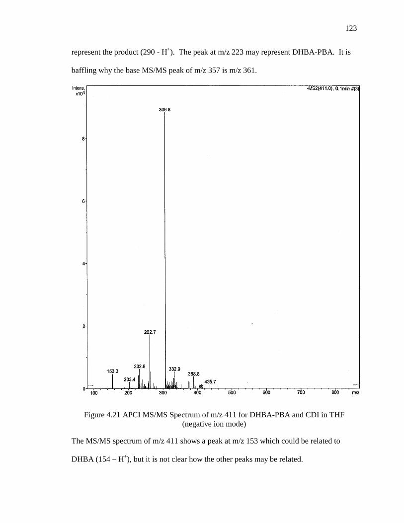
(negative ion mode) ........................................................................................................ 123
Figure 4.22 APCI MS/MS Spectrum of m/z 529 for DHBA-PBA and CDI in THF
(negative ion mode) ........................................................................................................ 124
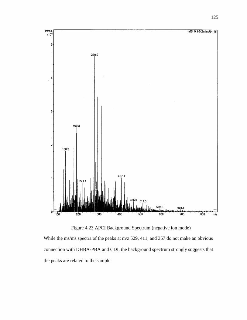
Figure 4.23 APCI Background Spectrum (negative ion mode) ...................................... 125
Figure 4.24 ESI Spectrum of DHBA-PBA (negative ion mode) .................................... 126
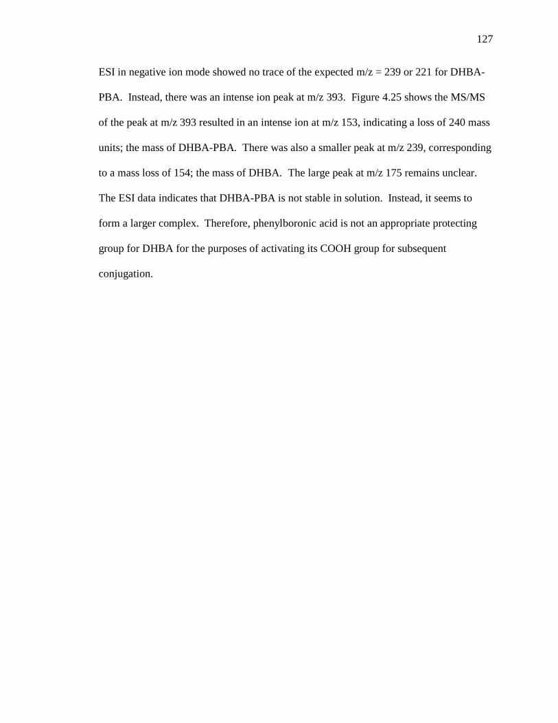
Figure 4.25 MS/MS Spectrum of m/z = 393 from ESI (negative ion mode) of DHBA-
PBA ................................................................................................................................. 128
Appendix Figure ...................................................................................................................
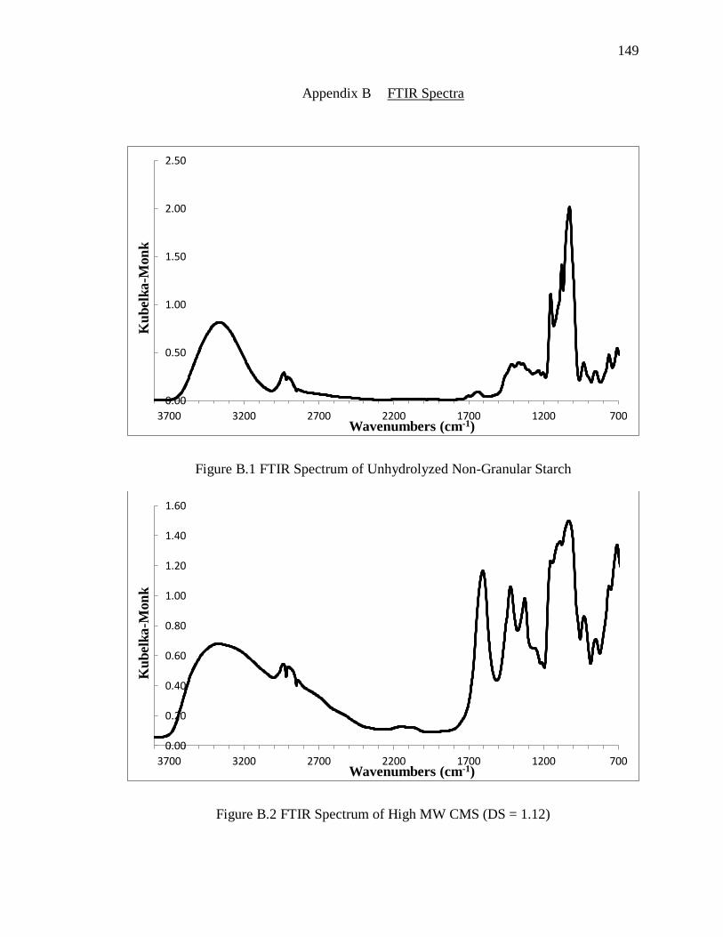
Figure B.1 FTIR Spectrum of Unhydrolyzed Non-Granular Starch ............................... 149
Figure B.2 FTIR Spectrum of High MW CMS (DS = 1.12) .......................................... 149
Figure B.3 FTIR Spectrum of One-Step Reaction CMS-Dopamine, time = 0, no EDC
added. .............................................................................................................................. 150
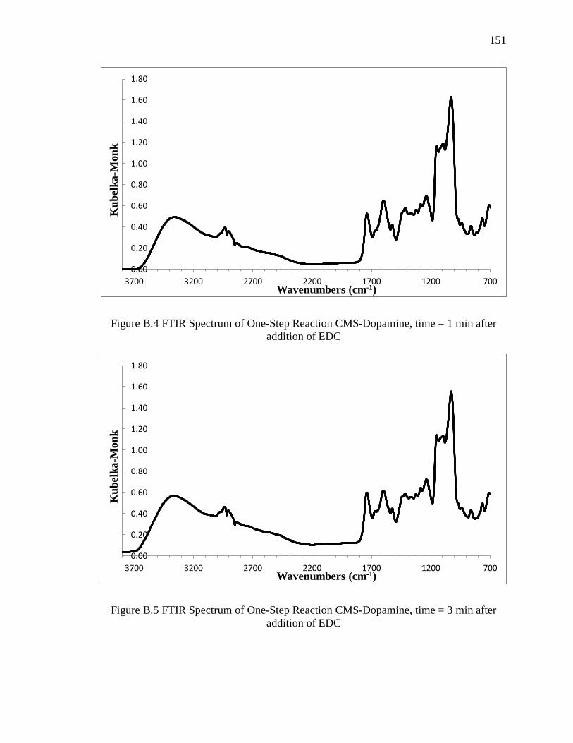
Figure B.4 FTIR Spectrum of One-Step Reaction CMS-Dopamine, time = 1 min after
addition of EDC .............................................................................................................. 151
Figure B.5 FTIR Spectrum of One-Step Reaction CMS-Dopamine, time = 3 min after
addition of EDC .............................................................................................................. 151
Page 15
xiii
xiii
Appendix Figure Page
Figure B.6 FTIR Spectrum of One-Step Reaction CMS-Dopamine, time = 5 min after
addition of EDC .............................................................................................................. 152
Figure B.7 FTIR Spectrum of One-Step Reaction CMS-Dopamine, time = 15 min after
addition of EDC .............................................................................................................. 152
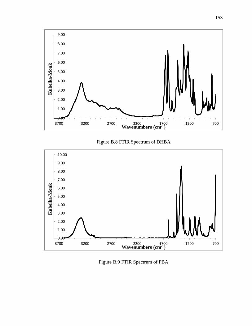
Figure B.8 FTIR Spectrum of DHBA ............................................................................. 153
Figure B.9 FTIR Spectrum of PBA ................................................................................ 153
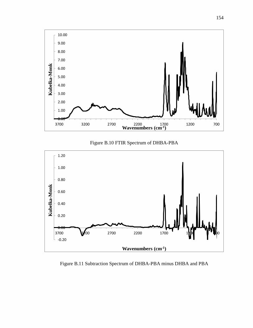
Figure B.10 FTIR Spectrum of DHBA-PBA .................................................................. 154
Figure B.11 Subtraction Spectrum of DHBA-PBA minus DHBA and PBA ................. 154
Figure C.1 H-NMR Spectrum of Low MW Starch in DMSO-d6................................... 155
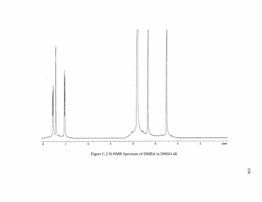
Figure C.2 H-NMR Spectrum of DMBA in DMSO-d6 ................................................. 156
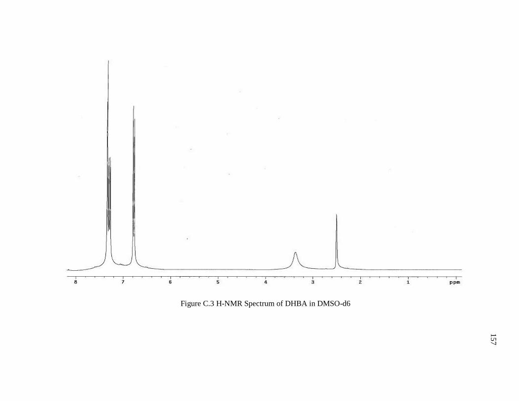
Figure C.3 H-NMR Spectrum of DHBA in DMSO-d6 .................................................. 157
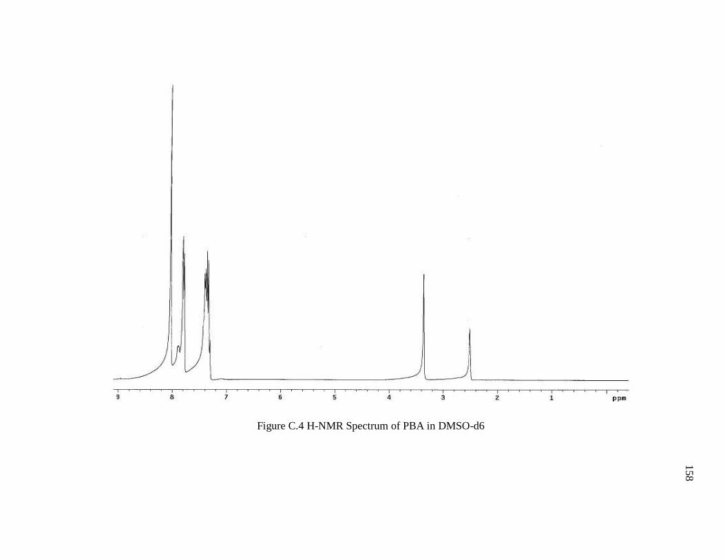
Figure C.4 H-NMR Spectrum of PBA in DMSO-d6 ...................................................... 158
Figure C.5 H-NMR Spectrum of DHBA-PBA in DMSO-d6 ......................................... 159
Figure C.6 Starch-DMBA (DS = 1.24) after BBr3 Demethylation Attempt #2 in DMSO-
d6..................................................................................................................................... 160
Figure C.7 CMS-Dopamine (two-step reaction with sodium borate, low MW, DScatechol =
0.015) in D2O .................................................................................................................. 161
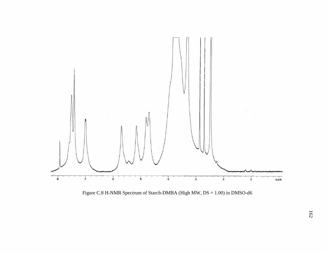
Figure C.8 H-NMR Spectrum of Starch-DMBA (High MW, DS = 1.00) in DMSO-d. .162
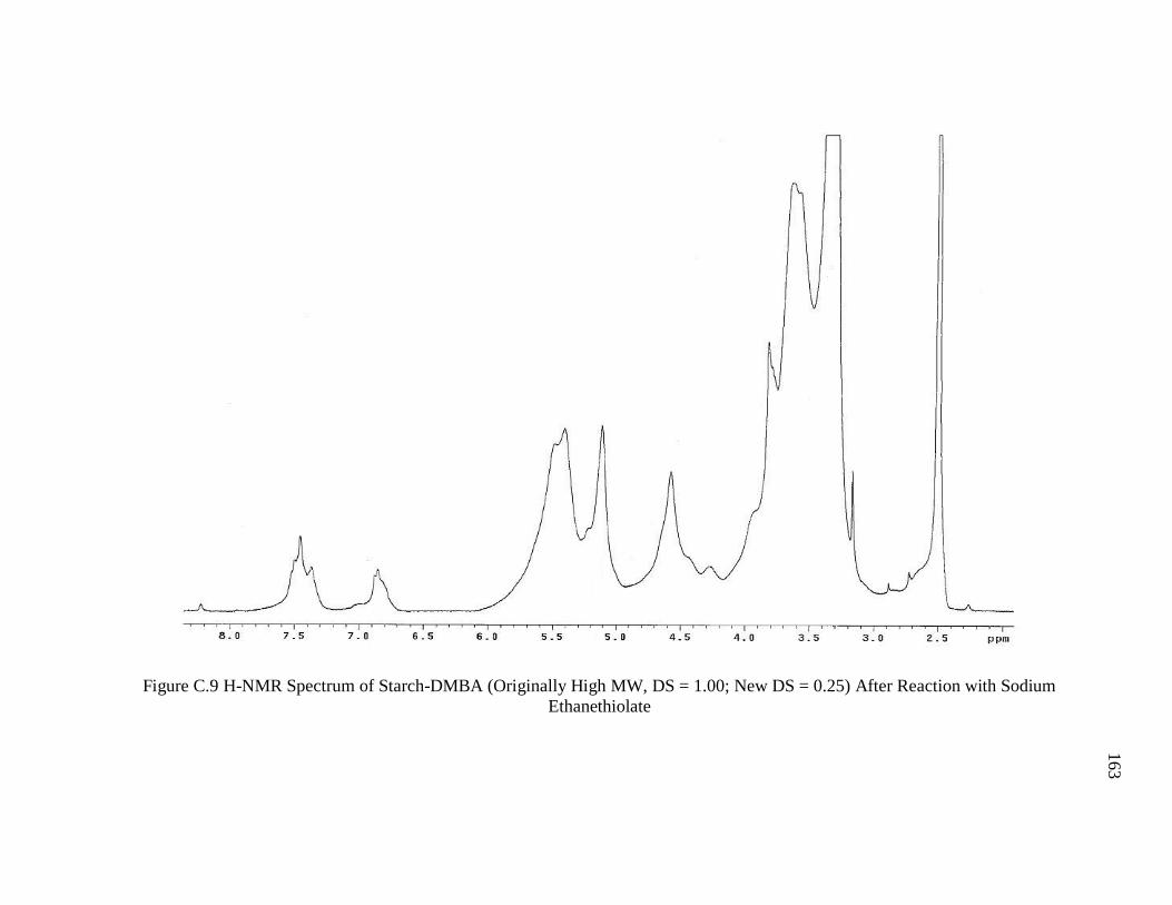
Figure C.9 H-NMR Spectrum of Starch-DMBA (Originally High MW, DS = 1.00; New
DS = 0.25) After Reaction with Sodium Ethanethiolate................................................. 163
Page 16
xiv
xiv
LIST OF ABBREVIATIONS
1-Ethyl-3-(3-dimethylaminopropyl)carbodiimide EDC
3,4-dihydroxybenzoic acid DHBA
3,4-dimethoxybenzoic acid DMBA
Ascorbic Acid AA
Atmospheric Pressure Chemical Ionization APCI
Anhydroglucose unit AGU
Carbonyldiimidazole CDI
Carboxylic acid COOH
Carboxymethyl starch CMS
Concentration of “x” [x]
Degree of substitution DS
Dichloromethane DCM
Dimethylacetamide DMA
Dimethylformamide DMF
Dimethylsulfoxide DMSO
Electron impact/chemical ionization EI/CI
Electrospray Ionization ESI
Ethanol EtOH
Page 17
xv
xv
Fourier transform infrared spectroscopy FTIR
Isopropanol IPA
Methanol MeOH
Molecular weight MW
N-hydroxysuccinimide NHS
Nuclear magnetic resonance spectroscopy NMR
Phenylboronic acid PBA
Proton NMR H-NMR
p-Toluenesulfonic acid chloride TosCl
Pyridine Py
Sodium monochloroacetate SMCA
Tetra-n-butylammonium fluoride TBAF
tert-Butyldimethylsilyl TBDMS
Ultraviolet-visible UV-vis
Page 18
xvi
xvi
ABSTRACT
de Kozlowski, Jeffrey K. M.S., Purdue University, December 2013. Development of a
Starch-Based Mussel-Mimetic Adhesive Polymer. Major Professor: Bernard Tao.
Mussel-mimetic adhesive polymers have gained lots of attention for their strong adhesive
strength, moisture resistance, and unique ability to crosslink. These properties are mainly
attributed to the high content of catecholic 3,4-dihydroxyphenylalanine (DOPA) in
mussel adhesive proteins. While there has been success in creating mussel-mimetic
synthetic polymers, less effort has been given to create a renewable, green, biocompatible
counterpart. This thesis explores the possibilities of starch-based mussel-mimetic
adhesives. Carboxymethyl starch of various molecular weights and degree of substitution
was synthesized and subsequent conjugation of dopamine to these polymers by 1-Ethyl-
3-(3-dimethylaminopropyl)carbodiimide was investigated. The polymers suffered from
very low substitution (DScatechol < 0.02) and easily precipitated from solution as an
insoluble product. The cause of precipitation was investigated and was shown to be
unrelated to autooxidation of conjugated dopamine by O2 and pH. Instead, EDC seemed
to be somehow responsible for the precipitation and most likely also for the very low
DScatechol due to competing reactions and instability of EDC intermediates. Lap-shear
strengths of the CMS-dopamine conjugates failed to exceed those of unmodified CMS.
Page 19
xvii
xvii
In search of another path to starch-catechol conjugates with higher DScatechol, 1,1’-
carbonyldiimidazole was employed for direct conjugation of bis-O-protected 3,4-
dihydroxybenzoic acid to unmodified starch. High DS was achieved with 3,4-
dimethoxybenzoic acid, but demethylation techniques were incompatible with starch and
its esters. Phenylboronic acid was then employed as an easily removable diol protecting
group for DHBA, but the complex was apparently not stable enough in solution for
selective activation of the carboxylic acid group of PBA-DHBA by either CDI or TosCl.
Further screening of different protecting groups or a new coupling chemistry is needed to
fully assess the possibilities of starch-catechol conjugates of high DScatechol.
Page 20
1
1
CHAPTER 1. INTRODUCTION
1.1 Objectives
The ultimate goal of this research was to create a novel starch-based adhesive inspired by
marine mussel adhesive chemistry by covalent coupling of catecholic moieties to high
amylose starch. The specific objectives of this project were to:
1. Modify the molecular weight of high amylose starch through controlled
hydrolysis.
2. Synthesize carboxymethyl starch of various degrees of substitution.
3. Conjugate dopamine to carboxymethyl starch in aqueous reaction with EDC
coupling agent and characterize the new polymer.
4. Evaluate the effectiveness of the catechol-substituted carboxymethyl starch as an
adhesive.
5. Investigate alternative, non-aqueous methods for creating catechol-functionalized
starch.
1.2 Organization
This thesis is divided into five chapters. The second chapter is a literature review to
provide motivation for renewable adhesives, the basics of adhesion and mussel adhesion
chemistry, and an overview of progress made in developing mussel-mimetic adhesives.
Page 21
2
2
Chapter three discusses the materials and methods used in the research. The results of the
research project are presented and discussed in chapter four. Finally, chapter five
provides a summary of the findings of this research and considerations for future work.
Page 22
3
3
CHAPTER 2. LITERATURE REVIEW
2.1 Adhesives
2.1.1 Introduction
On the most basic level, adhesives afford humanity the ability to join together dissimilar
objects. With such a vast number of applications possible from such a simple concept,
one can imagine the demand for adhesives and the market size they occupy. Adhesives
have a significant value in construction, packaging, transportation, automotive, consumer,
rigid bonding, and medical industries (Bosik, 2012).
Figure 2.1 Share of Adhesives and Sealants Market Value by Industry Application (Bosik,
2012)
Page 23
4
4
Globally, the adhesives and sealants market was worth about $22 billion in 2011. North
America, Europe, and Asia represent the three largest adhesives and sealants markets;
North America being the largest at $11.2 billion in 2011. About $7 billion of the North
American market is represented by adhesives, specifically. Of this $7 billion, the market
is mostly dominated by synthetic resin and rubber adhesives which account for about
$6.5 billion, while natural based glues and adhesives account for the remainder.
However, growth in almost all subcategories of the synthetic resin and rubber adhesives
market has been relatively stagnant while certain subcategories of the natural based glues
and adhesives market have seen significant growth; most notably protein adhesives, with
a compound annual growth rate of nearly 8% from 2002 to 2011 (Bosik, 2012).
2.1.2 Motivation for Green Adhesives
The majority of current adhesives are based on petroleum-derived materials. While
petro-based adhesives have advantages of superior bonding strength and some water
resistance, they are non-renewable and may pose environmental and health issues (Li et
al., 2012).
The main environmental/health concern with traditional adhesives is volatile organic
compounds (VOCs). One common VOC is formaldehyde, a major contributor to indoor
air pollution that may pose risk of cancer and respiratory complications like asthma
(Deschamps, 2010; Li et al., 2012; Ruffing, Smith, & Brown, 2010). Urea formaldehyde
and phenol formaldehyde are widely used as adhesives for construction of composite
wood panels such as particle board or fiber board which are used for interior wood panels
Page 24
5
5
of homes and for furniture. Additionally, adhesive formulations may contain other VOCs
that pose environmental and health concerns such as trichloroethane and toluene which
are used as solvents for adhesive application (Li et al., 2012). Environmental regulations
are becoming more stringent with regards to VOC limits and green construction programs
such as LEED offer credits for use of low-emitting products in new construction (Bosik,
2012). Therefore, high-performance natural based glues will become increasingly
important.
Other ecological factors that should be considered include the degree of treatment
necessary for waste from adhesives processing and the ability to reuse the materials onto
which the adhesive is ultimately applied (Onusseit, VonByern, & Grunwald, 2010;
Shuttleworth, Clark, Mantle, & Stansfield, 2010). Finally, it should not be overlooked
that as petroleum stocks are gradually depleted, the cost of petro-chemicals will rise
accordingly, further driving the need for renewable “green” adhesives.
2.1.3 Concepts
Although the concept of adhesion is very simple, the science and mechanistic
understanding of adhesives can be very complicated. Some basic terminology, concepts,
and theory related to adhesives are provided in the following section.
Adhesion is the force responsible for the joining of dissimilar surfaces. The objects being
joined are referred to as the adherends and the substance responsible for adhesion is
referred to as the adhesive. Adhesion cannot be discussed without cohesion, as it plays
Page 25
6
6
an equally important role in adhesives. Cohesion can be thought of as internal adhesion,
or the force that joins similar surfaces.
Higher energy exists at the surfaces of solid and liquid materials due to the inability of
surface molecules to interact symmetrically with surrounding molecules as those in the
material bulk do; resulting in an imbalance of intermolecular interactions for surface
molecules. This energy associated with the potential for intermolecular interaction of
surface molecules is known as the surface energy of a material.
When two dissimilar surfaces are in contact there exists an interfacial energy. After work
is applied to separate these two surfaces, two new surfaces with distinct surface energies
are the result, but the interfacial energy is no longer present. The sum of the surface
energies of two materials minus the interfacial energy gives rise to the term work of
adhesion.
While work of adhesion provides a fundamental measure of adhesive intermolecular
forces, it is not a straight forward answer to the practical strength of adhesion between
two surfaces because the mechanical response of the adhesive, substrate, and adhesive-
substrate interface also factor into adhesive strength. In most cases this “practical
adhesion” is measured by conducting one or several types of stress tests where the
adhesive strength is equal to the amount of stress required for adhesive failure. Common
stress tests include shear tests and tensile tests. Adhesive strength is often reported as the
Page 26
7
7
result of a stress test. Expressions relating adhesive strength to work of adhesion can be
used to gain a more fundamental picture of a given adhesive system (Packham, 2005).
An important concept in the practical application of adhesives is the weak boundary layer.
The weak boundary layer arises from any weakly covalent layer of matter associated with
a surface that prevents the immediate contact of adhesive and adherend and ultimately
leads to premature adhesive failure. For example, wet surfaces pose a problem for
adhesives due to the presence of a weak layer of water molecules (Anderson et al., 2010).
Additionally, dirt, grease, dust, or other impurities that can coat surfaces may act as a
weak boundary layer (Waite, 2002). Therefore, surface preparation/treatment is often a
necessary operation prior to adhesive application.
2.1.4 Mechanisms of Adhesion
The mechanisms responsible for adhesion can be broadly split into mechanical and
chemical adhesive forces.
2.1.4.1 Mechanical Adhesion
Mechanical adhesion refers to interlocking between adherend surfaces on the microscopic
level. Although mechanical forces have been shown to be significant in a few specific
applications, it is generally disregarded as an important mechanism of adhesion (Pocius,
2002).
2.1.4.2 Chemical Adhesion
Chemical adhesion refers to a host of different forces, chemical in nature, responsible for
adhesion. These forces include ionic bonds from electrostatic interactions, physical
Page 27
8
8
bonds from van der Waals interactions, and chemical bonds from electron pair sharing
(Pocius, 2002).
Electrostatic forces are those that arise between two charged atoms or molecules. Atoms
or molecules with like charges repel each other while those with dissimilar charges attract.
Electrostatic forces are considered the second strongest interaction between atoms or
molecules (Pocius, 2002).
Van der Waals interactions are based mainly on differences in electron densities on or
between molecules. These interactions include dipole-dipole (including hydrogen
bonding) and dipole-induced dipole interactions, and dispersion forces (Pocius, 2002).
Electron pair sharing interactions encompass both covalent and donor-acceptor
interactions. Covalent bonds are the result of electron pair sharing between atoms in a
molecule. Covalent bonds are considered the strongest interaction between atoms or
molecules. Acid-base interactions are a special case of donor-acceptor interactions where
interaction occurs between an electron-deficient Lewis acid and the lone electron pair of
a Lewis base. Acid-base interactions have been extensively studied and deemed to play
an important role in adhesion (Pizzi & Mittal, 2003; Pocius, 2002).
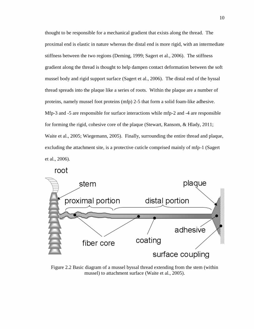
2.1.5 Adhesion in Nature – Mussel Adhesion
In nature, a wide variety of organisms naturally produce unique adhesives to aid their
survival. One such adhesive that has been extensively studied is that of the marine
mussel from the genus Mytilus (Bruce P. Lee, 2006; Waite, Andersen, Jewhurst, & Sun,
Page 28
9
9
2005). Marine mussels depend on adhesion in order to permanently cling to substrates
such as rock in order to endure the harsh environment of the shores where they reside.
Such an adhesive must be able to function on rough, untreated surfaces submerged in
turbulent marine waters (Sagert, Sun, & Waite, 2006). Significant research and interest
in developing materials based on the marine mussel adhesive spur from its ability to
quickly set, effectively displace water from the attachment surface, withstand a range of
temperatures and salinity, and adhere to practically any type of surface (Crisp, Walker,
Young, & Yule, 1985; Deming, 1999; Lin et al., 2007; Waite, 2002). The utility of
mussel adhesives have yet to be rivaled by any synthetic adhesives (Sever, Weisser,
Monahan, Srinivasan, & Wilker, 2004).
In order to scout out surfaces for adhesion, mussels have an organ called a foot. At the
base of the foot is a stem-like structure known as the byssus where a collection of
collagenous threads meet. The individual threads are known as byssal threads and are
responsible for the adhesion of the mussel to surfaces. Byssal threads extend radially
from the underside of the organism to the surface of attachment. The area of a byssal
thread furthest from the mussel is known as the distal end which ends with the plaque,
where adhesion between thread and surface occurs (Waite et al., 2005).
The thread and plaque are made up of a variety of proteins. The threads themselves are
primarily composed of collagen-like proteins, preCol-P and preCol-D. PreCol-P
predominates at the proximal end (closest to the byssus) and preCol-D at the distal end of
the byssal thread (Deming, 1999). The difference in amount of preCol-P and preCol-D is
Page 29
10
10
thought to be responsible for a mechanical gradient that exists along the thread. The
proximal end is elastic in nature whereas the distal end is more rigid, with an intermediate
stiffness between the two regions (Deming, 1999; Sagert et al., 2006). The stiffness
gradient along the thread is thought to help dampen contact deformation between the soft
mussel body and rigid support surface (Sagert et al., 2006). The distal end of the byssal
thread spreads into the plaque like a series of roots. Within the plaque are a number of
proteins, namely mussel foot proteins (mfp) 2-5 that form a solid foam-like adhesive.
Mfp-3 and -5 are responsible for surface interactions while mfp-2 and -4 are responsible
for forming the rigid, cohesive core of the plaque (Stewart, Ransom, & Hlady, 2011;
Waite et al., 2005; Wiegemann, 2005). Finally, surrounding the entire thread and plaque,
excluding the attachment site, is a protective cuticle comprised mainly of mfp-1 (Sagert
et al., 2006).
Figure 2.2 Basic diagram of a mussel byssal thread extending from the stem (within
mussel) to attachment surface (Waite et al., 2005).
Page 30
11
11
In all of the mussel foot proteins, there exist post- or co- translationally modified amino
acids. These modified amino acids include 3,4-dihydroxyphenylalanine (DOPA), o-
phosphoserine, 4-hydroxyproline, 3,4-dihydroxyproline, and 4-hydroxyarginine
(Wiegemann, 2005). DOPA and phosphoserine are implied in the actual adhesive
properties of the plaque based on their abilities to interact strongly with metals and metal
oxides (Haemers, Koper, & Frens, 2003). DOPA has received the most attention because
it is universally present in mussel foot proteins as well as the adhesion proteins of some
other organisms (Stewart et al., 2011). Furthermore, DOPA content is especially high in
the interfacial plaque proteins mfp-3 and -5, where it is present up to about 20 mol% and
30 mol%, respectively (Sagert et al., 2006). DOPA is also found up to about 15 mol% in
mfp-1 where it is thought to facilitate cohesion and hardening of the protective cuticle (H.
Lee, Scherer, & Messersmith, 2006; Rischka et al., 2010)
2.2 DOPA Chemistry
DOPA is a post-translationally modified amino acid that arises from hydroxylation of
tyrosine. DOPA is generally understood to be the primary facilitator of both adhesion
and cohesion of mussel foot proteins. However, the mechanisms for adhesion and
cohesion are still not fully established. Moreover, DOPA's adhesive mechanisms are
generally less understood than its cohesive mechanisms (Bruce Lee, Dalsin, &
Messersmith, 2006). DOPA is also attributed to the water-resistant adhesion of mussel
proteins (M. Yu, Hwang, & Deming, 1999) and the ability of mussel plaques to adhere to
a wide variety organic and inorganic surfaces.
Page 31
12
12
2.2.1. Redox and Metals
The underlying principle behind the overall adhesive and cohesive properties of mussel
adhesives is the oxidation/reduction of DOPA and the maintenance of a balance between
oxidized and reduced forms. DOPA readily undergoes oxidation to its highly reactive
DOPA-quinone and –semiquinone forms, especially in basic conditions (Haemers et al.,
2003). In vivo, it is thought that the mussel employs the enzyme catechol oxidase to
catalyze DOPA oxidation. In vitro, the enzyme tyrosinase has been used to facilitate
oxidation of DOPA in addition to chemical oxidants (Haemers et al., 2003; Yamada et al.,
2000)
Experiments have shown that both increasing pH and increasing presence of oxidizing
agents leads to decrease in mussel adhesive performance on metal oxide surfaces while
increasing adhesive strength on organic surfaces, specifically amine-functionalized
surfaces (H. Lee et al., 2006; J. Yu, Wei, Danner, Israelachvili, & Waite, 2011).
Therefore, on metal oxide surfaces, it is supposed that in its reduced form DOPA aids in
adhesive interactions while the oxidized form is primarily involved in cohesive
interactions (Wilker, 2011). The relationship between oxidized DOPA and its adhesion
to organic surfaces is less clear.
As with any adhesive, a balance of cohesion and adhesion is necessary for optimal
adhesive performance. Thus, balance between oxidized and reduced forms of DOPA
must be maintained in order for proper adhesion to exist. In nature, it has been suggested
that this balance is maintained by secretion of thiol-rich mfp-6 along with the DOPA-rich
Page 32
13
13
mfps. Through oxidation of the thiol groups, DOPA-quinone can be reduced to restore
an optimal balance of adhesive and cohesive forms of DOPA (J. Yu, Wei, Danner,
Ashley, et al., 2011).
Mussels are able to accumulate metals in their byssal threads. The plaque, specifically,
contains metal ions including copper, iron, zinc, and manganese concentrated up to
100,000 times greater than in ocean water. These metal ions play many important roles
in DOPA chemistry. First, transition metals provide a route of cross-linking between
DOPA-containing proteins (Monahan & Wilker, 2004). More specifically, it is proposed
that iron atoms are primarily responsible for protein cross-linking by creating a tris-
DOPA complex (Sever et al., 2004; Zeng, Hwang, Israelachvili, & Waite, 2010).
Another important role of transition metal ions is they facilitate oxidation of DOPA by
aligning the aromatic side chain of DOPA molecules thus lowering the energy of
oxidation. Finally, transition metal ions provide a center for interactions between DOPA-
proteins and surfaces, thereby facilitating adhesion (Brooksby, Schiel, & Abell, 2008).
2.2.2 DOPA in Adhesion
DOPA in its reduced form is primarily responsible for adhesive interactions with surfaces
(Wilker, 2011). Polar interactions between the hydroxyl groups present on the side chain
of DOPA and the adhesion surface are presumably the reason for these interactions
(Frank & Belfort, 2001). DOPA’s hydroxyl groups can participate in hydrogen binding,
as either donor or acceptor, with electrophilic groups along polar, hydrophilic surfaces.
Additionally, it has been shown that DOPA’s hydroxyl groups are able to strongly
Page 33
14
14
complex with metals and minerals on inorganic surfaces (Wiegemann, 2005).
Furthermore, the hydrogen bonding activity of DOPA’s catecholic side chain may allow
DOPA to compete with water for interactions on polar surfaces (Bruce Lee et al., 2006).
It is proposed that π-π orbital interactions between the aromatic ring of DOPA and other
aromatic groups may account for the ability of mussels to adhere to organic surfaces
(Frank & Belfort, 2001; Wiegemann, 2005). It has also been suggested that covalent
bonds formed by reaction of oxidized DOPA with organic functional groups could also
be responsible for adhesion of mussel plaques to organic surfaces. An example of this is
DOPA-quinone forming a covalent bond with surface amine groups via Michael
addition-type reactions (Bruce Lee et al., 2006; H. Lee et al., 2006; J. Yu, Wei, Danner,
Israelachvili, et al., 2011). The catechol side group of DOPA is also attributed to the
mucoadhesive properties of mussel adhesive proteins (Schnurrer & Lehr, 1996).
2.2.3 DOPA in Cohesion
The oxidized form of DOPA (DOPA-quinone) is primarily responsible for cross-linking
interactions that give rise to the cohesive properties of mussel adhesives. Yu et al.
proposed many possible mechanisms for DOPA-mediated cross-linking including aryl
coupling, metal chelation, imine formation, and Michael addition (M. Yu et al., 1999). In
aryl coupling, DOPA-quinone is partially reduced by unoxidized DOPA resulting in two
reactive radicals that form a diDOPA complex. Similar to the way that metal ions such
as copper facilitate adhesion by cross-linking DOPA and surface functional groups, they
can also cause cross-linking between DOPA residues not involved in surface interactions,
Page 34
15
15
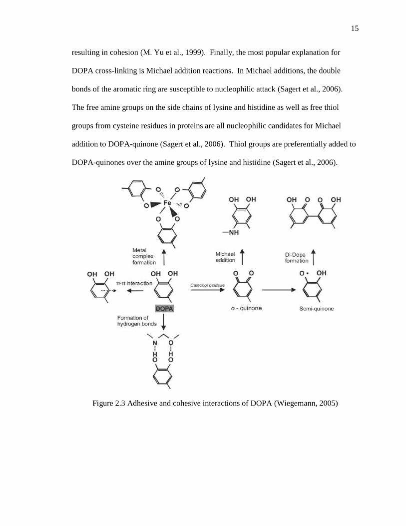
resulting in cohesion (M. Yu et al., 1999). Finally, the most popular explanation for
DOPA cross-linking is Michael addition reactions. In Michael additions, the double
bonds of the aromatic ring are susceptible to nucleophilic attack (Sagert et al., 2006).
The free amine groups on the side chains of lysine and histidine as well as free thiol
groups from cysteine residues in proteins are all nucleophilic candidates for Michael
addition to DOPA-quinone (Sagert et al., 2006). Thiol groups are preferentially added to
DOPA-quinones over the amine groups of lysine and histidine (Sagert et al., 2006).
Figure 2.3 Adhesive and cohesive interactions of DOPA (Wiegemann, 2005)
Page 35
16
16
2.3 Mussel-Mimetic Adhesive Polymers
There are essentially two approaches for creating mussel-mimetic adhesive polymers.
The first approach is to directly incorporate DOPA or another catechol-containing
molecule onto the backbone of a polymer. This can be accomplished by reacting a
functional group of the catechol monomer with a functional group on the polymer. The
second approach incorporates DOPA or one of its analogs onto a monomer which is
subsequently polymerized, usually with another monomer, in order to achieve polymers
of various catechol contents.
Attaching DOPA to another molecule without affecting the functionality of DOPA
requires that its catechol group is excluded from any reactions. DOPA exists in mfps as a
member of a chain of amino acids linked by peptide (amide) bonds which are formed
between the amine group or N-terminus of one amino acid and the carboxyl group or C-
terminus of another. Amide bonds can just as well be made between the amine or
carboxyl groups of DOPA and reactive groups of a monomer or polymer to form DOPA-
functionalized polymers.
Because the adhesion/cohesion roles of DOPA are attributed to the catechol group, other
catechol-containing molecules can, and have been successfully used to create mussel-
mimetic adhesives. For simplicity, in many cases it is actually preferable to use a
catecholic molecule having just one reactive group on its side-chain as opposed to two
reactive groups like DOPA. Some of these DOPA analogs include 3,4-
dihydroxyphenethylamine (dopamine) , 3,4-dihydroxybenzoic acid, caffeic acid, and 3,4-
Page 36
17
17
dihydroxybenzaldehyde. Many researchers have successfully functionalized different
polymers with catecholic groups using a variety of approaches. The following section
summarizes the work that has been done so far based on the conjugation chemistry used.
2.3.1. Catechol-Functionalized Polymers
2.3.1.1 Benzotriazoles
Benzotriazoles have been widely used as organic coupling reagents. In the case of
peptides, benzotriazoles function by activating the carboxyl group, making it prone to
nucleophilic attack by an amine group (Scott, 2009). Because each amino acid in the
reaction has one carboxylic acid and one amine group, the amine group of the activated
amino acid requires blocking to ensure the desired reaction occurs.
Utilizing this chemistry, several researchers from the Messersmith Research Group
developed DOPA-functionalized adhesive hydrogels. In each case tert-butyloxycarbonyl
(Boc)-protected DOPA was reacted with triethylamine (Et3N), 1-hydroxybenzotriazole
(HOBt), and O-(benzotriazol-1-yl)-N,N,N',N'-tetramethyluronium hexafluorophosphate
(HBTU) in a solvent system of dichloromethane (DCM) and dimethylformamine (DMF)
to covalently link the carboxyl group of DOPA to the amine groups of various polymers.
Above 80% coupling efficiency was achieved when linking DOPA to linear and four-
armed poly(ethylene glycol) (PEG4) (BP Lee, Dalsin, & Messersmith, 2002). A
following study synthesized DOPA-PEG4 using the same materials and reported DOPA
content of 6% by weight (Burke, Ritter-Jones, Lee, & Messersmith, 2007). DOPA was
Page 37
18
18
also grafted onto a triblock copolymer consisting of PEG sandwiched between
methacrylated poly(lactic acid) and glycine, referred to as G-PPM, in which case 2%
DOPA content by weight was achieved (BP Lee et al., 2006). Guvendiren et al.
developed modified methacrylic triblock copolymers consisting of poly(methacrylic acid)
(PMAA) sandwiched between poly(methyl methacrylate) (PMMA) end blocks with
DOPA contents of 0, 20, and 40 mol % . In this case, the N-terminus of DOPA methyl
ester was incorporated into the PMAA portion of the triblockcopolymer (Guvendiren,
Messersmith, & Shull, 2008).
In a similar manner, Messersmith et al. functionalized four-arm PEG amine with 3,4-
dihydroxyhydrocinnamic acid (DHCA). The group utilized their mussel-inspired
adhesive to aid in extrahepatic islet transplantation in mice. The adhesive remained intact
with adipose tissue for up to one year and resulted in minimal inflammatory response; a
testament to the capability of catechol-functionalized polymer adhesives (Brubaker,
Kissler, Wang, Kaufman, & Messersmith, 2010).
2.3.1.2 Carbodiimide
A popular chemistry for the conjugation of dopamine with carboxyl-containing polymers
is 1-ethyl-3-(3-dimethylaminopropyl)carbodiimide hydrochloride (EDC) cross-linking.
EDC is a water-soluble zero-length crosslinker that can function between pH 3.5 – 8 at
room temperature (Hattori, Yang, & Takahashi, 1995; Nakajima & Ikada, 1995); making
it convenient to use in many biochemical reactions. Peptide bond formation by EDC
Page 38
19
19
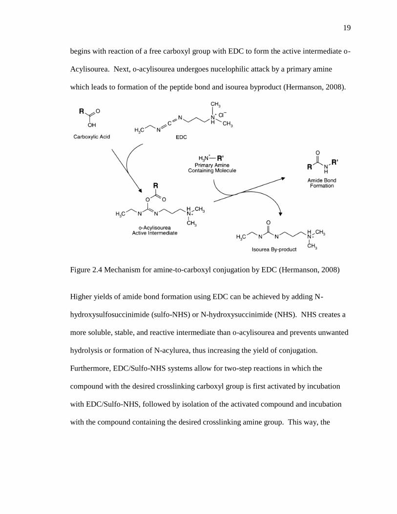
begins with reaction of a free carboxyl group with EDC to form the active intermediate o-
Acylisourea. Next, o-acylisourea undergoes nucelophilic attack by a primary amine
which leads to formation of the peptide bond and isourea byproduct (Hermanson, 2008).
Figure 2.4 Mechanism for amine-to-carboxyl conjugation by EDC (Hermanson, 2008)
Higher yields of amide bond formation using EDC can be achieved by adding N-
hydroxysulfosuccinimide (sulfo-NHS) or N-hydroxysuccinimide (NHS). NHS creates a
more soluble, stable, and reactive intermediate than o-acylisourea and prevents unwanted
hydrolysis or formation of N-acylurea, thus increasing the yield of conjugation.
Furthermore, EDC/Sulfo-NHS systems allow for two-step reactions in which the
compound with the desired crosslinking carboxyl group is first activated by incubation
with EDC/Sulfo-NHS, followed by isolation of the activated compound and incubation
with the compound containing the desired crosslinking amine group. This way, the
Page 39
20
20
reaction is more controlled and undesired self-polymerization can be avoided
(Hermanson, 2008).
Wu et al. covalently bound dopamine to poly(acrylic acid) (PAA) using EDC chemistry.
The group determined the catechol content of their conjugate to be about 12%. The
dopamine-PAA ultimately served as a cohesive agent to form stable, multilayer, thiol-
modified films composed of poly(allylamine hydrochloride) (Wu et al., 2011).
Most recently, Karabulut et al. employed EDC to covalently modify carboxymethyl
cellulose nanofibrils (CNFC) with dopamine in order to create strong layer-by-layer films
with improved adhesion. The group was able to functionalize about 76% of the carboxyl
groups with dopamine. Interestingly, the group found that the addition of NHS to the
EDC cross-linking reaction did not enhance the extent of conjugation. Modification of
CNFC with dopamine was found to lower the colloidal stability of the polymer
dispersions, most likely due to the loss of carboxyl groups and consequent loss of overall
charge in the system (Karabulut, Pettersson, Ankerfors, & Wagberg, 2012).
Liu and Li grafted dopamine onto soy protein isolate (SPI) using EDC chemistry in effort
to enhance the SPI for wood bonding. By controlling the concentrations of EDC and
dopamine, the two researchers obtained SPI-dopamine conjugates with varying dopamine
content. Plywood samples bonded with the conjugates exhibited significantly greater
shear strengths and water resistance compared to regular SPI and actually exhibited
performance comparable to traditional urea- or phenyl-formaldehyde resins. The group
Page 40
21
21
found that higher dopamine content led to greater shear strength and moisture resistance
in prepared wood samples. Additionally, the group showed that the hydroxyl groups of
unoxidized dopamine are necessary for good adhesive strength and moisture resistance
(Liu & Li, 2002).
2.3.1.3 Schiff Base
DOPA’s amine group has also been utilized in conjugation via Schiff base reactions.
When a primary amine reacts with aldehyde, ketone, or glyoxal groups they may form a
Schiff base/imine intermediate which can then be further converted to a secondary amine
bond (Hermanson, 2008).
Figure 2.5 Simplified Mechanism of Amide Bond Formation via Schiff Base
Hoffman et al. designed bioadhesive bone glues comprised of starch or dextran, chitosan,
and DOPA. First, starch or dextran was oxidized to provide reactive aldehyde groups.
For systems including DOPA, the starch/dextran was oxidized in its presence in order to
promote Schiff base reactions between the alehyde groups of the oxidized starch/dextran
and the amine group of DOPA. Finally, the starch/dextran-DOPA complex was oxidized
in the presence of low molecular weight chitosan. Cross-linking was achieved in two
Page 41
22
22
ways: aldehyde groups of oxidized starch/dextran interacted with amino groups of
chitosan, and/or the oxidized DOPA (DOPA-quinone) reacted with the amino groups of
chitosan. Unfortunately, no data was offered concerning the DOPA content of the final
adhesive (Hoffmann et al., 2009). Similarly, an adhesive polymer with up to 12% DOPA
content by weight was developed by modifying oxidized dextran with DOPA, followed
by conjugation with star PEG to form a hydrogel (Shazly et al., 2010).
Most recently, Schiff base reaction was used by Ni et al. to form a catechol-chitosan
conjugate from 3,4-dihydroxy benzaldehyde and chitosan. In this case, the aldehyde
group of the 3,4-dihydroxy benzaldehyde was utilized for Schiff base reaction with the
amine groups on chitosan. The researchers reported a degree of substitution of 52% with
respect to conjugated catechol groups. The catechol-chitosan conjugate demonstrated
adhesion to iron nanoparticles over which a layer of the conjugate could be formed. Free
catechol groups on the outside of the catechol-chitosan-iron nanoparticles were then used
to immobilize enzyme. These nanoparticles dispersed well in aqueous solution and could
be easily recovered by magnets (Ni et al., 2012).
2.3.2 Polymerization of Catechol-Functionalized Monomers
2.3.2.1 Condensation
Mehdizadeh et al. synthesized a mussel-inspired adhesive in a one-step polycondensation
reaction with monomers of citric acid, PEG, and dopamine or L-DOPA. The reaction
Page 42
23
23
was carried out under vacuum at 140-160°C for the duration of time required for a
particular degree of polymerization. Catechol content of the polymers could be varied by
adding different amounts of dopamine or L-DOPA to the reaction mixture. The polymers
formed from the reaction were then crosslinked to form adhesive hydrogels by oxidizing
the catechol moieties with sodium periodate. The citric acid units served two important
roles: to promote biodegradable ester bonds and to act as a conjugation site for the amine
group of dopamine or L-DOPA (Mehdizadeh, Weng, Gyawali, Tang, & Yang, 2012).
These adhesives exhibited wet tissue strength up to 8 times greater than traditional fibrin
glue.
Kaneko et al. developed an adhesive copolymer based on polycoumarates, DHCA and 4-
hydroxycinnamic acid (HCA). DHCA and HCA monomers were polymerized at 200°C
in the presence of sodium acid phosphate catalyst and absence of oxygen. The
poly(DHCA-co-4HCA) adhesive demonstrated impressive adhesive strength compared to
conventional super glue on inorganic and organic surfaces. However, no information was
provided concerning the compatibility of the adhesive with wet surfaces (Kaneko et al.,
2011).
2.3.2.2 Ring-Opening Addition of N-Carboxyanhydrides
Deming et al. synthesized synthetic polypeptides containing lysine and DOPA by
creating N-carboxyanhydrides of lysine and DOPA by phosgenation, followed by ring-
opening addition which was initiated by sodium tert-butoxide. For these reactions, the
Page 43
24
24
hydroxyl groups of DOPA and the amine side-group of lysine were protected by
carbobenzoxyl groups which were removed after polymerization. Synthetic poly(lysine-
DOPA) polypeptides with up to 20% DOPA content were synthesized and showed
adhesive strengths ten times greater than poly(L-lysine). Adhesive strength of the
poly(lysine-DOPA) polypeptides could be further enhanced by crosslinking via oxidation
of DOPA. Lap-shear adhesion tests of the polymer demonstrated relatively weak
adhesive capability between plastics (>0.5 MPa), but much greater adhesion between
glass and metal (up to~2.5 MPa and ~5 MPa, respectively) (M. Yu & Deming, 1998).
2.3.2.3 Reactive Anhydride or Acid Chloride
Lee et al. combined the adhesive strategies of both geckos and mussels to create a
reversible, wet-dry adhesive. Poly dimethyl siloxane was microfabricated into an array
of pillars which were then coated with a mussel-mimetic adhesive. The mussel-mimetic
adhesive was formed by first synthesizing dopamine methacrylate via nucleophilic attack
of the amine group of dopamine on methacrylic anhydride. The reaction was able to take
place in a high pH aqueous environment without oxidation of dopamine because a high
concentration of sodium borate was provided to protect the catechol group. Dopamine
methacrylate was then copolymerized with methoxyethyl acrylate to yield poly(dopamine
methacrylamide-co-methoxyethyl acrylate). The mussel-mimetic adhesive was designed
based on the criteria that catechol content of the synthetic adhesive must be high (~27
mol %), and the polymer should have low solubility in water (H. Lee, Lee, &
Messersmith, 2007).
Page 44
25
25
Lee et al also demonstrated conjugation of DOPA with methacroyl chloride by
nucleophilic attack of the amine group of DOPA with the acid chloride group of
methacroyl chloride in an alkaline aqueous environment containing borax to protect
DOPA from oxidation. To synthesize the polymer, the hydroxyl groups of DOPA’s
catechol side chain were first blocked by t-butyldimethylsilyl chloride. The DOPA-
methacrylate monomers were then photopolymerized with PEG-diacrylate to form an
adhesive hydrogel. DOPA content was found to be about 15 µgram per gram of gel (BP
Lee, Huang, Nunalee, Shull, & Messersmith, 2004).
2.3.2.4 Vinyl Polymerization
Westwood et al. synthesized mussel-mimetic styrene based polymers by copolymerizing
styrene and 3,4-dimethoxystyrene using n-Butyllithium to initiate polymerization of the
vinyl groups. Synthesis and recovery of the copolymer was followed by demethylation
of the methoxy groups by BBr3 to furnish the active catechol. By varying the feed ratio
of 3,4-dimethoxystyrene:styrene, polymers of varying catechol content could be obtained.
Lap-shear adhesive tests were carried out with poly[(3,4-dihydroxystyrene)-co-styrene]
of MW = 16,000 and 3,4-dihydroxystyrene:styrene = 3.4:96.6 which showed
significantly stronger adhesion than pure styrene polymers of comparable MW.
Adhesion of the copolymer was also tested after crosslinking by treatment with various
oxidizing agents. Adhesive strength was shown to increase upon crosslinking, with a
maximum adhesive strength of 1.2 MPa attained (Westwood, Horton, & Wilker, 2007).
Page 45
26
26
2.3.3 Characterizing DOPA-polymer Conjugates
2.3.3.1 DOPA Content
A relatively simple and common method for DOPA-content determination of water-
soluble DOPA compounds is a colorimetric test based on the work of Waite and Benedict
(Waite & Benedict, 1984). When catecholic compounds are oxidized, they produce a
red-orange color which is quantified by UV-vis absorbance at 500 nm (BP Lee et al.,
2006). Typically, a nitrite reagent and NaOH are used to induce oxidation (Guvendiren
et al., 2008; Huang, Lee, Ingram, & Messersmith, 2002; BP Lee et al., 2002; BP Lee et
al., 2004). This method is especially useful in situations where other aromatic groups
may be present as it is specific to catechols.
As with most aromatic compounds, catechol has a characteristic UV-vis absorbance at
280 nm. While this method is not fit for quantifying DOPA in compounds containing
other aromatic amino acids and DNA (due to absorbance overlap), it has been used for
DOPA quantification in DOPA- and dopamine-functionalized polymers where the
catechol moiety is the sole contributor to absorbance at 280 nm (BP Lee et al., 2006; Wu
et al., 2011). However, care must be taken to prevent any oxidation of the sample for this
method to yield accurate results.
DOPA content can also been determined with proton nuclear magnetic resonance
spectroscopy (1H NMR). The three protons in the aromatic ring of DOPA have a distinct
Page 46
27
27
chemical shift around 6.4-6.9 in deuterium oxide (BP Lee et al., 2006; Shazly et al., 2010;
Wu et al., 2011). From a standard curve, the integral value of the peaks may then be used
to determine the concentration of catechol in solution and ultimately the catechol content
of the conjugate (% wt.). Additionally, if the monomer units of the polymer have a
unique proton signal, then a direct value for the degree of catechol substitution may be
obtained by calculating the ratio of the integral values of catechol to monomer (% mol).
2.3.3.2 Verification of Conjugation
In many instances, it may be useful to verify that conjugation between catechol and
polymer is achieved through the desired chemical bond. Spectroscopic analysis of
catechol-modified compounds provides a relatively easy way to assess successful
conjugation thanks to the ability to detect specific bond types. Conjugation can also be
confirmed by comparing the abundance of free attachment sites before and after
conjugation reaction.
FTIR spectroscopy was used to verify the amide bond formed by cross-linking dopamine
to poly(acrylic acid) by EDC, evident by the presence of an amide II band (Wu et al.,
2011). Conjugation of dopamine to carboxymethyl cellulose nanofibrils by Karabulut et
al. was evident by loss of intensity of the peak for asymmetric stretching vibrations of
deprotonated carboxyl groups and the appearance of Amide I bands (Karabulut et al.,
2012).
Page 47
28
28
Proton NMR can also be used to detect specific bonds. Shazly et al. used 1H NMR to
verify whether Schiff base reaction occurred between oxidized dextran and DOPA by
detecting the resulting imine proton (Shazly et al., 2010).
2.3.4 Performance of Mussel-Inspired Adhesives
In most cases, incorporation of DOPA or catechol-containing moieties into polymer or
copolymer systems was associated with increased moisture resistance and higher
adhesive strengths (Guvendiren et al., 2008; Karabulut et al., 2012; BP Lee et al., 2006;
H. Lee et al., 2007; Liu & Li, 2002; Mehdizadeh et al., 2012). In only one instance did
incorporation of DOPA have a negative effect on the adhesive (Hoffmann et al., 2009).
However, it should be noted that in the later study the adhesive was applied to bovine
femora, whereas titanium, wood, or polymer surfaces were used in the other studies.
Therefore, the benefits of mussel-inspired adhesives may be dependent on the application
and surface type.
In general, higher catechol content in mussel-mimetic polymers is associated with greater
adhesive capabilities (BP Lee et al., 2006; Liu & Li, 2002). As previously noted, mussel
foot proteins can contain up to 30 mol % DOPA. Most of the adhesives mentioned in this
review fail to achieve such an extent of catechol functionalization so it is not clear
whether the adhesive benefits of catechol moieties continue with increasing catechol
content indefinitely. There may be some optimal degree of catechol functionalization for
mussel-mimetic adhesives. Furthermore, the optimal catechol content may depend on the
nature of the particular adhesive system.
Page 48
29
29
As with most adhesives, molecular weight can greatly affect the adhesive abilities of
DOPA-functionalized polymers. For a synthetic poly(lysine-DOPA) peptide, the
adhesive strength demonstrated by the polymer with MW = 255,000 was over twice that
of MW = 98,000 (M. Yu & Deming, 1998). Therefore, molecular weight should be fine-
tuned for maximum adhesion of mussel-mimetic polymers.
The adhesive performance of mussel-inspired adhesives has been shown to be dependent
on pH and oxidation state of the incorporated catechol group. Increasing pH was
associated with weaker adhesive strengths to titanium; presumably due to oxidation of
DOPA’s catechol group (Guvendiren et al., 2008). Similarly, oxidized DOPA-adhesives
were shown to exhibit lower work of adhesion when compared to their unoxidized
counterparts (BP Lee et al., 2006). This work indicates that the reduced form of the
catechol group is necessary for strong adhesion of mussel-mimetic polymers to titanium
surfaces. However, crosslinking of mussel-mimetic polymers by oxidation of the
incorporated catechol groups has been shown to significantly enhance the adhesive
strength and moisture resistance of mussel-mimetic adhesives (Westwood et al., 2007; M.
Yu & Deming, 1998). A balance of reduced and crosslinked forms of catechol in mussel-
mimetic adhesives is therefore necessary to ensure there is an adequate amount of both
adhesive interactions, between the hydroxyl groups of catechol and the adherend, and
cohesive interactions, through intermolecular crosslinking, for maximum moisture-
resistance and adhesive strength.
Page 49
30
30
2.3.5 DOPA for Adhesive Polymer Crosslinking
In addition to its adhesive functionality, DOPA is also attractive for facilitating cohesive
forces within adhesive polymer networks through oxidation of its catechol side group.
Oxidized DOPA can undergo various reactions, therefore a number of cross-linking
pathways exist depending on the chemical make-up of the adhesive polymer.
2.3.5.1 Chemical Oxidants
Periodate has widely been used to induce crosslinking of mussel-inspired adhesives
through oxidation of their catechol groups.
Lee et al. developed adhesive polymers based on DOHA, PEG, and polycaprolactone
(PCL). Periodate was used to induce cross-linking of the adhesive polymers. The group
found that there was an optimal degree of crosslinking for maximum adhesive strength
and work of adhesion (Murphy, Vollenweider, Xu, & Lee, 2010).
Messersmith et al. prepared periodate-loaded liposomes as a means to crosslink DOPA-
modified PEG polymers. The prepared liposomes had a bilayer melting transition of 37°
C (physiological conditions). The researchers were able to thermally trigger the release
of periodate in a mixture of liposomes and DOPA-modified PEG to create an adhesive
hydrogel. The cross-linked DOPA-PEG hydrogel exhibited significantly greater shear
strength than a commercially available fibrin adhesive when applied to porcine skin.
Page 50
31
31
This technology could be applied in the biomedical field as a unique method for inducing
cross-linking of catechol-functionalized tissue adhesives (Burke et al., 2007).
Other oxidizing agents such as H2O2, NaOH, can also be used to trigger catechol
crosslinking (Guvendiren et al., 2008).
2.3.5.2 Metals
In the work by Karabulut et al., the researchers used iron ions to induce catechol
coordination complexes and thus increase the internal film strength of their dopamine-
carboxymethyl cellulose nanofirbil conjugates. The presence of Fe3+
increased the wet
adhesion force of the conjugate to inorganic surfaces up to three times when compared to
water or NaCl solution. Additionally, the group observed stable wet adhesion of a single
layer CNFC-dopamine film to a polystyrene petri dish after solvent-casting in FeCl3,
whereas a pure CNFC film failed to adhere (Karabulut et al., 2012).
Westwood et al. found that dichromate anion was an extremely effective crosslinking
agent for poly[(dihydroxystyrene)-co-styrene] polymers. In lap-shear tests of their
polymer, those crosslinked by dichromate ions resulted in the highest adhesive forces.
Crosslinking of the catechol-containing polystyrene adhesive was also demonstrated
using permanganate ion; however, it was much less effective than dichromate or
periodate (Westwood et al., 2007).
Page 51
32
32
2.3.5.3 Enzymes
Yamada et al. developed adhesive systems based on chitosan, dopamine, and tyrosinase.
The group showed that a dilute solution of chitosan, in the presence of dopamine and
tyrosinase, could cross-link to form a viscous, water-resistant, adhesive gel. The cross-
linking was attributed to the oxidation of dopamine by tyrosinase to dopamine-quinone
which then presumably underwent Schiff base or Michael-type reactions with the amine
groups of chitosan. Adhesive strength increased by increasing chitosan concentration,
molecular weight of chitosan and concentration of amino groups. On glass slides, shear
strengths of up to 400 kPa were achieved; greater than that of chitosan cross-linked with
glutaraldehyde, a typical chemical cross-linking agent (Yamada et al., 2008; Yamada et
al., 2000).
Another enzymatic method for the cross-linking of catechol-containing polymers that has
been used is horseradish peroxidase (HRP) in conjunction with hydrogen peroxide (H2O2)
(BP Lee et al., 2002). HRP is an oxidoreductase which functions by catalyzing the
transfer of electrons from, in this case, a catechol group to an oxidizing agent such as
H2O2. This method has been used to form hydrogels from tyramine-functionalized
polysaccharides as well (Ogushi, Sakai, & Kawakami, 2007).
2.4 Opportunity for Catechol-Functionalized Biopolymers
While there has been moderate success in developing, and in some cases applying,
DOPA-based adhesives using synthetic polymers, there has been relatively little focus on
catechol-functionalized biopolymers.
Page 52
33
33
The most available renewable biopolymers are polysaccharides, proteins, and natural
rubber. Of polysaccharides, those most commonly used for adhesives are starch,
cellulose, dextran, and chitosan. In fact, the majority of natural adhesives used today are
those based on starch and dextran, used in the packaging and paper industries. Important
adhesive proteins include those of both animal origin, such as casein and gelatin, and
plant origin, such as soy protein. The traditional use of animal glues in book making
continues to this day (Onusseit, VonByern, & I, 2010). Soy protein-based adhesives are
becoming increasingly popular, especially thanks to renewed demand for its application
in the composite wood industry (Deschamps, 2010). The demand for biocompatible,
non-toxic, moisture resistant adhesives in the medical field has driven the discovery of a
variety of protein and/or polysaccharide-based adhesives as well. Natural rubber
(polyisoprene) is predominantly used in the production of band-aids and adhesive tape
(Onusseit, VonByern, & Grunwald, 2010).
Polysaccharides in particular are an interesting source for adhesives for many reasons.
First, they are the most abundant biopolymer in nature and are therefore readily available
and relatively cheap as a raw material. Polysaccharides also come in a variety of
molecular sizes and can be easily and cheaply modified in a number of different ways to
impart desirable characteristics. Furthermore, polysaccharides are generally
biocompatible and biodegradable. These characteristics may be especially desirable for
adhesives in medical applications or construction as they present a less significant threat
to human health and the environment compared to their petroleum-based counterparts.
However, one major drawback of polysaccharide adhesives is their tendency to hydrate
Page 53
34
34
and lose adhesion in humid conditions. Functionalization of polysaccharides with
catechol groups may ameliorate this problem.
While catechol-functionalized chitin, dextran, and carboxymethyl cellulose have been
investigated, starch has so far failed to gain attention as a target for catechol-
functionalization.
2.5 Starch and Starch Adhesives
2.5.1 Introduction
Starch serves as an energy storage molecule for plants and is consequently one of the
most abundant natural polymers on earth. Starch lends itself as an attractive biomaterial
for many reasons. First, starch is readily obtainable and renewable from a variety of
agricultural products including maize, potatoes, and rice, to name a few. Second, starch
is non-toxic and biodegradable. Finally, chemical and physical modification of starch is
relatively easy and can generally be carried out in gentle aqueous or dry conditions
(Humphreys & Solarek, 2001). The unique properties of starch have led to its application
in a variety of industries including: food, paper, adhesives, fuel, pharmaceuticals, and
personal care products.
2.5.2 History of Starch Adhesives
The adhesive abilities of starch have been exploited for some time. The earliest uses of
starch as an adhesive began with the Egyptians who used wheat starch as both a sizing
Page 54
35
35
agent to coat the surface of papyrus and as a joint adhesive to combine individual papyrus
strips (BeMiller & Whistler, 2009). There was a Roman treatise that detailed the
production of starch and starch has also been detected in the paper of Chinese documents
that date back as far as 1700 years ago. The ability and versatility of starch and modified
starch adhesives have been realized since then and now include: corrugated cardboard,
gypsum wall board, paper bags, carton and case sealing, bottle labeling, cigarettes, paper
and board tube winding, laminated paper board, envelopes, wallpaper, textile sizing,
pharmaceuticals, personal care products, building materials, and paints, inks, and toners
(BeMiller & Whistler, 2009; "Measuring the dynamic viscosity of starch adhesives in the
paper and packaging industry," 2011).
2.5.3 Structure
Starch is extracted from its agricultural sources as a powder of water-insoluble starch
granules. The properties, shape, and composition of the starch granules may vary greatly
depending on the botanical source. Starch granules are composed mainly of two
macromolecules; amylose and amylopectin. Amylose is a primarily linear polymer of
glucose units linked by α-(1,4) glycosidic bonds. Amylose chains are very long and may
have a molecular weight typically ranging from 100,000-1,000,000 Daltons.
Amylopectin is also an α-(1,4)-glucan polymer, but it contains many α-(1,6) branch
points. The branches may also have additional branches to give a “branch-on-branch”
structure. The range of molecular weight of amylopectin is about ten times greater than
that of amylose, but because of branching, amylopectin is more spatially consolidated
(Gozzo, Glittenberg, & Hofer, 2009). Amylopectin is the main constituent of native
Page 55
36
36
starch granules, but genetic modification of crops has resulted in the ability to attain
starch of various proportions of amylose and amylopectin (BeMiller & Whistler, 2009).
Figure 2.6 Basic structures of amylose and amylopectin (Nuffield Foundation)
Figure 2.7 "Branch-on-branch" structure of amylopectin (Thompson, 2000)
Hydroxyl groups along the backbone of starch molecules facilitate hydrogen bonding and
other Van der Waals forces which cause inter- and intramolecular interactions among
starch molecules. These interactions of amylose and amylopectin are responsible for the
insolubility of native starch granules, gelatinization, and retrogradation. The network of
hydrogen bonding in starch granules prohibits penetration of water and thereby prevents
Page 56
37
37
hydration of starch molecules. Cooking the starch overcomes the hydrogen bonds and
leads to gelatinization, where the starch becomes hydrated and a new weaker network of
hydrogen bonds is formed. Retrogradation occurs when hydrated starch reverts to
crystalline structure. Retrogradation can be facilitated either by tight association of the
linear amylose chains or by formation of double helices between adjacent amylopectin
branches (Gozzo et al., 2009). Clearly, starch hydroxyl groups also provide reactive sites
for interactions with other molecules as well.
2.5.4 Modified Starch
It is often desirable to modify starch in order to take advantage of its beneficial properties
(ie. non-toxic) or to reduce its negative properties (ie. water insoluble), depending on the
application. Starch modification can drastically alter properties including: solubility,
rheology, hydrophobicity/hydrophilicity, ionic charge, and retrogradation (Gozzo et al.,
2009).
The abundance of hydroxyl groups available along the backbone of starch molecules
makes modification relatively easy. Starch modifications usually entail substitution of
some desired functional group onto the available hydroxyl groups of the glucose
monomers via esterification, oxidation, or etherification reactions (BeMiller & Whistler,
2009). The properties imparted by modifications of starch may depend greatly on the
average number of substituted sites per glucose molecule, known as the degree of
substitution (DS). For each glucose molecule in starch, three hydroxyl groups are
available for reaction while the other two are involved in glycosidic linkages. Thus the
Page 57
38
38
maximum possible DS is three. Additionally, the distribution of substituent groups
affects modified starch properties (Richardson & Gorton, 2003).
2.5.5 Carboxymethyl Starch
One type of modified starch that has been used for adhesive purposes is carboxymethyl
starch (CMS). CMS was first synthesized in 1924 and has since been applied in a wide
range of industries including food, pharmaceuticals, petroleum, paper, textile, and
adhesives (El-Sheikh, 2010; Zhang & Wu, 1992).
Synthesis of CMS traditionally involves reacting native starch with monochloroacetate in
aqueous alkaline solution (T Heinze & Koschella, 2005) to perform an SN2 reaction on
the hydroxyl groups of starch. The reaction occurs in two steps as follows:
1. Starch-OH + NaOH ↔ Starch-O-Na + H2O
2. Starch-O- + ClCH2COONa → Starch-O-CH2COONa + NaCl
Along with the possible side reaction:
3. NaOH + ClCH2COONa ↔ OH-CH2COONa + NaCl
Many processing methods have been developed for optimization and control of the
carboxymethylation of starch (T Heinze & Koschella, 2005). Commercially produced
CMS based on one-step aqueous methods generally have a DS below 0.3, but much
higher DS, around 1, is possible and has been achieved through heterogenous reaction of
starch in non-aqueous media such as alcohols (T Heinze, Liebert, Heinze, & Schwikal,
2004; Kwon et al., 1997; Volkert, Loth, Lazik, & Engelhardt, 2004). Also, CMS with
Page 58
39
39
very high DS (above 2) has been achieved by multiple-step heterogenous reactions in
non-aqueous media (T Heinze et al., 2004; Tijsen, Kolk, Stamhuis, & Beenackers, 2001).
The DS of carboxymethyl starch is mostly responsible for the extent of its properties.
Some of the most useful properties of carboxymethyl starch is that it is cold-water soluble
and forms viscous solutions (T Heinze & Koschella, 2005).
2.5.6 CMS Bioconjugates
The available carboxylic acid groups of CMS and its ability to completely solubilize in
cold water and physiological conditions make it a useful platform for conjugation with
other biomolecules like proteins and amino acids. More specifically, creation of amide
bonds between the carboxyl groups of starch and primary amines of peptides or other
molecules is possible.
As previously mentioned, EDC is one of the most commonly used cross-linking reagents.
EDC has been used to successfully create amide bonds between proteins, peptides or
amino acids and a number of other materials including: other proteins (Hoare &
Koshland, 1967), PAAc, PEG-dioglycolic acid, fumaric and maleic acids (Nakajima &
Ikada, 1995), heparin (X. Yu et al., 2005), uronic acid-/amine-containing polysaccharides
(Danishefsky & Siskovic, 1971; X. Yu et al., 2005), and carboxymethylated
polysaccharides (Hoare & Koshland, 1967; Kobayashi, Yanagihara, & Ichishima, 1989).
However, there have been relatively few studies using EDC to alter the functionality of
CMS, especially outside the realm of food applications.
Page 59
40
40
Whey protein was conjugated to carboxymethylated potato starch (degree of modification
= .034) in an aqueous EDC solution, resulting in 6% protein content for the conjugate.
This resulted in decreased swelling and solubility of starch granules (Hattori et al., 1995).
Also, zein has been conjugated to carboxymethyl (degree of modification = .06) corn
starch films in ethanol and acetone via EDC by Takahashi et al in order to increase the
hydrophobicity of CMS films (Takahashi, Ogata, Yang, & Hattori, 2002). The film
conjugate had a protein content of 0.4-0.6%. Clearly, there is an opportunity for catechol
conjugation to CMS.
2.6 Future Studies in Mussel-Inspired Biopolymer Adhesives
The efforts made so far to create mussel-mimetic biopolymer adhesives have produced
some promising results and should encourage future research endeavors to enhance and
better understand these adhesives. This area of research is still relatively young so there
are multiple approaches for further study.
Screening catechol-functionalized polysaccharides and proteins for their adhesive
abilities is one approach for further study. In addition to carboxymethyl starch, there are
a host of other economical modified and unmodified polysaccharides, such as those of
bacterial origin like xanthan gum and curdlan, which have yet to be functionalized with
catecholic groups. Likewise, the only protein source that has been catechol-
functionalized is soy protein isolate. Industrial sources of protein like whey and zein
protein could also be functionalized.
Page 60
41
41
Another direction for future study is optimization of the adhesive capabilities of catechol-
functionalized biopolymers. The chemistries available for conjugation of catechol groups
to polymeric backbones have been established, but few studies go further to examine the
effects of the characteristics of the adhesive polymers on adhesion. Characteristics of
interest could include the molecular weight distribution of the polymer, degree of
catechol substitution, presence of other functional groups, and extent of cross-linking.
In-depth studies taking into consideration all of these parameters would give better
insight into the behavior of catechol-functionalized biopolymer adhesives and perhaps
suggest, roughly, common guidelines for optimal adhesion.
Finally, the type of polymer and its characteristics for optimal adhesion may vary
depending on surface type. Analysis of the factors previously mentioned on different
types of surfaces may also yield important findings to enhance the understanding of
catechol-functionalized biopolymer adhesives.
Combining the catechol-based adhesive strategies of nature with the abundance, diversity,
and natural adhesive abilities of biopolymers could lead to a new class of
environmentally friendly adhesives that surpass their predecessors in performance.
Page 61
42
42
CHAPTER 3. MATERIALS AND METHODS
3.1 Materials
Chemicals
Commonly used chemicals were reagent grade or better. Hi-maize® 260 starch was
obtained from Granary Bulk Foods, Appleton, WI. This particular starch was chosen
because it is an easily attainable, unmodified, high-amylose starch. The reason behind
using high-amylose starch was that linear molecules are better able to associate with one
another, thus making better adhesives than branched molecules.
Equipment
Adam Moisture Analyzer, Adam Equipment, Danbury, CT. Chemglass CG-1929-X11,
Chemglass, Vineland, NJ. Chemglass CG-1950, Chemglass, Vineland, NJ. Chemglass
Tempstir, Chemglass, Vineland, NJ. Hitachi U-2910 Spectrophotometer, Hitachi High
Technologies America, Inc., Pleasanton, California. Shimadzu LC-10AT VP HPLC,
Shimadzu Corperation, Kyoto, Japan. Dawn Heleos-II, Wyatt Technology, Santa
Barbara, California. Optilab rEX, Wyatt Technology, Santa Barbara, California. 300
Mhz Varian NMR, Agilent Technologies, Santa Clara, California. Thermo Nicolet
Nexus 670 FTIR, Thermo Scientific, Waltham, MA. Waring 700G laboratory blender,
Page 62
43
43
Waring Laboratory & Science, Torrington, CT. VWR 1410D, VWR Scientific Products,
Radnor, Pennsylvania.
Donations
Fungal α-amylase from Aspergillus orzyae was kindly supplied by Deerland enzymes,
Kennesaw, GA.
Buffers
Acetate Buffer with CaCl (100 mM, pH=5)
Add 5.8 mL glacial acetic acid to 500 mL of distilled water
Adjust pH to 5 with 1 M NaOH
Add 0.74 g CaCl dihydrate and dissolve
Bring total volume to 1 liter with distilled water.
Store in fridge
HEPES Buffer (100 mM, pH=7.4)
Add 5.9575 g dry HEPES to 200 mL distilled water
Adjust pH to 7.4 with small additions of 15% NaOH
Adjust total volume to 250 mL with distilled water
MES Buffer (100 mM, pH=4.5)
Add 19.5240 g MES hydrate to 900 mL distilled water
Adjust pH to 4.5 with small additions of 15% NaOH
Adjust total volume to 1 L with distilled water
Page 63
44
44
Phosphate Buffer (1 M, pH 7)
Add 5.8362 g monosodium phosphate, monohydrate and 15.4660 g disodium
phosphate, heptahydrate to 80 mL of distilled water
Adjust pH as needed with small additions of 15% NaOH or 6 M HCl
Adjust total volume to 100 mL with distilled water
Dilute buffer as needed
Phosphate Buffer with Saturated Borate (500 mM phosphate buffer, pH = 8.8)
Dilute 1 M phosphate buffer (pH 7) 1:1 with distilled water
Heat solution to just under boiling and begin adding sodium borate while stirring
Continue adding sodium borate in decreasing amounts until it is obviously no
longer dissolving
Remove solution from heat and let slowly cool to room temperature, excess
sodium borate will fall out of solution
3.2 Preparation of Hydrolyzed Non-Granular Starch
3.2.1 Enzymatic Hydrolysis Time Study
In order to have some control over the extent of starch hydrolysis, a time study was
conducted as follows. In a 50 mL Erlenmeyer flask, 1.2 grams of Hi-Maize® 260 corn
starch was dissolved in 30 mL 90% DMSO/10% water by heating the solution to 98°C
and maintaining this temperature while stirring with magnetic stir bar for about 2 hours,
or until the solution became transparent and bits of starch were no longer visible. While
Page 64
45
45
dissolving the starch, the flask was covered with tin foil to prevent loss of liquid to
evaporation.
Once the starch was dissolved, the solution was transferred to a 125 mL Erlenmeyer flask
and placed in an incubator at 50°C where it was allowed to slowly cool over the course of
about an hour to the temperature of the incubator. Meanwhile, 45 mL of acetate buffer
was placed in the incubator and also allowed to reach 50°C. Once both the dissolved
starch solution and the acetate buffer reached 50°C, the dissolved starch solution was
placed in an incubated flask shaker at 50°C and 200 rpm. While the dissolved starch
solution was being stirred, the acetate buffer was slowly added to it using a glass funnel.
After the acetate buffer was added to the starch solution, the mixture was left for about 30
minutes to ensure thorough mixing.
A 1 mg/mL α-amylase solution was prepared in acetate buffer and a 24 µL aliquot was
taken from this and added to the dissolved starch/acetate buffer mixture to begin the
hydrolysis reaction with an enzyme loading of 0.01 mg enzyme/g starch. Samples of 3
mL were taken at various time intervals, inactivated by addition of 50 µL 6 M HCl, and
added to a 15 mL centrifuge tube with 12 mL of 100% ethanol to precipitate the starch.
The starch was recovered by centrifugation and washed with 15 mL 80% ethanol until the
pH of the supernatant was neutral. A final wash with acetone was performed, the
supernatant discarded, and the starch placed in an incubator at 60°C to dry.
Page 65
46
46
The dry samples were then prepared for molecular weight distribution analysis by HPLC-
SEC-RI-MALS by placing 2 mg of sample per 1 mL distilled water in a screw-top glass
vial. The vial was then placed in the pressure cooker for 1 hour to dissolve as much
sample as possible.
3.2.2 Gram-Scale Preparation of Non-Granular Hydrolyzed Starch
3.2.2.1 Starch Dissolution
In a glass batch reactor with temperature controlled water jacket and overhead stirrer, 10
g of Hi-maize® 260 starch was dispersed in 200 mL of 90% DMSO/10% distilled water.
This mixture was stirred at 300 rpm and kept at about 98°C for at least 2 hours or until
the solution became transparent and visible bits of starch were no longer apparent. The
dissolved starch solution was then allowed to cool to 50°C.
3.2.2.2 Enzymatic Hydrolysis of Dissolved Starch
To the dissolved starch solution, 300 mL of acetate buffer at 50°C was slowly added with
stirring to produce a mixture of 40% dissolved starch solution and 60% acetate buffer.
The mixture was allowed to stir at 300 rpm for an hour in order to ensure it was
completely mixed and the temperature was uniform throughout the mixture. The pH of
this mixture was 6.0.
An enzyme loading of 0.02 mg enzyme/g starch was achieved by adding 200 µL of 1
mg/mL fungal α-amylase prepared in acetate buffer to the acetate buffer/DMSO mixture.
Page 66
47
47
The mixture was kept at 50°C and stirred at 300 rpm for the duration of the hydrolysis
reaction.
3.2.2.3 Starch Recovery
Starch was recovered from the hydrolysis mixture by ethanol precipitation. The 500 mL
of hydrolysis mixture was poured into 2 L of 100% ethanol in a 4 L separatory flask for a
final concentration of 80% ethanol. This solution was stirred for about 10 minutes before
being allowed to completely settle. Once settled, the top layer of starch-free liquid was
removed and discarded. The remaining layer of precipitated starch was then transferred
to centrifuge bottles and centrifuged at 6,000g for 5 minutes at room temperature and the
supernatant was discarded.
To wash the recovered starch, the solids were placed in a Warring blender with 300 mL
of 80% ethanol and breifly homogenized. The mixture was poured into a beaker with stir
bar and allowed to stir for 15 minutes. Then the mixture was split equally into two
centrifuge bottles and centrifuged at 6,000g for 5 minutes at room temperature. The
supernatant was discarded and the washing procedure was repeated for a total of three
washes with 80% ethanol. Following the washes in 80% ethanol, the starch was washed
once with 100% methanol and then finally with acetone to help dehydrate the starch so
that it could be quickly dried.
Page 67
48
48
After the starch was washed with acetone and recovered by centrifugation, it was
deposited onto a watch-glass and placed in an oven at 60°C overnight. The starch was
then removed from the incubator, blended into powder using a Warring blender, and
stored in a desiccator. After 24 hours in the desiccator, the starch was blend again to
ensure it was entirely ground to a fine powder.
3.2.3 Molecular Weight Distribution of Hydrolyzed Starch
Analysis of the molecular weight distribution of hydrolyzed starch was carried out on a
Sephacryl S-500 column using HPLC-SEC with tandem refractive index and multi-angle
laser light scattering detectors at a flow rate of 1.3 mL/min. Samples were prepared by
adding the appropriate mass of dried non-granular starch to the corresponding volume of
distilled water to achieve a concentration of 2 mg/mL. This mixture was placed into a
pressure cooker and cooked for one hour to dissolve the starch as much as possible. 1
mL of the solution was taken and filtered through a 0.5 µm nylon filter before injection.
Data was analyzed using ASTRA software. Light scattering analysis was conducted
based on the Debye model with a Fit Degree of 2 and a dn/dc value of 0.146.
Additionally, the mass data was fit to a polynomial model for lowest statistical error.
Page 68
49
49
3.3 Carboxymethyl Starch
3.3.1 Synthesis
3.3.1.1 One-Step Non-Granular CMS
3.3.1.1.1 Batch
Carboxymethylation of non-granular starch was based on the methods of Heinze et al. (T
Heinze et al., 2004). In a 300 mL glass batch reactor with temperature-controlled water
jacket and overhead stirrer, 5 g of dry starch (~11% moisture) was dispersed in 150 mL
isopropanol. Then 13 mL of 15% NaOH was added and the mixture was allowed to mix
at 300 rpm at room temperature for an hour. Next, the temperature of the reactor was
increased to 50°C and 6.11 g of sodium monochloroacetate were added. The reaction
was then allowed to proceed for an allotted amount of time.
After the allotted amount of time, the reaction mixture was collected and the solids were
separated from the reaction liquid using a fritted glass Buchner funnel (porosity F). The
solids were then dissolved in water and pH was adjusted to neutral by addition of 8.5 M
acetic acid. Unreacted bits of starch were then removed by centrifugation. The CMS
solution was then precipitated in four volumes of ethanol and centrifuged for recovery.
The recovered CMS was then washed twice more by dissolving the material in water
followed by precipitation in ethanol or until the supernatant tested negative by the silver
Page 69
50
50
nitrate test for chloride ions. It should be noted that sometimes recovery of the
precipitated material was difficult, and in these instances, the pH of the precipitated
starch mixtures was dropped slightly with drop-wise addition of 0.5 M HCl to aid
precipitation.
After the last wash, the CMS pellet was briefly blended in methanol via Warring blender
and allowed to sit for 15 minutes before being recovered by centrifugation. Finally, a
Warring blender was used to briefly blend the CMS in acetone. The mixture was
transferred to a beaker with stir bar and allowed to stir for half an hour before it was
centrifuged to recover the CMS. The CMS was then collected on a watch-glass, and
placed in an incubator at 60°C overnight. The dry CMS was then ground using a
Warring blender and placed in a desiccator. After desiccation, the CMS was ground
again to ensure it was a fine powder.
3.3.1.1.2 Fed-Batch
3.3.1.1.2.1 Additional SMCA
Fed-batch synthesis of non-granular CMS was initially carried out in the same manner as
the batch method, however after 4 hours an amount of SMCA equivalent to that added for
the first four hours was added and the reaction was allowed to proceed overnight (about
16 hours).
Page 70
51
51
3.3.1.1.2.2 Additional SMCA and NaOH
This reaction was carried out identically to 3.3.1.1.2.1; However; when it came time to
add additional SMCA, NaOH pellets were also added. The mass of NaOH pellets added
was equivalent to mass of NaOH added by the initial volume of 15% NaOH in order to
avoid changing the reaction volume and water content.
3.3.1.2 Multi-Step Granular CMS
Preparation of granular carboxymethyl starch was based on the multi-step methods of
Tijsen et al. (Tijsen et al., 2001). Granular Hi-Maize 260® was first washed three times
by suspending 15 grams of starch in 250 mL of distilled water, stirring for 15 minutes,
and removing the liquid via fritted glass Buchner funnel (porosity F). Finally the starch
was dispersed in acetone for 15 minutes and filtered in a similar fashion. The dehydrated
starch was then put on a watch-glass and placed in an oven at 60°C for one hour before
being stored in a vacuum desiccator overnight. The carboxymethylation of the washed
granular starch was then carried out with the following parameters:
Table 3.1 Parameters for Multi-Step Carboxymethylation of Granular Starch
First Step Second Step Third Step
WAGU 0.04 0.04 0.04
WH2O 0.10 0.05 0.05
NaOH:AGU 1 1.6 1.3
SMCA:AGU 1 1.6 1.3
Reaction Time (hr) 2.5 7 26.5
Page 71
52
52
where,
WAGU = mass fraction of starch (kg/kg)
WH2O = mass fraction of water in reaction medium (including starch moisture)
First, the appropriate volumes of isopropanol and distilled water were combined and
mixed in a glass batch reactor with temperature controlled water jacket and overhead
stirrer. The starch was then added and stirred for 15 minutes at 350 rpm. The
appropriate mass of NaOH pellets were then added and allowed to dissolve for at least 12
hours at 40°C in order to completely activate the starch. Next, the prescribed amount of
sodium monochloroacetate was added to start the reaction and the reaction temperature
was kept at 40°C. After the defined reaction time elapsed, the slurry was removed from
the reactor and filtered through a fritted glass Buchner funnel (porosity F) to remove the
reaction liquid. The granular CMS was then suspended in 95% isopropanol and the pH
was adjusted to neutral with 8.5 M acetic acid. The suspension was then filtered and the
granular CMS was washed by suspending it in 95% isopropanol for 15 minutes and
filtering off the liquid. The wash was continued in this way until the filtrate tested
negative for silver nitrate test for chloride ion. Finally, the granular CMS was suspended
in acetone, filtered, transferred to a watch-glass, and put in an oven at 60°C for one hour
before being stored in a vacuum desiccator overnight.
Subsequent carboxymethylation steps were carried out following the same procedure,
with a reaction temperature of 40°C in each step. This procedure was also applied to
hydrolyzed non-granular starch.
Page 72
53
53
3.3.2 Degree of Substitution
Determination of the degree of substitution of carboxymethyl starch was based on the
titration methods of Eyler et al. for determination of degree of substitution of
carboxymethyl cellulose (Eyler, Klug, & Diephuis, 1947) and Stojanovic et al.
(Stojanovic, Jeremic, Jovanovic, & Lechner, 2005). 2 g of dry CMS was converted to its
acid form after being dispersed in a mixture of 60 mL acetone and 6 mL of 6M HCl for
half an hour. This mixture was then filtered through a fritted glass Buchner funnel
(porosity F) and the liquid was removed via vacuum filtration. The CMS was then
washed with 90% acetone until the filtrate was neutral and tested negative for silver
nitrate test for chloride ion, indicating removal of excess HCl.
The CMS was then suspended in acetone for 15 minutes to dehydrate the material before
being filtered through a fritted glass Buchner funnel (porosity F). The solids were then
deposited onto a watch glass and allowed to dry at 60°C in an oven for one hour before
being stored in a vacuum desiccator overnight. The moisture content of the CMS was
determined using a moisture analyzer. For this procedure, the moisture content was
about 11% after overnight desiccation, as verified by multiple moisture analyses by an
Adam Moisture Analyzer.
0.5 grams of acidified CMS was dissolved in 20 mL of 0.2 M NaOH. 50 mL of water
were then added and the solution was transferred to a 100 mL volumetric flask where
water was added up to the 100 mL line. This solution was inverted three times to
completely mix. A blank was also prepared in the same manner excluding CMS.
Page 73
54
54
25 mL of the solution was transferred to an Erlenmeyer flask and diluted with 75 mL
water. A drop of phenolphthalein was added for indicator. The solution was then titrated
with 0.05 M HCl until the solution lost all pink color. DS was calculated as follows:
where,
nCOOH = moles of carboxylic acid = (Vb - V) * CHCl * 4
Vb = volume of HCl used to titrate blank (mL)
V = volume of HCl used to titrate sample (mL)
CHCl = Concentration of HCl (mol/dm3) = 50 mol/dm
3 for 0.05 M HCl = 0.00005
mol/mL
4 = ratio of total solution volume to volume taken for titration (100mL / 25mL)
mds = mass of dry sample (g) = ms – ms * % moisture
It should be noted that Vb and V were calculated as the average of three titrations.
3.4 Starch-Catechol Conjugates
3.4.1 CMS-Dopamine Synthesis by EDC
3.4.1.1 One-step Reaction
High MW CMS (DS=1.12) was first dissolved in 0.1 M MES buffer (pH=4.5) at a
concentration of 10 mg/mL. The pH was then adjusted to just under 5.0 by dropwise
addition of 6 M HCl. Next, dry dopamine-HCl was added to the solution. Once the
Page 74
55
55
dopamine was completely dissolved, dry EDC was slowly added to solution in order to
start the conjugation reaction. The reaction was stopped after the allotted time by
precipitation of the conjugate in four volumes of alcohol. The product was separated by
centrifugation and washed with 80% ethanol until the supernatant no longer visibly
changed color with addition of NaIO4. A final wash with acetone was then performed
and the conjugate was dried at 50°C in an oven.
3.4.1.2 One-Step Anaerobic Reaction
60 mL of 0.1 M MES buffer in a 125 mL Erlenmeyer vacuum flask was sparged with N2
for 15 minutes, stoppered and put under vacuum for 30 minutes to degass. 600 mg of
low MW CMS (DS=0.5) was then added while continually sparging the reaction medium
with N2. The pH of the mixture was then adjusted to 4.5 before adding dopamine to
achieve 5:1 [dopamine]:[COOH]. The pH was then adjusted to 4.0 by slow dropwise
addition of 6 M HCl and the mixture was sparged with N2 for an additional 15 minutes
before putting the reaction vessel under vacuum for 30 minutes to degass. At this point,
the mixture tested negative for dissolved O2 by resazurin indicator. Finally, EDC was
added under N2 to achieve 2:1 [EDC]:[COOH]. The reaction mixture was then capped
under N2 and the reaction was allowed to proceed until precipitation occured.
Page 75
56
56
3.4.1.3 One-Step Reaction with Ascorbic Acid
3.4.1.3.1 Reaction 1
200 mg of high MW CMS (DS=1.12) was dissolved in 20 mL 0.1 M MES buffer. Once
dissolved, ascorbic acid was added to obtain 2:1 ascorbic acid:COOH. Dopamine was
then added to obtain 1:1 dopamine:COOH. The pH of the mixture was adjusted to 4.6 by
dropwise addition of 6 M HCl. Finally, EDC was added to obtain 1:1 EDC:COOH. The
reaction was allowed to proceed for 4 hours. The polymer was precipitated with 4
volumes of EtOH and recovered by centrifugation. The supernatant was discarded, and
the solids were dissolved in water. The polymer was again precipitated with excess
ethanol and dropwise addition of 6 M HCl to aid precipitation, followed by centrifugation.
The last few steps were repeated once more, followed by two washes with 80%
EtOH/H2O and a final wash with acetone before being dried at 50°C.
3.4.1.3.2 Reaction 2
200 mg of low MW CMS (DS=0.5) was dissolved in 20 mL of 0.1 M MES buffer. Once
CMS was dissolved, ascorbic acid was added to obtain 10:1 ascorbic acid:COOH. The
pH was then adjusted to 4.2 before adding an amount of dopamine to achieve 10:1
dopamine:COOH. After adding dopamine, the pH was 3.9. Finally, EDC was added to
achieve 2:1 EDC:COOH. The reaction was allowed to proceed for 4 hours. The polymer
was precipitated with 4 volumes of EtOH and recovered by centrifugation. The
supernatant was discarded, and the solids were dissolved in water. The polymer was
Page 76
57
57
again precipitated with excess ethanol and dropwise addition of 6 M HCl to aid
precipitation, followed by centrifugation. The last few steps were repeated once more,
followed by two washes with 80% EtOH/H2O and a final wash with acetone before being
dried at 50°C.
3.4.1.4 Two-Step Reaction
3.4.1.4.1 Reaction 1: Method based on Wang et al.
High MW CMS (DS = 1) was dissolved in 0.1 phosphate buffer (pH=7) to obtain a 10
mg/mL solution. After CMS was dissolved, the pH was adjusted to 5.7 by dropwise
addition of 6 M HCl. NHS (2:1 NHS:COOH) was then added and allowed to completely
dissolve, followed by addition of EDC (2:1 EDC:COOH). This solution was allowed to
stir at room temperature for 45 minutes to activate the acid groups of CMS while being
sparged with N2. After 45 minutes the pH was adjusted to 5.8 by dropwise addition of 15%
NaOH. Finally dopamine HCl (4:1 dopamine:COOH) was added under N2 and the pH of
the solution was adjusted to 5.5 if needed. This mixture was sparged for 5 more minutes
with N2 before the mixture was capped and the headspace flushed with N2. The reaction
was then allowed to proceed at room temperature for at least 12 hours. The reaction
mixture was then precipitated in excess acetone and centrifuged to recover the solids.
The solids were then dissolved in a small volume of water before being precipitated again
with acetone followed by centrifugation. These steps were repeated once more for a total
of three precipitations. Dilute HCl was added to aid in recover of precipitated material
Page 77
58
58
when needed. After the last precipitation, the product was dissolved in water and
lyophilized.
3.4.1.4.2 Reaction 2: Removal of Excess EDC
CMS was first dissolved in 0.1 M MES buffer (pH=4.5). Once the CMS was thoroughly
dissolved, dry NHS was added and allowed to completely dissolve. Dry EDC was then
added to begin the activation reaction. The reaction proceeded for 45 minutes before
purifying the activated CMS by centrifugal ultrafiltration. The activated CMS was
washed by addition of distilled water, followed by ultrafiltration until the pH of the
filtrate was neutral, indicating complete removal of buffer salt and ideally the free EDC
and NHS as well.
Once thoroughly washed, a final wash with 0.1 M HEPES buffer (pH=7.4) or 0.1 M MES
buffer (pH=4.5) was performed. The concentrated activated CMS was then reconstituted
in 0.1 M HEPES buffer (pH=7.4) or 0.1 M MES buffer (pH=4.5) to the desired reaction
volume and dopamine was added slowly. The reaction was allowed to proceed for 24
hours before the conjugate was precipitated in four volumes of 100% ethanol. The
precipitated conjugate was separated by centrifugation and washed with 80% ethanol
until the supernatant no longer changed color with addition of NaIO4. A final wash with
acetone was then performed and the conjugate was dried overnight at 60°C.
Page 78
59
59
3.4.1.4.3 Reaction 3: Two-Step Reaction with Protection of Dopamine by Borate
A 4% solution of carboxymethyl starch in 0.5 M phosphate buffer was prepared and the
pH was adjusted to 5.5. NHS was added to this solution to give 10:1 NHS:COOH. The
pH of the solution after addition of NHS dropped to 4.5. An equimolar amount of EDC
was then added and the pH rose above 6 before falling. Bubbles and heat were observed
during the activation of concentrated CMS. The reaction was allowed to proceed for 45
minutes. The pH of the solution was kept around 5.5 by dropwise addition of 15% NaOH.
During the activation of CMS, a solution of dopamine was prepared in phosphate buffer
with saturated sodium borate in a three-neck flask. The volume of the solution was three
times that of the CMS-activation mixture so that the final polymer volume would be 10
mg/mL and the amount of dopamine added was enough to give 10:1 dopamine:COOH.
To prepare the solution, the pH of the buffer was first adjusted from 8.8 to 9.2 by addition
of 15% NaOH. The buffer was then sparged vigorously with N2 for 15 minutes, followed
by vacuum to remove O2. Dopamine HCl was then added to the buffer under N2.
The activated CMS solution was then added to the dopamine solution and the pH was
gradually adjusted to 7.2 by dropwise addition of 15% NaOH. The reaction was allowed
to proceed for 20 hours. 100 mL of reaction mixture was then dialyzed (6-8k MWCO)
against 4 L distilled water for four days, with two complete exchanges per day, or until
the dialysis water no longer showed any absorbance by UV-vis. The pH of the dialysis
water was maintained at about 3.5 throughout dialysis to remove the borate complex and
prevent auto-oxidation of dopamine.
Page 79
60
60
3.4.2 Starch-Dopamine Synthesis by CDI
3.4.2.1 In DMSO
400 mg of hydrolyzed starch was added to 20 mL of DMSO and allowed to completely
dissolve. CDI was then added at a 2:1 molar ratio to starch hydroxyl groups and allowed
to react for 1.5 hours before precipitating the polymer into 4 volumes of acetone and
proceeding with three washes of the polymer by dissolution in a small volume of DMSO
followed by precipitation with acetone. The polymer was then dissolved in 20 mL fresh
DMSO. Next dopamine was added at a 2:1 molar excess to original starch hydroxyl
content. This mixture was then allowed to react for 48 hours.
The product was then precipitated in 4 volumes of ethanol, washed three times by
dissolution in a small volume of DMSO followed by precipitation by excess ethanol, and
dried at 50°C.
3.4.2.2 In DMF
600 mg hydrolyzed starch was added to 20 mL DMF and stirred overnight. CDI was
then added at a 2:1 molar ratio to starch hydroxyl groups and allowed to react for 1.5
hours before precipitating the activated polymer into 4 volumes of acetone and washing
the polymer three times by dissolution in a small volume of DMSO followed by
precipitation with acetone. The polymer was then dissolved in 20 mL fresh DMF. Once
Page 80
61
61
dissolved, dopamine was added at a 2:1 molar excess to original starch hydroxyl content.
This mixture was then allowed to react for 18 hours.
The product was then precipitated in 4 volumes of ethanol, washed three times by
dissolution in a small volume of DMSO followed by precipitation by excess ethanol, and
dried at 50°C.
3.4.3 Starch-Benzoic Acid Conjugates
3.4.3.1 Starch-DMBA
3.4.3.1.1 Synthesis
To a flame-dried round-bottom flask, 3,4-dimethoxybenzoic acid (DMBA) and N,N’-
carbonyldiimidazole were added in equimolar amounts to DMSO that was dried over
molecular sieves. After initial vigorous bubbling due to CO2 production, the flask was
stoppered and the headspace was flushed with nitrogen before being placed in an
incubated flask shaker at 60°C for at least 16 hours. Meanwhile, nongranular starch was
added to dry DMSO in a separate round-bottom flask, stoppered, and placed in the shaker
along with the DMBA/CDI solution.
After DMBA was activated by CDI, the activated DMBA solution was added to the
starch solution, stoppered, and the headspace flushed with nitrogen and placed back in
the flask shaker for another 24 hours at 70°C.
Page 81
62
62
To recover the product, the solution was added to 4 volumes of isopropanol to precipitate
the polymer which was then recovered by centrifugation. The product was then dissolved
in a small volume of DMF followed by precipitation in excess isopropanol and recovery
by centrifugation again. To aid recovery, a 0.5 M HCl was added drop-wise until floccs
were evident before centrifugation. Precipitation was carried out once more before the
recovered polymeric material was washed twice with methanol and finally with acetone,
recovering the material by centrifugation between each wash. The acetone-washed
product was then dried in an oven at 50° C under vacuum. The color of the dried product
varied from light tan to brown depending on the DS.
3.4.3.1.2 Demethylation of Starch-DMBA
3.4.3.1.2.1 BBr3 Demethylation
An oven-dried three neck round-bottom flask with magnetic stir bar was fitted with a
rubber septum in one of the side necks, an addition funnel (capped with rubber septum)
with pressure-equalized arm in the middle neck, and a rubber tube adapter in the other
neck. The apparatus was then connected to a Schlenk line via the tube adapter and
evacuated at least three times by flame drying under vacuum and back-filling with argon.
Once the flask was evacuated and flame-dried, dried starch-DMBA conjugate was added,
followed by addition of a given volume of DMF dried over molecular sieves. The
polymer was allowed to completely dissolve before the flask was placed on ice and
allowed to reach 0°C. Once the mixture was cooled, 1 M BBr3 in hexanes was added to
Page 82
63
63
the addition funnel and added dropwise to the solution. The reaction was then allowed to
warm to room temperature over the course of at least 16 hours.
The reaction mixture was then added to a volume of 0.5M HCl equal to the reaction
volume. To this mixture, excess cold methanol was added to precipitate the polymer.
Once the precipitated material settled, the supernatant was decanted and the remaining
methanol was pumped down using a Rotovap so that a small volume of polymer
dissolved in DMF remained. Excess methanol was then added to precipitate the polymer
again and the procedure just described was repeated twice more followed by a final
precipitation by acetone before drying under vacuum.
3.4.3.1.2.2 Demethylation by Sodium Ethanethiolate
A 50 mL three-neck round bottom flask fitted with a rubber tube adapter and a rubber
septum in the side necks and a reflux condenser (capped with septum) in the middle neck
was flame dried five times and evacuated with argon. 0.3 grams of high MW starch-
DMBA (DS = 1) and 30 mL of dry DMF were added under argon. The starch-DMBA
was allowed to dissolve at room temperature. Once dissolved, dry sodium ethanethiolate
was added to achieve 6:1 ethanethiolate:DMBA group. The reaction mixture was then
heated to 100°C and left under reflux for 20 hours. The mixture was allowed to reach
room temperature before addition of 1 mL 0.5 M HCl. Next the entire mixture was
poured into excess MeOH. The precipitated material was sparse and cloudy.
Page 83
64
64
The precipitated material was set in a freezer and allowed to settle before the top layer
was poured off and the remaining liquid was pumped down by Rotovap. Once most
liquid was removed, the solids dissolved back into the small volume of DMF remaining.
An additional 2 mL of 0.5M HCl was added to this solution which caused the solution to
“break”, resulting in a dark red colored solution with pink precipitate. The precipitated
material was dissolved in a small volume of fresh DMF and precipitated with MeOH. A
small volume of additional 0.5 M HCl was added to aid in precipitation. The precipitated
material was allowed to settle before discarding the liquid layer and pumping down the
remaining liquid so that what remined was the product dissolved in a small volume of
DMF. From this solution, the product was precipitated with methanol again, washed with
methanol, and then dried under vacuum. The dried product was brown and only about 25
mg were recovered.
3.4.3.2 Starch-DHBA
3.4.3.2.1 Protection of DHBA with PBA
Equimolar amounts of dry DHBA and PBA were co-ground using pestle and mortar. The
mixture was then placed in a round-bottom flask and put in an oven at 120°C for one
hour. After an hour the mixture was put under vacuum at 80°C for at least 2 hours and
then cooled to room temperature in a vacuum desiccator.
Page 84
65
65
3.4.3.2.2 Synthesis of Starch-DHBA-PBA by CDI
Conjugation of DHBA-PBA to starch using CDI was carried out using the same method
as starch-DMBA conjugation.
3.4.3.2.3 Synthesis of Starch-DHBA-PBA by TosCl
Based on the methods of Heinze et al. (Thomas Heinze, Liebert, & Koschella, 2006), A
2.5% (w/v) solution of starch in DMAc or DMF with 7.5% LiCl (w/v) was prepared by
heating a mixture of starch and solvent to 130°C while stirring in a round-bottom flask
for one hour before letting the mixture cool to 100°C. At this point anhydrous LiCl was
added and the mixture was stirred until the solution was clear. The mixture was then
allowed to cool to room temperature under N2. Once the solution reached room
temperature, a volume of pyridine was added to achieve 2:1 Py:TosCl. Once the pyridine
was added, TosCl was added to achieve 2:1 TosCl:AGU. Once the TosCl dissolved,
DHBA-PBA was added and allowed to dissolve (1:1 DHBA-PBA:TosCl). The
headspace of the flask was then flushed with N2 and the mixture was placed in a flask
shaker at 80°C and allowed to react for 24 hours.
Page 85
66
66
3.4.4 Characterization of Catechol-Functionalized Starch Polymers
3.4.4.1 Catechol Content
3.4.4.1.1 Colorimetric Assay
To quantify the catechol content of CMS-dopamine conjugates, a colorimetric assay
developed by Arnow (Arnow, 1937) was used. A 0.5 M HCl solution was made by
diluting 4.17 mL of concentrated HCl (12 M) to 100 mL with distilled water. A 1 M
NaOH solution was made by dissolving 4.0010 g of NaOH pellets in 100 mL of distilled
water. Nitrite reagent was made by dissolving 10.0037 g sodium nitrite and 10.0260 g
sodium molybdate in 100 mL of distilled water. To construct a standard curve, three
separate solutions of 1 mM dopamine were prepared by dissolving approximately 0.0190
g dopamine HCl in 100 mL of distilled water before being used in a series of dilutions.
The standard curve was constructed as follows:
Page 86
67
67
Table 3.2 Dilution Series for Arnow Assay Calibration Curve
1 mM
dopa (µL)
0 10 20 30 40 50 60 70 80 90 100
Water
(µL)
100 90 80 70 60 50 40 30 20 10 0
0.5 M HCl
(µL)
300 300 300 300 300 300 300 300 300 300 300
Nitrite rgt.
(µL)
300 300 300 300 300 300 300 300 300 300 300
1 M NaOH
(µL)
400 400 400 400 400 400 400 400 400 400 400
To perform the assay, 1 mM dopamine and water were combined in a 1.5 mL centriguge
tube and mixed by vortex for five seconds. Next, 300 µL of 0.5 M HCl was added and
the solution was mixed again by vortex for 5 seconds. Then 300 µL of nitrite reagent
was added and the solution was mixed again in the same manner, producing a yellow
color. Finally, 400 µL of 1 M NaOH was added and the solution was mixed again as
before, producing a red color in presence of dopamine. 1 mL of the oxidized dopamine
solution was then pipetted into a quarts cuvette and its absorbance at 499 nm was
recorded.
Page 87
68
68
Figure 3.1 Standard Curve for Colorimetric Dopamine Assay
The slope of the standard curve was determined to be 11.90 ± 0.22.
Catechol content of CMS-dopamine conjugates were assessed by dissolving enough
washed, dry conjugate in distilled water to make a 1 mg/mL solution. A 100 µL aliquot
of the solution was then used for the assay unless dilution was necessary.
To estimate the degree of substitution of dopamine, the following calculation was used:
Where “1100/µL CMS-dopamine solution” is a dilution factor, [CMS-dopamine solution]
is the concentration (mg/mL) of CMS-dopamine prepared for the assay before dilution,
DSCMS is the DS of the CMS used in the conjugation reaction,162 is the MW of AGU, 44
Page 88
69
69
is the MW of COOH – H from the substituted OH, and 1000 is to convert to mM CMS
monomer.
3.4.4.1.2 H-NMR
10 mg of starch-benzoic acid conjugate was dissolved in 1 mL of DMSO-d6. The
solution was centrifuged to remove any residual material. The solution was then
transferred to an NMR tube (Wilmad, 535-PP-8) for analysis on a 300 MHz Varian NMR.
Starch: δ(ppm) = 3.4-4.0 (H-2,3,4,5,6), 4.55 (OH-6), 5.1 (H-1), 5.4 (OH-2), 5.5 (OH-3)
(Chi et al., 2008; Peng & Perlin, 1987; Wesslen & Wesslen, 2002).
DMBA: δ(ppm) = 3.8 (CH3), 7.0-7.6 (H aromatic)
DHBA: δ(ppm) = 6.6-7.6 (H aromatic)
PBA: δ(ppm) = 7.2-8.2 (H aromatic)
Starch-DMBA: 3.4-4.0 (H-2,3,4,5,6 from starch & CH3 from DMBA), 4.6-5.0 (OH-6
and unknown), 5.2 (H-1), 5.7 (OH-3), 6.9-7.7 (H aromatic).
Residual solvents: δ(ppm) = 2.09 (acetone), 2.50 (DMSO),2.73, 2.89, 7.95 (DMF), 3.16,
4.01 (MeOH), 3.33 (H2O)
The DS of starch-DMBA was determined by taking the ratio of the integral of the
aromatic H signal to the integral of the H-1 signal.
The DS of starch-DHBA-PBA was found by taking the ratio of the integral of the
aromatic H signal corresponding to DHBA at 7.0 to the integral of the H-1 signal.
Page 89
70
70
3.4.4.2 Verification of Conjugation Bond-Type by FTIR
FTIR experiments were conducted on a Nicolet Nexus 670 FTIR spectrometer equipped
with Thermo Smart Diffuse Reflectance FT-IR accessory. Samples were prepared by
grinding 25-50 mg of sample with 500 mg KBr using a Wig-L-Bug grinding mill. The
powder was collected and allowed to dry in a vacuum desiccator overnight before being
analyzed. Spectra were obtained with 128 scans and a resolution of 4 and processed
using OMNIC software.
3.4.4.3 Characterization of Dihydroxybenzoic acid-Phenylboronic acid Esters
3.4.4.3.1 Gravimetric Analysis
The mass of the PBA, DHBA mixture was recorded after grinding, before being put in
the oven. Once the mixture was reacted and dried, the mass was again recorded. For a
complete reaction, the final theoretical mass was calculated based on the initial mass, the
loss of water, and the new molecular weight of DHBA-PBA (MW = 240). The yield was
calculated as the ratio of the theoretical molecular weight of DHBA-PBA (assuming 100%
conversion) to the actual final mass.
Page 90
71
71
3.4.4.3.2 Mass Spectrometry
To verify that only phenylboronic acid esters of DHBA were being formed, dry samples
were analyzed by EI/CI mass spectrometry.
For ESI and APCI MS analysis of DHBA-PBA and its product with CDI, 20 mg of CDI
and/or 30 mg of DHBA-PBA were dissolved in about 0.5 mL of DMSO or THF in a
gaskted, screw-cap, 1 mL centrifuge tube. Samples were directly injected. Samples were
analyzed within 24 hours of preparation.
3.4.5 Lap-Shear Adhesive Test
Lap-Shear testing was performed with an Instron materials testing machine. Polished
aluminum strips of dimensions 10 cm 1.25 cm were used as the adherends for the tests.
300 mg/mL solutions of CMS or CMS-dopamine in distilled water were prepared, using a
sonicator to aid dissolution. 22.5 µL aliquots of the solution were applied to each
adherend over an area roughly 1.25 cm 1.25 cm. A 15 µL aliquot of distilled water
was then applied to one adherend. The areas of the adherends over which the adhesive
was applied were then overlapped and allowed to cure for 1 hour at room temperature
before being placed in an oven at 55°C for 24 hours, and finally cooled for 1 hour at
room temperature. For each test, three samples were evaluated and the adhesive force
averaged.
Page 91
72
72
CHAPTER 4. RESULTS AND DISCUSSION
4.1 Enzymatic Hydrolysis of Hi-Maize® 260
Enzymatic hydrolysis by α-amylase was investigated for reducing the molecular weight
of starch because it is quick, easy to control, environmentally friendly, and could be
carried out while starch was dispersed in DMSO. Successful enzymatic hydrolysis of
starch in DMSO was affected by factors including: DMSO/buffer ratio, type of buffer,
presence of CaCl, and pH.
Initially, hydrolysis was attempted by adding α-amylase (1 mg/g starch) in 20 mL of 1 M
phosphate buffer (pH=7) to 200 mL of a 5% solution of starch in 90/10 DMSO/H2O at
50°C. Hydrolysis was not obvious by HPLC-SEC-RI-MALLS and it appeared that the
buffer salts may have fallen out of solution. The pH of the reaction mixture was 8.25
which was in the upper range for stability of the enzyme.
To lower the pH and hopefully keep the buffer salts in solution, the parameters were kept
the same as above, but a 50/50 mixture of 1 M phosphate buffer containing enzyme and 5%
starch solution in 90% DMSO was prepared to commence hydrolysis. The pH of this
mixture was 8, but again it appeared that there was significant precipitation of buffer salts.
Page 92
73
73
In attempt to keep the buffer salts in solution and also keep the pH lower, the same
reaction described above was carried out, except the buffer capacity was reduced to 0.1
M and the pH was manually adjusted by HCl to 7.35. While it appeared that the
components of the reaction mixture stayed in solution better than previously, there was
still no significant starch hydrolysis.
After searching the literature for a procedure detailing enzymatic starch hydrolysis in
DMSO, a procedure was adopted from Megazyme’s “Total Starch Assay”. The
procedure called for a 60/40 ratio of 0.1 M acetate buffer (pH=5) containing CaCl to
DMSO/starch solution. Applying this to a 5% starch solution in 90% DMSO/H2O
yielded an overall pH of 6 and the components of the reaction mixture seemed to stay in
solution, although some cloudiness was obvious. Initial reactions using these parameters
were run overnight using enzyme loadings of 1 mg α-amylase/g starch, 0.1 α-amylase/g
starch, and 0.01 α-amylase/g starch. The next morning, four volumes of ethanol were
added to the reaction mixtures to precipitate the starch. It was immediately obvious that
there was significant starch hydrolysis because no material could be precipitated from the
reaction containing 1 mg α-amylase/g starch, indicating that all of the high molecular
weight starch had been hydrolyzed into smaller units which could not be separated via
precipitation by 80% ethanol. Thus, this procedure was adopted for further use and the
lowest enzyme loading was chosen because hydrolysis of starch progressed slowly
enough to be controlled and studied.
Page 93
74
74
4.1.1 Enzymatic Hydrolysis Time Study
The results from the small-scale enzymatic hydrolysis study can be seen in Figure 4.1.
Figure 4.1 Elution Profile of Hydrolyzed Starch from Hydrolysis Time Study
From the chromatogram, it can be seen that the elution time for starch increases as a
function of hydrolysis time. This verifies that the starch is in fact being hydrolyzed
because longer elution times coincide with smaller molecules based on the concept of
size exclusion chromatography. The lack of peaks beyond 90 minutes indicates that the
product was free of the hydrolysis products maltotriose, maltose, and glucose after
precipitation and washing.
The first small peak to be eluted represents amylopectin eluted in the void volume. It
should be noted that the amylopectin peak diminishes as a function of hydrolysis time.
Page 94
75
75
While α-amylase cannot debranch the amylopectin molecules, it can hydrolyze the side
chains to the point that the amylopectin more closely resembles amylose.
The corresponding molecular weight and statistical error can be seen in Table 4.1. The
recorded molecular weight after a reaction time of 30 minutes is inconsistent with the rest
of the data as it indicates that the average molecular weight actually increased. However,
the chromatogram clearly indicates that the peak for the 30 minute sample elutes later
than the 10 minute peak as expected. It is unclear why such a high molecular weight was
calculated for the 30 minute sample, but issues with starch-starch interactions and starch-
column interactions causing reproducibility issues have been reported (Chen & Bergman,
2007).
Table 4.1 Molecular Weight of Major Elution Peaks
4.1.2 Gram-Scale Preparation of Hydrolyzed Non-Granular Starch
The molecular weight distribution of starch as a function of hydrolysis time was again
analyzed once the procedure was scaled up. The results can be seen in Figure 4.2.
Sample Mw Stat Error
0 min 1182000 0.9
10 min 426200 1
30 min 644800 1
60 min 290600 2
17 hr 63010 9
Page 95
76
76
Figure 4.2 Elution Profile for Hydrolyzed Starch from Gram-Scale Preparation
Table 4.2 Molecular Weights of Major Elution Peaks
Again, it can be seen that the molecular weight decreases as a function of time. The rate
of hydrolysis also seems to be very similar to that of the small scale hydrolysis study.
Based on the hydrolysis experiments, about 50-60 kDa is the lowest molecular weight
attainable after 24 hour hydrolysis under the reaction conditions specified and the
washing procedure. The precipitation method used may have selected for starch
molecules about 50k Da and higher.
Sample Mw Stat Error
15 min 763700 1
30 min 536700 3
60 min 284600 2
21 hr 57700 10
Page 96
77
77
For simplicity, only unhydrolyzed and 24 hour-hydrolyzed starch was used as starting
material for further modification and functionalization. Unhydrolyzed starch and 24-hour
hydrolyzed starch will be referred to as high MW and low MW starch, respectively from
here on.
4.2 Carboxymethylation of Hi-Maize® 260
4.2.1 One-Step Non-Granular CMS
The first attempts at making carboxymethyl starch were done based on the process of
Heinze et al., but non-granular starch was used. The reaction parameters included
reaction temperature of 55°C and 5 hour reaction time. The earliest attempts were done
in Erlenmeyer flasks which were placed in an incubated shaker. These trials resulted in
agglomerated, sticky, yellow masses that were difficult to handle. Additionally, there
were chunks of unreacted starch which were evident when attempting to dissolve the
product in water. By lowering the reaction temperature to a maximum of 50°C,
shortening the reaction time to 4 hours, finely grinding and drying the non-granular starch,
and moving the reaction into a glass reactor with overhead stirrer and temperature control,
it became possible to synthesize carboxymethyl starch from non-granular starch without
complete gelatinization of the product or significant color development. There was also
significantly less unreacted material; however, some still remained.
Page 97
78
78
Table 4.3 Summary of One-Step, Non-Granular, Batch CMS Reactions
Oddly, the high MW non-granular starch resulted in a higher DS than the low MW non-
granular starch. This may have been a function of the high MW non-granular starch
having higher amylopectin content and/or a difference in the nature of the solid structure
of low MW vs high MW non-granular starch.
4.2.2 Fed-Batch Non-Granular CMS
A fed-batch strategy was adopted in attempt to synthesize CMS of very high DS.
4.2.2.1 Additional SMCA
The reaction resulted in an unimpressive DS of 1.3 considering the total amount of
SMCA added. While recovering the CMS from this reaction, it was found that the pH of
the reaction mixture was nearly neutral, due to the second addition of SMCA. Because
the carboxymethylation reaction depends on alkaline conditions to activate the starch
hydroxyl groups for nucleophilic substitution, it was thought that perhaps the
neutralization of the reaction mixture by addition of only SMCA may have been the
reason for the relatively low DS. Therefore, the reaction was carried out with addition of
both SMCA and NaOH.
MW% starch
(kg/kg)
% water
(kg/kg)NaOH:AGU SMCA:AGU Time (hr) Temp (°C) DS
1 Low 3.4 5 1.7 1.7 4 50 0.49
2 Low 3.4 10 1.7 1.7 4 50 0.71
3 High 3.4 10 1.7 1.7 4 50 1.11
Page 98
79
79
4.2.2.2 Additional SMCA and NaOH
The DS of the CMS synthesized by this method was not drastically different than the
previous method. The addition of more NaOH may have favored the side reaction
towards sodium glycolate and actually resulted in a lower DS.
Table 4.4 Summary of Fed-Batch Non-Granular CMS Reactions
Based on the conditions used, a fed-batch strategy with non-granular starch does not
seem to be an effective way to greatly increase DS of non-granular CMS.
4.2.3 Single and Multi-step Granular CMS
Performing carboxymethylation of starch in granular form presents many benefits over
non-granular starch. Starch granules basically provide a neat package of starch in a
particle of defined size. While intuitively it would seem like the starch granule would
provide a significant mass transfer barrier between the starch molecules and the reactants,
the liquid phase is apparently able to adequately penetrate the starch granules. The water
fraction of the reaction medium is not only useful for dissolution of the sodium hydroxide
and sodium chloroacetate, but also helps to swell the starch granules, allowing greater
access of reactants to the starch molecules.
One of the main benefits of granular starch in carboxymethylation reactions is that the
starch reacts uniformly. Very little, if any, unreacted chunks of starch remained after
MW% starch
(kg/kg)
% water
(kg/kg)NaOH:AGU SMCA:AGU Time 1 (hr) Time 2 (hr) Temp (°C) DS
1 High 3.4 10 1.7 1.7 4 16 50 1.30
2 High 3.4 10 1.7 1.7 4 16 50 1.16
Page 99
80
80
reaction of granular starch, whereas a small fraction of the added starch remained
unreacted in the case of non-granular starch. Granular carboxymethyl starch is also much
easier to process. Granular CMS can be easily filtered and washed whereas non-granular
CMS agglomerates and clogs filters, requiring multiple precipitations, or preparatory
chromatography to thoroughly wash. Additionally, due to the uniform granule size, and
undisrupted molecular structure of granular CMS, a ready-to-use powder can be obtained
simply by drying in an oven after washing. Non-granular CMS, on the other hand, forms
various sized chunks of agglomerated material when recovered from precipitation,
making thorough grinding necessary or ultimately requiring lyophilization which requires
water and time.
While granular CMS has many clear benefits over non-granular CMS, the molecular
weight of the starting material cannot be varied. It is fortunate then, that processing
conditions for non-granular starch were worked out because it was necessary to
carboxymethylate enzyme-hydrolyzed starch.
Synthesis of CMS with DS greater than 2 has been achieved using multiple-step reaction
strategies (T Heinze et al., 2004; Tijsen et al., 2001). In order to successfully perform
multi-step carboxymethylation of starch, a delicate balance of parameters must be
maintained in order to avoid gelation of the starch granules while still achieving high
reaction efficiency. The most important parameters to control are temperature, water
content, and reaction time. If any of these parameters are too high, gelation of CMS can
occur.
Page 100
81
81
4.2.3.1 Method 1
In the first trial, the material was successfully reacted twice, but completely gelatinized
when a third attempt was made. The product from the second step was somewhat
swollen and would easily gelatinize in an environment with moderate water content. The
DS of the product from the second step of the first trial was determined to have a DS of
about 1.70. For the second trial, some material was set aside after the first step to check
DS and the rest was reacted further. The DS of the product from the first step was
determined to be about 1.00. On the second and third step of the second attempt, the
water content was halved compared to the other steps, but the product of the second step
was semi-agglomerated and full gelation still occurred on the third step.
Table 4.5 Summary of Multi-Step Granular CMS Reactions using Method 1
4.2.3.2 Method 2
The second approach was done using the exact same parameters and following, as closely
as possible, the procedure of Tijsen et al. While the product from these reactions never
broke out of granular form, the DS after three steps was only 0.87. The most likely
reason for this is the difference in starch type. Higher water content is actually required
to obtain carboxymethyl corn starch with a DS comparable to that of potato starch (Tijsen
et al., 2001). Additionally, Hi-Maize 260® is a high-amylose variety of corn starch.
Step
% starch
(kg/kg)
% water
(kg/kg) NaOH:AGU SMCA:AGU Time (hr) Temp (°C) DS
1 1 3.4 10 1.7 1.7 5 40 -
2 3.4 10 1.7 1.7 5 40 1.70
3 3.4 10 1.7 1.7 5 40 -
2 1 3.4 10 1.7 1.7 5 40 1.03
2 3.4 5 1.7 1.7 5 40 -
3 3.4 5 1.7 1.7 5 40 -
Page 101
82
82
Highly branched amylopectin molecules swell easily in water, whereas linear amylose
molecules maintain rigid structure. Therefore, swelling of Hi-Maize 260® granules may
require more water than for normal corn or potato starch to obtain DS values comparable
to that obtained by Tijsen et al. One potential drawback to this reaction strategy is that
depolymerization may result from long contact times between NaOH and starch at
elevated temperature before addition of SMCA.
No further attempts to synthesize CMS with DS higher than 2 were made due to time
constraints and because it was outside the scope of the project. Optimization of multi-
step reaction parameters, specifically for high amylose corn starch, would be necessary to
attain carboxymethyl high-amylose starch with DS greater than 2. Adjusting the water
content of the reactions seems to be a good starting point.
4.3 Starch-Catechol Conjugates
4.3.1 Synthesis of CMS-dopamine by EDC
Early attempts to create a dopa-functionalized conjugate were made by trying to
conjugate DOPA to carboxymethyl cellulose using EDC as crosslinker. Carboxymethyl
cellulose was used as a place-holder while CMS synthesis methods were developed and
applied. While the initial conjugation attempts were not very successful, it quickly
became obvious that dopamine was a much better candidate for conjugation to carboxylic
acid groups than DOPA for a couple of reasons. First, the solubility of dopamine is much
higher than that of DOPA in water. The solubility of DOPA in water is limited to less
Page 102
83
83
than 10 mg/mL which places a limit on the concentration of carboxylic acid groups in the
reaction if there is to be an excess of DOPA. The higher water solubility of dopamine
allows greater concentrations of carboxylic acid groups to be used while still providing
dopamine in excess. Second, dopamine does not have a carboxylic acid group. Without
the carboxylic acid group, dopamine does not run the risk of self-polymerization by EDC.
Additionally, the lack of a carboxylic acid group on dopamine means there is no chance
for repulsion from the carboxylic acid groups of CMS, or steric hindrance of the amine
group.
Early reactions of CMS with dopamine suffered from an inadequate washing method and
therefore afford mostly only qualitative observation. Some quantitative observation of
these reactions is included in Appendix A for comparison amongst other samples with
similar concentrations of dopamine and purified in the same way.
4.3.1.1 One-step Reaction
Many procedures for EDC crosslinking advise using an excess (up to 10X) of both EDC
and the second molecule to be conjugated for effective conjugation. Therefore, initial
attempts to create CMS-dopamine were carried out using an excess of EDC and
dopamine. Upon addition of EDC to the solution of dopamine and CMS, the pH slowly
began to rise and the reaction mixture gradually became cloudy over the course of about
15 minutes, due to precipitated material. The change in pH is consistent with
observations in reactions between EDC and other carboxylic acids and indicates the
Page 103
84
84
formation of O-acylisourea (Mojarradi, 2011). The reason for material precipitating out
of solution was thought to be caused either by solubility change imparted on CMS by
dopamine conjugation, or crosslinking of CMS as a result of di-DOPA links formed by
oxidation of conjugated dopamine molecules, leading to extremely large polymers.
The product was extremely insoluble; it could not be dissolved in water, polar organic
solvents, or nonpolar organic solvents, even when heat was applied. The only methods
effective at dissolving the material were raising the pH significantly above neutral, or by
autoclaving the material in water; both of which resulted in a brown solution due to
oxidation of dopamine. Various measures were taken to gain more insight into what
might have been causing precipitation and how to avoid it.
4.3.1.1.1 Lower EDC and dopamine
By lowering EDC:COOH and dopamine:COOH to 0.5, precipitation could be avoided.
However, an equimolar ratio of EDC and dopamine to carboxyl groups still caused
precipitation. Using lower EDC:COOH and dopamine:COOH presumably led to less
conjugated dopamine, thus lowering the chance of oxidized dopamine groups interacting
to form di-DOPA crosslinks between adjacent polymers.
4.3.1.1.2 Anhydride Formation by EDC
To rule out the possibility that EDC was directly cross-linking CMS through formation of
anhydrides between neighboring COOH groups, the reaction was carried out without
Page 104
85
85
dopamine using high MW CMS (DS = 1.7) and EDC:COOH = 1, a condition that
resulted in precipitated material when carried out with dopamine. No precipitated
material resulted, ruling out the possibility that EDC was directly responsible for
precipitation.
4.3.1.1.3 Lower pH
Initial attempts to create CMS-dopamine conjugates allowed the pH to drift over 5 once
EDC was added, making auto-oxidation of dopamine more likely. To address the
possibility of high pH causing dopamine oxidation and subsequent precipitation, the pH
of the reaction was adjusted so that it would not exceed 4.5 after addition of EDC. It was
found that precipitation still resulted, regardless of the lower pH.
4.3.1.1.4 Lower Molecular Weight
By using CMS of lower molecular weight, cross-linked material should be more likely to
stay in solution because the individual molecules are much smaller to begin with. In
addition to a lower molecular weight, the CMS used for these experiments had a DS of
0.5; roughly half the DS of the high MW CMS initially used.
Table 4.6 Summary of Reactions with Low MW CMS
MW DS mg/mL EDC:COOH dopamine:COOH pH Precipitation
1 Low 0.5 10 2 4 < 4.5 Y
2 Low 0.5 10 1 2 < 4.5 N
3 Low 0.5 5 2 4 < 4.5 Y
4 Low 0.5 10 2 2 < 4.5 Y
6 Low 0.5 10 1 20 < 4.5 N
Page 105
86
86
Based on the results in Table 4.6, a greater EDC:COOH could be used with the lower
molecular weight CMS with no precipitation. However, excess EDC:COOH still caused
precipitation. It is not completely clear whether the ability to use a higher relative
concentration of EDC can be attributed to the lower molecular weight, or the lower DS of
the CMS used.
Another important point is that precipitation of CMS-dopamine conjugate during reaction
seems to be dependent on the concentration of EDC, but not of dopamine. The same
dopamine concentration could be used with different outcomes, depending on the
concentration of EDC used. As seen in Table 4.6, when using 2:1 dopamine:COOH and
1:1 EDC:COOH precipitation did not occur, yet precipitation was obvious when
EDC:COOH was increased to 2:1 under the same conditions. Even when using 20:1
dopamine:COOH, precipitation did not occur with 1:1 EDC:COOH. These results
indicated two possibilities: precipitation of the polymer depended on a critical amount of
conjugated and oxidized dopamine, which was limited by the concentration of EDC, or
that EDC was in some other way responsible for the observed precipitation.
4.3.1.1.5 Removal of O2
Another factor possibly causing auto-oxidation of conjugated dopamine was the presence
of O2 in the reaction mixture. The reaction was therefore carried out with care to exclude
O2 to check whether reducing the presence of O2 in the reaction mixture would prevent
polymer precipitation. Despite low pH and absence of O2, precipitation still occurred.
Page 106
87
87
Therefore, O2 was not responsible for precipitation of the polymer. The fact that
precipitation of the polymer proceeded despite conditions preventing auto-oxidation of
dopamine (low pH and absence of O2) in the presence of excess EDC led to question
whether there was some interaction between EDC and conjugated dopamine that could be
causing precipitation of the polymer.
4.3.1.1.6 Addition of Ascorbic Acid
To take even further measures to prevent possible oxidation of conjugated dopamine,
addition of ascorbic acid to the reaction was investigated. While there are many chemical
anti-oxidants, ascorbic acid was chosen because it is water-soluble, cheap, readily
available, does not interact with catechol, and lacks nucleophilic thiol or amine groups
that could interfere with conjugation of dopamine by EDC.
Reaction 3.4.1.3.1, utilizing 2:1 AA:dopamine prevented precipitation of the polymer, but
the product had a yellow hue, indicating conjugation of ascorbic acid. Therefore, it was
not clear whether the antioxidant capabilities of ascorbic acid were responsible for
preventing precipitation, or competing conjugation between dopamine and ascorbic acid
to CMS resulted in an overall lower amount of conjugated dopamine; essentially
preventing precipitation in the same manner as lowering the amount of EDC and
dopamine in the reaction.
Page 107
88
88
A small amount of the recovered, purified material was dissolved in water, diluted, and
analyzed by UV-vis spectroscopy for evidence of ascorbic acid conjugation. The results
can be seen in Figure 4.3.
Figure 4.3 UV-Vis Spectrum of CMS-dopamine-AA
According to literature, UV absorbance by ascorbic acid can range from about 240 nm to
270 nm due to contribution from both its neutral and ionic forms (Markarian & Sargsyan,
2011). Figure 4.3 shows a broad peak between 240 nm and 290 nm, indicating
conjugation of ascorbic acid in addition to dopamine. Moreover, while ascorbic acid
lacks amine or thiol groups, it is still able to behave as a strong nucleophile because of its
ability to exist as an enolate ion (Kesinger & Stevens, 2009). Therefore, it makes sense
that ascorbic acid was actually able to participate in conjugation.
Page 108
89
89
In order to reduce the conjugation of ascorbic acid to CMS and retain its benefits as an
antioxidant, it would be desirable to use the lowest concentration of AA possible while
still preventing precipitation. Reaction 3.4.1.3.1 was carried out with various
AA:dopamine and evaluated with respect to precipitation. The results can be seen in
Table 4.7.
Table 4.7 Identification of Lowest AA:dopamine for Prevention of Precipitation
ascorbic acid:dopamine Precipitation?
0.1 Y
0.25 Y
0.5 Y
1 N
2 N
According to the results, at least 1:1 ascorbic acid:dopamine was required to prevent
precipitation of the CMS conjugate.
Using this information, an attempt was made to synthesize a CMS-dopamine conjugate
with high dopamine substitution according to reaction 3.4.1.3.2. In addition to using the
lowest ascorbic acid concentration necessary for preventing precipitation, the pH of the
reaction was kept at or just below the pKa of ascorbic acid, 4.2, in order to keep ascorbic
acid in its fully protonated, neutral state, thereby decreasing its capabilities as a
nucleophile to conjugate to CMS. The substitution of dopamine on the final product was
determined to be about 0.04; very low. Additionally, UV-vis revealed a broad peak
Page 109
90
90
corresponding to ascorbic acid. Therefore, it was determined that while ascorbic acid is
effective at preventing precipitation during the reaction of dopamine and EDC with CMS,
it competed with dopamine for conjugation and ultimately led to a very low degree of
catechol substitution. It remains unclear whether the route by which ascorbic acid
prevented precipitation was by preventing oxidation of conjugated dopamine or by
competing with dopamine for conjugation to CMS.
4.3.1.2 Two-Step Reaction
It was clear that using EDC to couple dopamine to CMS was potentially bound by the
inability to use excess EDC without causing precipitation. The use of NHS in EDC
conjugation reactions is commonly used to create a more stable intermediate and increase
the reaction efficiency.
Without excess EDC:COOH, Wang et al. successfully conjugated dopamine to alginic
acid, another carboxylic acid-containing polysaccharide, with graft ratios of about 30-40%
using EDC/NHS chemistry (Wang et al., 2012). Attempts to synthesize CMS-dopamine
conjugates with higher dopamine content were made based on the success of Wang et al.
Initial reactions were conducted using NHS to assess its effect on precipitation of
polymer from the reaction mixture.
Page 110
91
91
Table 4.8 Results of NHS Addition to EDC Reaction with Respect to Precipitation
From Table 4.8, it can be seen that under reaction conditions that previously resulted in
precipitation, the addition of NHS prevented precipitation. However, excess
EDC/NHS:COOH, in the case of high MW CMS with very high DS still resulted in
precipitation.
4.3.1.2.1 Replication of Method by Wang et al.
An attempt was made to replicate the work of Wang et al. The reaction resulted in a
DScatechol of 0.018. Unfortunately, the catechol content of the CMS-dopamine conjugate
was much lower compared to the alginate-dopamine conjugate of Wang et al. Details of
the reaction by Wang et al. are slim so it is difficult to compare the methods to those used
for this project in order to form some idea about the discrepancy in DScatechol. The source
of COOH groups and buffer strength are the only obvious differences.
4.3.1.2.2 Removal of Excess Reactants
Based on the finding that excess NHS and EDC could still cause precipitation, reactions
were carried out by first washing the activated CMS free of reactants before reaction with
dopamine.
MW DS mg/mL CMS EDC:COOH NHS:COOH dopa:COOH pH Precipitation
1 Low 0.5 10 2 2 10 < 4.5 N
2 High 0.84 10 1 1 2 < 4.5 N
3 High 0.84 10 2 2 4 < 4.5 N
4 High 0.84 10 2 - 4 < 4.5 Y
5 High 1.7 10 2 2 4 < 4.5 Y
Page 111
92
92
The original plan to remove excess EDC and NHS from activated CMS was to precipitate
the activated intermediate with acetone for subsequent reaction. However, the
precipitated intermediate formed a gel and would not dissolve for conjugation with
dopamine. Therefore, filtration was used to isolate activated CMS and avoid bringing the
polymer out of solution. Precipitation was effectively avoided using this method even
though the dopamine was clearly oxidized at high pH, suggesting precipitation was not
caused by oxidized dopamine.
Quantitative results for these reactions were inaccurate due to improper purification.
Results are included in Table A.3 for comparison to reactions carried out with similar
purification steps.
4.3.1.2.3 High pH Conjugation with Dopamine Protection by Borate
When amide bond formation by EDC/NHS is carried out in a two-step method, it is
advisable for each step is carried out at a different pH. For activation of COOH by EDC
and NHS, the optimal pH is around 4.5-6 while the displacement of the NHS ester by an
amine occurs most efficiently at pH 7-8 ("NHS and Sulfo-NHS,"). Low pH was
maintained throughout the previous reactions in this thesis due to the tendency of
dopamine to auto-oxidize in neutral to alkaline conditions. Therefore, high pH could
have been preventing high efficiency amide bond formation between CMS and dopamine.
Page 112
93
93
Reactions of dopamine in high pH can be successfully performed when a high
concentration of sodium borate is present. Sodium borate can form a stable complex with
the hydroxyl groups of dopamine at elevated pH, essentially protecting it from oxidation
(BP Lee et al., 2004). Conjugation of dopamine to CMS was carried out in two steps, a
low pH activation step followed by high pH conjugation. Additionally, very high
concentrations of EDC, NHS, and dopamine were used. No oxidation or precipitation
resulted, but the DScatechol of the CMS-dopamine conjugate was only 0.015; no higher
than what could be achieved with EDC and NHS at equal molar concentrations to COOH
at low pH
4.3.1.3 Summary of EDC Reactions
Table 4.9 Results for Conjugation of Dopamine to CMS by EDC in Aqueous Solution
*EDC added 15 minutes before dopamine
aMethod based on Wang et al.
bBuffer with Sodium Borate
From Table 4.9 it can be seen that despite various reaction strategies to increase the
catechol content of dopamine-CMS and regardless of the MW or DS of CMS, the
MW DSCMS mg/mL CMS EDC:COOH NHS:COOH dopamine:COOH pH 1 pH 2 DScatechol
1* High 0.84 10 2 - 4 < 4.5 - 0.021
2 Low 0.50 10 1 - 10 5 - 0.006
1 Low 0.50 10 2 2 10 < 4.5 < 4.5 0.021
2 Low 0.50 10 2 2 10 5 5 0.016
3a High 1.00 10 2 2 4 5.5 5.5 0.018
4b Low 0.50 10 10 10 10 5.5 7.2 0.015
One-Step
Two-Step
Page 113
94
94
DScatechol never exceeded 0.02 for any conjugates. Also, excess EDC was successfully
used when added to CMS 15 minutes before addition of dopamine.
4.3.1.4 FTIR Characterization of CMS-Dopamine
For comparison, FTIR spectra of low MW starch and its carboxymethylated counterpart
are included in Figure 4.4 and 4.5, respectively. The CMS-dopamine conjugates in
Figures 4.6-4.8 were synthesized from the CMS of Figure 4.5.
Figure 4.4 FTIR Spectrum of Low MW Starch
The spectrum in Figure 4.4 is typical for starch. The broad peak between 3000 cm-1
and
3700 cm-1
represents OH stretching from hydrogen bonding between starch molecules.
The band at 2932 cm-1
represents CH2 symmetrical stretching. The peak at 1637 cm-1
represents bending vibrations of starch-bound water molecules. The bands between 1000
cm-1
and 1150 cm-1
are assigned to C-Ostretching vibrations from C-O-C and C-O-H
0.00
0.10
0.20
0.30
0.40
0.50
0.60
0.70
0.80
0.90
1.00
700 1200 1700 2200 2700 3200 3700
Ku
belk
a-M
on
k
Wavenumbers (cm-1)
Page 114
95
95
within the glucose units of starch. Finally, the bands at 762 cm-1
and 856 cm-1
are
assigned to stretching vibrations along the starch backbone (Lawal, Lechner, & Kulicke,
2008; Yaacob, Amin, Hashim, & Abu Bakar, 2011).
Figure 4.5 FTIR Spectrum of Low MW CMS (DS = 0.50)
New peaks can be seen near 1600 cm-1
, 1420 cm-1
, and 1330 cm-1
and are characteristic
of the carboxymethyl moiety of CMS (Lawal et al., 2008). The band at 1600 cm-1
is
indicative of the carbonyl stretch of the -COO-Na
+ group while the other two bands are
related to symmetrical and asymmetrical stretching of –COO- (Yaacob et al., 2011).
0.00
0.20
0.40
0.60
0.80
1.00
1.20
700 1200 1700 2200 2700 3200 3700
Ku
belk
a-M
on
k
Wavenumbers (cm-1)
Page 115
96
96
Figure 4.6 Representative FTIR Spectrum of CMS-Dopamine by One-Step Reaction with
EDC
After conjugation with dopamine, a new peak appears at 1732 cm-1
which represents
conversion of -COO-Na
+ to –COOH (Bendahou, Dufresne, Magnin, Mortha, & Kaddami,
2014), in addition to disappearance of the bands at 1330 cm-1
and 1420 cm-1
due to acidic
conditions of the reaction and dialysis. Bands at 1674 cm-1
and 1521 cm-1
can be
assigned to Amide I and II bands, respectively, from the formation of amide bonds
between dopamine and CMS. Additionally, it can be seen that the band originally at
1600 cm-1
has slightly downshifted, indicating conjugation on the –COO- group.
0.00
0.50
1.00
1.50
2.00
2.50
3.00
3.50
4.00
4.50
700 1200 1700 2200 2700 3200 3700
Ku
belk
a-M
on
k
Wavenumbers (cm-1)
Page 116
97
97
Figure 4.7 Representative FTIR Spectrum of CMS-Dopamine by EDC/NHS Low pH
Similar to Figure 4.6, new bands are apparent at 1733 cm-1
, 1670 cm-1
as a shoulder peak,
and 1522 cm-1
, as well as a downshifted peak at 1596 cm-1
.
4.3.1.5 Adhesive Strength of CMS-Dopamine
Adhesive capabilities of CMS-dopamine conjugates were tested to see whether even low
catechol content would increase adhesive performance.
0.00
0.50
1.00
1.50
2.00
2.50
3.00
3.50
4.00
700 1200 1700 2200 2700 3200 3700
Ku
belk
a-M
on
k
Wavenumbers (cm-1)
Page 117
98
98
Table 4.10 Results of Lap-Shear Tests
The results from the lap-shear tests show that the adhesive strength of the CMS-
dopamine conjugates failed to achieve higher adhesive performance than catechol-free
CMS. In one case, the adhesive strength was actually lower which could have occurred
due to oxidation of dopamine. Additionally, it can be seen that the low MW, low DS
CMS exhibits significantly higher adhesive strength than the CMS with higher MW and
higher DS. However, it is unclear whether MW, DS, or both variables are responsible for
this result. Moreover, the higher adhesive strength of low MW CMS could have been a
factor of its lower amylopectin content.
4.3.2 Starch-Catechol Conjugates
The synthesis of CMS-dopamine conjugates by EDC in aqueous solution suffered from
low catechol substitution. One complicating factor may have been water as the solvent.
Water competes with dopamine for activated carboxyl groups by hydrolyzing the active
intermediates back to carboxylic acid groups. Generally, coupling reactions in organic
solvents yield products with higher substitution. In order to overcome the drawbacks of
the aqueous conjugation of dopamine to EDC, a new reaction strategy was devised
utilizing organic solvents and a more efficient coupling agent.
MW DSCMS DScatechol Lap Shear Strength (MPa)
1 High 0.84 0 0.98 ± 0.17
2 High 0.84 0.021 0.97 ± 0.18
3 Low 0.5 0 1.83 ± 0.12
4 Low 0.5 0.021 0.96 ± 0.46
Page 118
99
99
N,N’-carbonyldiimidazole (CDI) is a well-established coupling agent that functions by
activating carboxylic acid or hydroxyl groups, making them prone to nucleophilic attack,
even by other hydroxyl groups (Thomas Heinze et al., 2006; Hermanson, 2008). The
coupling mechanism of CDI functions differently depending on whether a carboxylic
acid or hydroxyl group is first activated. CDI has been successfully used to facilitate the
conjugation of various carboxylic acid-containing molecules to starch and cellulose with
high substitution (Thomas Heinze et al., 2006). Figure 4.8 shows the mechanisms by
which CDI crosslinking can occur with respect to polysaccharides and carboxylic acid-
containing molecules. CDI is attractive as a coupling agent for polysaccharides because
of its reactivity towards hydroxyl groups; therefore not requiring prior modification, its
benign and easy to remove reaction by-products, low cost, high efficiency, and
compatibility with DMSO as solvent.
Page 119
100
100
Figure 4.8 Reaction pathways of CDI leading to carbonate (path A) or ester linkages of
polysaccharides (Thomas Heinze et al., 2006)
Page 120
101
101
Initially, CDI was used as the coupling agent in a two-step reaction between hydrolyzed
starch and dopamine in DMSO or DMF. The goal was to directly attach dopamine to the
hydroxyl groups on the backbone of starch via carbamate bond as depicted in Figure 4.9.
Figure 4.9 Direct conjugation of amine-containing molecule to hydroxylic molecule via
CDI.
The product from these reactions was resistant to dissolution in DMSO which indicated
cross-linking had occurred between starch molecules. Cross-linking may have resulted
from the formation of carbonate ester links between hydroxyl molecules of starch or by
the amine and hydroxyl groups of dopamine both coupling to the activated hydroxyl
groups of starch, essentially making a dopamine bridge between starch molecules.
One interesting observation was that starch became soluble in DMF upon addition of CDI
over the course of an hour, indicating its activation by CDI. However, there were
solubility issues after the activated starch was isolated, washed, and added to fresh DMF
Page 121
102
102
for conjugation. Again, this was most likely due to cross-linking. While CDI was
apparently very reactive towards starch, a different approach was needed to avoid
unwanted reactions.
4.3.2.1 Starch-DMBA
The issues with the initial attempts to make starch-dopamine made two things clear: First,
that activating starch with CDI tended to make cross-linked material and second, that the
hydroxyl groups on catechol should be protected to avoid their involvement in CDI
coupling. Therefore, a bis-O-protected catecholic monomer should first be activated by
CDI then added to starch for conjugation to its hydroxyl groups. 3,4-dimethoxybenzoic
acid seemed like a good choice for this strategy because its COOH group could be
activated with CDI and its hydroxyl groups are methylated, eliminating the need to
develop a protection scheme. Westwood et al. were successfully able to completely
demethylate 3,4-dimethoxystyrene so it seemed conceivable that the same strategy could
be used for starch-DMBA (Westwood et al., 2007).
Page 122
103
103
4.3.2.1.1 Synthesis
Table 4.11 Results of Starch-DMBA Synthesis
As can be seen in Table 4.11, starch-DMBA conjugates with a range of DS could be
achieved easily by varying DMBA/CDI:AGU. All of the starch-DMBA conjugates were
soluble in DMSO, DMF and pyridine.
4.3.2.1.2 Characterization
A representative NMR spectra of starch-DMBA can be seen in Figure 4.10.
MW starch [DMBA]/[CDI]:[AGU]
Activation
time (hr)
Conjugation
time (hr) DS
1 Low 3 16 24 1.20
2 High 3 40 48 1.27
3 Med 3 20 20 1.00
4* Med 3 20 24 1.12
5 Low 6 20 24 1.91
6 Low 1 2 16 0.30
Page 123
104
Figure 4.10 Representative H-NMR Spectrum of Starch-DMBA Conjugate
Page 124
105
105
The triplet peaks at δ(ppm) = 6.9-7.7 and the decrease in starch OH peaks indicate
successful conjugation of DMBA.
Figure 4.11 Representative FTIR spectrum of starch-DMBA conjugate (DS = 1.27)
The FTIR spectrum in Figure 4.10 verifies attachment of DMBA to starch. The band at
1712 cm-1
can be assigned to the carbonyl stretching vibration of the ester bond formed
between starch and DMBA. The sharp peaks that arise between 1400 cm-1
and 1600 cm-1
can be assigned to C=C stretching of the aromatic ring. Peaks at 1222 cm-1
and 1272 cm-
1 most likely arise from the aryl-O stretch of DMBA. Finally, the strong peak at 764 cm
-1
is characteristic of ortho-disubstituted aromatic molecules.
0.00
0.10
0.20
0.30
0.40
0.50
0.60
0.70
0.80
0.90
700 1200 1700 2200 2700 3200 3700
Ku
belk
a-M
on
k
Wavenumbers (cm-1)
Page 125
106
106
4.3.2.1.3 Demethylation
4.3.2.1.3.1 By BBr3
Demethylation of starch-DMBA in DMF by BBr3 was unsuccessful as evident by H-
NMR and by visual lack of color when the recovered polymer was subjected to oxidation
by NaIO4 or base. A summary of the parameters used in three different trials can be seen
in Table 4.12.
The first attempt resulted in a solid mass. Oddly, the material was able to dissolve in
fresh DMF or slowly dissolve into the original solution when left in open air.
The second attempt was made by using a lower polymer concentration and keeping the
reaction on ice. These precautions prevented a solid mass from forming, but the
recovered product was still not demethylated.
A third attempt at demethylation by BBr3 was made using lower molecular weight starch-
DMBA to see whether the lower molecular weight would result in greater reactivity
while being less likely to fall out of solution. Additionally, the DS of the polymer was
significantly higher than those previously used in order to increase the chance that BBr3
would interact with the O-methyl groups of DMBA. Starch-DMBA of high DS was also
made to see if it would lead to solubility of the polymer in lewis-base-devoid solvents
such as chloroform or DCM, but this was not the case. Lastly, the solvent used was a 1:1
Page 126
107
107
mixture of DMF/DCM in order to decrease possible interactions between DMF and BBr3.
This attempt resulted in a suspension of precipitated material and no demethylation.
Table 4.12 Summary of Conditions for BBr3 Demethylation Attempts
There are many factors that possibly complicated the reaction. First, BBr3 is able to
complex with the lone pair on the nitrogen atom of DMF, thus interfering with the
desired reaction (Gore, Blears, & Danyluk, 1965). Second, there are many other sites on
starch-DMBA that are susceptible to interactions with BBr3 besides the O-methyl groups
of DMBA. The oxygen atoms of the glycosidic linkages and hydroxyl groups on starch
as well as the ester bond between starch and DMBA would also be capable of interacting
with BBr3 through their lone pairs. However, it does not appear that glycosidic linkages
or ester bonds participated in the reaction because there was no evident depolymerization
or decrease in DS of the recovered polymer.
It seems that reversible cross-linking between starch molecules was responsible for the
solid material formed when the temperature was allowed to reach room temperature.
Two possibilities seem most likely for this phenomenon. First, BBr3 may have
coordinated hydroxyl groups between different starch molecules, effectively cross-
linking the starch. Second, boric acid is one of the degradation products of BBr3 and its
MW DS
Concentration
(mg/mL) BBr3:DMBA Temperature
1 High 1.24 30 3.5 0 - r.t.
2 High 1.24 10 1.5 0
3 Low 1.91 10 10 0 - r.t.
Page 127
108
108
ability to cross-link starch is well known; however, this option seems less likely because
proper precautions were taken to exclude water from the reaction. In either case, above-
freezing temperatures were apparently necessary for these interactions to take place.
4.3.2.1.3.2 By Sodium Ethanethiolate
Sodium ethanethiolate is a strong nucleophile that has been used to completely
demethylate aryl methyl ethers when refluxed in DMF (Cutler, Majetich, Tian, &
Spearing, 1997; Feutrill & Mirrington, 1970; Suzuki, Tanemura, Horaguchi, & Kaneko,
2006). Sodium ethanethiolate was attractive as a demethylating agent for starch-DMBA
because of its effectiveness in DMF, a good solvent for starch-DMBA.
Attempts to demethylate starch by sodium ethanethiolate resulted in low yields of
polymer with significantly reduced DSDMBA. The low yield of polymer after
demethylation can most likely be attributed to significant depolymerization by sodium
ethanethiolate attacking the glycosidic bonds of starch. The reduced DSDMBA of the
product was evident by H-NMR; the ratio of the integral of the aromatic signal to H-1 of
starch was significantly lower and the intensity of the starch OH peaks were significantly
higher. The H-NMR spectra of starch-DMBA (high MW, DS = 1) and “demethylated”
starch-DMBA are included in Appendix C for comparison. Additionally, there was no
sign of bis-demethylation as there was no visible oxidation upon addition of NaIO4 or
base.
Page 128
109
109
4.3.2.2 Starch-DHBA
While there were obvious benefits and relatively high efficiency of CDI coupling DMBA
to starch, the main drawback was the difficulty in specifically demethylating the
conjugated DMBA groups due to the other chemical groups on starch capable of
interacting with the demethylating agents. Therefore, a new method of catechol
protection was pursued. Previous work utilizing boric acid to protect dopamine in
aqueous solution led to a search for methods to protect DHBA using borate chemistry.
4.3.2.2.1 Protection of 3,4-Dihydroxybenzoic Acid by Phenylboronic Acid
Researchers have successfully synthesized stable, protected catechols using boronic acids
(Kaupp, Naimi-Jamal, & Stepanenko, 2003; Ketuly & Hadi, 2010). Most notably, Kaupp
et al. protected various molecules, including catechol, with stoichiometric amounts of
phenylboronic acid in solid-state. Complete conversion of catechol to its protected
phenylboronic ester form was achieved by either ball-milling equimolar amounts of
phenylboronic acid and catechol at 80°C for one hour or by co-grinding followed by
heating to 115°C for one hour and drying under vacuum at 80°C (Kaupp et al., 2003).
By applying the method of Kaupp et al., DHBA could be protected near 100% with
phenylboronic acid using similar conditions. Conversion was verified by gravimetric
analysis and EI/CI mass spectrometry. Yields were >95%.
Page 129
110
110
Figure 4.12 EI MS Spectrum of DHBA-PBA
The mass spectra can be seen in Figures 4.12 and 4.13 and verify formation of the
boronate ester (m/z = 240). The peak at m/z = 223 represents the loss of a hydroxyl from
the COOH group.
Page 130
111
111
Figure 4.13 CI MS Spectrum of DHBA-PBA
The peak at 269 represents the methane adduct of DHBA-PBA.
Page 131
112
112
4.3.2.2.2 Synthesis of Starch-DHBA-PBA by CDI
Upon addition of CDI to an equimolar amount of DHBA-PBA dissolved in DMSO, there
was vigorous bubbling as expected. However, there was also a burst of vibrant yellow
color in the solution which quickly faded to mostly colorless with a slightly green hue.
There were no visible changes once the DHBA-PBA-CDI solution was added to a
solution of starch in DMSO.
The reactions of CDI-activated DHBA-PBA with starch led to a water-soluble product
with very low catechol content. The highest DScatechol achieved was 0.018 according to
the colorimetric assay. H-NMR revealed the presence of many other peaks in the NMR
spectrum, indicating the reaction was not specific for coupling of DHBA-PBA to starch.
The only way a different product could have resulted was from disassociation of the
DHBA-PBA boronate ester.
Page 132
113
Figure 4.14 Representative H-NMR Spectrum of Starch-DHBA-PBA Conjugate
Page 133
114
114
To investigate the source of the color change upon addition of CDI to DHBA-PBA, CDI
was added to the both DHBA and PBA solutions in DMSO separately using
concentrations similar to those used in the reaction. There was no visible color change
when CDI was added to the individual components. This suggests that the color change
was specific towards DHBA-PBA. The color change phenomenon also seemed to be
specific to nitrogen-containing heterocyclic compounds like imidazole and pyridine.
Addition of imidazole to a solution of DHBA-PBA produced a similar effect as CDI, but
when DHBA-PBA was dissolved in pyridine the solution remained yellow instead of the
color fading. Pyridine is known to coordinate with boron compounds that are sufficiently
Lewis acidic. While phenylboronic acid is not a strong enough lewis acid to coordinate
pyridine (Flores-Parra & Contreras, 2000), catechol ester derivatives of boronic acids
exhibit greater lewis acidity due to conjugation between the aromatic ring of catechol and
its oxygens (Hall, 2011). Therefore, the color produced by dissolution of DHBA-PBA in
pyridine may have been the result of pyridine coordination by the boron atom in DHBA-
PBA. It seemed odd then that solutions of imidazole or CDI with DHBA-PBA lose their
initial yellow color. Assuming there is coordination between DHBA-PBA and imidazole,
maybe there is a subsequent reaction or rearrangement that occurs, indicated by the loss
of color.
4.3.2.2.3 Synthesis of Starch-DHBA-PBA by TosCl
TosCl has been successfully used to conjugate various carboxylic acids to
polysaccharides with efficiency, including fatty acids to starch and cellulose, bioactive
Page 134
115
115
molecules to dextran, and abietic acid to pullulan to name a few (Thomas Heinze et al.,
2006). The mechanism is thought to proceed via activation of the carboxylic acid by
TosCl to form a mixture of anhydrides and acid chlorides that are then reactive toward
the hydroxyl groups of polysaccharides (Thomas Heinze et al., 2006). The general
mechanism for activation of carboxylic acids and their conjugation to hydroxyl groups by
TosCl can be seen in Figure 4.13.
Figure 4.15 General mechanism for activation of carboxylic acids by TosCl and their
conjugation to hydroxylic compounds (Thomas Heinze et al., 2006)
Because complication with coupling DHBA-PBA to starch was thought to be
complicated by possible interactions between the imidazole rings of CDI and boron,
Page 135
116
116
TosCl was investigated as a potential alternative because it lacks chemical groups that
could potentially interact with boron.
Initial reactions did not include pyridine and resulted in complete hydrolysis of starch in
as little time as a few hours. Pyridine, or some other moderately strong Lewis base, was
therefore necessary as a hydrogen acceptor for the reaction to proceed for any length of
time without significant depolymerization of starch. It should also be noted that there
was no color change to the solutions upon addition of TosCl.
Reactions resulted in a water soluble product that tested positive for catechol; however,
the DScatechol was very low and the H-NMR spectrum revealed many unexpected peaks,
suggesting that other reactions were occurring. It is possible that tosylation of starch
occurred under the reaction conditions used (Thomas Heinze et al., 2006). Additionally,
if the boronate ester complex was dissociated, the catecholic hydroxyl groups may have
become tosylated, leading to possible self-polymerization reactions, unwanted hydroxyl-
hydroxyl coupling to starch, and/or combinations of both.
Based on the fact that attempted coupling of DHBA-PBA to starch by two different, but
robust, coupling agents failed to result in starch-catechol conjugates of high DS and also
caused unwanted and undefined starch conjugates, it seems that the boronate ester may be
too labile of a protecting group for successful conjugation of DHBA to starch.
Page 136
117
Figure 4.16 H-NMR Spectrum of Starch-DHBA-PBA by TosCl
Page 137
118
118
4.3.2.2.4 Investigation of DHBA-PBA Stability
The low DS of DHBA-PBA to starch by CDI and TosCl and the unexpected peaks in
their H-NMR spectra indicated instability of the boronate ester. Especially in the case of
CDI, where for O-protected DHBA, the only possible reaction should have occurred
between the carboxylic acid of DHBA and the hydroxyl groups of starch. Therefore, the
DHBA-PBA complex must have been disassociating.
Mass spectrometry experiments were conducted to gain insight of whether the DHBA-
PBA complex was inherently unstable in solution, or if CDI was directly disrupting the
complex. The activated DHBA-PBA imidazolide has a mass of 290.
Page 138
119
119
Figure 4.17 APCI MS Spectrum of DHBA-PBA and CDI in THF (positive ion mode)
For equal molar amounts of DHBA-PBA and CDI in THF, positive ion APCI revealed
that there was some product formed, indicated by the small peak at m/z 291 (290 + H)+,
as well as starting material, indicated by the large peak at m/z 223 (240 + H - H2O)+.
Page 139
120
120
Additionally, the peak at m/z 223 could result from 290 + H – imidazole, further
implicating presence of the active DHBA-PBA imidazolide.
Figure 4.18 APCI MS Background Spectrum (positive ion mode)
Comparing the APCI spectrum of the product to the background spectrum, the peaks at
m/z 291 and m/z 223 are clearly related to the sample.
Page 140
121
121
Figure 4.19 APCI Spectrum of DHBA-PBA and CDI in THF (negative ion mode)
Negative ion APCI showed large peaks for higher MW material; most notably at m/z 529,
411, and 357. Tandem MS spectra were obtained for these peaks showing evidence of
ions related to the starting material, suggesting oligomerization.
Page 141
122
122
Figure 4.20 APCI MS/MS Spectrum of m/z 357 for DHBA-PBA and CDI in THF
(negative ion mode)
MS/MS of the peak at m/z 357 showed some evidence of the components in the reaction.
The peak at m/z 289 represents a mass loss of 68, the mass of imidazole, or could also
Page 142
123
123
represent the product (290 - H+). The peak at m/z 223 may represent DHBA-PBA. It is
baffling why the base MS/MS peak of m/z 357 is m/z 361.
Figure 4.21 APCI MS/MS Spectrum of m/z 411 for DHBA-PBA and CDI in THF
(negative ion mode)
The MS/MS spectrum of m/z 411 shows a peak at m/z 153 which could be related to
DHBA (154 – H+), but it is not clear how the other peaks may be related.
Page 143
124
124
Figure 4.22 APCI MS/MS Spectrum of m/z 529 for DHBA-PBA and CDI in THF
(negative ion mode)
Based on the ms/ms spectrum of peak m/z 529, there is no obvious relationship between
the ions and the components of the reaction of DHBA-PBA with CDI.
Page 144
125
125
Figure 4.23 APCI Background Spectrum (negative ion mode)
While the ms/ms spectra of the peaks at m/z 529, 411, and 357 do not make an obvious
connection with DHBA-PBA and CDI, the background spectrum strongly suggests that
the peaks are related to the sample.
Page 145
126
126
Based on the EI/CI MS data from the dry sample of DHBA-PBA it was clear that the dry
compound was stable. ESI MS was used to gain insight on the stability of DHBA-PBA
in solution.
Figure 4.24 ESI Spectrum of DHBA-PBA (negative ion mode)
Page 146
127
127
ESI in negative ion mode showed no trace of the expected m/z = 239 or 221 for DHBA-
PBA. Instead, there was an intense ion peak at m/z 393. Figure 4.25 shows the MS/MS
of the peak at m/z 393 resulted in an intense ion at m/z 153, indicating a loss of 240 mass
units; the mass of DHBA-PBA. There was also a smaller peak at m/z 239, corresponding
to a mass loss of 154; the mass of DHBA. The large peak at m/z 175 remains unclear.
The ESI data indicates that DHBA-PBA is not stable in solution. Instead, it seems to
form a larger complex. Therefore, phenylboronic acid is not an appropriate protecting
group for DHBA for the purposes of activating its COOH group for subsequent
conjugation.
Page 147
128
128
Figure 4.25 MS/MS Spectrum of m/z = 393 from ESI (negative ion mode) of DHBA-
PBA
Page 148
129
129
CHAPTER 5. CONCLUSIONS AND FUTURE CONSIDERATIONS
5.1 Molecular Weight Reduction of High-Amylose Starch
The molecular weight of high amylose starch was effectively and uniformly reduced
through hydrolysis by fungal α-amylase in a mixture of DMSO and acetate buffer. This
is a unique method for hydrolysis of high-amylose starch unable to undergo traditional
hydrolysis through high temperature gelatinization and subsequent enzyme or acid
treatment.
5.2 Synthesis of Carboxymethyl Starch
Heterogenous reaction of granular and non-granular starch was successfully carried out to
produce CMS with DS as high as 1.7. There are little to no reports of
carboxymethylation of non-granular starch in literature. The results reported in this
thesis show that non-granular starch can undergo heterogenous reaction conditions
similar to those used for non-granular starch to produce CMS with relatively high DS.
However, non-granular CMS may be more prone to gelatinization during synthesis so
special care should be taken with respect to water content and temperature to avoid
unwanted gelatinization. To achieve greater DS for granular high-amylose starch, tighter
optimization of parameters in a multiple-step reaction strategy should be investigated.
Because high-amylose starch granules are most likely more resistant to penetration by
Page 149
130
130
water due to the nature of amylose vs. amylopectin, temperature, reaction time and/or
water content should be adjusted so that the product of the reaction has a DS similar to
that obtained for other varieties of starch while avoiding gelatinization. Each parameter
should be evaluated separately with respect to DS in order to determine what factors have
the greatest impact on DS and whether some relationship can be drawn between those
parameters and properties of the starch granule such as amylose/amylopectin ratio.
Based on the results from the different multi-step carboxymethylation procedures, time,
water content, and SMCA/NaOH:AGU all greatly affect both the DS of the product and
its tendency to gelatinize.
5.3 Synthesis of CMS-Dopamine
After struggling with solubility issues, CMS-dopamine conjugates with very low
DScatechol (≤ 0.02) were synthesized using EDC. Experiments showed that precipitation of
the reaction product occurred when excess EDC was used. Precipitation did not appear
to be caused by cross-linking induced by auto-oxidation of conjugated dopamine, as
evident from occurrence of precipitation despite anaerobic, low pH conditions. Addition
of ascorbic acid as an antioxidant allowed high concentrations of EDC to be used without
precipitation of the product, but did not result in significantly higher DS of dopamine.
Moreover, it was evident by UV-vis that ascorbic acid was actually acting as a
nucleophile and conjugating to CMS. Addition of NHS to the reaction did not greatly
increase the DScatechol, but did allow higher concentrations of EDC to be used. When
sodium borate was added, very high concentrations of all reactants could be used at
Page 150
131
131
elevated pH, but ultimately did not increase the DScatechol. Therefore, EDC was not an
effective coupling agent for CMS and dopamine.
Based on the observations made from numerous reactions of CMS, dopamine, and EDC
under various conditions, the combination of EDC and dopamine is responsible for
precipitation of the polymer. One way in which cross-linking could have been mediated
by excess EDC is if activated COOH groups on neighboring CMS molecules were
bridged by dopamine molecules through nucleophilic attack by both its amine and a
hydroxyl group. High concentration of EDC would increase the availability of activated
COOH groups and potential cross-linking sites. This would explain why both EDC and
dopamine had to be present for precipitation to occur and also why precipitation occurred
under non-oxidizing conditions, but was prevented when borate was employed to protect
the hydroxyl groups of dopamine.
There are a couple of explanations possible for the low DScatechol of CMS-dopamine
conjugates. First, EDC may have been interacting with dopamine in addition to
carboxylic acid groups of CMS as indicated by increased pH upon addition of EDC to a
dopamine solution. This would lower the likelihood of successful coupling of dopamine
to carboxylic acid groups. Second, EDC-activated carboxylic acid groups are able to
rearrange to form a stable, covalently attached N-acylurea group resistant to displacement
by nucleophilic attack. Obviously, this would reduce the potential DScatechol of CMS-
dopamine because there would be a lower number of potentially reactive carboxylic acid
Page 151
132
132
groups. N-acylurea has been reported as the major product in the coupling of tyramine to
hyaluronan (Mojarradi, 2011).
Future studies for this reaction should include clearly identifying the reason behind
precipitation of polymer in the presence of excess EDC; specifically looking for signs of
dopamine’s ability to bridge two activated COOH groups . This could be investigated by
replacing CMS with glucuronic acid and looking for either glucuronic acid oligomers, or
glucuronic acid-dopamine conjugates connected through ester bonds, indicating the
ability of the hydroxyl groups of dopamine to participate in nucleophilic attack of EDC-
activated COOH groups.
It would also be useful to further investigate the reasons for low DScatechol, especially
potential interaction between dopamine and EDC. If an interaction between EDC and
dopamine existed similar to that documented for EDC and tyramine, then the expected
product would be dopamine-O-EDC or dopamine-O,O’-EDC, where the hydroxyl groups
of dopamine are activated in the same manner as carboxylic acids. Along this line of
reasoning, then it would also be conceivable that dopamine oligomers would be present
due to substation of dopamine on dopamine-O-EDC.
Finally, it would be helpful to understand whether the efficiency of the coupling reaction
is dictated by the ability of EDC/NHS to activate COOH groups or their subsequent
substitution with dopamine. NHS esters are relatively stable and detectable by UV-vis at
260 nm, therefore NHS content of activated CMS could be determined through
Page 152
133
133
separation of the polymer from excess reactants followed by detection via HPLC. Then,
by comparing the initial NHS ester content to the final dopamine content, the limiting
reaction could be identified.
If it can be shown that EDC-dopamine interactions do exist and conversion of COOH to
its activated NHS ester is highly efficient, without significant conversion to N-acylurea, a
two-step reaction should be adopted where CMS is first converted to its active NHS-ester
form by reaction with EDC and NHS in low pH. Excess NHS and EDC should then be
quickly removed from the activated CMS by preparative chromatography before being
combined with borate-protected dopamine in high pH. Only then, could an increase in
the coupling efficiency of dopamine to CMS by EDC possibly be realized.
5.4 Synthesis of Catechol-Starch Conjugates in Non-Aqueous Reactions
Because of the presence of hydroxyl groups on both starch and catecholic monomers, O-
protection of the catechol group is necessary because coupling of molecules to
unmodified starch must occur between its hydroxyl groups and a reactive site on the
catecholic molecule. The specificity and high efficiency of CDI-mediated coupling of
carboxylic acid-containing molecules with starch was obvious from the high DScatechol
achieved with starch-DMBA conjugates. Additionally, this pathway removes the need
for prior modification of starch as it can be conveniently carried out in DMSO or other
solvents such as DMA/LiCl and DMF/LiCl capable of dissolving polysaccharides.
Page 153
134
134
However, while high DScatechol is easily achieved with CDI, O-demethylation of starch-
catechol polymers is inherently tricky because of the susceptibility of glycosidic linkages
and esters to common O-demethylation techniques. Additionally, the relative insolubility
of starch limits the choice of solvent for deprotection which, in the case of BBr3
demethylation, may have determined the success of the procedure.
If demethylation of starch-DMBA by BBr3 was pursued further, two main issues should
be addressed. First, in order to rule out solvent effects, it would be ideal to create a starch
polymer soluble in water-immiscible solvents traditionally used in BBr3 demethylations
such as DCM or chloroform. Second, complete functionalization of starch should be
attained in order to eliminate the possibility that the hydroxyl groups interact with BBr3
and lead to cross-linking. It would be best to first check whether starch-DMBA of low
MW with DS near 3 would be capable of dissolving in chloroform or DCM because that
would address both issues. Otherwise, starch could be first functionalized with a
chemical group capable of imparting enhanced solubility in the solvents mentioned,
followed by conjugation of DMBA. Even then, demethylation by BBr3 is most likely not
a viable route due to potential interactions with glycosidic or ester oxygen molecules,
unless the favorable position of the catechol oxygens to complex trigonal boron
compounds favors selective bis-O-demethylation of the DMBA groups.
Instead of O-methylated catechol molecules, less stable protecting groups are preferable
due to ease of removal. Boronate esters of DHBA were easily synthesized, but
apparently not stable enough to be compatible with CDI or TosCl chemistry. Results
Page 154
135
135
from mass spectrometry experiments suggest that there was a small amount of the desired
product, but a higher MW complex of DHBA-PBA may exist when in solution. While
adding to general knowledge of boronate chemistry, greater insight into the stability of
DHBA-PBA boronate esters and possible dimerization would be beneficial if a more
stable boronate ester could be achieved for application purposes. Future studies could
include seeing whether the carboxylic acid of DHBA plays a role in DHBA-PBA
instability. This could be done by evaluating the stability of non-carboxylic catechol
boronate esters using the methods described in this thesis. Additionally, the stability of
DHBA-boronate esters derived from boronic acids besides phenylboronic acid could be
evaluated to see whether the boronic acid side group has an influence. It would be
interesting to determine the coordination state of the boron atom when in solution as a
boronate ester. This information could be useful in understanding whether the boronate
ester is somehow destabilized by interactions of boron with solvent or other molecules in
solution, favoring rearrangement and formation of a higher MW complex.
Future studies could investigate other protection/deprotection techniques for DHBA that
are specific enough to deprotect the catechol while leaving starch and its esters intact.
Acetonide and ketal groups are commonly used for protecting catechol and are easily
removed, but have proved inefficient when applied to DOPA (Sever & Wilker, 2001).
While it is possible that these protective groups may be more effective when used with
DHBA instead of DOPA, there are other protecting groups that should first be
investigated.
Page 155
136
136
TBDMS has been successfully used to protect DOPA for synthesis of DOPA-containing
polypeptides (Nakonieczna, Przychodzen, & Chimiak, 1994; Sever & Wilker, 2001).
Deprotection methods for TBDMS ethers are somewhat easier and the reactants are less
harsh than those for demethylations. For instance, TBDMS can be removed by
combination of concentrated HCl and heat or in the presence of TBAF at room
temperature (Matos-Perez & Wilker, 2012). Clearly, hot concentrated HCl could
potentially hydrolyze starch and/or the ester bond between starch and DHBA, but perhaps
proper tweaking of the reaction conditions could minimize the undesired reactions.
While TBAF would not cause substantial depolymerization, it has been shown to
significantly reduce the DS of cellulose acetate by disruption of the ester bond (Xu &
Edgar, 2012). However, according to the proposed reaction mechanism of TBAF and
acetyl esters by the researchers just mentioned, benzoate esters would be more stable to
cleavage by TBAF. Therefore, synthesis of starch-DHBA with TBDMS protection
followed by deprotection with TBAF may be a viable pathway towards synthesis of a
highly substituted starch-catechol conjugate.
Somewhat more convincing is the work by Gu et al. in which chloroacetyl esters of small
carbohydrate molecules were selectively cleaved by TBAF, even in the presence of other
esters, including benzoate ester (Gu, Fang, & Du, 2011). Assuming complete protection
of DHBA by chloroacetic anhydride is possible, its conjugation to starch followed by
cleavage with TBAF may be a promising route towards catechol-functionalized starch of
high DS. In any case, tailoring TBAF deprotection to be selective towards the protected
Page 156
137
137
catechol and not the starch ester will require attention to various parameters including
reaction time, molar excess of TBAF, reaction temperature, and solvent choice.
Finally, an alternative method could be to completely modify starch with carboxylic acid
groups so that it could be activated by CDI, followed by conjugation to dopamine -
assuming the nucleophilicity of the amine group is sufficiently more reactive than the
hydroxyl groups so that protection is not necessary and the major product formed is the
carbamate bond. This approach was used by Bruneel et al. to couple 1-aminopropan-2-ol
to pullulan (Bruneel & Schacht, 1994).
5.5 Closing Remarks
The reactions investigated in this thesis require further insight and optimization for
synthesizing starch-catechol conjugates for adhesive use. EDC coupling between
dopamine and CMS in aqueous media is not a viable route towards catechol-
functionalized starch due to very low substitution. Reactions of starch and protected
DHBA with CDI in organic media yield high substitution, but selective deprotection of
the protected DHBA is difficult due to other ester and ether bonds present in the polymer.
While other potential routes exist and it may be scientifically feasible to create catechol-
functionalized starch, these strategies are necessarily more complex and require more
material and expense. From an applications stand-point, an adhesive requiring more
processing, materials, and expense can greatly impact the economic viability and
environmental benefits of such an adhesive.
Page 157
138
LIST OF REFERENCES
Page 158
138
138
LIST OF REFERENCES
Anderson, T., Yu, J., Estrada, A., Hammer, M., Waite, J., & Israelachvili, J. (2010). The
Contribution of DOPA to Substrate-Peptide Adhesion and Internal Cohesion of
Mussel-Inspired Synthetic Peptide Films. Advanced Functional Materials, 20(23),
4196-4205. doi: 10.1002/adfm.201000932
Arnow, L. (1937). Colorimetric determination of the components of 3,4-
dihydroxyphenylalanine tyrosine mixtures. Journal of Biological Chemistry,
118(2), 531-537.
BeMiller, J. N., & Whistler, R. L. (2009). Starch - chemistry and technology Food
science and technology (pp. xx, 879 p.).
Bendahou, K., Dufresne, A., Magnin, A., Mortha, G., & Kaddami, H. (2014). Control of
size and viscoelastic properties of nanofibrillated cellulose from palm tree by
varying the TEMPO-mediated oxidation time (Vol. 99, pp. 74-83): Carbohydrate
Polymers.
Bosik, D. (2012). Adhesives and Sealants: Global Markets, 3rd Edition. Rockville,
Maryland: SBI.
Brooksby, P., Schiel, D., & Abell, A. (2008). Electrochemistry of catechol terminated
monolayers with Cu(II), Ni(II) and, Fe(III) cations: A model for the marine
adhesive interface. Langmuir, 24(16), 9074-9081. doi: 10.1021/la8007816
Brubaker, C., Kissler, H., Wang, L., Kaufman, D., & Messersmith, P. (2010). Biological
performance of mussel-inspired adhesive in extrahepatic islet transplantation.
Biomaterials, 31(3), 420-427. doi: 10.1016/j.biomaterials.2009.09.062
Bruce P. Lee, J. L. D., Phillip B. Messersmith. (2006). Biomimetic Adhesive Polymers
Based on Mussel Adhesive Proteins. In J. A. C. A.M. Smith (Ed.), Biological
Adhesives (pp. 257-273): Springer.
Bruneel, D., & Schacht, E. (1994). Chemical modification of pullulan .3. Succinoylation.
Polymer, 35(12), 2656-2658. doi: 10.1016/0032-3861(94)90395-6
Page 159
139
139
Burke, S., Ritter-Jones, M., Lee, B., & Messersmith, P. (2007). Thermal gelation and
tissue adhesion of biomimetic hydrogels. Biomedical Materials, 2(4), 203-210.
doi: 10.1088/1748-6041/2/4/001
Chen, M., & Bergman, C. (2007). Method for determining the amylose content,
molecular weights, and weight- and molar-based distributions of degree of
polymerization of amylose and fine-structure of amylopectin. Carbohydrate
Polymers, 69(3), 562-578. doi: 10.1016/j.carbpol.2007.01.018
Chi, H., Xu, K., Wu, X., Chen, Q., Xue, D., Song, C., . . . Wang, P. (2008). Effect of
acetylation on the properties of corn starch. Food Chemistry, 106(3), 923-928. doi:
10.1016/j.foodchem.2007.07.002
Crisp, D., Walker, G., Young, G., & Yule, A. (1985). Adhesion and substrate choice in
mussels and barnacles. Journal of Colloid and Interface Science, 104(1), 40-50.
doi: 10.1016/0021-9797(85)90007-4
Cutler, H., Majetich, G., Tian, X., & Spearing, P. (1997). Syntheses of 3,7-dimethyl-8-
hydroxy-6-methoxyisochroman the 3,7-dimethyl-6-hydroxy-8-methoxy isomer,
and their ester and ether derivatives: Plant growth regulatory activity. Journal of
Agricultural and Food Chemistry, 45(4), 1422-1429. doi: 10.1021/jf960618l
Danishefsky, I., & Siskovic, E. (1971). Conversion of carboxyl groups of
mucopolysaccharides into amides of amino acid esters. Carbohydrate Research,
16(1), 199-&. doi: 10.1016/S0008-6215(00)86114-5
Deming, T. (1999). Mussel byssus and biomolecular materials. Current Opinion in
Chemical Biology, 3(1), 100-105. doi: 10.1016/S1367-5931(99)80018-0
Deschamps, N. (2010). Biorenewable Chemicals World Market. Rockville, Maryland:
SBI.
El-Sheikh, M. (2010). Carboxymethylation of maize starch at mild conditions.
Carbohydrate Polymers, 79(4), 875-881. doi: 10.1016/j.carbpol.2009.10.013
Eyler, R., Klug, E., & Diephuis, F. (1947). Determination of degree of substitution of
sodium carboxymethylcellulose. Analytical Chemistry, 19(1), 24-27. doi:
10.1021/ac60001a007
Feutrill, G., & Mirrington, R. (1970). Demethylation of aryl methyl ethers with
thioethoxide ion in dimethyl formamide. Tetrahedron Letters(16), 1327-&.
Flores-Parra, A., & Contreras, R. (2000). Boron coordination compounds derived from
organic molecules of biological interest. Coordination Chemistry Reviews, 196,
85-124. doi: 10.1016/S0010-8545(99)00053-3
Page 160
140
140
Frank, B., & Belfort, G. (2001). Atomic force microscopy for low-adhesion surfaces:
Thermodynamic criteria, critical surface,tension, and intermolecular forces.
Langmuir, 17(6), 1905-1912. doi: 10.1021/la0011533
Gore, E., Blears, D., & Danyluk, S. (1965). Hindered internal rotation in boron halide
complexes of n,n-dimethylformamide. Canadian Journal of Chemistry, 43(8),
2135-&. doi: 10.1139/v65-288
Gozzo, A., Glittenberg, D., & Hofer, R. (2009). Starch: A Versatile Product from
Renewable Resources for Industrial Applications. Sustainable Solutions For
Modern Economies, 238-263. doi: 10.1039/9781847552686-00238
Gu, G., Fang, M., & Du, Y. (2011). Efficient and selective removal of chloroacetyl group
promoted with tetra-n-butylammonium fluoride (TBAF). Carbohydrate Research,
346(17), 2801-2804. doi: 10.1016/j.carres.2011.09.033
Guvendiren, M., Messersmith, P., & Shull, K. (2008). Self-assembly and adhesion of
DOPA-modified methacrylic triblock hydrogels. Biomacromolecules, 9(1), 122-
128. doi: 10.1021/bm700886b
Haemers, S., Koper, G., & Frens, G. (2003). Effect of oxidation rate on cross-linking of
mussel adhesive proteins. Biomacromolecules, 4(3), 632-640. doi:
10.1021/bm025707n
Hall, D. (2011). Boronic acids preparation and applications in organic synthesis,
medicine and materials (pp. 1 online resource (1 v.)).
Hattori, M., Yang, W., & Takahashi, K. (1995). Functional-changes of carboxymethyl
potato starch by conjugation with whey proteins. Journal of Agricultural and
Food Chemistry, 43(8), 2007-2011. doi: 10.1021/jf00056a009
Heinze, T., & Koschella, A. (2005). Carboxymethyl ethers of cellulose and starch - A
review. Macromolecular Symposia, 223, 13-39. doi: 10.1002/masy.200550502
Heinze, T., Liebert, T., Heinze, U., & Schwikal, K. (2004). Starch derivatives of high
degree of functionalization 9: carboxymethyl starches. Cellulose, 11(2), 239-245.
doi: 10.1023/B:CELL.0000025386.68486.a4
Heinze, T., Liebert, T., & Koschella, A. (2006). Esterification of polysaccharides
Springer laboratory (pp. xv, 232 p.).
Hermanson, G. T. (2008). Bioconjugate techniques (pp. 1 v.).
Page 161
141
141
Hoare, D., & Koshland, D. (1967). A method for quantitative modification and estimation
of carboxylic acid groups in proteins. Journal of Biological Chemistry, 242(10),
2447-&.
Hoffmann, B., Volkmer, E., Kokott, A., Augat, P., Ohnmacht, M., Sedlmayr, N., . . .
Ziegler, G. (2009). Characterisation of a new bioadhesive system based on
polysaccharides with the potential to be used as bone glue. Journal of Materials
Science-Materials in Medicine, 20(10), 2001-2009. doi: 10.1007/s10856-009-
3782-5
Huang, K., Lee, B., Ingram, D., & Messersmith, P. (2002). Synthesis and characterization
of self-assembling block copolymers containing bioadhesive end groups.
Biomacromolecules, 3(2), 397-406. doi: 10.1021/bm015650p
Humphreys, R., & Solarek, D. (2001). Adhesives from renewable raw materials: Design
of adhesives based on starch technology. Wood Adhesives 2000, 3-9.
Kaneko, D., Wang, S., Matsumoto, K., Kinugawa, S., Yasaki, K., Chi, D., & Kaneko, T.
(2011). Mussel-mimetic strong adhesive resin from bio-base polycoumarates.
Polymer Journal, 43(10), 855-858. doi: 10.1038/pj.2011.77
Karabulut, E., Pettersson, T., Ankerfors, M., & Wagberg, L. (2012). Adhesive Layer-by-
Layer Films of Carboxymethylated Cellulose Nanofibril Dopamine Covalent
Bioconjugates Inspired by Marine Mussel Threads. Acs Nano, 6(6), 4731-4739.
doi: 10.1021/nn204620j
Kaupp, G., Naimi-Jamal, M., & Stepanenko, V. (2003). Waste-free and facile solid-state
protection of diamines, anthranilic acid, diols, and polyols with phenylboronic
acid. Chemistry-a European Journal, 9(17), 4156-4160. doi:
10.1002/chem.200304793
Kesinger, N., & Stevens, J. (2009). Covalent interaction of ascorbic acid with natural
products. Phytochemistry, 70(17-18), 1930-1939. doi:
10.1016/j.phytochem.2009.09.028
Ketuly, K., & Hadi, A. (2010). Boronate Derivatives of Functionally Diverse Catechols:
Stability Studies. Molecules, 15(4), 2347-2356. doi: 10.3390/molecules15042347
Kobayashi, M., Yanagihara, S., & Ichishima, E. (1989). Preparation and characterization
of taka-amylase-a attached to carboxymethyl dextran by water-soluble
carbodiimide. Agricultural and Biological Chemistry, 53(12), 3133-3138.
Kwon, K., Auh, J., Kim, J., Park, K., Park, C., & Ko, C. (1997). Physicochemical
properties and functionality of highly carboxymethylated starch. Starch-Starke,
49(12), 499-505. doi: 10.1002/star.19970491207
Page 162
142
142
Lawal, O., Lechner, M., & Kulicke, W. (2008). Single and multi-step carboxymethylation
of water yam (Dioscorea alata) starch: Synthesis and characterization.
International Journal of Biological Macromolecules, 42(5), 429-435. doi:
10.1016/j.ijbiomac.2008.02.006
Lee, B., Chao, C., Nunalee, F., Motan, E., Shull, K., & Messersmith, P. (2006). Rapid gel
formation and adhesion in photocurable and biodegradable block copolymers with
high DOPA content. Macromolecules, 39(5), 1740-1748. doi:
10.1021/ma0518959
Lee, B., Dalsin, J., & Messersmith, P. (2002). Synthesis and gelation of DOPA-Modified
poly(ethylene glycol) hydrogels. Biomacromolecules, 3(5), 1038-1047. doi:
10.1021/bm025546n
Lee, B., Dalsin, J., & Messersmith, P. (2006). Biomimetic Adhesive Polymers Based on
Mussel Adhesive Proteins. In J. A. C. A.M. Smith (Ed.), Biological Adhesives (pp.
257-273): Springer.
Lee, B., Huang, K., Nunalee, F., Shull, K., & Messersmith, P. (2004). Synthesis of 3,4-
dihydroxyphenylalanine (DOPA) containing monomers and their co-
polymerization with PEG-diacrylate to form hydrogels. Journal of Biomaterials
Science-Polymer Edition, 15(4), 449-464. doi: 10.1163/156856204323005307
Lee, H., Lee, B., & Messersmith, P. (2007). A reversible wet/dry adhesive inspired by
mussels and geckos. Nature, 448(7151), 338-U334. doi: 10.1038/nature05968
Lee, H., Scherer, N., & Messersmith, P. (2006). Single-molecule mechanics of mussel
adhesion. Proceedings of the National Academy of Sciences of the United States
of America, 103(35), 12999-13003. doi: 10.1073/pnas.0605552103
Li, W., Kong, X., Ruan, M., Ma, F., Jiang, Y., Liu, M., . . . Zuo, X. (2012). Green waxes,
adhesives and lubricants (Retraction of vol 368, pg 4869, 2010). Philosophical
Transactions of the Royal Society a-Mathematical Physical and Engineering
Sciences, 370(1970), 3269-3269. doi: 10.1098/rsta.2012.0141
Lin, Q., Gourdon, D., Sun, C., Holten-Andersen, N., Anderson, T., Waite, J., &
Israelachvili, J. (2007). Adhesion mechanisms of the mussel foot proteins mfp-1
and mfp-3. Proceedings of the National Academy of Sciences of the United States
of America, 104(10), 3782-3786. doi: 10.1073/pnas.0607852104
Liu, Y., & Li, K. (2002). Chemical modification of soy protein for wood adhesives.
Macromolecular Rapid Communications, 23(13), 739-742. doi: 10.1002/1521-
3927(20020901)23:13<739::AID-MARC739>3.0.CO;2-0
Page 163
143
143
Markarian, S., & Sargsyan, H. (2011). Electronic absorption spectra of ascorbic acid in
water and water-dialkylsulfoxide mixtures. Journal of Applied Spectroscopy,
78(1), 6-10.
Matos-Perez, C., & Wilker, J. (2012). Ambivalent Adhesives: Combining Biomimetic
Cross-Linking with Antiadhesive Oligo(ethylene glycol). Macromolecules, 45(16),
6634-6639. doi: 10.1021/ma300962d
Measuring the dynamic viscosity of starch adhesives in the paper and packaging industry.
(2011): Anton Paar.
Mehdizadeh, M., Weng, H., Gyawali, D., Tang, L., & Yang, J. (2012). Injectable citrate-
based mussel-inspired tissue bioadhesives with high wet strength for sutureless
wound closure. Biomaterials, 33(32), 7972-7983. doi:
10.1016/j.biomaterials.2012.07.055
Mojarradi, H. (2011). Coupling of Substances Containing a Primary Amine to
Hyaluronan via Carbodiimide-Mediated Amidation. Uppsala University.
Monahan, J., & Wilker, J. (2004). Cross-linking the protein precursor of marine mussel
adhesives: Bulk measurements and reagents for curing\. Langmuir, 20(9), 3724-
3729. doi: 10.1021/la0362728
Murphy, J., Vollenweider, L., Xu, F., & Lee, B. (2010). Adhesive Performance of
Biomimetic Adhesive-Coated Biologic Scaffolds. Biomacromolecules, 11(11),
2976-2984. doi: 10.1021/bm1007794
Nakajima, N., & Ikada, Y. (1995). Mechanism of amide formation by carbodiimide for
bioconjugation in aqueous-media. Bioconjugate Chemistry, 6(1), 123-130. doi:
10.1021/bc00031a015
Nakonieczna, L., Przychodzen, W., & Chimiak, A. (1994). A new convenient route for
the synthesis of dopa peptides. Liebigs Annalen Der Chemie(10), 1055-1058.
NHS and Sulfo-NHS. In T. Scientific (Ed.).
Ni, K., Zhou, X., Zhao, L., Wang, H., Ren, Y., & Wei, D. (2012). Magnetic Catechol-
Chitosan with Bioinspired Adhesive Surface: Preparation and Immobilization of
omega-Transaminase. Plos One, 7(7). doi: 10.1371/journal.pone.0041101
Ogushi, Y., Sakai, S., & Kawakami, K. (2007). Synthesis of enzymatically-gellable
carboxymethylcellulose for biomedical applications. Journal of Bioscience and
Bioengineering, 104(1), 30-33. doi: 10.1263/jbb.104.30
Page 164
144
144
Onusseit, H., VonByern, J., & Grunwald, I. (2010). Renewable (Biological) Compounds
in Adhesives for Industrial Applications. Biological Adhesive Systems: From
Nature To Technical and Medical Application, 189-199. doi: 10.1007/978-3-
7091-0286-2_12
Onusseit, H., VonByern, J., & I, G. (2010). Renewable (Biological) Compounds in
Adhesives for Industrial Applications. Biological Adhesive Systems: From Nature
To Technical and Medical Application, 189-199. doi: 10.1007/978-3-7091-0286-
2_12
Packham, D. E. (2005). Handbook of adhesion (2nd ed.). Hoboken, N.J.: John Wiley.
Peng, Q., & Perlin, A. (1987). Observations on NMR-spectra of starches in
dimethylsulfoxide, iodine-complexing, and solvation in water dimethylsulfoxide.
Carbohydrate Research, 160, 57-72. doi: 10.1016/0008-6215(87)80303-8
Pizzi, A., & Mittal, K. L. (2003). Handbook of adhesive technology (pp. xii, 1024 p.).
Pocius, A. V. (2002). Adhesion and adhesives technology : an introduction (2nd ed.).
Munich Cincinnati: Hanser ; Distributed in the USA by Hanser/Gardner
Publications.
Richardson, S., & Gorton, L. (2003). Characterisation of the substituent distribution in
starch and cellulose derivatives. Analytica Chimica Acta, 497(1-2), 27-65. doi:
10.1016/j.aca.2003.08.005
Rischka, K., Richter, K., Hartwig, A., Kozielec, M., Slenzka, K., Sader, R., . . . I, G.
(2010). Bio-inspired Polyphenolic Adhesives for Medical and Technical
Applications. Biological Adhesive Systems: From Nature To Technical and
Medical Application, 201-211. doi: 10.1007/978-3-7091-0286-2_13
Ruffing, T., Smith, P., & Brown, N. (2010). Resin Suppliers' Perspectives on the
"Greening" of the North American Interior Wood Composite Panel Market.
Forest Products Journal, 60(2), 119-125.
Sagert, J., Sun, C., & Waite, H. (2006). Chemical Subtleties of Mussel and Polychaete
Holdfasts. In J. A. C. A.M. Smith (Ed.), Biological Adhesives (pp. 125-140):
Springer.
Schnurrer, J., & Lehr, C. (1996). Mucoadhesive properties of the mussel adhesive protein.
International Journal of Pharmaceutics, 141(1-2), 251-256. doi: 10.1016/0378-
5173(96)04625-X
Scott, P. J. H. (2009). Linker strategies in solid-phase organic synthesis (pp. 1 online
resource (xxviii, 677 p.)).
Page 165
145
145
Sever, M., Weisser, J., Monahan, J., Srinivasan, S., & Wilker, J. (2004). Metal-mediated
cross-linking in the generation of a marine-mussel adhesive. Angewandte Chemie-
International Edition, 43(4), 448-450. doi: 10.1002/anie.200352759
Sever, M., & Wilker, J. (2001). Synthesis of peptides containing DOPA (3,4-
dihydroxyphenylalanine). Tetrahedron, 57(29), 6139-6146. doi: 10.1016/S0040-
4020(01)00601-9
Shazly, T., Baker, A., Naber, J., Bon, A., Van Vliet, K., & Edelman, E. (2010).
Augmentation of postswelling surgical sealant potential of adhesive hydrogels.
Journal of Biomedical Materials Research Part a, 95A(4), 1159-1169. doi:
10.1002/jbm.a.32942
Shuttleworth, P., Clark, J., Mantle, R., & Stansfield, N. (2010). Switchable adhesives for
carpet tiles: a major breakthrough in sustainable flooring. Green Chemistry, 12(5),
798-803. doi: 10.1039/b922735k
Stewart, R., Ransom, T., & Hlady, V. (2011). Natural Underwater Adhesives. Journal of
Polymer Science Part B-Polymer Physics, 49(11), 757-771. doi:
10.1002/polb.22256
Stojanovic, Z., Jeremic, K., Jovanovic, S., & Lechner, M. (2005). A comparison of some
methods for the determination of the degree of substitution of carboxymethyl
starch. Starch-Starke, 57(2), 79-83. doi: 10.1002/star.200400342
Suzuki, T., Tanemura, K., Horaguchi, T., & Kaneko, K. (2006). Synthesis and structure
of 8-hydroxy-6-methoxy-3,7-dimethylisochromane and its analogues.
Tetrahedron, 62(15), 3739-3751. doi: 10.1016/j.tet.2006.01.079
Takahashi, K., Ogata, A., Yang, W., & Hattori, M. (2002). Increased hydrophobicity of
carboxymethyl starch film by conjugation with zein. Bioscience Biotechnology
and Biochemistry, 66(6), 1276-1280.
Thompson, D. (2000). On the non-random nature of amylopectin branching.
Carbohydrate Polymers, 43(3), 223-239. doi: 10.1016/S0144-8617(00)00150-8
Tijsen, C., Kolk, H., Stamhuis, E., & Beenackers, A. (2001). An experimental study on
the carboxymethylation of granular potato starch in non-aqueous media.
Carbohydrate Polymers, 45(3), 219-226. doi: 10.1016/S0144-8617(00)00243-5
Volkert, B., Loth, F., Lazik, W., & Engelhardt, J. (2004). Highly substituted
carboxymethyl starch. Starch-Starke, 56(7), 307-314. doi:
10.1002/star.200300266
Page 166
146
146
Waite, J. (2002). Adhesion a la Moule. Integrative and Comparative Biology, 42(6),
1172-1180. doi: 10.1093/icb/42.6.1172
Waite, J., Andersen, N., Jewhurst, S., & Sun, C. (2005). Mussel adhesion: Finding the
tricks worth mimicking. Journal of Adhesion, 81(3-4), 297-317. doi:
10.1080/00218460590944602
Waite, J., & Benedict, C. (1984). Assay of dihydroxyphenylalanine (DOPA) in
invertebrate structural proteins. Methods in Enzymology, 107, 397-413.
Wang, X., Jiang, Z., Shi, J., Liang, Y., Zhang, C., & Wu, H. (2012). Metal-Organic
Coordination-Enabled Layer-by-Layer Self-Assembly to Prepare Hybrid
Microcapsules for Efficient Enzyme Immobilization. Acs Applied Materials &
Interfaces, 4(7), 3476-3483. doi: 10.1021/am300559j
Wesslen, K., & Wesslen, B. (2002). Synthesis of amphiphilic amylose and starch
derivatives. Carbohydrate Polymers, 47(4), 303-311. doi: 10.1016/S0144-
8617(01)00196-5
Westwood, G., Horton, T., & Wilker, J. (2007). Simplified polymer mimics of cross-
linking adhesive proteins. Macromolecules, 40(11), 3960-3964. doi:
10.1021/ma0703002
Wiegemann, M. (2005). Adhesion in blue mussels (Mytilus edulis) and barnacles (genus
Balanus): Mechanisms and technical applications. Aquatic Sciences, 67(2), 166-
176. doi: 10.1007/s00027-005-0758-5
Wilker, J. (2011). Biomaterials: Redox and adhesion on the rocks. Nature Chemical
Biology, 7(9), 579-580.
Wu, J., Zhang, L., Wang, Y., Long, Y., Gao, H., Zhang, X., . . . Xu, J. (2011). Mussel-
Inspired Chemistry for Robust and Surface-Modifiable Multilayer Films.
Langmuir, 27(22), 13684-13691. doi: 10.1021/la2027237
Xu, D., & Edgar, K. (2012). TBAF and Cellulose Esters: Unexpected Deacylation with
Unexpected Regioselectivity. Biomacromolecules, 13(2), 299-303. doi:
10.1021/bm201724s
Yaacob, B., Amin, M., Hashim, K., & Abu Bakar, B. (2011). Optimization of Reaction
Conditions for Carboxymethylated Sago Starch. Iranian Polymer Journal, 20(3),
195-204.
Page 167
147
147
Yamada, K., Aoki, T., Ikeda, N., Hirata, M., Hata, Y., Higashida, K., & Nakamura, Y.
(2008). Application of chitosan solutions gelled by melB tyrosinase to water-
resistant adhesives. Journal of Applied Polymer Science, 107(4), 2723-2731. doi:
10.1002/app.27339
Yamada, K., Chen, T., Kumar, G., Vesnovsky, O., Topoleski, L., & Payne, G. (2000).
Chitosan based water-resistant adhesive. Analogy to mussel glue.
Biomacromolecules, 1(2), 252-258. doi: 10.1021/bm0003009
Yu, J., Wei, W., Danner, E., Ashley, R., Israelachvili, J., & Waite, J. (2011). Mussel
protein adhesion depends on interprotein thiol-mediated redox modulation.
Nature Chemical Biology, 7(9), 588-590. doi: 10.1038/NCHEMBIO.630
Yu, J., Wei, W., Danner, E., Israelachvili, J., & Waite, J. (2011). Effects of Interfacial
Redox in Mussel Adhesive Protein Films on Mica. Advanced Materials, 23(20),
2362-+. doi: 10.1002/adma.201003580
Yu, M., & Deming, T. (1998). Synthetic polypeptide mimics of marine adhesives.
Macromolecules, 31(15), 4739-4745. doi: 10.1021/ma980268z
Yu, M., Hwang, J., & Deming, T. (1999). Role of L-3,4-dihydroxyphenylalanine in
mussel adhesive proteins. Journal of the American Chemical Society, 121(24),
5825-5826. doi: 10.1021/ja990469y
Yu, X., Bichtelen, A., Wang, X., Yan, Y., Lin, F., Xiong, Z., . . . Lu, Q. (2005).
Collagen/chitosan/heparin complex with improved biocompatibility for hepatic
tissue engineering. Journal of Bioactive and Compatible Polymers, 20(1), 15-28.
doi: 10.1177/0883911505049653
Zeng, H., Hwang, D., Israelachvili, J., & Waite, J. (2010). Strong reversible Fe3+-
mediated bridging between dopa-containing protein films in water. Proceedings
of the National Academy of Sciences of the United States of America, 107(29),
12850-12853. doi: 10.1073/pnas.1007416107
Zhang, J., & Wu, D. (1992). Characteristics of the aqueous-solution of carboxymethyl
starch ether. Journal of Applied Polymer Science, 46(2), 369-374. doi:
10.1002/app.1992.070460218
Page 168
148
148
APPENDICES
Page 169
148
148
Appendix A Results for Improperly Washed CMS-Dopamine Conjugates
Table A.1 Results for One-Step CMS-Dopamine Reaction
Table A.2 Results for Two-Step CMS-Dopamine Reaction
Table A.3 Results for Two-step CMS-Dopamine Reaction – Washed Intermediate
MW DS [CMS] (mg/mL) EDC:COOH NHS:COOH dopa:COOH pH DSdopa Precipitation?
1 Low 0.5 10 1 - 2 < 4.5 0.05 N
2 Low 0.5 10 1 - 20 < 4.5 0.09 N
MW DS [CMS] (mg/mL) EDC:COOH NHS:COOH dopa:COOH pH DSdopa Precipitation?
1 Low 0.5 10 2 2 10 < 4.5 0.16 N
2 High 0.84 10 1 1 2 < 4.5 0.09 N
3 High 0.84 10 2 2 4 < 4.5 0.14 N
MW DS [CMS] (mg/mL) EDC:COOH NHS:COOH dopa:COOH pH 1 pH 2 DSdopa Precipitation?
1 High 0.84 10 5 5 10 4.5 - 0.16 N
2 Low 0.52 10 2 2 10 4.75 6.9 0.19 N
3 High 1.7 10 2 2 5 4.75 6.6 0.35 N
4 High 1.7 10 2 2 5 4.9 7 0.4 N
Page 170
149
149
Appendix B FTIR Spectra
Figure B.1 FTIR Spectrum of Unhydrolyzed Non-Granular Starch
Figure B.2 FTIR Spectrum of High MW CMS (DS = 1.12)
0.00
0.50
1.00
1.50
2.00
2.50
700 1200 1700 2200 2700 3200 3700
Ku
belk
a-M
on
k
Wavenumbers (cm-1)
0.00
0.20
0.40
0.60
0.80
1.00
1.20
1.40
1.60
700 1200 1700 2200 2700 3200 3700
Ku
belk
a-M
on
k
Wavenumbers (cm-1)
Page 171
150
150
Figures A.3-A.7 show the FTIR spectra of a one-step reaction with 10 mg/mL low MW
CMS (DS = 0.7), 2:1 EDC:COOH, and 2:1 dopamine:COOH, ultimately resulting in
precipitation. Samples were taken at various times and the reaction stopped by addition
of excess mercaptoethanol to quench EDC, followed by precipitation in 4 volumes of
ethanol and extensive washing of the solid product with 80% ethanol before a final wash
with acetone and dried at 50°C.
Figure B.3 FTIR Spectrum of One-Step Reaction CMS-Dopamine, time = 0, no EDC
added.
0.00
0.20
0.40
0.60
0.80
1.00
1.20
700 1200 1700 2200 2700 3200 3700
Ku
belk
a-M
on
k
Wavenumbers (cm-1)
Page 172
151
151
Figure B.4 FTIR Spectrum of One-Step Reaction CMS-Dopamine, time = 1 min after
addition of EDC
Figure B.5 FTIR Spectrum of One-Step Reaction CMS-Dopamine, time = 3 min after
addition of EDC
0.00
0.20
0.40
0.60
0.80
1.00
1.20
1.40
1.60
1.80
700 1200 1700 2200 2700 3200 3700
Ku
belk
a-M
on
k
Wavenumbers (cm-1)
0.00
0.20
0.40
0.60
0.80
1.00
1.20
1.40
1.60
1.80
700 1200 1700 2200 2700 3200 3700
Ku
belk
a-M
on
k
Wavenumbers (cm-1)
Page 173
152
152
Figure B.6 FTIR Spectrum of One-Step Reaction CMS-Dopamine, time = 5 min after
addition of EDC
Figure B.7 FTIR Spectrum of One-Step Reaction CMS-Dopamine, time = 15 min after
addition of EDC
0.00
0.20
0.40
0.60
0.80
1.00
1.20
700 1200 1700 2200 2700 3200 3700
Ku
belk
a-M
on
k
Wavenumbers (cm-1)
0.00
0.10
0.20
0.30
0.40
0.50
0.60
0.70
0.80
0.90
700 1200 1700 2200 2700 3200 3700
Ku
belk
a-M
on
k
Wavenumbers (cm-1)
Page 174
153
153
Figure B.8 FTIR Spectrum of DHBA
Figure B.9 FTIR Spectrum of PBA
0.00
1.00
2.00
3.00
4.00
5.00
6.00
7.00
8.00
9.00
700 1200 1700 2200 2700 3200 3700
Ku
belk
a-M
on
k
Wavenumbers (cm-1)
0.00
1.00
2.00
3.00
4.00
5.00
6.00
7.00
8.00
9.00
10.00
700 1200 1700 2200 2700 3200 3700
Ku
belk
a-M
on
k
Wavenumbers (cm-1)
Page 175
154
154
Figure B.10 FTIR Spectrum of DHBA-PBA
Figure B.11 Subtraction Spectrum of DHBA-PBA minus DHBA and PBA
0.00
1.00
2.00
3.00
4.00
5.00
6.00
7.00
8.00
9.00
10.00
700 1200 1700 2200 2700 3200 3700
Ku
belk
a-M
on
k
Wavenumbers (cm-1)
-0.20
0.00
0.20
0.40
0.60
0.80
1.00
1.20
700 1200 1700 2200 2700 3200 3700
Ku
belk
a-M
on
k
Wavenumbers (cm-1)
Page 176
155
Appendix C H-NMR Spectra
Figure C.1 H-NMR Spectrum of Low MW Starch in DMSO-d6
Page 177
156
Figure C.2 H-NMR Spectrum of DMBA in DMSO-d6
Page 178
157
Figure C.3 H-NMR Spectrum of DHBA in DMSO-d6
Page 179
158
Figure C.4 H-NMR Spectrum of PBA in DMSO-d6
Page 180
159
Figure C.5 H-NMR Spectrum of DHBA-PBA in DMSO-d6
Page 181
160
Figure C.6 Starch-DMBA (DS = 1.24) after BBr3 Demethylation Attempt #2 in DMSO-d6
Page 182
161
Figure C.7 CMS-Dopamine (two-step reaction with sodium borate, low MW, DScatechol = 0.015) in D2O
Page 183
162
Figure C.8 H-NMR Spectrum of Starch-DMBA (High MW, DS = 1.00) in DMSO-d6
Page 184
163
Figure C.9 H-NMR Spectrum of Starch-DMBA (Originally High MW, DS = 1.00; New DS = 0.25) After Reaction with Sodium
Ethanethiolate
Page 186
164
164
VITA
Jeffrey Kazimir de Kozlowski grew up in Columbia, South Carolina. He obtained his
Bachelor of Science degree in Biosystems Engineering from Clemson University where
he graduated Magna Cum Laude in 2010. After an internship at Savannah River National
Laboratory, travelling in Europe, and working as a research assistant at the Medical
University of South Carolina, he began his Master’s program at Purdue University
beginning in August 2011. His future plans are to enter the biotechnology industry
working as a Fermentation Research Associate for DuPont Industrial Biosciences.