+ All Categories
Home > Documents > Development of Combining of Human Bronchial Mucosa Models ... · for Exposure of Air Pollution...

Development of Combining of Human Bronchial Mucosa Models ... · for Exposure of Air Pollution...

Date post: 09-Apr-2019
Category:
Upload: vuhanh
View: 215 times
Download: 0 times
Share this document with a friend
17
RESEARCH ARTICLE Development of Combining of Human Bronchial Mucosa Models with XposeALI® for Exposure of Air Pollution Nanoparticles Jie Ji 1 *, Anna Hedelin 1 , Maria Malmlo ¨f 2,3 , Vadim Kessler 4 , Gulaim Seisenbaeva 4 , Per Gerde 2,3 , Lena Palmberg 1 1 Lung and Airway Research, Institute of Environmental Medicine, Karolinska Institutet, Stockholm, Sweden, 2 Inhalation Sciences Sweden AB, Stockholm, Sweden, 3 Experimental Asthma and Allergy Research, Institute of Environmental Medicine, Karolinska Institutet, Stockholm, Sweden, 4 Inorganic Bionanotechnology Unit, Department of Chemistry and Biotechnology, Swedish University of Agricultural Sciences (SLU), Uppsala, Sweden * [email protected] Abstract Background Exposure to agents via inhalation is of great concerns both in workplace environment and in the daily contact with particles in the ambient air. Reliable human airway exposure systems will most likely replace animal experiment in future toxicity assessment studies of inhaled agents. Methods In this study, we successfully established a combination of an exposure system (XposeALI) with 3D models mimicking both healthy and chronic bronchitis-like mucosa by co-culturing human primary bronchial epithelial cells (PBEC) and fibroblast at air-liquid interface (ALI). Light-, confocal microscopy, scanning- and transmission electron microscopy, transepithe- lial electrical resistance (TEER) measurement and RT-PCR were performed to identify how the PBEC differentiated under ALI culture condition. Both models were exposed to palla- dium (Pd) nanoparticles which sized 6–10 nm, analogous to those released from modern car catalysts, at three different concentrations utilizing the XposeALI module of the Preci- seInhale ® exposure system. Results Exposing the 3D models to Pd nanoparticles induced increased secretion of IL-8, yet the chronic bronchitis-like model released significantly more IL-8 than the normal model. The levels of IL-8 in basal medium (BM) and apical lavage medium (AM) were in the same ranges, but the secretion of MMP-9 was significantly higher in the AM compared to the BM. Conclusion This combination of relevant human bronchial mucosa models and sophisticated exposure system can mimic in vivo conditions and serve as a useful alternative animal testing tool PLOS ONE | DOI:10.1371/journal.pone.0170428 January 20, 2017 1 / 17 a1111111111 a1111111111 a1111111111 a1111111111 a1111111111 OPEN ACCESS Citation: Ji J, Hedelin A, Malmlo ¨f M, Kessler V, Seisenbaeva G, Gerde P, et al. (2017) Development of Combining of Human Bronchial Mucosa Models with XposeALI® for Exposure of Air Pollution Nanoparticles. PLoS ONE 12(1): e0170428. doi:10.1371/journal.pone.0170428 Editor: Amitava Mukherjee, VIT University, INDIA Received: September 16, 2016 Accepted: January 4, 2017 Published: January 20, 2017 Copyright: © 2017 Ji et al. This is an open access article distributed under the terms of the Creative Commons Attribution License, which permits unrestricted use, distribution, and reproduction in any medium, provided the original author and source are credited. Data Availability Statement: All relevant data are within the paper and its Supporting Information files. Funding: Support was provided to: LP by the Swedish Fund for Research without Animal Experiments (grant no. 22/10, 40/11, F35/12, F25/ 13, F34-14 and F36/15); LP, VK, PG by: Swedish Research Council (grant no. 521-2010-2801 and 2014-02767); LP by the Swedish Heart-lung Foundation (grant no. 20100180, 20120376, 20120818, 20150328, 20150329 and 20150330); and LP by the Karolinska Insitutet. The funders had
Transcript

RESEARCH ARTICLE

Development of Combining of Human

Bronchial Mucosa Models with XposeALI®for Exposure of Air Pollution Nanoparticles

Jie Ji1*, Anna Hedelin1, Maria Malmlof2,3, Vadim Kessler4, Gulaim Seisenbaeva4,

Per Gerde2,3, Lena Palmberg1

1 Lung and Airway Research, Institute of Environmental Medicine, Karolinska Institutet, Stockholm, Sweden,

2 Inhalation Sciences Sweden AB, Stockholm, Sweden, 3 Experimental Asthma and Allergy Research,

Institute of Environmental Medicine, Karolinska Institutet, Stockholm, Sweden, 4 Inorganic

Bionanotechnology Unit, Department of Chemistry and Biotechnology, Swedish University of Agricultural

Sciences (SLU), Uppsala, Sweden

* [email protected]

Abstract

Background

Exposure to agents via inhalation is of great concerns both in workplace environment and in

the daily contact with particles in the ambient air. Reliable human airway exposure systems

will most likely replace animal experiment in future toxicity assessment studies of inhaled

agents.

Methods

In this study, we successfully established a combination of an exposure system (XposeALI)

with 3D models mimicking both healthy and chronic bronchitis-like mucosa by co-culturing

human primary bronchial epithelial cells (PBEC) and fibroblast at air-liquid interface (ALI).

Light-, confocal microscopy, scanning- and transmission electron microscopy, transepithe-

lial electrical resistance (TEER) measurement and RT-PCR were performed to identify how

the PBEC differentiated under ALI culture condition. Both models were exposed to palla-

dium (Pd) nanoparticles which sized 6–10 nm, analogous to those released from modern

car catalysts, at three different concentrations utilizing the XposeALI module of the Preci-

seInhale® exposure system.

Results

Exposing the 3D models to Pd nanoparticles induced increased secretion of IL-8, yet the

chronic bronchitis-like model released significantly more IL-8 than the normal model. The

levels of IL-8 in basal medium (BM) and apical lavage medium (AM) were in the same

ranges, but the secretion of MMP-9 was significantly higher in the AM compared to the BM.

Conclusion

This combination of relevant human bronchial mucosa models and sophisticated exposure

system can mimic in vivo conditions and serve as a useful alternative animal testing tool

PLOS ONE | DOI:10.1371/journal.pone.0170428 January 20, 2017 1 / 17

a1111111111

a1111111111

a1111111111

a1111111111

a1111111111

OPENACCESS

Citation: Ji J, Hedelin A, Malmlof M, Kessler V,

Seisenbaeva G, Gerde P, et al. (2017) Development

of Combining of Human Bronchial Mucosa Models

with XposeALI® for Exposure of Air Pollution

Nanoparticles. PLoS ONE 12(1): e0170428.

doi:10.1371/journal.pone.0170428

Editor: Amitava Mukherjee, VIT University, INDIA

Received: September 16, 2016

Accepted: January 4, 2017

Published: January 20, 2017

Copyright: © 2017 Ji et al. This is an open access

article distributed under the terms of the Creative

Commons Attribution License, which permits

unrestricted use, distribution, and reproduction in

any medium, provided the original author and

source are credited.

Data Availability Statement: All relevant data are

within the paper and its Supporting Information

files.

Funding: Support was provided to: LP by the

Swedish Fund for Research without Animal

Experiments (grant no. 22/10, 40/11, F35/12, F25/

13, F34-14 and F36/15); LP, VK, PG by: Swedish

Research Council (grant no. 521-2010-2801 and

2014-02767); LP by the Swedish Heart-lung

Foundation (grant no. 20100180, 20120376,

20120818, 20150328, 20150329 and 20150330);

and LP by the Karolinska Insitutet. The funders had

when studying adverse effects in humans exposed to aerosols, air pollutants or particles in

an occupational setting.

Introduction

As more and more ethical concerns are raised over the use of animal models in medical

research, attempts are made to reduce and replace animal experiments. For primary contact

organs like the lung, in vivo exposure occurs at an air-liquid interface (ALI) close to the apical

cell surfaces while in vitro submerge exposure poorly represents the in vivo route of exposure.

Moreover, when exposing epithelial cells in vitro to particles under submerged condition, a

substantial fraction of the particles will either remain in the liquid or be lost to the lateral walls

of the culture vessel, which alter the dose of particles as well as the interaction with the cells.

Some in vitro techniques for exposing primary or cell line cultures in ALI have been described

in the literature[1–3], also several ALI cell cultures are commercially available and have been

used for inhalation toxicology research, for instance MucilAirTM-HF (Epithelix, Genève, Swit-

zerland) and EpiAirwayTM (MatTek, Ashland, USA).

Chronic exposure to particulate matter (PM) from traffic emissions or cigarette smoke is

associated with higher risk of morbidity and mortality related to cardiovascular diseases, can-

cer and pulmonary diseases including chronic obstructive pulmonary disease (COPD) with or

without chronic bronchitis [4, 5]. COPD affects up to 10% of the population world-wide and is

estimated to become the third most common cause of disease-related death in 2020 [6, 7]. The

disease is characterized by inflammation of the large airways (bronchitis), small airways (bron-

chiolitis) and destruction of the pulmonary parenchyma with formation of large airway spaces

(emphysema). Chronic bronchitis is a commonly neglected co-morbidity of COPD that fur-

ther increases morbidity and mortality in an independent manner [8]. Acute exacerbations of

COPD and chronic bronchitis have been associated with short-term exposure to air pollution

[9]. Therefore, using normal and chronic bronchitis-like models would give better insight

regarding effects of particle exposure in health and disease. According to previous studies,

treatment of epithelial cells with Interleukin-13 (IL-13) induced mucus-producing cells, meta-

plasia or hyperplasia as well as increased expression and production of MUC5AC[10]. These

features are major characteristics of chronic bronchitis. Therefore, treating cell cultured under

ALI conditions with IL-13, the development of a chronic bronchitis-like mucosa is expected.

XposeALI is an in vitro cell exposure system where cells cultivated under ALI conditions can

be exposed to aerosol of interest by being coupled to the PreciseInhale aerosol generator which

generates aerosols from a variety of dry powders through de-agglomeration [11]. This set-up

allows aerosol exposures of cell cultures to respirable particles. In addition, it only consumes

minimal amounts of test substance which is favorable if the quantities are limited [12]. A vari-

ety of aerosols, including PM can be generated and precisely dosed to expose cells under well-

controlled conditions.

In this methodological study, our primary goal was to develop a system which combined

normal and chronic bronchitis-like models of bronchial mucosa, cultured at ALI with the

newly developed XposeALI exposure module. These normal and chronic bronchitis-like mod-

els were exposed to aerosols of Pd nanoparticles, which are present in air pollution.

Human Bronchial Mucosa Models Combined with XposeALI® for Exposure

PLOS ONE | DOI:10.1371/journal.pone.0170428 January 20, 2017 2 / 17

no role in study design, data collection and

analysis, decision to publish, or preparation of the

manuscript.

Competing Interests: Anna Hedelin is an industrial

PhD student supported by Nynas AB. Per Gerde

and Maria Malmlof are shareholders of commercial

Inhalation Sciences Sweden AB (ISAB). The

salaries of PG and MM are paid by ISAB. PG is a

board member of ISAB. The PreciseInhale and

XposeALI patents are ISAB assignee. This does not

alter our adherence to PLOS ONE policies on

sharing data and materials.

Materials and Methods

Establishment of bronchial mucosa models

The bronchial mucosa model (3D model) was generated by co-culturing primary bronchial

epithelial cells (PBEC) from 3 different donors (All donors gave their informed and written

consent and the study was approved by the Ethics Committee of Karolinska Institutet, Stock-

holm, Sweden) and MRC-5 cell (human fetal lung fibroblast cell line (American Type Culture

Collection ATCC, Manassas, VA)) serving both as stromal cells and a feeding layer (S1 Infor-

mation), on 0.4 μm semiporous Transwell inserts (BD Falcon™, San Diego, USA) in twelve-

well plate. The whole procedure of establishing the model (S1 Fig) took around 30 days

(Table 1) including cell expansion, co-culture the cell on the insert and culturing at air-liquid

interface (S1 Information). Light-, confocal microscopy (S1 Information), scanning- and

transmission electron microscopy (S1 Information) and transepithelial electrical resistance

(TEER) measurement (S1 Information) were performed to characterized the morphology of

differentiated PBEC under air-liquid interface (ALI) culture condition. RT-PCR was also per-

formed to identify expression of different cell type markers (FOXJ1, MUC5AC, Club cell pro-

tein and KRT5) (S1 Information).

In order to produce a chronic bronchitis-like mucosa, 1 ng/ml or 10 ng/ml human IL-13

(R&D SYSTEMS1, UK) was added to the co-culture medium when airlifted. To evaluate the

effects of IL-13, RT-PCR and light microscopy were performed (S1 Information).

Pd nanoparticles preparation and characterization

The Pd nanoparticles were prepared by solvothermal technique, exploiting the Bradley reac-

tion, using a modified procedure [13]. Palladium acetylacetonate (99%, Aldrich CAS No

14024-61-4) ca. 0.2 g was dissolved in 20 ml of 99% acetophenone (>98% Aldrich CAS no 98-

86-2) and subjected to reflux in 4 h. The color of the solution then changed from reddish

brown to black. The formed particles were separated by centrifugation and washed by 3 por-

tions of 10 ml 99.5% ethanol and dried in vacuum.

To characterize the Pd nanoparticles, the SEM-EDS investigation of the samples was carried

out with a tabletop Hitachi TM 1000-μ-DeX scanning electron microscope. The size of indi-

vidual Pd nanoparticles was established by image analysis in TEM using Philips EM 420 trans-

mission electron microscope. The thus determined size was well correlating with that

determined by Debye-Scherrer formula applied on the X-ray powder diffraction data. X-ray

powder diffraction study was made with a multifunctional Bruker SMART Apex-II diffrac-

tometer operating with a MoKα radiation (l = 0.71073 Å). In solution (ethanol or KSFM

medium) the hydrodynamic size of the aggregates was determined by Laser reflection micros-

copy (NanoSight, Malvern Instruments Ltd, Malvern, UK)

Table 1. Timeline and different procedures involved in establishing the model.

Day 1–7 Day 8–14 Day 15 Day 16–30

PBEC expansion

MRC-5 expansion

Seed PBEC on inserts culture in submersion

Add MRC-5 on other side of inserts and culture in submersion

Culture at ALI

Add IL-13 in airlifted medium of chronic bronchitis-like model

doi:10.1371/journal.pone.0170428.t001

Human Bronchial Mucosa Models Combined with XposeALI® for Exposure

PLOS ONE | DOI:10.1371/journal.pone.0170428 January 20, 2017 3 / 17

Pd nanoparticles exposure using XposeALI (exposure system)

In order to mimic the in vivo exposure situation of the lung, the model was exposed to aerosol-

ized Pd nanoparticles under ALI condition. Pd nanoparticle aerosol was generated from small

batches of dry Pd nanoparticle powder using the high pressure aerosol generator of the Preci-

seInhale exposure platform. Triplicate model inserts were exposed to the aerosolized Pd nano-

particles at the same time using the XposeALI exposure module of the PreciseInhale system

(S2A and S2B Fig). Before exposure, medium was changed and the apical side of the model

was washed by PBS. Pd nanoparticles were loaded to the powder chamber of the PreciseInhale

aerosol generator for each exposure cycle. Compressed air of 100–140 bars was used to aero-

solize the particles into the 300 ml holding chamber. Generated aerosol was then pulled from

the holding chamber through a Casella light dispersion instrument and into the exposure man-

ifold, at a main flow rate of 90 ml/min. The main flow was diverted into three consecutive

branch flows of 10 ml/min for exposure of each cell culture insert. To control the dose, the

Casella signal was measured and correlated to the particles entering the exposure manifold, by

calculating the substance correlation factor (scf) for the Pd nanoparticle aerosol. The scf was

calculated by dividing the gravimetric deposition of Pd nanoparticle aerosol on an end filter

with the integrated Casella signal from a triplicate series of test exposures only using the main

flow. The scf for the Pd nanoparticles used in this study was 0.8648.

To maintain viable cells and throughout the exposures, the holding chamber was humidi-

fied by covering the inside walls with humid filter paper. During the exposures the model

inserts were in contact with airlifted medium from below. Sham exposures (exposures with

identical flow rate settings using only air and a clean exposure system) were done to control

for potential viability effects on the model induced by the exposure itself.

3 normal and 3 chronic bronchitis-like models (model treated with 1 ng/ml IL-13; the IL-

13 concentration was determined based on results obtained from RT-PCR and PAS staining)

from 3 different donors were exposed for different time durations in proportion to the desired

target dose (sham, low, medium and high doses). The exposure cycles took 20 s (low), 45 s

(medium) and 3 min (high). Corresponding sham exposures represented all exposure times.

After aerosol exposure, the models were continually incubated in 5% CO2 at 37˚C for 8 hours

or 24 hours. To get the apical medium (AM), the apical side of the epithelial layers was lavaged

with 180 μl pre-heated airlifted medium for 15 minutes at room temperature. Both AM and

airlifted medium from the basal chamber of the insert (BM) were collected after 8 hours or 24

hours incubation and frozen at -80˚C until further use.

Cell viability

The apical side of the insert was rinsed with 200 μl PBS to remove the mucus, then the fibro-

blasts were gently scraped from the model and the PBEC were trypsinized and resuspended.

The viability of resuspended PBEC were detected with trypan blue to estimate the effects of

both the exposure and the trypsinizing process and cell viability of more than 95% was

accepted. The PBEC was then treated with annexin V—PE/7-AAD (BD Pharmingen, San

Diego, USA) to detect apoptotic rate by collecting 2000 cells using flow cytometer (FACS For-

tessa; BD Biosciences, San Diego, USA).

Pd nanoparticles exposure dose and uptake

The analysis of Pd nanoparticle exposure dose in the model was carried out by an inductively

coupled plasma mass spectrometer (ICP-MS) [14] using solutions produced by digestion of

the model membrane with aqua regia and neutralized to pH = 3 by addition of a concentrated

(25%) solution of ammonia. To identify whether the Pd nanoparticles were immobilized on

Human Bronchial Mucosa Models Combined with XposeALI® for Exposure

PLOS ONE | DOI:10.1371/journal.pone.0170428 January 20, 2017 4 / 17

the cell surface or taken up by the PBEC, the model was exposed with Pd nanoparticles for 3

min and incubated for 2, 4, 8 or 24 h post-exposure and analyzed with TEM (Supporting

Information).

Inflammatory mediators

Measurements of interleukin 8 (IL-8/CXCL8) in BM from unexposed/exposed model and AM

from exposed model were performed using an in-house ELISA [15]. The detection limit of the

IL-8 assay was 50 pg/ml. Levels of matrix metallopeptidase 9 (MMP-9) and club cell protein in

BM from unexposed/exposed models and AM from exposed models were measured using a

purchased DouSet ELISA MMP-9 Kit and uteroglobin kit (R&D SYSTEMS1, UK), respec-

tively. The detection limits of the MMP-9 and club cell protein assay were 31.2 pg/ml.

Statistics

All data were expressed as medians and interquartile ranges (25th-75th percentiles). Within

model, the comparisons between different time points and different Pd nanoparticles exposure

doses were assessed by Friedman test and followed by Wilcoxon signed rank t test as a post hoctest. The comparisons between normal, 1 ng/ml and 10 ng/ml IL-13 treated models were per-

formed by Wilcoxon signed rank t test. A p-value<0.05 was considered as significant. All the

data were analyzed using STATISTICA9 (StatSoft, Inc. Uppsala, Sweden)

Results

Morphological characterization of 3D model

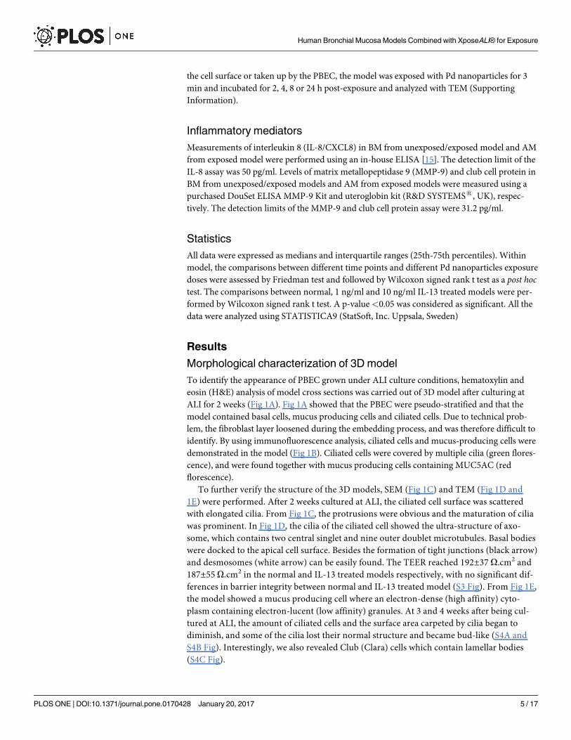
To identify the appearance of PBEC grown under ALI culture conditions, hematoxylin and

eosin (H&E) analysis of model cross sections was carried out of 3D model after culturing at

ALI for 2 weeks (Fig 1A). Fig 1A showed that the PBEC were pseudo-stratified and that the

model contained basal cells, mucus producing cells and ciliated cells. Due to technical prob-

lem, the fibroblast layer loosened during the embedding process, and was therefore difficult to

identify. By using immunofluorescence analysis, ciliated cells and mucus-producing cells were

demonstrated in the model (Fig 1B). Ciliated cells were covered by multiple cilia (green flores-

cence), and were found together with mucus producing cells containing MUC5AC (red

florescence).

To further verify the structure of the 3D models, SEM (Fig 1C) and TEM (Fig 1D and

1E) were performed. After 2 weeks cultured at ALI, the ciliated cell surface was scattered

with elongated cilia. From Fig 1C, the protrusions were obvious and the maturation of cilia

was prominent. In Fig 1D, the cilia of the ciliated cell showed the ultra-structure of axo-

some, which contains two central singlet and nine outer doublet microtubules. Basal bodies

were docked to the apical cell surface. Besides the formation of tight junctions (black arrow)

and desmosomes (white arrow) can be easily found. The TEER reached 192±37 O.cm2 and

187±55 O.cm2 in the normal and IL-13 treated models respectively, with no significant dif-

ferences in barrier integrity between normal and IL-13 treated model (S3 Fig). From Fig 1E,

the model showed a mucus producing cell where an electron-dense (high affinity) cyto-

plasm containing electron-lucent (low affinity) granules. At 3 and 4 weeks after being cul-

tured at ALI, the amount of ciliated cells and the surface area carpeted by cilia began to

diminish, and some of the cilia lost their normal structure and became bud-like (S4A and

S4B Fig). Interestingly, we also revealed Club (Clara) cells which contain lamellar bodies

(S4C Fig).

Human Bronchial Mucosa Models Combined with XposeALI® for Exposure

PLOS ONE | DOI:10.1371/journal.pone.0170428 January 20, 2017 5 / 17

mRNA expression of different cell type markers

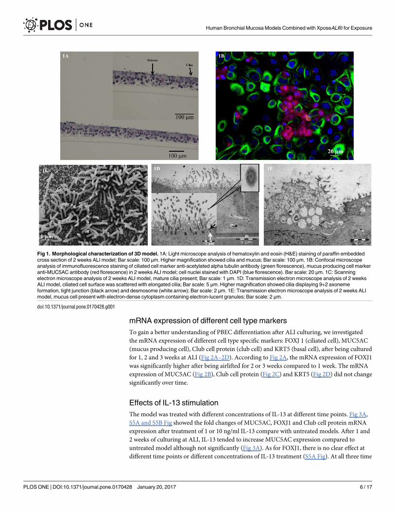
To gain a better understanding of PBEC differentiation after ALI culturing, we investigated

the mRNA expression of different cell type specific markers: FOXJ 1 (ciliated cell), MUC5AC

(mucus producing cell), Club cell protein (club cell) and KRT5 (basal cell), after being cultured

for 1, 2 and 3 weeks at ALI (Fig 2A–2D). According to Fig 2A, the mRNA expression of FOXJ1

was significantly higher after being airlifted for 2 or 3 weeks compared to 1 week. The mRNA

expression of MUC5AC (Fig 2B), Club cell protein (Fig 2C) and KRT5 (Fig 2D) did not change

significantly over time.

Effects of IL-13 stimulation

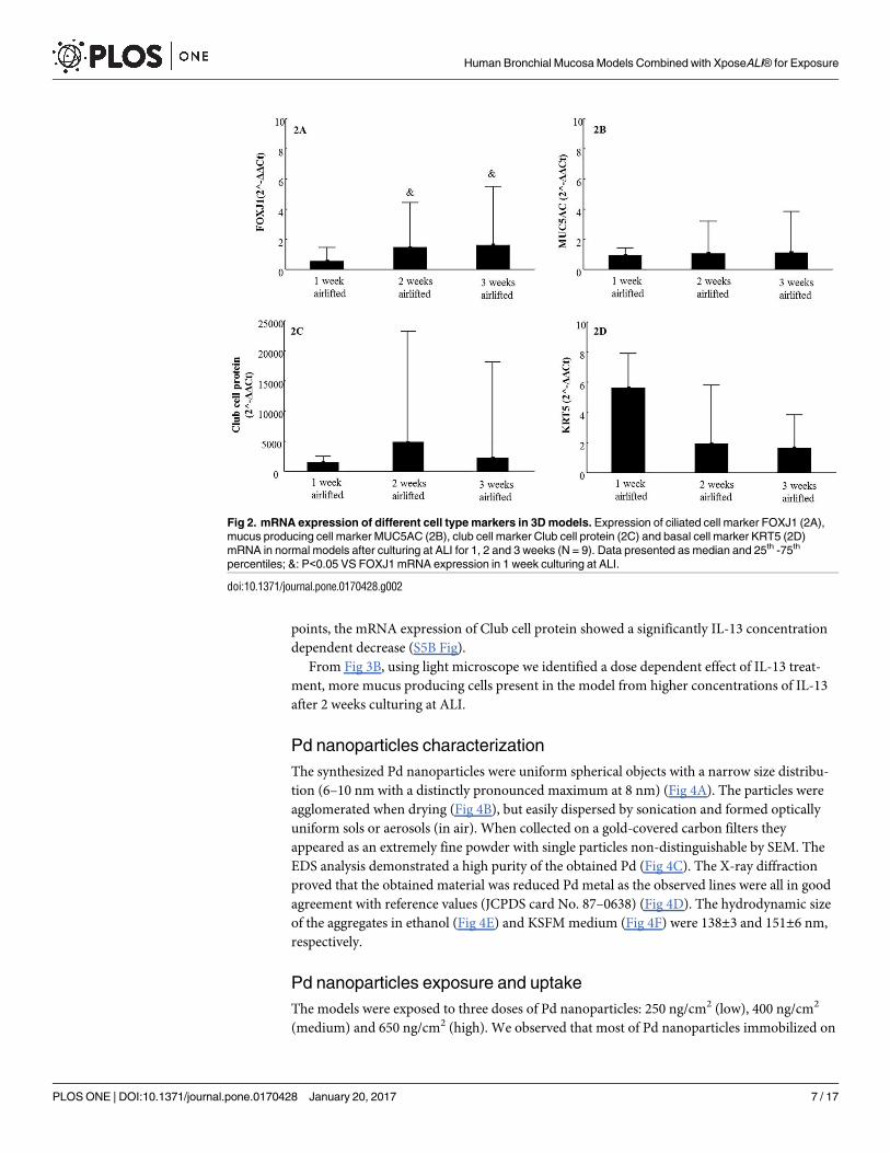
The model was treated with different concentrations of IL-13 at different time points. Fig 3A,

S5A and S5B Fig showed the fold changes of MUC5AC, FOXJ1 and Club cell protein mRNA

expression after treatment of 1 or 10 ng/ml IL-13 compare with untreated models. After 1 and

2 weeks of culturing at ALI, IL-13 tended to increase MUC5AC expression compared to

untreated model although not significantly (Fig 3A). As for FOXJ1, there is no clear effect at

different time points or different concentrations of IL-13 treatment (S5A Fig). At all three time

Fig 1. Morphological characterization of 3D model. 1A: Light microscope analysis of hematoxylin and eosin (H&E) staining of paraffin embedded

cross section of 2 weeks ALI model; Bar scale: 100 μm. Higher magnification showed cilia and mucus; Bar scale: 100 μm. 1B: Confocal microscope

analysis of immunofluorescence staining of ciliated cell marker anti-acetylated alpha tubulin antibody (green florescence), mucus producing cell marker

anti-MUC5AC antibody (red florescence) in 2 weeks ALI model; cell nuclei stained with DAPI (blue florescence). Bar scale: 20 μm. 1C: Scanning

electron microscope analysis of 2 weeks ALI model, mature cilia present; Bar scale: 1 μm. 1D: Transmission electron microscope analysis of 2 weeks

ALI model, ciliated cell surface was scattered with elongated cilia; Bar scale: 5 μm. Higher magnification showed cilia displaying 9+2 axoneme

formation, tight junction (black arrow) and desmosome (white arrow); Bar scale: 2 μm. 1E: Transmission electron microscope analysis of 2 weeks ALI

model, mucus cell present with electron-dense cytoplasm containing electron-lucent granules; Bar scale: 2 μm.

doi:10.1371/journal.pone.0170428.g001

Human Bronchial Mucosa Models Combined with XposeALI® for Exposure

PLOS ONE | DOI:10.1371/journal.pone.0170428 January 20, 2017 6 / 17

points, the mRNA expression of Club cell protein showed a significantly IL-13 concentration

dependent decrease (S5B Fig).

From Fig 3B, using light microscope we identified a dose dependent effect of IL-13 treat-

ment, more mucus producing cells present in the model from higher concentrations of IL-13

after 2 weeks culturing at ALI.

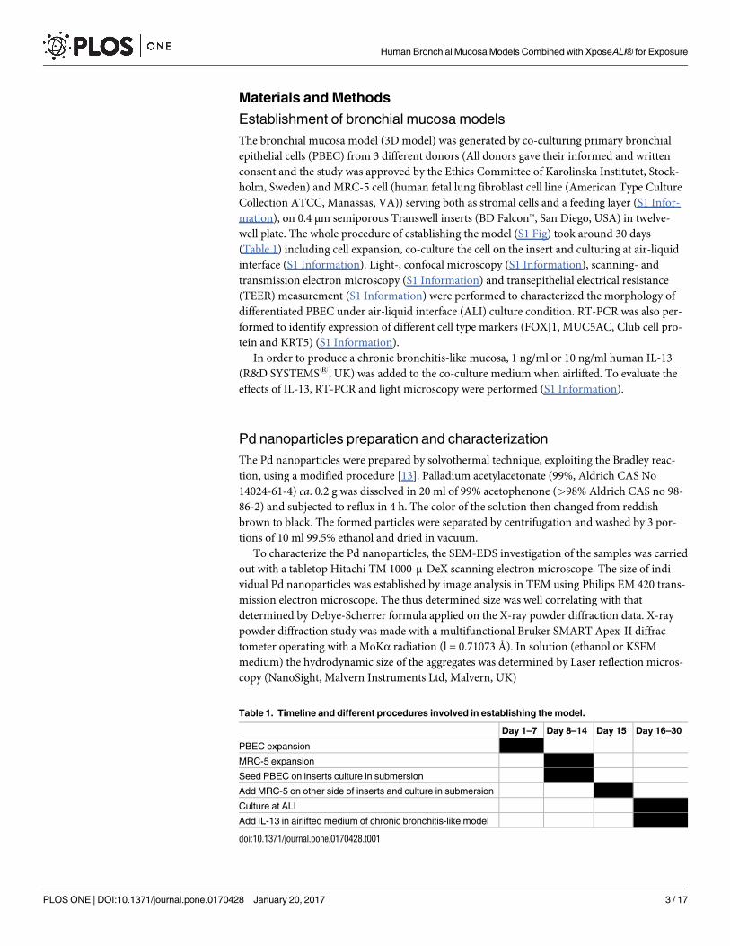
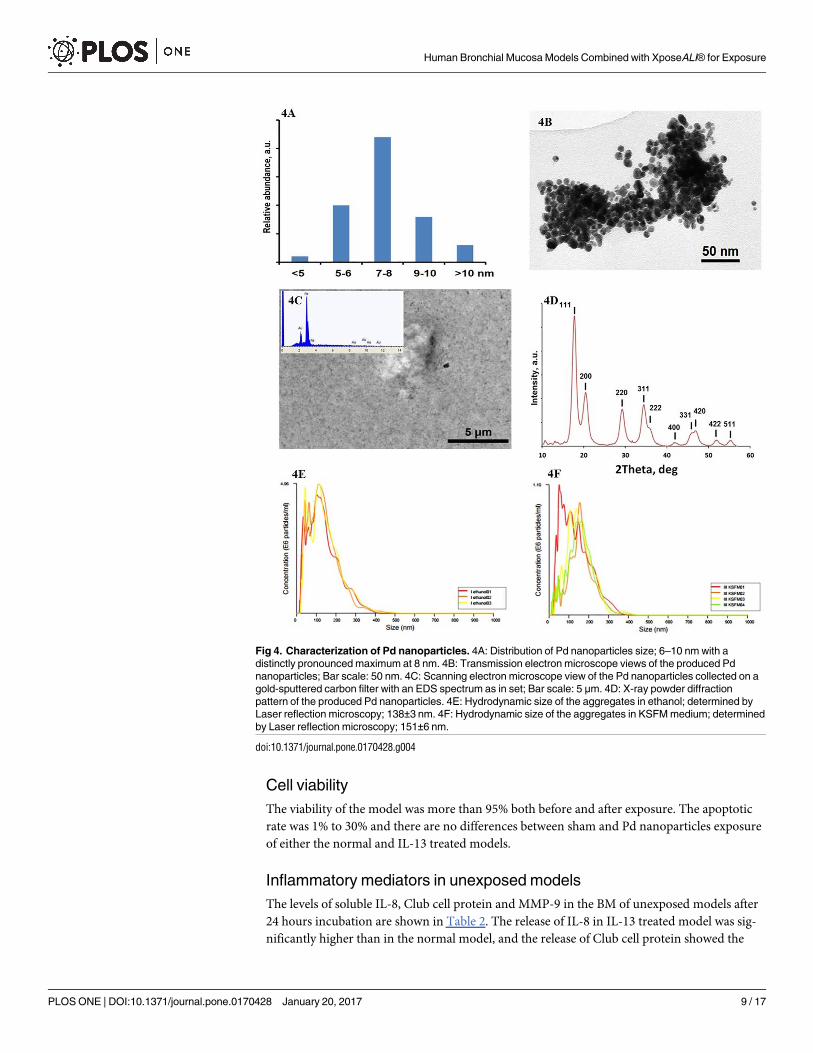
Pd nanoparticles characterization

The synthesized Pd nanoparticles were uniform spherical objects with a narrow size distribu-

tion (6–10 nm with a distinctly pronounced maximum at 8 nm) (Fig 4A). The particles were

agglomerated when drying (Fig 4B), but easily dispersed by sonication and formed optically

uniform sols or aerosols (in air). When collected on a gold-covered carbon filters they

appeared as an extremely fine powder with single particles non-distinguishable by SEM. The

EDS analysis demonstrated a high purity of the obtained Pd (Fig 4C). The X-ray diffraction

proved that the obtained material was reduced Pd metal as the observed lines were all in good

agreement with reference values (JCPDS card No. 87–0638) (Fig 4D). The hydrodynamic size

of the aggregates in ethanol (Fig 4E) and KSFM medium (Fig 4F) were 138±3 and 151±6 nm,

respectively.

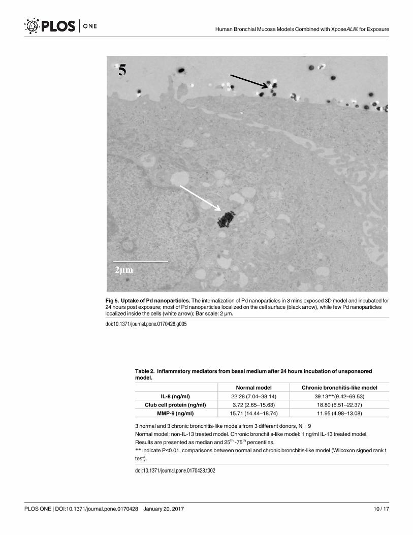
Pd nanoparticles exposure and uptake

The models were exposed to three doses of Pd nanoparticles: 250 ng/cm2 (low), 400 ng/cm2

(medium) and 650 ng/cm2 (high). We observed that most of Pd nanoparticles immobilized on

Fig 2. mRNA expression of different cell type markers in 3D models. Expression of ciliated cell marker FOXJ1 (2A),

mucus producing cell marker MUC5AC (2B), club cell marker Club cell protein (2C) and basal cell marker KRT5 (2D)

mRNA in normal models after culturing at ALI for 1, 2 and 3 weeks (N = 9). Data presented as median and 25th -75th

percentiles; &: P<0.05 VS FOXJ1 mRNA expression in 1 week culturing at ALI.

doi:10.1371/journal.pone.0170428.g002

Human Bronchial Mucosa Models Combined with XposeALI® for Exposure

PLOS ONE | DOI:10.1371/journal.pone.0170428 January 20, 2017 7 / 17

the cell surface while there were just a few Pd nanoparticles found within the cells incubated

for 24 hours after exposure and there were no differences between different time points (Fig

5). According to S6 Fig, after the initial deposition (S6A Fig), Pd nanoparticles were found to

be redistributed and accumulated at one spot (S6B Fig).

Fig 3. Effects of IL-13 stimulation on 3D models. 3A: Fold change of MUC5AC mRNA expression in ALI

models treated without (blank) and with 1 ng/ml (grey) and 10 ng/ml (black) IL-13 for 1, 2 and 3 weeks (N = 9);

Data presented as median and 25th -75th percentiles. 3B: Periodic acid—Schiff (PAS) staining of paraffin

embedded cross section of ALI models treated without and with 1 ng/ml and 10 ng/ml IL-13 for 2 weeks

visualized by light microscope; Bar scale: 100 μm.

doi:10.1371/journal.pone.0170428.g003

Human Bronchial Mucosa Models Combined with XposeALI® for Exposure

PLOS ONE | DOI:10.1371/journal.pone.0170428 January 20, 2017 8 / 17

Cell viability

The viability of the model was more than 95% both before and after exposure. The apoptotic

rate was 1% to 30% and there are no differences between sham and Pd nanoparticles exposure

of either the normal and IL-13 treated models.

Inflammatory mediators in unexposed models

The levels of soluble IL-8, Club cell protein and MMP-9 in the BM of unexposed models after

24 hours incubation are shown in Table 2. The release of IL-8 in IL-13 treated model was sig-

nificantly higher than in the normal model, and the release of Club cell protein showed the

Fig 4. Characterization of Pd nanoparticles. 4A: Distribution of Pd nanoparticles size; 6–10 nm with a

distinctly pronounced maximum at 8 nm. 4B: Transmission electron microscope views of the produced Pd

nanoparticles; Bar scale: 50 nm. 4C: Scanning electron microscope view of the Pd nanoparticles collected on a

gold-sputtered carbon filter with an EDS spectrum as in set; Bar scale: 5 μm. 4D: X-ray powder diffraction

pattern of the produced Pd nanoparticles. 4E: Hydrodynamic size of the aggregates in ethanol; determined by

Laser reflection microscopy; 138±3 nm. 4F: Hydrodynamic size of the aggregates in KSFM medium; determined

by Laser reflection microscopy; 151±6 nm.

doi:10.1371/journal.pone.0170428.g004

Human Bronchial Mucosa Models Combined with XposeALI® for Exposure

PLOS ONE | DOI:10.1371/journal.pone.0170428 January 20, 2017 9 / 17

Fig 5. Uptake of Pd nanoparticles. The internalization of Pd nanoparticles in 3 mins exposed 3D model and incubated for

24 hours post exposure; most of Pd nanoparticles localized on the cell surface (black arrow), while few Pd nanoparticles

localized inside the cells (white arrow); Bar scale: 2 μm.

doi:10.1371/journal.pone.0170428.g005

Table 2. Inflammatory mediators from basal medium after 24 hours incubation of unsponsored

model.

Normal model Chronic bronchitis-like model

IL-8 (ng/ml) 22.28 (7.04–38.14) 39.13**(9.42–69.53)

Club cell protein (ng/ml) 3.72 (2.65–15.63) 18.80 (6.51–22.37)

MMP-9 (ng/ml) 15.71 (14.44–18.74) 11.95 (4.98–13.08)

3 normal and 3 chronic bronchitis-like models from 3 different donors, N = 9

Normal model: non-IL-13 treated model. Chronic bronchitis-like model: 1 ng/ml IL-13 treated model.

Results are presented as median and 25th -75th percentiles.

** indicate P<0.01, comparisons between normal and chronic bronchitis-like model (Wilcoxon signed rank t

test).

doi:10.1371/journal.pone.0170428.t002

Human Bronchial Mucosa Models Combined with XposeALI® for Exposure

PLOS ONE | DOI:10.1371/journal.pone.0170428 January 20, 2017 10 / 17

same trend, although not significant. The MMP-9 levels in both models were in the same

range in both types of models.

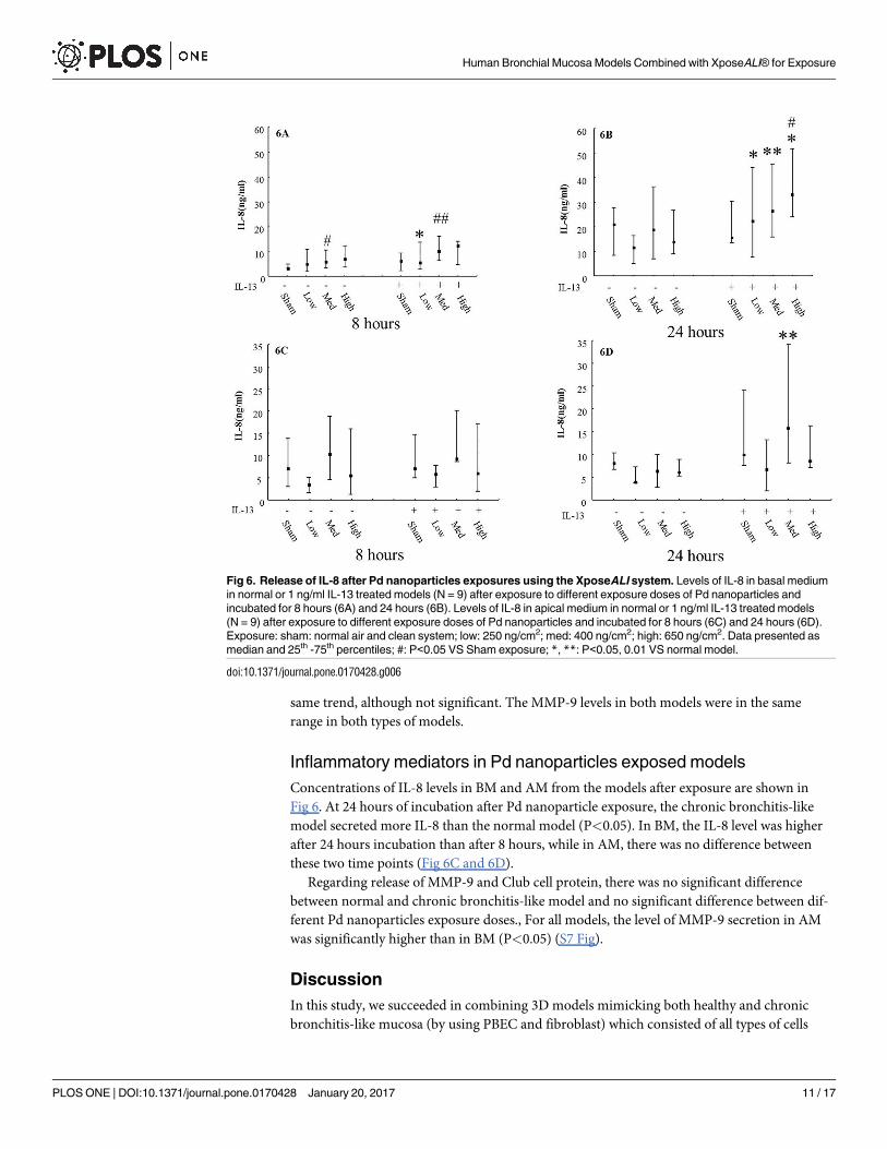
Inflammatory mediators in Pd nanoparticles exposed models

Concentrations of IL-8 levels in BM and AM from the models after exposure are shown in

Fig 6. At 24 hours of incubation after Pd nanoparticle exposure, the chronic bronchitis-like

model secreted more IL-8 than the normal model (P<0.05). In BM, the IL-8 level was higher

after 24 hours incubation than after 8 hours, while in AM, there was no difference between

these two time points (Fig 6C and 6D).

Regarding release of MMP-9 and Club cell protein, there was no significant difference

between normal and chronic bronchitis-like model and no significant difference between dif-

ferent Pd nanoparticles exposure doses., For all models, the level of MMP-9 secretion in AM

was significantly higher than in BM (P<0.05) (S7 Fig).

Discussion

In this study, we succeeded in combining 3D models mimicking both healthy and chronic

bronchitis-like mucosa (by using PBEC and fibroblast) which consisted of all types of cells

Fig 6. Release of IL-8 after Pd nanoparticles exposures using the XposeALI system. Levels of IL-8 in basal medium

in normal or 1 ng/ml IL-13 treated models (N = 9) after exposure to different exposure doses of Pd nanoparticles and

incubated for 8 hours (6A) and 24 hours (6B). Levels of IL-8 in apical medium in normal or 1 ng/ml IL-13 treated models

(N = 9) after exposure to different exposure doses of Pd nanoparticles and incubated for 8 hours (6C) and 24 hours (6D).

Exposure: sham: normal air and clean system; low: 250 ng/cm2; med: 400 ng/cm2; high: 650 ng/cm2. Data presented as

median and 25th -75th percentiles; #: P<0.05 VS Sham exposure; *, **: P<0.05, 0.01 VS normal model.

doi:10.1371/journal.pone.0170428.g006

Human Bronchial Mucosa Models Combined with XposeALI® for Exposure

PLOS ONE | DOI:10.1371/journal.pone.0170428 January 20, 2017 11 / 17

presentin vivo with controlled aerosol exposure module (XposeALI). In order to evaluate this

combined system, exposure to Pd nanoparticles induced an increased secretion of IL-8, where

chronic bronchitis-like model released significantly more IL-8 than normal models.

In vivo, human bronchial epithelium consists of 50–70% ciliated cells, up to 30% basal cells,

up to 25% goblet cells, and 11% Club (Clara) cells [16]. When culturing at ALI the epithelial

cells differentiated and we have identified all cell types listed above by light/confocal micro-

scope, SEM, TEM, mRNA expression, and protein secretion. The FOXJ1expression increased

in our models after 2 and 3 weeks of culturing at air-liquid interface compared with week 1,

indicating that the number of ciliated cell increased over time.

According to previous studies, in vitro stimulation with IL-13 induces mucus producing

cell hyperplasia which is a characteristic feature of chronic bronchitis [17]. The effects of differ-

ent concentrations of IL-13 in human cell culture model varied [10, 18]. Based on PAS stained

cross sections, we identified that at 2 weeks culturing at ALI, the model treated with 1 ng/ml

IL-13 have more mucus producing cells than untreated model. However, after 2 weeks cultur-

ing at ALI, in the models treated with 10 ng/ml IL-13 almost all cells were mucus producing

cells as identified by PAS stained cross sections, while the models treated with 1 ng/ml IL-13

showed different cell types, although more mucus producing cells compared with untreated

models. Moreover, MUC5AC mRNA expression was slightly increased after IL-13 treatment

for 2 weeks at ALI conditions. These findings indicate that the model which mimics chronic

bronchitis-like mucosa was created after stimulation with 1ng/ml IL-13 and cultured at ALI

for 2 weeks.

In the present study, we succeeded in combining the 3D models with controlled aerosol

exposures using the XposeALI exposure module and thereby managed to deposit nanoparticles

on a well differentiated cellular layer cultured at ALI. There are some commercially available

cell exposure systems, such as ALICE, Vitrocell and Cultex1 RFS [19–21]. It was an important

breakthrough when in vitro inhalation research was able to expose ALI cells to aerosols in a

manner resembling in vivo exposures with aerosols and particles deposited directly onto lung

cells. In this study, the unique performance of the XposeALI exposure module was linked to its

close integration with the PreciseInhale aerosol generator and dosing system [11, 12]. The

aerosolization in the PreciseInhale system was driven by compressed air with pressures up to

160 bar generating well dispersed particles through deagglomeration of loaded powder por-

tions. Also, nanoparticle powders with high cohesive energies were generally well dispersed by

the PreciseInhale system. Therefore, the amounts of powdered substance required were mini-

mal. A key feature of the combined exposure system was the dosing technics including the

Casella light dispersion that automatically controls the dose drawn from the generator to the

cell surface and steered by a computer at each experiment. Another advantage of the combined

exposure system was that XposeALI generated more concentrated aerosols which gave shorter

exposure times. Therefore, it minimized the time for cells being out of the incubator, which is

an important survival factor for human primary cells. In addition, sufficient deposition of

aerosol in the 3D models can be achieved even without using more complex methods such as

electrostatic deposition. In the combined 3D cell models cultured in inserts and XposeALIexposure system, the exposure hoods restrict the area available for particle exposure to only

the PBEC surface (not the insert walls) and the particles can be evenly distributed over the

whole PBEC surface. By using a reverse flow pattern over the PBEC surface from the periphery

to one central outlet, excess aerosol can be collected in filters located on top of the exposure

hoods, which reduce the risk of an unhealthy laboratory environment.

For both healthy subjects, and subjects with respiratory diseases, our chosen test substrates

of highly dispersed Pd nanoparticles present in polluted ambient air are of clinical importance.

Pd nanoparticle is a traffic-related particulate pollution, originating from the emission of

Human Bronchial Mucosa Models Combined with XposeALI® for Exposure

PLOS ONE | DOI:10.1371/journal.pone.0170428 January 20, 2017 12 / 17

particles constituting the active component of the catalytic converters in road vehicles [22].

The complex particles leaving the catalytic converters are about 10 nm in size resulting in a rel-

ative stable aerosol that usually deposit in the deep lung such as the alveolar region, but the

deposition will also occur higher up in the airway tree particularly at the bifurcations [23]. The

synthesized Pd nanoparticles in this study were uniform about 6–10nm in size, which could

represent in vivo situation.

After the exposure, levels of IL-8 from both AM and BM were detected. IL-8 is an important

chemoattractant and activator which is produced by airway epithelial cells. Increased level of

IL-8 has been identified in different compartments in smokers with and without COPD [24].

Moreover, IL-8 secretion has been well studied as an inflammatory biomarker in response to

particle exposures [25]. In this paper, we found in the BM of Pd nanoparticles exposed models,

the levels of IL-8 were significantly higher in chronic bronchitis-like model than in normal

model after 24 hours of incubation and showed the same tendency after 8 hours of incubation.

Also in unexposed models, IL-13 treated models secreted significantly more IL-8 than non-13

treated model. These results may indicate that the chronic bronchitis-like model is more sensi-

tive to Pd nanoparticles exposure than the normal model. Besides, individuals with COPD have

been shown to have an increased sensitivity to particulate matter in air pollution [26] which

confirms that our combined exposure system in vitro can emulate the real situation in vivo.

In our previous study, we demonstrated that submerged PBEC cultures exposed to medium

dispersed Pd nanoparticles secretion of IL-8 tend to increase at higher Pd nanoparticle con-

centration [13]. In line with this finding, we showed that Pd nanoparticle exposures dose-

dependently increased the release of IL-8 in BM of both the normal and chronic bronchitis-

like models. These results may demonstrate that exposure to Pd nanoparticles can cause

inflammatory response in both normal and chronic bronchitis-like models after 8 hours, and

the effects of Pd nanoparticles exposure were increased with the dose. After 24 hours incuba-

tion, the influence of Pd nanoparticles exposure on IL-8 level disappeared in the normal

model, but still existed in the chronic bronchitis-like model. This may occur because of the

normal model being able to self-regulate and recover from inflammation caused by Pd nano-

particles exposures after 24 hours. However, for the chronic bronchitis-like model, the recov-

ery process may not be as efficient as in the normal model and may need longer time.

MMP-9 is a type IV collagenase that is related to remodeling [24] and will not be produced

by resident lung cells until lesions occur to the bronchial surface in form of mechanical trauma

or exposure to toxic substances or inflammatory mediators. Metaplastic epithelial cells pro-

duce the highest amounts of MMP-9, whereas production by fibroblasts is minimal [26]. We

found that levels of MMP-9 in AM were much higher than in BM. This difference was

observed despite the fact that the AM was obtained by a 15 min lavage periods of the 3D mod-

els’ apical surface at 8 or 24 hours after exposure, while the samples of BM contained the

cumulative secretion of MMP-9 for 8 or 24 hours. These results suggest that Pd nanoparticles

caused epithelial MMP-9 secretion as response to damage that requires extracellular matrix

(ECM) remodeling, probably for quick re-epithelialization [27]. These results were also in con-

trast to IL-8 levels, as there were no differences in IL-8 levels between BM and AM. This might

be due to size differences as the molecular weight of IL-8 (8.4 kDa) is much smaller than of

MMP-9 (92 kDa). Therefore, IL-8 could more easily penetrate through confluent cell layer and

the insert membrane. Besides, from the basal fibroblasts the contribution to the basal medium

were probably higher from IL-8 compared to MMP-9 [28]. A third explanation for this could

also be that there were differences in the direction of secretion between IL-8 and MMP-9.

Analysis of the models with TEM showed very little uptake of the particles by the PBEC.

This was in marked contrast to our previous study using the same cells and particles, but with

cells cultured under submerged condition and particles dispersed in cell culture medium [13].

Human Bronchial Mucosa Models Combined with XposeALI® for Exposure

PLOS ONE | DOI:10.1371/journal.pone.0170428 January 20, 2017 13 / 17

When the particles were added to the medium, an uptake was observed already within 1 hour

[13]. The lack of uptake in our 3D model cultured under ALI conditions could be explained by

the presence of mucociliary clearance. After the initial deposition, particles were found to be

redistributed and accumulated at one spot which probably was caused by the beating of cilia.

The lack of a protective mucus layer in the submerged culture could also explain the difference

in uptake of Pd nanoparticles by PBEC.

Conclusion

In present study, we succeeded in combining relevant human airway wall models with con-

trolled aerosol exposures using the XposeALI exposure module. In addition, the modification

of the 3D model leading to mucus producing cell metaplasia and hyperplasia, which are phe-

nomena observed in airway diseases such as COPD and chronic bronchitis, are suited for

mimicking interactions between nanoparticle exposures and the innate immune response in

chronic bronchitis patients. For both healthy subjects, and subjects with respiratory diseases,

our chosen test substrate, Pd nanoparticles present in polluted ambient air, is of clinical impor-

tance. With further development and validation, this exposure setting could form part of an invitro testing strategy to reduce the requirement for animal inhalation studies.

Supporting Information

S1 Fig. A diagrammatic representation of the main steps in establishing the model. S1A:

Apical seeding of PBEC into a 0.4 μm semiporous transwell insert; S1B: Basolateral seeding of

fibroblast; S1C: Removed medium and only added airlifted medium in the basolateral cham-

ber; S1D: Differentiation when culturing at ALI. All steps were performed under sterile condi-

tions and cells were cultured at 37˚C and 5% CO2.

(TIF)

S2 Fig. The XposeALI exposure system. The XposeALI cell exposure module (S2A) and a

close up of the transwell insert with cells (down), the exposure hood (middle) and the exposure

filter holder (up) (S2B).

(TIF)

S3 Fig. Transepithelial electrical resistance (TEER) of 3D model. Transepithelial electrical

resistance (TEER) of normal (blank) and 1ng/ml IL-13 treated (black) models. Data present as

median and 25th -75th percentiles.

(TIF)

S4 Fig. Morphological characterization of 3D model. S4A &S4B: Scanning electron micro-

scope analysis of 3 and 4 weeks ALI model, cilia begin to lost their normal structure and

became bud-like; Bar scale: 1 μm. S4C: Transmission electron microscope analysis of 2 weeks

ALI model, club cell which contain lamellar bodies was present; Bar scale: 2 μm. Higher mag-

nification showed concentric/parallel lamellae, Bar scale: 1 μm.

(TIF)

S5 Fig. Effects of IL-13 stimulation on 3D models. S5A & S5B: Fold change of FOXJ1 and

club cell protein mRNA expression in ALI models treated without (blank) and with 1 ng/ml

(grey) and 10 ng/ml (black) IL-13 for 1, 2 and 3 weeks (N = 9); Date present as median and

25th -75th percentiles. ‡,‡‡: P<0.05, 0.01 VS Club cell protein mRNA expression in non-IL-13

treated models. tm¤: P<0.01 VS Club cell protein mRNA expression in 1ng/ml IL-13 treated

models.

(TIF)

Human Bronchial Mucosa Models Combined with XposeALI® for Exposure

PLOS ONE | DOI:10.1371/journal.pone.0170428 January 20, 2017 14 / 17

S6 Fig. Illustration of mucociliary clearance. S6A: The models were exposed to high dose

(650 ng/cm2) of Pd nanoparticles and even distribution was observed. S6B: The Pd nanoparti-

cle redistributed and accumulated at one spot within 24 hours, indicating existing mucociliar

clearance.

(TIF)

S7 Fig. Release of MMP-9 after Pd nanoparticles exposures using the XposeALI system.

Levels of MMP-9 in basal medium in normal or 1 ng/ml IL-13 treated models (N = 9) after

exposure to different deposited doses of Pd nanoparticles and incubated for 8 hours (S7A) and

24 hours (S7B). Levels of MMP-9 in apical medium in normal or 1 ng/ml IL-13 treated models

(N = 9) after exposure to different concentrations of Pd nanoparticles and incubated for 8

hours (S7C) and 24 hours (S7D). Exposure: sham: normal air and clean system; low: 250 ng/

cm2; med: 400 ng/cm2; high: 650 ng/cm2. Data presented as median and 25th -75th percentiles;

##: P<0.01 VS Sham exposure; �: P<0.05 VS normal model.

(TIF)

S1 Information. Supporting text.

(DOCX)

Acknowledgments

The authors would like to thank Dr Mattias Svensson, Dr Maciej Kupczyk, Dr Anh Thu

Nguyen Hoang, Dr Kjell Hultenby, Katja Jansen and Xiaomiao Xiong for valuable technical

assistance.

Author Contributions

Conceptualization: JJ AH MM VK GS PG LP.

Data curation: JJ AH MM VK GS PG LP.

Formal analysis: JJ AH MM VK GS PG LP.

Funding acquisition: LP.

Investigation: JJ AH MM VK GS PG LP.

Methodology: JJ AH MM VK GS PG LP.

Project administration: LP.

Resources: VK GS PG LP.

Supervision: LP.

Validation: JJ AH MM VK GS PG LP.

Visualization: JJ AH MM VK GS PG LP.

Writing – original draft: JJ AH MM VK GS PG LP.

Writing – review & editing: JJ AH MM VK GS PG LP.

References1. Bitterle E, Karg E, Schroeppel A, Kreyling WG, Tippe A, Ferron GA, et al. Dose-controlled exposure of

A549 epithelial cells at the air-liquid interface to airborne ultrafine carbonaceous particles.

Human Bronchial Mucosa Models Combined with XposeALI® for Exposure

PLOS ONE | DOI:10.1371/journal.pone.0170428 January 20, 2017 15 / 17

Chemosphere. 2006; 65(10):1784–90. Epub 2006/06/10. doi: 10.1016/j.chemosphere.2006.04.035

PMID: 16762398

2. Scheffler S, Dieken H, Krischenowski O, Forster C, Branscheid D, Aufderheide M. Evaluation of E-ciga-

rette liquid vapor and mainstream cigarette smoke after direct exposure of primary human bronchial epi-

thelial cells. Int J Environ Res Public Health. 2015; 12(4):3915–25. Epub 2015/04/10. doi: 10.3390/

ijerph120403915 PMID: 25856554

3. Mathis C, Poussin C, Weisensee D, Gebel S, Hengstermann A, Sewer A, et al. Human bronchial epithe-

lial cells exposed in vitro to cigarette smoke at the air-liquid interface resemble bronchial epithelium

from human smokers. Am J Physiol Lung Cell Mol Physiol. 2013; 304(7):L489–503. Epub 2013/01/29.

doi: 10.1152/ajplung.00181.2012 PMID: 23355383

4. Eisner MD, Anthonisen N, Coultas D, Kuenzli N, Perez-Padilla R, Postma D, et al. An official American

Thoracic Society public policy statement: Novel risk factors and the global burden of chronic obstructive

pulmonary disease. Am J Respir Crit Care Med. 2010; 182(5):693–718. doi: 10.1164/rccm.200811-

1757ST PMID: 20802169

5. Brook RD, Rajagopalan S, Pope CA 3rd, Brook JR, Bhatnagar A, Diez-Roux AV, et al. Particulate mat-

ter air pollution and cardiovascular disease: An update to the scientific statement from the American

Heart Association. Circulation. 2010; 121(21):2331–78. doi: 10.1161/CIR.0b013e3181dbece1 PMID:

20458016

6. Buist AS, McBurnie MA, Vollmer WM, Gillespie S, Burney P, Mannino DM, et al. International variation

in the prevalence of COPD (the BOLD Study): a population-based prevalence study. Lancet. 2007; 370

(9589):741–50. Epub 2007/09/04. doi: 10.1016/S0140-6736(07)61377-4 PMID: 17765523

7. Murray CJ, Lopez AD. Alternative projections of mortality and disability by cause 1990–2020: Global

Burden of Disease Study. Lancet. 1997; 349(9064):1498–504. Epub 1997/05/24. doi: 10.1016/S0140-

6736(96)07492-2 PMID: 9167458

8. Hurst JR. Exacerbation phenotyping in chronic obstructive pulmonary disease. Am J Respir Crit Care

Med. 2011; 184(6):625–6. Epub 2011/09/17. doi: 10.1164/rccm.201106-1136ED PMID: 21920921

9. Atkinson RW, Anderson HR, Sunyer J, Ayres J, Baccini M, Vonk JM, et al. Acute effects of particulate

air pollution on respiratory admissions: results from APHEA 2 project. Air Pollution and Health: a Euro-

pean Approach. Am J Respir Crit Care Med. 2001; 164(10 Pt 1):1860–6. doi: 10.1164/ajrccm.164.10.

2010138 PMID: 11734437

10. Atherton HC, Jones G, Danahay H. IL-13-induced changes in the goblet cell density of human bronchial

epithelial cell cultures: MAP kinase and phosphatidylinositol 3-kinase regulation. Am J Physiol Lung

Cell Mol Physiol. 2003; 285(3):L730–9. doi: 10.1152/ajplung.00089.2003 PMID: 12794003

11. Gerde P, Ewing P, Lastbom L, Ryrfeldt A, Waher J, Liden G. A novel method to aerosolize powder for

short inhalation exposures at high concentrations: isolated rat lungs exposed to respirable diesel soot.

Inhal Toxicol. 2004; 16(1):45–52. Epub 2004/01/28. doi: 10.1080/08958370490258381 PMID:

14744664

12. Selg E, Ewing P, Acevedo F, Sjoberg CO, Ryrfeldt A, Gerde P. Dry Powder Inhalation Exposures of the

Endotracheally Intubated Rat Lung, Ex Vivo and In Vivo: The Pulmonary Pharmacokinetics of Flutica-

sone Furoate. J Aerosol Med Pulm D. 2013; 26(4):181–9.

13. Wilkinson KE, Palmberg L, Witasp E, Kupczyk M, Feliu N, Gerde P, et al. Solution-engineered palla-

dium nanoparticles: model for health effect studies of automotive particulate pollution. ACS Nano. 2011;

5(7):5312–24. doi: 10.1021/nn1032664 PMID: 21650217

14. Kanitsar K, Koellensperger G, Hann S, Limbeck A, Puxbaum H, Stingeder G. Determination of Pt, Pd

and Rh by inductively coupled plasma sector field mass spectrometry (ICP-SFMS) in size-classified

urban aerosol samples. J Anal Atom Spectrom. 2003; 18(3):239–46.

15. Larsson K, Tornling G, Gavhed D, Muller-Suur C, Palmberg L. Inhalation of cold air increases the num-

ber of inflammatory cells in the lungs in healthy subjects. Eur Respir J. 1998; 12(4):825–30. Epub 1998/

11/17. PMID: 9817153

16. Schamberger AC, Staab-Weijnitz CA, Mise-Racek N, Eickelberg O. Cigarette smoke alters primary

human bronchial epithelial cell differentiation at the air-liquid interface. Sci Rep-Uk. 2015;5.

17. Zhen G, Park SW, Nguyenvu LT, Rodriguez MW, Barbeau R, Paquet AC, et al. IL-13 and epidermal

growth factor receptor have critical but distinct roles in epithelial cell mucin production. Am J Respir Cell

Mol Biol. 2007; 36(2):244–53. Epub 2006/09/19. doi: 10.1165/rcmb.2006-0180OC PMID: 16980555

18. Yoshisue H, Hasegawa K. Effect of MMP/ADAM inhibitors on goblet cell hyperplasia in cultured human

bronchial epithelial cells. Biosci Biotechnol Biochem. 2004; 68(10):2024–31. Epub 2004/10/27. PMID:

15502346

19. Aufderheide M, Halter B, Mohle N, Hochrainer D. The CULTEX RFS: a comprehensive technical

approach for the in vitro exposure of airway epithelial cells to the particulate matter at the air-liquid

Human Bronchial Mucosa Models Combined with XposeALI® for Exposure

PLOS ONE | DOI:10.1371/journal.pone.0170428 January 20, 2017 16 / 17

interface. Biomed Res Int. 2013; 2013:734137. Epub 2013/03/20. doi: 10.1155/2013/734137 PMID:

23509768

20. Aufderheide M, Mohr U. A modified CULTEX system for the direct exposure of bacteria to inhalable sub-

stances. Exp Toxicol Pathol. 2004; 55(6):451–4. Epub 2004/09/24. doi: 10.1078/0940-2993-00348

PMID: 15384250

21. Lenz AG, Karg E, Lentner B, Dittrich V, Brandenberger C, Rothen-Rutishauser B, et al. A dose-con-

trolled system for air-liquid interface cell exposure and application to zinc oxide nanoparticles. Part

Fibre Toxicol. 2009; 6:32. Epub 2009/12/18. doi: 10.1186/1743-8977-6-32 PMID: 20015351

22. Rauch S, Hemond HF, Barbante C, Owari M, Morrison GM, Peucker-Ehrenbrink B, et al. Importance of

automobile exhaust catalyst emissions for the deposition of platinum, palladium, and rhodium in the

northern hemisphere. Environ Sci Technol. 2005; 39(21):8156–62. Epub 2005/11/22. PMID: 16294849

23. Geiser M, Kreyling WG. Deposition and biokinetics of inhaled nanoparticles. Part Fibre Toxicol. 2010;

7:2. Epub 2010/03/09. doi: 10.1186/1743-8977-7-2 PMID: 20205860

24. Ji J, von Scheele I, Bergstrom J, Billing B, Dahlen B, Lantz AS, et al. Compartment differences of inflam-

matory activity in chronic obstructive pulmonary disease. Respir Res. 2014; 15:104. Epub 2014/08/27.

doi: 10.1186/s12931-014-0104-3 PMID: 25155252

25. Monteiller C, Tran L, MacNee W, Faux S, Jones A, Miller B, et al. The pro-inflammatory effects of low-

toxicity low-solubility particles, nanoparticles and fine particles, on epithelial cells in vitro: the role of sur-

face area. Occup Environ Med. 2007; 64(9):609–15. Epub 2007/04/06. doi: 10.1136/oem.2005.024802

PMID: 17409182

26. Sint T, Donohue JF, Ghio AJ. Ambient air pollution particles and the acute exacerbation of chronic

obstructive pulmonary disease. Inhal Toxicol. 2008; 20(1):25–9. Epub 2008/02/01. doi: 10.1080/

08958370701758759 PMID: 18236218

27. Atkinson JJ, Senior RM. Matrix metalloproteinase-9 in lung remodeling. Am J Respir Cell Mol Biol.

2003; 28(1):12–24. doi: 10.1165/rcmb.2002-0166TR PMID: 12495928

28. Stan MS, Sima C, Cinteza LO, Dinischiotu A. Silicon-based quantum dots induce inflammation in

human lung cells and disrupt extracellular matrix homeostasis. FEBS J. 2015; 282(15):2914–29. doi:

10.1111/febs.13330 PMID: 26032556

Human Bronchial Mucosa Models Combined with XposeALI® for Exposure

PLOS ONE | DOI:10.1371/journal.pone.0170428 January 20, 2017 17 / 17


Recommended