journa l homepage: www.e lsev ier .com/ locate /carbpol
evelopment of mucoadhesive thiolated chitosan nanoparticles foriomedical applications
. Anithaa, N. Deepaa, K.P. Chennazhia, S.V. Naira, H. Tamurab, R. Jayakumara,∗
Amrita Centre for Nanoscience and Molecular Medicine Amrita Institute of Medical Sciences and Research Centre, Amrita Vishwa Vidyapeetham University, Kochi 682041, IndiaFaculty of Chemistry, Materials and Bioengineering, Kansai University, Osaka 564-8680, Japan
r t i c l e i n f o
rticle history:eceived 16 June 2010eceived in revised form 9 July 2010ccepted 9 July 2010
a b s t r a c t
The main objective of this work was to develop nanoparticles (NPs) of a mucoadhesive polymer based onchitosan for biomedical applications. Here, we developed thiolated chitosan (TCS) using thioglycolic acid(TGA) and chitosan in the presence of 1-ethyl-3-3(3-dimethylaminopropyl) carbodiimide hydrochloride(EDC) as catalyst. The prepared TCS was characterized using FT-IR and the degree of thiol substitution was
found out by Ellman’s method. The TCS nanoparticles (TCS-NPs) were developed using ionic cross-linkingreaction with pentasodium tripolyphosphate (TPP). The prepared TCS-NPs were characterized by DLS,AFM, FT-IR, TG/DTA, etc. In vitro cytocompatibility and cell uptake studies were also carried out. Thesestudies suggest that the prepared NPs show less toxicity towards normal and cancer cells and they areeasily taken up by both the normal and cancer cells. So the prepared TCS-NPs could be used for drug and
s.
ytocompatibilityrug delivery
gene delivery application
. Introduction
Development of bioadhesive/mucoadhesive controlled releaseystems has been the subject of many studies in recent years.t is widely accepted that limiting the clearance by increas-ng the viscosity of a drug formulation and also prolonging theontact time through mucoadhesive interactions will result inn increased bioavailability of the drug or whatever therapeu-ics (Margit, Constantia, & Bernkop-Schnürch, 2003). Polymericnhancers/mucoadhesive polymers would be a promising candi-ate for these kinds of applications. They can act as permeationnhancers and thereby they find application in the oral delivery ofrotein drugs (Lichen, Jieying, Chunbai, Liming, & Cui, 2009). Theucoadhesion, or the attachment of a natural or synthetic polymer
o a biological substrate is a practical method of drug immobi-ization or localization and it is an important aspect of controlledrug delivery (Wu et al., 2009). While the subject of mucoadhe-ion is not new, there has been increased interest in recent yearsn using mucoadhesive polymers for drug delivery (Ranabir, Lila,
Sontosh, 2009). Substantial effort has recently been focused onlacing a drug or a formulation in a particular region of the body forxtended periods of time. This is needed not only for targeting ofrugs but also for better control of systemic drug delivery (Ranabir
et al., 2009) and the drugs that are absorbed through the mucosallining of tissues can enter directly into the blood stream and not beinactivated by enzymatic degradation in the gastrointestinal tract(Ranabir et al., 2009). In earlier studies, various natural and syn-thetic polymers were explored as mucoadhesive excipients (Wu etal., 2009). The mucoadhesive properties of these polymers or themechanism of mucoadhesion can be explained on the basis of for-mation of non-covalent bonds such as hydrogen bonds and ionicinteractions between the mucoadhesive polymer and the mucuslayer (Wu et al., 2009). Several polymeric bioadhesive drug deliverysystems have been fabricated and studied in the past. Acrylic-basedhydrogels is an example of bioadhesive synthetic polymers whichare used for these kinds of applications (Ranabir et al., 2009).
Chitin is a biopolymer, used for various biomedical applica-tions (Jayakumar, Nair, & Tamura, 2009). Chitosan, a biopolymerobtained by the alkaline deacetylation of chitin, is used in thecurrent work. This is a natural polysaccharide of �-(1,4)-linked2-amino-2-deoxy-d-glucopyranose. Because of its biocompatibil-ity and biodegradation properties (Bernkop-Schnürch, Schwarz, &Steininger, 1999; Jayakumar, Chennazhi, et al., 2010; Jayakumar,Deepthy, Manzoor, Nair, & Tamura, 2010; Jayakumar, Nwe, Tokura,& Tamura, 2007; Jayakumar, Prabaharan, Nair, & Tamura, 2010;Jayakumar, Prabaharan, Nair, Tokura, et al., 2010; Jayakumar,
Reis, & Mano, 2007), it has been used in a variety of biomedi-cal applications. A number of clinical studies using chitosan havebeen reported including its use as cell scaffolds in tissue engi-neering, nerve regeneration tubes, cartilage regeneration (Freier,Koh, Kazazian, & Shoichet, 2005; Khor and Lim, 2003; VandeVord
A. Anitha et al. / Carbohydrate Polymers 83 (2011) 66–73 67
nthesi
eaJ2PJMeiaSwSipPa(t(
Lsaai(hsipwB(aepctfe2P
psatpl
Fig. 1. Reaction scheme for the sy
t al., 2002) and also in drug delivery applications (Anitha etl., 2009; Dev, Binulal, et al., 2010; Dev, Jithin, et al., 2010;ayakumar, Chennazhi, et al., 2010; Jayakumar, Deepthy, et al.,010; Jayakumar, Prabaharan, Nair, & Tamura, 2010; Jayakumar,rabaharan, Nair, Tokura, et al., 2010; Jayakumar, Nwe, et al., 2007;ayakumar, Prabaharan, Reis, & Mano, 2005; Jayakumar, Reis, &
ano, 2006; Mathew et al., 2010). Furthermore, it has been usedxtensively as a biomaterial, owing to its immunostimulatory activ-ties (Mwale et al., 2005), anticoagulant properties, antimicrobialnd antifungal action (Gorbach et al., 1994; Rabea, Badawy, Stevens,magghe, & Steubaut, 2003) and for its action as a promoter ofound healing in the field of surgery (Madhumathi et al., 2010;
udheesh Kumar et al., 2010; Wang, Du, Fan, Liu, & Hu, 2005) andn a number of pharmaceutical preparations, primarily for the pur-ose of controlled drug delivery (Felt, Buri, & Gurny, 1998; Khan,eh, & Chng, 2000) such as, mucosal (Freier et al., 2005; Gorbach etl., 1994; Mwale et al., 2005), buccal (Rabea et al., 2003), and ocularWang et al., 2005) delivery of drugs. NPs based on chitin and chi-osan are developed and used for different biomedical applicationsAnitha et al., 2009; Jayakumar, Prabaharan, Nair, & Tamura, 2010).
TCS was investigated in this study for biomedical applications.iteratures show that the mucoadhesive properties of TCS aretrongly improved in comparison to unmodified chitosan (Khan etl., 2000). When compared with other modified chitosan materi-ls, TCS has numerous advantageous features such as significantlymproved mucoadhesive and permeation enhancing propertiesKast & Bernkop-Schnürch, 2001). And these polymers possessydrophilic properties due to the presence of thiol groups on theiride chains (Ronny, Brigitta, Adolf, & Andreas Bernkop, 2008), its relatively safe and non-toxic (Ronny et al., 2008), its cationicolyelectrolyte character provides strong electrostatic interactionith negatively charged molecules like DNA or drug (Kast andernkop-Schnürch, 2001), it displays enzyme inhibitory propertiesRonny et al., 2008). The mucoadhesive property of TCS permitssustained interaction between the macromolecule being deliv-
red and the membrane epithelia, promoting more efficient uptakeroperties (Ronny et al., 2008) and it has the ability to open inter-ellular tight junctions facilitating transport into the cell also viahe basolateral membrane (Ronny et al., 2008). Because of theseavorable properties TCS have been used for drug and gene deliv-ry (Jayakumar, Chennazhi, et al., 2010; Jayakumar, Deepthy, et al.,010; Jayakumar, Prabaharan, Nair, & Tamura, 2010; Jayakumar,rabaharan, Nair, Tokura, et al., 2010).
In the case of thiolated polymers, the actual mechanism ofermeation enhancement can be explained based on the electro-
tatic interaction between positively charged thiolated polymernd negatively charged sites in the tight junctions, which will leado drug transport via transiently opened tight junctions. Theseolymers can tightly adhere to the intestinal mucus layer for a pro-
onged time through covalent bonding with mucin glycoproteins
s of TCS from chitosan using TGA.
via thiol–disulfide exchange reactions, hence providing a steeperdrug concentration gradient at the absorption sites and exert-ing an additional permeation enhancing effect (Bernkop-Schnürch,Hornof, & Guggi, 2004). Also polymers with thiol groups, so-called“thiomers” display in situ gelling properties due to the forma-tion of inter- and intramolecular disulfide bonds at physiologicalpH values. Recently, it has been shown that polymers with thiolgroups provide much higher adhesive properties than those poly-mers generally considered to be mucoadhesive (Bernkop-Schnürchet al., 1999). The enhancement of mucoadhesion can be explainedby the formation of covalent bonds between the polymer andthe mucus layer, which are stronger than non-covalent bonds(Bernkop-Schnürch et al., 1999).
In this work TCS was developed from chitosan with the helpof TGA. The prepared TCS was characterized by FT-IR and degreeof thiol substitution was found out using Ellman’s protocol. TCS-NPs were prepared by ionic cross-linking reaction. Characterizationof the same was done using different characterization techniques.As a preliminary work before going in to biomedical applications,cell compatibility studies of the prepared NPs were tested by MTTassay in different cell lines (both normal and cancer). In additioncell uptake studies of TCS-NPs were also conducted by rhodamineconjugation of TCS-NPs.
2. Experimental
2.1. Materials
Chitosan (molecular weight 100–150 kDa and degree ofdeacetylation 80%) was purchased from Koyo Chemical Co., Ltd.,Japan, TPP, dialysis tubings (molecular weight cut-off 12 kDa),Triton X-100, EDC and MTT [3-(4,5-dimethylthiazol-2-yl)-2,5-diphenyl tetrazolium bromide] were purchased from SigmaAldrich, TGA from Merck, breast cancer cell line (MCF-7), fibrob-last cell line (L929) and mouse embryonic fibroblast (NIH3T3) forcell culture experiments were purchased from NCCS, Pune and allother chemicals used are of analytical grade.
2.2. Synthesis of TCS
TCS was synthesized based on the reported literatures(Bernkop-Schnürch et al., 1999, 2004; Lichen et al., 2009; Margitet al., 2003). The reaction scheme for TCS synthesis is shown inFig. 1. The coupling reaction of chitosan with TGA was mediatedby a carbodiimide, EDC. Briefly 500 mg of chitosan was dissolved
in 50 ml 1% acetic acid. Thereafter EDC dissolved in 1 ml deminer-alized water was added to a final concentration of 125 mM. Afterproper mixing of these two, 500 mg TGA was added and the pH ofthe medium was adjusted to 5. The synthesis was performed at apH 5.0, in order to avoid the formation of disulfide bonds between
68 A. Anitha et al. / Carbohydrate Polymers 83 (2011) 66–73
e show
ttgctumlicciasegdu
2dg
prmsnfcrvfg(ttt
Fig. 2. Reaction schem
he polymer chains by oxidation during the coupling reaction. Athat pH, the amount of the reactive form for the oxidation of thiolroups (thiolate anions) is low and the formation of disulfide bondsan be minimized (Bernkop-Schnürch et al., 2004). Then the reac-ion mixture was incubated for 4 h in dark at room temperaturender constant stirring. In order to isolate the TCS from the reactionixture, the polymer solutions were dialyzed in tubings of cellu-
ose membrane with a molecular weight cut-off of 12 kDa for 3 daysn dark against 5 mM HCl, then twice against the same medium butontaining 1% NaCl to reduce the ionic interactions between theationic polymer and the anionic sulfhydryl groups. For maintain-ng the pH of the medium as 4, the samples were again dialyzedgainst 5 mM HCl for 2 days. Control samples were made in theame way without adding TGA. During dialysis, the sample contain-rs were kept in dark at 4 ◦C for avoiding the oxidation of sulfhydrylroups. After dialysis, the samples and controls were lyophilized byrying the frozen aqueous polymer. The lyophilized product wassed for further studies.
.3. Quantification of thiol group content (determination ofegree of thiol substitution/determination of free sulfhydrylroups) by Ellman’s method
The degrees of thiol group substitution in these modifiedolymers were determined spectrophotometrically using Ellman’seagent as described in literature (Margit et al., 2003). Ell-an’s method is based on the reaction of Ellman’s reagent with
ulfhydryl bearing compounds. Ellman’s reagent (5,5′-dithio-bis-2-itrobenzoic acid or DTNB) is a versatile water-soluble compound
or quantitating free sulfhydryl groups in solution. A solution of thisompound produces a measurable yellow-colored product when iteacts with sulfhydryl groups. Consequently, Ellman’s reagent isery useful as a sulfhydryl assay reagent because of its specificityor –SH groups at neutral pH, DTNB reacts with free sulfhydryl
roups to yield a mixed disulfide and 2-nitro-5-thiobenzoic acidTNB) as shown in Fig. 2. The target of DTNB in this reaction ishe conjugate base (R–S−) of a free sulfhydryl group. Therefore,he rate of this reaction is dependent on several factors such ashe reaction pH, the pKa of the sulfhydryl and steric and electro-
ing Ellman’s method.
static effects. TNB is the ‘colored’ species produced in this reactionand has a high molar extinction coefficient in the visible range.Sulfhydryl groups can be estimated in a sample way, by compar-ing with a standard curve composed of known concentrations of asulfhydryl-containing compound such as TGA.
Briefly, a 2 mg/ml solution of polymer was prepared in milli-pore water. Then 250 �l aliquots were added to 250 �l of 0.5 Mphosphate buffer (pH 8.0) and to 500 �l of Ellman’s reagent(0.3 mg/ml of DTNB in 0.5 mol/l phosphate buffer pH 8.0). The reac-tion was allowed to proceed for 2 h at room temperature and theabsorbance was measured at a wavelength of 420 nm. Control sam-ples were elaborated with non-modified chitosan. The amount ofthiol moieties was calculated from the corresponding standardcurve elaborated between 0.25 and 2 mM of thioglycolic acid solu-tion in water (Bernkop-Schnürch, Kast, & Guggi, 2003).
2.4. Characterization of TCS
FT-IR spectra of chitosan and TCS were recorded on Perkin-Elmer Spectrum RX1 Fourier transforms infrared spectrophotome-ter using KBr method. The new amide bond formation and thiolgroup substitution in TCS can be confirmed by FT-IR, based on thepresence of the characteristic peaks of newly formed amide bondand thiol groups.
2.5. Preparation of TCS-NPs
TCS-NPs were obtained as a result of ionic cross-linking reac-tion with a cross-linker, TPP (Anitha et al., 2009; Devika & Varsha,2006). The TCS prepared was dissolved in water and kept for stir-ring. NPs were obtained as result of the addition of TPP solution tothe aqueous solution of polymer; TCS to TPP weight ratio used is3:1. As a result of ionic cross-linking, a turbid solution was obtained,
which was kept for half an hour stirring. The resultant TCS-NPswere separated from the stable suspension by centrifugation at13,000 rpm for 1 h at 10 ◦C. Then the pellet was redispersed inwater and lyophilized, the lyophilized sample was used for furthercharacterization and studies.
rate Polymers 83 (2011) 66–73 69
2
2
dN
2
sD
2
NtiT
2
tsiTf
2
sw1um
2
TsiiTfte
2
Mcopawatim
2nfib
the fluorescence microscope. The experiment was performed intriplicates.
Table 1Cell viability (%) values of NIH3T3, L929 and MCF-7 for the different concentrationsof TCS-NPs.
.6.1. Measurement of particle size by DLS measurementsThe mean size and size distribution of the prepared NPs were
etermined by DLS measurements using DLS-ZP/Particle SizericompTM 380 ZLS.
.6.2. Stability studies—determination of zeta potentialThe surface charge and thereby the stability of the TSC-NP
ystem was determined by zeta potential measurements using aLS-ZP/Particle Sizer NicompTM 380 ZLS.
.6.3. Measurement of particle size and morphology by AFMThe size and morphological characteristics of the prepared TCS-
Ps were further confirmed by AFM (JEOL JSPM-5200). For AFM,he NP suspension was successively diluted in millipore water andt was spread on an aluminium disc and dried at room temperature.he resulting sample was used for AFM imaging.
.6.4. FT-IRIn order to confirm the formation of TCS from TGA, i.e. to study
he chemical interaction between each of these constituents, FT-IRpectra was taken by Perkin-Elmer Spectrum RX1 Fourier transformnfrared spectrophotometer using KBr method. The freeze driedCS-NPs were mixed with KBr and pressed in to a pellet and keptor scanning.
.6.5. Thermal studiesThe thermal behavior of the chitosan, TCS and TCS-NPs were
tudied using TG/DTA analysis. TG/DTA analysis was performedith SII TG/DTA 6200 EXSTAR between 20 and 500 ◦C with a
0 ◦C/min heating rate under nitrogen atmosphere. This study wassed to study the moisture content in each of them and their ther-al degradation behavior.
.7. Cell culture
L929, NIH3T3 and MCF-7 cell lines were used in this study.he cells were maintained in minimum essential medium (MEM)upplemented with 10% fetal bovine serum (FBS). The cells werencubated in CO2 incubator at 37 ◦C with 5% CO2. After reach-ng confluency, the cells were detached from the flask withrypsin-EDTA. The cell suspension was centrifuged at 3000 rpmor 3 min and then re-suspended in the growth medium for fur-her studies. After third passage, the cells were split and used forxperiments.
.8. In vitro cytocompatibility studies
In vitro cytocompatibility of the prepared TCS-NP was done byTT assay in two normal (L929 and NIH3T3) cell lines and one
ancer cell line (MCF-7). MTT assay is a colorimetric test basedn the selective ability of viable cells to reduce MTT in to pur-le colored formazan crystals. These formazan crystals have anbsorbance maxima at 570 nm. The formazan crystals producedere dissolved in a solubilisation buffer and absorbance was taken
t 570 nm. The absorbance obtained is directly proportional tohe number of viable cells. Based on that, a graph was plotted.e. the percentage cell viability versus TCS-NP concentration in
g/ml.MTT assay was done for a range of concentrations from 0.2 to
mg/ml with normal cell cultured wells without any particles asegative control. Then the particles along with cells were incubated
or 48 h. Then MTT solution (10%) was added and kept for 4 h ofncubation and then solubilisation buffer was added and again incu-ated for 1 h. After complete dissolvation of MTT, the absorbance
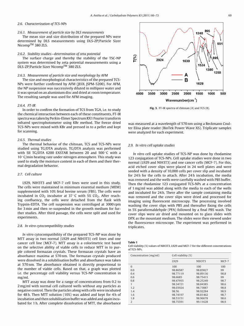
Fig. 3. FT-IR spectra of chitosan (A) and TCS (B).
was measured at a wavelength of 570 nm using a Beckmann Coul-ter Elisa plate reader (BioTek Power Wave XS). Triplicate sampleswere analyzed for each experiment.
2.9. In vitro cell uptake studies
In vitro cell uptake studies of TCS-NP was done by rhodamine123 conjugation of TCS-NPs. Cell uptake studies were done in twonormal (L929 and NIH3T3) and one cancer cells (MCF-7). For this,acid etched cover slips were placed in 24 well plates and wereseeded with a density of 10,000 cells per cover slip and incubatedfor 24 h for the cells to attach. After 24 h incubation, the mediawas removed and the wells were carefully washed with PBS buffer.Then the rhodamine 123 conjugated TCS-NPs at a concentrationof 1 mg/ml was added along with the media to each of the wellsand incubated for 24 h. There after the sample containing mediawas removed and the cover slips were dried and processed forimaging using fluorescent microscopy. The processing involvedwashing the cover slips with PBS and thereafter fixing the cellsin 5% paraformaldehyde (PFA) followed by a final PBS wash. Thecover slips were air dried and mounted on to glass slides withDPX as the mountant medium. The slides were then viewed under
70 A. Anitha et al. / Carbohydrate Polymers 83 (2011) 66–73
(C) AF
3
3
tacabo
Fig. 4. (A) DLS, (B) and
. Results and discussion
.1. Preparation and characterization of TCS
TCS was synthesized using TGA as the thiolating moiety andhe characterization was done using FT-IR. TCS was obtained asresult of the formation of amide bond between NH2 groups of
hitosan and carboxyl groups of TGA via EDC catalyzed reactionnd the new amide bond formation between chitosan and TGA cane confirmed by FT-IR. The degree of thiol substitution was foundut using Ellman’s protocol and it was found to be 60%.
Fig. 5. (A) and (B) TGA and DTA of (A) c
M images of TCP-NPs .
Fig. 3 represents the combined FT-IR spectra of chitosan and TCS.In the spectrum of chitosan, the characteristic absorption peaksare at about 3410 cm−1 (�O–H and �N–H), 2924 cm−1 (�C–H), 1623,1513 cm−1 (ıN–H), 1088 cm−1 (�C–N), 651 cm−1 (ıNH2 ), 1380 cm−1
(ıC–H), 1248 cm−1 (ıO–H), 1153 cm−1 (ıC–O–C), 651 cm−1 (due toamino groups) and 895 cm−1 (epimeric �-ıC–H of cyclic pyranosylrings) could be easily observed. In TCS all the peaks in chitosan were
present, except the peaks corresponding to amino (NH2) groups,since amino groups in chitosan were reacted with carboxyl groupsof TGA resulting in an amide bond, so the additional peaks of thisnewly formed amide bond and peaks of thiol groups (from the TGA
hitosan, (B) TCS and (C) TCS-NPs.
A. Anitha et al. / Carbohydrate Polymers 83 (2011) 66–73 71
towar
mac2
3
tTic
3
NT5
3
acFs
3m
tpTasa
3
i(1(p1
o
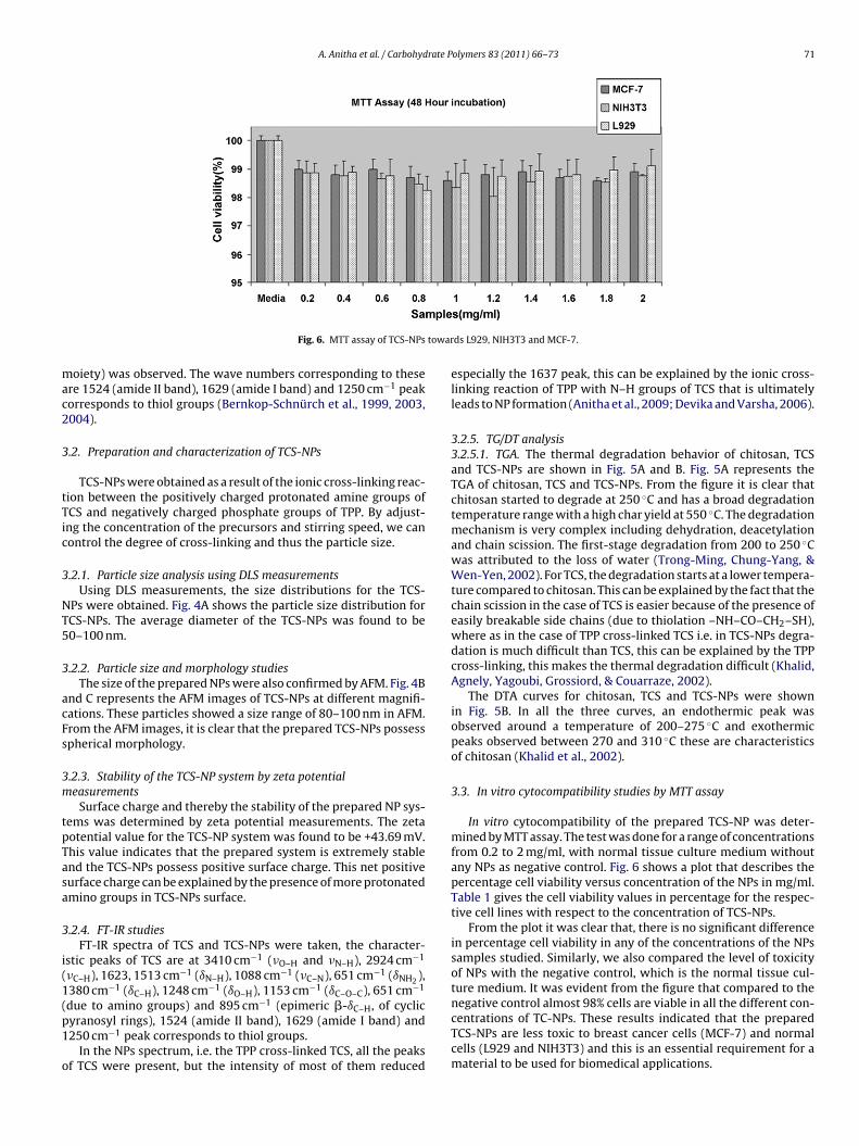
Fig. 6. MTT assay of TCS-NPs
oiety) was observed. The wave numbers corresponding to thesere 1524 (amide II band), 1629 (amide I band) and 1250 cm−1 peakorresponds to thiol groups (Bernkop-Schnürch et al., 1999, 2003,004).
.2. Preparation and characterization of TCS-NPs
TCS-NPs were obtained as a result of the ionic cross-linking reac-ion between the positively charged protonated amine groups ofCS and negatively charged phosphate groups of TPP. By adjust-ng the concentration of the precursors and stirring speed, we canontrol the degree of cross-linking and thus the particle size.
.2.1. Particle size analysis using DLS measurementsUsing DLS measurements, the size distributions for the TCS-
Ps were obtained. Fig. 4A shows the particle size distribution forCS-NPs. The average diameter of the TCS-NPs was found to be0–100 nm.
.2.2. Particle size and morphology studiesThe size of the prepared NPs were also confirmed by AFM. Fig. 4B
nd C represents the AFM images of TCS-NPs at different magnifi-ations. These particles showed a size range of 80–100 nm in AFM.rom the AFM images, it is clear that the prepared TCS-NPs possesspherical morphology.
.2.3. Stability of the TCS-NP system by zeta potentialeasurements
Surface charge and thereby the stability of the prepared NP sys-ems was determined by zeta potential measurements. The zetaotential value for the TCS-NP system was found to be +43.69 mV.his value indicates that the prepared system is extremely stablend the TCS-NPs possess positive surface charge. This net positiveurface charge can be explained by the presence of more protonatedmino groups in TCS-NPs surface.
.2.4. FT-IR studiesFT-IR spectra of TCS and TCS-NPs were taken, the character-
stic peaks of TCS are at 3410 cm−1 (�O–H and �N–H), 2924 cm−1
due to amino groups) and 895 cm−1 (epimeric �-ıC–H, of cyclicyranosyl rings), 1524 (amide II band), 1629 (amide I band) and250 cm−1 peak corresponds to thiol groups.
In the NPs spectrum, i.e. the TPP cross-linked TCS, all the peaksf TCS were present, but the intensity of most of them reduced
ds L929, NIH3T3 and MCF-7.
especially the 1637 peak, this can be explained by the ionic cross-linking reaction of TPP with N–H groups of TCS that is ultimatelyleads to NP formation (Anitha et al., 2009; Devika and Varsha, 2006).
3.2.5. TG/DT analysis3.2.5.1. TGA. The thermal degradation behavior of chitosan, TCSand TCS-NPs are shown in Fig. 5A and B. Fig. 5A represents theTGA of chitosan, TCS and TCS-NPs. From the figure it is clear thatchitosan started to degrade at 250 ◦C and has a broad degradationtemperature range with a high char yield at 550 ◦C. The degradationmechanism is very complex including dehydration, deacetylationand chain scission. The first-stage degradation from 200 to 250 ◦Cwas attributed to the loss of water (Trong-Ming, Chung-Yang, &Wen-Yen, 2002). For TCS, the degradation starts at a lower tempera-ture compared to chitosan. This can be explained by the fact that thechain scission in the case of TCS is easier because of the presence ofeasily breakable side chains (due to thiolation –NH–CO–CH2–SH),where as in the case of TPP cross-linked TCS i.e. in TCS-NPs degra-dation is much difficult than TCS, this can be explained by the TPPcross-linking, this makes the thermal degradation difficult (Khalid,Agnely, Yagoubi, Grossiord, & Couarraze, 2002).
The DTA curves for chitosan, TCS and TCS-NPs were shownin Fig. 5B. In all the three curves, an endothermic peak wasobserved around a temperature of 200–275 ◦C and exothermicpeaks observed between 270 and 310 ◦C these are characteristicsof chitosan (Khalid et al., 2002).
3.3. In vitro cytocompatibility studies by MTT assay
In vitro cytocompatibility of the prepared TCS-NP was deter-mined by MTT assay. The test was done for a range of concentrationsfrom 0.2 to 2 mg/ml, with normal tissue culture medium withoutany NPs as negative control. Fig. 6 shows a plot that describes thepercentage cell viability versus concentration of the NPs in mg/ml.Table 1 gives the cell viability values in percentage for the respec-tive cell lines with respect to the concentration of TCS-NPs.
From the plot it was clear that, there is no significant differencein percentage cell viability in any of the concentrations of the NPssamples studied. Similarly, we also compared the level of toxicityof NPs with the negative control, which is the normal tissue cul-ture medium. It was evident from the figure that compared to the
negative control almost 98% cells are viable in all the different con-centrations of TC-NPs. These results indicated that the preparedTCS-NPs are less toxic to breast cancer cells (MCF-7) and normalcells (L929 and NIH3T3) and this is an essential requirement for amaterial to be used for biomedical applications.
72 A. Anitha et al. / Carbohydrate Polymers 83 (2011) 66–73
F ld andM
3
TcFtwci
ig. 7. Cell uptake studies of rhodamine conjugated TCS-NPs in L929 (a–c, bright fieCF-7 (m–o, bright field and p–r, fluorescent images) cells.
.4. In vitro cell uptake studies using fluorescent imaging
In vitro cellular uptake studies of rhodamine 123 conjugatedCS-NPs were studied by visualizing the bright green fluorescenceoming from the rhodamine dye using fluorescence microscopy.
ig. 7 shows the microscopic images of fluorescence study. Con-rol cells without any NPs showed no fluorescence. Cells incubatedith rhodamine conjugated TCS-NPs showed green fluorescence
onfirming the internalization of the TCS-NPs inside the cells. Also,t is evident from the fluorescence images (Fig. 7) that even after
d–f, fluorescent images), NIH3T3 (g–i, bright field and j–l, fluorescent images) and
incubation with particles; the cells have retained its normal mor-phology which further demonstrates the biocompatibility of theTCS-NP system.
4. Conclusions
A mucoadhesive polymer, TCS was synthesized and character-ized. Degree of thiol substitution was found out using Ellman’sprotocol and it was found to be 60%. The prepared TCS-NPs were
rate P
cpmstomNcum
A
GtBpIRaFrCaM
R
A
B
B
B
D
D
D
F
F
G
J
J
A. Anitha et al. / Carbohyd
haracterized using DLS, AFM, FT-IR and TG/DTA studies. Therepared NPs possess a size range of 80–110 nm and sphericalorphology. Stability studies were done by zeta potential mea-
urements and the system was found to be extremely stable andhe NPs possess positive surface charge. In vitro cytocompatibilityf the prepared NPs were studied using MTT assay towards nor-al and cancer cell lines. These results showed that the preparedPs are less toxic (almost 98% cells are viable in all the differentoncentrations TCS-NPs samples). So the prepared TCS-NPs can besed as an efficient biomaterial for biomedical applications such asucoadhesive drug delivery, gene delivery, etc.
cknowledgments
The authors are thankful to Department of Biotechnology (DBT),ovt. of India, for their financial support for this work under
he Nanoscience and Nanotechnology Initiative program (Ref. No.T/PR10882/NNT/28/142/2008). This work was also partially sup-orted by Nanomission, Department of Science and Technology,
ndia under the Nanoinitiative Program monitored by Prof. C.N.R.ao. One of the authors A. Anitha is thankful to Council of Scientificnd Industrial Research (CSIR), India for providing Senior Researchellowship (SRF award-Ref. No. 9/963 (0005)2K10-EMR-I) for car-ying out her research work. The authors are also thankful to Koyohemicals Co., Ltd., Japan for providing Chitosan. The authors arelso thankful to Mr. Sajin P. Ravi, Mr. Girish C.M., Mr. Sarath andr. Sudheesh Kumar P.T. for their help in AFM and TG/DTA studies.
eferences
nitha, A., Divya Rani, V. V., Krishna, R., Sreeja, V., Selvamurugan, N., Nair, S. V.,et al. (2009). Synthesis, characterization, cytotoxicity and antibacterial studiesof chitosan, O-carboxymethyl and N,O-carboxymethyl chitosan nanoparticles.Carbohydrate Polymers, 78, 672–677.
ernkop-Schnürch, A., Hornof, M. D., & Guggi, D. (2004). Thiolated chitosans. Euro-pean Journal of Pharmaceutics and Biopharmaceutics, 57, 9–17.
ernkop-Schnürch, A., Kast, C. E., & Guggi, D. (2003). Permeation enhancing polymersin oral delivery of hydrophilic macromolecules: Thiomer/GSH systems. Journalof Controlled Release, 93, 95–103.
ernkop-Schnürch, A., Schwarz, V., & Steininger, S. (1999). Polymers with thiolgroups: A new generation of mucoadhesive polymers. Pharmaceutical Research,16, 876–881.
ev, A., Binulal, N. S., Anitha, A., Nair, S. V., Furuike, T., Tamura, H., et al. (2010). Prepa-ration of poly (lactic acid)/chitosan nanoparticles for anti-HIV drug deliveryapplications. Carbohydrate Polymers, 80, 833–838.
ev, A., Jithin, C. M., Sreeja, V., Tamura, H., Patzke, G. R., Hussain, F., et al. (2010).Novel carboxymethyl chitin nanoparticles for cancer drug delivery applications.Carbohydrate Polymers, 79, 1073–1079.
evika, R. B., & Varsha, B. P. (2006). Studies on effect of pH on cross-linking of chitosanwith sodium tripolyphosphate: A technical note. AAPS Pharmaceutical Scienceand Technology, 7, 1–6.
elt, O., Buri, P., & Gurny, R. (1998). Chitosan: A unique polysaccharide for drugdelivery. Drug Development and Industrial Pharmacy, 24, 979–993.
reier, T., Koh, H. S., Kazazian, K., & Shoichet, M. S. (2005). Controlling cell adhe-sion and degradation of chitosan films by N-acetylation. Biomaterials, 26,5872–5878.
orbach, V. I., Krasikova, I. N., Lukyanov, P. A., Loenko, Y. N., Soloveva, T. F., Ovood,Y. S., et al. (1994). New glycolipids (chitooligosaccharide derivatives) possess-ing immunostimulating and antitumor activities. Carbohydrate Research, 260,73–82.
ayakumar, R., Chennazhi, K. P., Muzzarelli, R. A. A., Tamura, H., Nair, S. V., & Selva-murugan, N. (2010). Chitosan conjugated DNA nanoparticles in gene therapy.Carbohydrate Polymers, 79, 1–8.
ayakumar, R., Deepthy, M., Manzoor, K., Nair, S. V., & Tamura, H. (2010). Biomed-ical applications of chitin and chitosan based nanomaterials—A short review.Carbohydrate Polymers, 82, 227–232.
olymers 83 (2011) 66–73 73
Jayakumar, R., Nair, S. V., & Tamura, H. (2009). Biomedical applications of chitin.Asian Chitin Journal, 5, 1–4.
Jayakumar, R., Nwe, N., Tokura, S., & Tamura, H. (2007). Sulfated chitin and chi-tosan as novel biomaterials. International Journal of Biological Macromolecules,40, 175–181.
Jayakumar, R., Prabaharan, M., Nair, S. V., & Tamura, H. (2010). Novel chitin andchitosan nanofibers in biomedical applications. Biotechnology Advances, 28,142–150.
Jayakumar, R., Prabaharan, M., Nair, S. V., Tokura, S., Tamura, H., & Selvamu-rugan, N. (2010). Novel carboxymethyl derivatives of chitin and chitosanmaterials and their biomedical applications. Progress in Materials Science, 55,675–709.
Jayakumar, R., Prabaharan, M., Reis, R. L., & Mano, J. F. (2005). Graft copoly-merized chitosan—Present status and applications. Carbohydrate Polymers, 62,142–158.
Jayakumar, R., Reis, R. L., & Mano, J. F. (2006). Chemistry and applications of phos-phorylated chitin and chitosan. E-Polymers, 35, 1–16.
Jayakumar, R., Reis, R. L., & Mano, J. F. (2007). Synthesis and characterization ofpH-sensitive thiol-containing chitosan beads for controlled drug delivery appli-cations. Drug Delivery, 14, 9–17.
Kast, C. E., & Bernkop-Schnürch, A. (2001). Thiolated polymers–thiomers: Devel-opment and in vitro evaluation of chitosan–thioglycolic acid conjugates.Biomaterials, 22, 2345–2352.
Khalid, M. N., Agnely, F., Yagoubi, N., Grossiord, J. L., & Couarraze, G. (2002). Waterstate characterization, swelling behavior, thermal and mechanical propertiesof chitosan based network. European Journal of Pharmaceutical Sciences, 15,425–432.
Khan, T. A., Peh, K. K., & Chng, H. S. (2000). Mechanical, bioadhesive strength andbiological evaluations of chitosan films for wound dressing. Journal of Pharmacy& Pharmaceutical Sciences, 3, 303–311.
Khor, E, & Lim, L. Y. (2003). Implantable applications of chitin and chitosan. Bioma-terials, 24, 2339–2349.
Lichen, Y., Jieying, D., Chunbai, H., Liming, C., & Cui, T. (2009). Drug permeability andmucoadhesion properties of thiolated trimethyl chitosan nanoparticles in oralinsulin delivery. Biomaterials, 30, 5691–5700.
Madhumathi, K., Sudheesh Kumar, P. T., Abhilash, S., Sreeja, V., Tamura, H., Manzoor,K., et al. (2010). Development of novel chitin/nanosilver composite scaffolds forwound dressing applications. Journal of Materials Science: Materials in Medicine,21, 807–813.
Margit, D. H., Constantia, E. K., & Bernkop-Schnürch, A. (2003). In vitro evaluationof the viscoelastic properties of chitosan–thioglycolic acid conjugates. EuropeanJournal of Pharmaceutics and Biopharmaceutics, 55, 185–190.
Mathew, E. M., Jithin, C. M., Manzoor, K., Nair, S. V., Tamura, H., & Jayakumar,R. (2010). Folate conjugated carboxymethyl chitosan–manganese doped zincsulphide nanoparticles for targeted drug delivery and imaging of cancer cells.Carbohydrate Polymers, 80, 442–448.
Mwale, F., Iordanova, M., Demers, C. N., Steffen, T., Roughley, P., & Antoniuo, J. (2005).Biological evaluation of chitosan salts cross-linked to genipin as a cell scaffoldfor disk tissue engineering. Tissue Engineering, 11, 130–140.
Rabea, E. I., Badawy, M. E., Stevens, C. V., Smagghe, G., & Steubaut, W. (2003). Chitosanas antimicrobial agent: Applications and mode of action. Biomacromolecules, 4,1457–1465.
Ranabir, C., Lila, K. N., & Sontosh, M. (2009). Formulation development of oralmucoadhesive coated terbutaline sulphate tablets using some natural materialsextracted from edible fruits available in India. Iranian Journal of PharmaceuticalSciences, 5, 3–12.
Ronny, M., Brigitta, L., Adolf, M. S., & Andreas Bernkop, S. (2008). Thiolated chi-tosan nanoparticles: Transfection study in the Caco-2 differentiated cell culture.Nanotechnology, 19, 1–9.
Sudheesh Kumar, P. T., Abhilash, S., Manzoor, K., Nair, S. V., Tamura, H., & Jayaku-mar, R. (2010). Preparation and characterization of novel �-chitin/nanosilvercomposite scaffolds for wound dressing applications. Carbohydrate Polymers, 80,761–767.
Trong-Ming, D., Chung-Yang, C., & Wen-Yen, C. (2002). Studies on the degradationbehavior of chitosan-g-poly(acrylic acid) copolymers. Tamkang Journal of Scienceand Engineering, 5, 235–240.
VandeVord, P. J., Matthew, H. W., De Silva, S. P., Mayton, L., Wu, B., & Wooley, P. H.(2002). Evaluation of the biocompatibility of a chitosan scaffold in mice. Journalof Biomedical Materials Research, 59, 585–590.
Wang, X. H., Du, Y. M., Fan, L. H., Liu, H., & Hu, Y. (2005). Chitosan-metal complexesas antimicrobial agent: Synthesis and characterization and structure-activitystudy. Polymer Bulletin, 55, 105–113.
Wu, Z. M., Zhang, X. G., Zheng, C., Li, C. X., Zhang, S. M., Donga, R. N., et al. (2009).Disulfide-cross linked chitosan hydrogel for cell viability and controlled proteinrelease. European Journal of Pharmaceutical Sciences, 37, 198–206.


![Research Article Enhanced Oral Delivery of Docetaxel Using … · 2019. 7. 31. · taxanes is inhibiting microtubule depolymerization [ , ]. ... easily entrapped in thiolated chitosan-pMMA](https://static.documents.pub/doc/80x56/60c8b8422dbab75fc839ded4/research-article-enhanced-oral-delivery-of-docetaxel-using-2019-7-31-taxanes.jpg)


