Full length article Development of osteopromotive poly (octamethylene citrate glycerophosphate) for enhanced bone regeneration q Yun He a,1 , Qiyao Li b,⇑,1 , Chuying Ma b , Denghui Xie c , Limei Li a , Yitao Zhao c , Dingying Shan b , Sarah K. Chomos b , Cheng Dong b , John W. Tierney b,d , Lin Sun e , Di Lu a , Li Gui f,⇑ , Jian Yang b,⇑ a Biomedicine Engineering Research Centre, Kunming Medical University, Kunming 650500, Yunnan, China b Department of Biomedical Engineering, Materials Research Institute, The Huck Institutes of the Life Sciences, The Pennsylvania State University, University Park, PA, USA c Academy of Orthopedics, Guangdong Province, Guangdong Provincial Key Laboratory of Bone and Joint Degenerative Diseases, The Third Affiliated Hospital of Southern Medical University, Guangzhou 510280, Guangdong, China d Department of Biomedical Engineering, College of Engineering and Computing, University of South Carolina, Columbia, SC, 29201, USA e Department of Cardiology, The Second Affiliated Hospital, Kunming Medical University, Kunming 650101, Yunnan, China f Department of Endocrinology, The Third People’s Hospital of Yunnan Province, Kunming 650011, Yunnan, China article info Article history: Received 24 November 2018 Received in revised form 5 March 2019 Accepted 23 March 2019 Available online 27 March 2019 Keywords: Citrate-based biomaterials Glycerophosphate Osteogenic differentiation Mechanical property Biodegradation abstract The design and development of bioactive materials that are inherently conducive for osteointegration and bone regeneration with tunable mechanical properties and degradation remains a challenge. Herein, we report the development of a new class of citrate-based materials with glycerophosphate salts, b-glycerophosphate disodium (b-GP-Na) and glycerophosphate calcium (GP-Ca), incorporated through a simple and convenient one-pot condensation reaction, which might address the above challenge in the search of suitable orthopedic biomaterials. Tensile strength of the resultant poly (octamethylene citrate glycerophosphate), POC-bGP-Na and POC-GP-Ca, was as high as 28.2 ± 2.44 MPa and 22.76 ± 1.06 MPa, respectively. The initial modulus ranged from 5.28 ± 0.56 MPa to 256.44 ± 22.88 MPa. The mechanical properties and degradation rate of POC-GP could be controlled by varying the type of salts, and the feed- ing ratio of salts introduced. Particularly, POC-GP-Ca demonstrated better cytocompatibility and the cor- responding composite POC-GP-Ca/hydroxyapatite (HA) also elicited improved osteogenic differentiation of human mesenchymal stem cells (hMSCs) in vitro, as compared to POC-bGP-Na/HA and POC/HA. The superior in-vivo performance of POC-GP-Ca/HA microparticle scaffolds in promoting bone regeneration over POC-bGP-Na/HA and POC/HA was further confirmed in a rabbit femoral condyle defect model. Taken together, the tunability of mechanical properties and degradation rates, together with the osteo- promotive nature of POC-GP polymers make these materials, especially POC-GP-Ca well suited for bone tissue engineering applications. Statement of Significance The design and development of bioactive materials that are inherently conducive for osteointegration and bone regeneration with tunable mechanical properties and degradation remains a challenge. Herein, we report the development of a new class of citrate-based materials with glycerophosphate salts, b-glycerophosphate disodium (b-GPNa) and glycerophosphate calcium (GPCa), incorporated through a simple and convenient one-pot condensation reaction. The resultant POC-GP polymers showed signifi- cantly improved mechanical property and tunable degradation rate. Within the formulation investigated, POC-GPCa/HA composite further demonstrated better bioactivity in favoring osteogenic differentiation of hMSCs in vitro and promoted bone regeneration in rabbit femoral condyle defects. The development of POC-GP expands the repertoire of the well-recognized citrate-based biomaterials to meet the ever- increasing needs for functional biomaterials in tissue engineering and other biomedical applications. Ó 2019 Acta Materialia Inc. Published by Elsevier Ltd. All rights reserved. https://doi.org/10.1016/j.actbio.2019.03.050 1742-7061/Ó 2019 Acta Materialia Inc. Published by Elsevier Ltd. All rights reserved. q Part of the Drug Delivery for Musculoskeletal Applications Special Issue, edited by Robert S. Hastings and Professor Johnna S. Temenoff. ⇑ Corresponding authors. E-mail addresses: qfl[email protected](Q. Li), [email protected](L. Gui), [email protected](J. Yang). 1 These authors contributed equally. Acta Biomaterialia 93 (2019) 180–191 Contents lists available at ScienceDirect Acta Biomaterialia journal homepage: www.elsevier.com/locate/actabiomat
Development of osteopromotive poly (octamethylene citrateglycerophosphate) for enhanced bone regenerationq
https://doi.org/10.1016/j.actbio.2019.03.0501742-7061/� 2019 Acta Materialia Inc. Published by Elsevier Ltd. All rights reserved.
q Part of the Drug Delivery for Musculoskeletal Applications Special Issue, edited by Robert S. Hastings and Professor Johnna S. Temenoff.⇑ Corresponding authors.
Yun He a,1, Qiyao Li b,⇑,1, Chuying Ma b, Denghui Xie c, Limei Li a, Yitao Zhao c, Dingying Shan b,Sarah K. Chomos b, Cheng Dong b, John W. Tierney b,d, Lin Sun e, Di Lu a, Li Gui f,⇑, Jian Yang b,⇑aBiomedicine Engineering Research Centre, Kunming Medical University, Kunming 650500, Yunnan, ChinabDepartment of Biomedical Engineering, Materials Research Institute, The Huck Institutes of the Life Sciences, The Pennsylvania State University, University Park, PA, USAcAcademy of Orthopedics, Guangdong Province, Guangdong Provincial Key Laboratory of Bone and Joint Degenerative Diseases, The Third Affiliated Hospital of SouthernMedical University, Guangzhou 510280, Guangdong, ChinadDepartment of Biomedical Engineering, College of Engineering and Computing, University of South Carolina, Columbia, SC, 29201, USAeDepartment of Cardiology, The Second Affiliated Hospital, Kunming Medical University, Kunming 650101, Yunnan, ChinafDepartment of Endocrinology, The Third People’s Hospital of Yunnan Province, Kunming 650011, Yunnan, China
a r t i c l e i n f o
Article history:Received 24 November 2018Received in revised form 5 March 2019Accepted 23 March 2019Available online 27 March 2019
The design and development of bioactive materials that are inherently conducive for osteointegrationand bone regeneration with tunable mechanical properties and degradation remains a challenge.Herein, we report the development of a new class of citrate-based materials with glycerophosphate salts,b-glycerophosphate disodium (b-GP-Na) and glycerophosphate calcium (GP-Ca), incorporated through asimple and convenient one-pot condensation reaction, which might address the above challenge in thesearch of suitable orthopedic biomaterials. Tensile strength of the resultant poly (octamethylene citrateglycerophosphate), POC-bGP-Na and POC-GP-Ca, was as high as 28.2 ± 2.44 MPa and 22.76 ± 1.06 MPa,respectively. The initial modulus ranged from 5.28 ± 0.56 MPa to 256.44 ± 22.88 MPa. The mechanicalproperties and degradation rate of POC-GP could be controlled by varying the type of salts, and the feed-ing ratio of salts introduced. Particularly, POC-GP-Ca demonstrated better cytocompatibility and the cor-responding composite POC-GP-Ca/hydroxyapatite (HA) also elicited improved osteogenic differentiationof human mesenchymal stem cells (hMSCs) in vitro, as compared to POC-bGP-Na/HA and POC/HA. Thesuperior in-vivo performance of POC-GP-Ca/HA microparticle scaffolds in promoting bone regenerationover POC-bGP-Na/HA and POC/HA was further confirmed in a rabbit femoral condyle defect model.Taken together, the tunability of mechanical properties and degradation rates, together with the osteo-promotive nature of POC-GP polymers make these materials, especially POC-GP-Ca well suited for bonetissue engineering applications.
Statement of Significance
The design and development of bioactive materials that are inherently conducive for osteointegration andbone regeneration with tunable mechanical properties and degradation remains a challenge. Herein, wereport the development of a new class of citrate-based materials with glycerophosphate salts,b-glycerophosphate disodium (b-GPNa) and glycerophosphate calcium (GPCa), incorporated through asimple and convenient one-pot condensation reaction. The resultant POC-GP polymers showed signifi-cantly improved mechanical property and tunable degradation rate. Within the formulation investigated,POC-GPCa/HA composite further demonstrated better bioactivity in favoring osteogenic differentiation ofhMSCs in vitro and promoted bone regeneration in rabbit femoral condyle defects. The development ofPOC-GP expands the repertoire of the well-recognized citrate-based biomaterials to meet the ever-increasing needs for functional biomaterials in tissue engineering and other biomedical applications.
� 2019 Acta Materialia Inc. Published by Elsevier Ltd. All rights reserved.
Y. He et al. / Acta Biomaterialia 93 (2019) 180–191 181
1. Introduction
Over 2.2 million bone grafting procedures are performed annu-ally worldwide [1] in orthopedics, neurosurgery and dentistry, ren-dering bone substitutes increasingly necessary and appliedclinically. The development of fully synthetic bone substitutes thatare readily available, cost-effective, and osteo-promotive is partic-ularly encouraged in the clinical field [2]. Despite significant pro-gress, currently available synthetic materials are limited by theirinabilities to mimic native tissue composition, weak mechanicalstrength, significant inflammatory responses, poor bone integra-tion, and slow bone regeneration [3]. Inspired by natural bone, anative biocomposite comprised of collagen as the major organiccomponent providing the naturally derived polymeric frameworkfor inorganic calcium phosphate (CaP) mineral nucleation, inor-ganic bioceramics, such as hydroxyapatite (HA), and tricalciumphosphate (TCP), have been introduced into biodegradable syn-thetic polymers to create biomimetic composites aimed at betterresembling the native bone composition [4–7].
Citrate, highly accumulated in natural bone, has been found tostrongly bind to bone inorganic minerals, playing indispensableroles in stabilizing the mineral crystals [8] and controlling crystalsize [9]. As a robust multifunctional monomer, citrate chemistryenables the formation of a 3D cross-linked network structure whenreacted with different diols [10,11] as well as providing rich pen-dant carboxyl and hydroxyl groups to allow the incorporation ofup to 65 % weight of HA, closely resembling the inorganic compo-sition of natural bone. During the past decade, a series of citrate-based material/HA composites have been developed in our laband have demonstrated impressive in vivo performance in variousanimal models for bone regeneration, such as poly(octamethylenecitrate)/HA (POC/HA) for osteochondral defect healing [12], click-able POC/HA (POC-click/HA) for long segmental radial bone regen-eration [13] and for cranial defect repair [14], citrate-basedpolymer blends/HA (CBPB/HA) for the repair of lateral femoral con-dyle defect [15], injectable citrate-based mussel-inspired tissuebioadhesives/HA composites (iCMBA/HA) for comminuted radialbone regeneration [16], as well as N-methyldiethanolamine(MDEA) modified click-crosslinked POC-click/HA (POC-M-click/HA) for spinal fusion [17]. Excitingly, one of our recent studies[18] demonstrated released citrate could promote the differentia-tion of human mesenchymal stem cells (hMSCs) towards activebone forming cells by regulating energy-producing metabolicpathways. The newly discovered citrate metabonegenic regulationmechanism not only highlights the significance of citrate-basedcomposites for orthopedic applications, but also provides valuableguidance to design materials with improved osteo-promotive func-tionality. For example, phosphoserine, the native organic phos-phate donor in bone, when incorporated in citrate polymer or inits solute form, demonstrates concerted action with citrate leadingto accelerated bone formation and maturation [18]. The abovestudies suggest polymers incorporating phosphates can synergizewith citrate to further promote osteogenic differentiation ofhMSCs, pointing toward the specific chemical formulation ofcitrate materials to influence in vivo bioactivity.
Mechanical properties are also an important factor of materialsthat greatly affect tissue engineering efficacy, especially for bonerepair [19]. Numerous efforts have been made in our lab toimprove the mechanical properties of citrate-based materials,especially for orthopedic applications. Introduction of urethanegroups into citrate-based polyester materials has proven to be aneffective way for improving mechanical strength but sacrificesthe valuable pendant groups for further biofunctionalization andHA chelation [14,20]. Biodegradable elastomers employing clickchemistry as an additional crosslinking mechanism have alsodemonstrated improved mechanical properties; however, these
materials display a delayed degradation rate compared with POC,making them unsuitable for applications requiring fast-degradingscaffolds [17,21]. The relatively complex click-monomer synthesisalso makes the click-polymers less attractive. Therefore, balancingthe mechanical properties and biodegradation rate while introduc-ing biofunctionality into polymer networks in a more convenientway remains as a challenge.
b-glycerophosphate disodium (bGP-Na), widely known as anosteogenic medium supplement and a weak base, has been usedto initiate the gelation of chitosan or collagen to generatethermosensitive injectable hydrogels [22]. Another GP salt,glycerophosphate calcium (GP-Ca), representing as a mixture ofb-isomer (80 %) and rac-a-isomer (20 %), has long been used as afood supplement for both calcium and phosphate. Given the osteo-promotive benefits of both salts in their solute and conjugatedform [23–25], we incorporated two types of GP salts into thecitrate-based materials through a one-pot polycondensation reac-tion by taking advantage of the reactive nature of citric acid, to pre-pare POC-GP, in order to further improve the bioactivity of citratematerials to promote stem cell osteogenic differentiation. At thesame time, our results show that GP incorporation greatlyincreased the mechanical properties of the resultant polymer. Bychanging the salt type and feeding ratio and polymerizing themonomers in a very convenient, one-pot polycondensation, thedegradation rate of the resulting polymers can also be easily tuned,providing an alternative way to balance mechanical properties,degradation, and bioactivity. A rabbit femoral condyle defectmodel was used to evaluate the efficacy of POC-GP/HA for boneregeneration. It was observed that the osteopromotive capabilitiesof GP-doped POC were significantly improved compared with POCand autograft control groups.
2. Materials and methods
2.1. Materials
HA [(particle size: >75 lm (0.5 %), 45–75 lm (1.4 %), <45 lm(98.1 %)] was purchased from Fluka (St Louis, MO). 1,8-octanediol(98 %) and citric acid (99.5 %) were purchased from Alfa Aesar. b-glycerophosphate-Na (bGPNa), Glycerophosphate-Ca (GPCa), andall remaining chemicals were purchased from Sigma-Aldrich (StLouis, MO) and used as received unless stated otherwise.
2.2. Synthesis of pre-polymers
POC pre-polymer was synthesized by bulk polymerization usingcitric acid and 1,8-octanediol in 1.0:1.1 feeding ratio. Briefly,monomers were stirred mechanically and melted under nitrogenprotection at 160 �C in a silicon oil bath. Then the temperature ofthe system was lowered to 140 �C, followed by stirring for 1–2 hto create the pre-polymer. The reaction was quenched by 1,4-dioxane and purified in deionized water followed by lyophilization.The purified pre-polymer was dissolved in 1,4-dioxane (30 wt/wt%)for further use. POC-bGP-Na and POC-GP-Ca pre-polymers weresynthesized via the above method with citric acid, 1,8-octanediol,b-glycerophosphate disodium (b-GP-Na), and glycerophosphatecalcium (GP-Ca) in different feeding ratios as shown in Table 1,except that dialysis was performed to purify the pre-polymerbefore freeze drying.
2.3. Preparation of polymer films, polymer/HA composites andpolymer/HA microparticle scaffolds
To prepare polymer films, pre-polymer solutions were cast inTeflon dishes and the solvent was allowed to evaporate. The dried
Table 1Formulations of pre-polymer synthesis.
Citric acid (M) 1,8-Octandiol (M) bGP-Na or GP-Ca (M)
182 Y. He et al. / Acta Biomaterialia 93 (2019) 180–191
pre-polymer was then thermally crosslinked at 80 �C for 3 daysand then at 120 �C with vacuum for 1 day to obtain a crosslinkedpolymer film. To prepare polymer/HA composites, a 30 % pre-polymer solution in 1,4-dioxane was mixed with hydroxyapatite(HA) by continuously stirring. After the mixture turned dough-like, it was then pressed into thin sheets. The composite sheetswere cut into round disks then thermally crosslinked at 80 �C for3 days and at 120 �C with vacuum for 1 day. To prepare the poly-mer/HA microparticles, porous composite scaffolds were first pre-pared by mixing pre-polymer solution, HA and NaCl (250–425 mmsalt size) followed by casting in Teflon dishes. After the solventevaporated, the scaffolds were thermally crosslinked as describedabove and then soaked in de-ionized (DI) water to leach the salt.Next, the porous sponge-like scaffolds were freeze-dried, groundand sieved into different particle sizes (<150 mm; 150–250 mm;250–425 mm; >425 mm).
2.4. Material characterizations
2.4.1. Structural characterizationTo check the chemical structural difference between POC, POC-
bGP-Na and POC-GP-Ca, Fourier transform infrared (FT-IR) analysiswas operated by casting polymer solution in 1,4-dioxane on KBrpellets using Bruker Vertex V70 spectrometer. FT-IR spectra wererecorded over a wavelength range of 400–4000 cm�1. 31P NMR(161.9 MHz) spectra of pre-polymer solutions were obtained usingBruker DPX 400 NMR Spectroscopy. Either D2O or DMSO were usedas solvents. All chemical shifts were reported in ppm (d).
2.4.2. Contact angle measurementsThe water-in-air contact angles on crosslinked polymer films
were measured at room temperature within 10 s after water wasdropped on the film surface by a Rame-Hart goniometer and imag-ing system (Rame-Hart Inc., Mouttain Lake, NJ) using the sessiledrop method. Four independent measurements at different siteswere averaged. The change of water-in-air contact angle with timewas monitored over 60 s after water dropping.
2.4.3. Tensile mechanical testsTensile tests of polymer films were conducted on an Instron
5966 machine equipped with a 1000 N load cell. Samples werecut into rectangular strips and pulled until failure at a speed of500 mmmin�1 to obtain stress–strain curves. The initial slope(0–10 %) of the stress–strain curve was used to determine the ini-tial modulus of each sample. Six specimens were averaged for eachsample, and the results were presented as mean ± standarddeviation.
2.4.4. In vitro degradation and release studiesDegradation properties were studied in vitro with polymer film
samples placed in tubes containing 10 mL of NaOH solution (0.05M) or phosphate buffered saline (PBS, pH = 7.4) and incubated at37 �C. Samples were weighed before degradation to find the initialmass (W0). At each time point, samples were washed thoroughlywith deionized water 3 times followed by lyophilization. After
lyophilization, samples were weighed to measure the remainingmass (Wt). Six parallel specimens were averaged for each sample,and the results were presented as means ± standard deviation.The percent mass loss was calculated based on the followingequation:
Mass Loss %ð Þ ¼ ð W0 �Wtð Þ=W0Þ � 100: ð1Þ
2.4.5. Phosphorus release studiesCumulative phosphorus release was investigated by inductively
coupled plasma optical emission spectroscopy (ICP-OES 730-ES,Varian, USA) to confirm the presence of bGP-Na and GP-Ca in therelease media. Films weighing about 50 mg were soaked in 10mL of PBS (pH 7.4) solution and incubated at 37 �C. After 14, 21,and 28 days, the release media of each sample were mixed with10 wt% HNO3 solution at 1:1 ratio and then subjected to ICP-OESmeasurements.
2.4.6. Morphology of microparticle scaffolds and hMSCsTo observe morphology of microparticle scaffolds and differen-
tiating hMSCs, scanning electron microscopy (SEM, Zeiss Sigma)was used to capture and analyze images at 10 kV. All samples werecoated with 5 nm iridium using a Leica sputter coater beforeimaging.
2.5. Cell culture
In vitro cell studies were performed with human mesenchymalstem cells (hMSCs) in passage 5–7. Growth media (GM) was com-posed of Dulbecco’s Modified Essential Medium (DMEM) supple-mented with 10 % fetal bovine serum and 100 units/mL ofpenicillin-streptomycin. Osteogenic media (OG) was composed ofgrowth media supplemented with 0.5 mM ascorbate-2-phosphate, 100 nM dexamethasone, and 10 mM b-glycerophosphate disodium. Solid samples were submerged in70 % ethanol for 1 h and sterilized by UV irradiation for 1 h on eachsurface. For liquid samples, solutions were sterilized by filteringthrough a sterile syringe filter (0.2 lm cellulose acetate, VWR,PA). For all cell studies, cells were kept in a humidified incubatorat 37 �C with 5 % CO2. Both GM and OM were replaced every otherday.
2.6. In vitro cytocompatibility evaluation
hMSCs were seeded at 5000 cells/cm2 and 3000 cells/cm2 forcell cytotoxicity and cell proliferation, respectively. At each timepoint, media was removed and hMSCs were rinsed once with PBSsolution. 200 lL of cell counting kit-8 (CCK-8) solution was addedto each well and incubated at 37 �C for 30 min. The absorbancewas measured at a wavelength of 450 nm with a Tecan microplatereader.
2.7. hMSCs differentiation study
To evaluate the hMSCs differentiation on polymer/HA compos-ite disks and microparticle scaffolds, samples were sterilized andtransferred to 48-well plates. hMSCs were seeded onto samplesat a density of 3000 cells/cm2 in growth media. Once cells reached80 % confluency, growth media was removed and replaced with theestablished osteogenic media or growth media as a general control.To evaluate the effect of GP salt concentrations on osteogenicdifferentiation, hMSCs were also seeded onto a 48-well plateat a concentration of 10,000 cells/mL in growth medium. Oncecells reached 80 % confluency, the growth media was replacedwith a reductive osteogenic media (DMEM with 0.5 mMascorbate-2-phosphate, 100 nM dexamethasone but without
Y. He et al. / Acta Biomaterialia 93 (2019) 180–191 183
b-glycerophosphate disodium) supplemented with 0.2 mM of dif-ferent GP salts.
After differentiation for 7, 14, and 21 days, cells were washed 3times with PBS solution and lysed with 250 mL RIPA buffer for alka-line phosphatase (ALP) assay and DNA quantification. The ALPactivity was measured according to the method published previ-ously [18]. The DNA content was tested by Pico Green dsDNA testkit according to manufacturer’s instruction. To observe the mineraland ECM formation and cell morphology on polymer/HAcomposites under SEM, cells were harvested and fixed in 2.5 %glutaraldehyde for 48 h followed by a serial dehydration process,critical-point drying, and iridium sputter coating (Leica Sputter-coater). Gene expression of Runx-2 (runt-related transcriptionfactor 2), Col1a1 (encoding collagen type 1 alpha 1), and SPP1(encoding osteopontin) were evaluated by real-time PCR. TotalRNA was extracted from harvested cells on day 14 with QIAGENRNeasy kit (Hilden, Germany) and transcribed into cDNA withHigh-Capacity cDNA Reverse Transcription Kits (Applied Biosys-temsTM, CA). Then real-time PCR was carried out using a QuantStu-dio Real-Time PCR system (Applied BiosystemsTM, CA). The primersand TaqMan probe used for human Runx-2, Col1a1, and SPP1 wereHs00231692_m1, Hs00164004_m1, and Hs00959010_m1.
2.8. Surgical procedures for animal study
An established femoral condyle defect model on New Zealandwhite rabbits was used for in vivo evaluation according to themethod described elsewhere [26,27]. Briefly, a defect with 5 mmin diameter and 5 mm in depth was created at the lateral femoralcondyle of each rabbit under continuous saline buffer irrigation,then powders were packed into the defect and the musculatureand skin incision were closed with nylon suture. Both the leftand right condyles of femur were used. Rabbits were randomlyassigned into four groups with different bone implants in leftand right legs respectively: 1) sham group (untreated); 2) POC/HA microparticle scaffold group; 3) POC-bGP-Na/HA microparticlescaffold group, and 4) POC-GP-Ca/HA microparticle scaffold group.The concentrations of HA in the composites are all 65 wt%. The par-ticle scaffold with a mixture of different sizes were used for animalstudies. After 4, 8, and 12 weeks, 4 rabbits/group were euthanizedand subjected to micro-CT analyses. Then the medial femoral con-dyle and the surrounding tissues were removed, fixed in 4 % phos-phate buffered paraformaldehyde solution, sectioned, and stainedfor histological analysis [28]. A total of 48 New Zealand white rab-bits weighing approximately 2.5 kg were used and approved byKunming Medical University in compliance with all regulatoryguidelines. The anesthesia (Pentobarbital Sodium, 30 mg/kg) forall animals was administered through marginal ear veins on thelateral aspect.
2.9. Micro-CT analysis
In order to quantify new bone formation within defects, quan-titative imaging of harvested femurs was performed using amicro-CT imaging system (l-CT80 scanner; Scanco Medical AG,Brüttisellen, Switzerland) with a 3D Gaussian filter constrained atr = 1.0 and support = 2 for partial suppression of the noise in thevolumes. All the micro-CT data was processed with Mimics 21.0software at a threshold of 2500 to discriminate new bone tissuefrom surrounding tissues and polymer/HA scaffold and a cylindri-cal region of interest (ROI) with a diameter of 5.0 � 3.0 mm waschosen to evaluate the newly regenerated bone. Bone volume/totalvolume (BV/TV) was calculated. All analyses were repeated withfour specimens.
2.10. Histological analysis
Tissues were sectioned and fixed in 4 % phosphate bufferedparaformaldehyde solution for histological assessment. The sec-tions were stained with hematoxylin and eosin (H&E) and Mas-son’s trichrome (Hematoxylin Staining Solution, FZ-025, EosinStaining Solution, FZ-028, Masson’s trichrome stain solution, FZ-037, Hebei Bio-High Technology Development CO., LTD, China.)to observe the new bone formation and collagen deposition,respectively, within the wound areas. Digital images from eachexperimental group were acquired by an optical microscope(40�, 100�, and 400� magnification).
2.11. Statistical analysis
All quantitative data is presented as mean ± SD with a mini-mum of three independent samples and analyzed by one-wayanalysis of variance (ANOVA). P values < 0.05 were regarded as sta-tistically significant.
3. Results and discussion
3.1. POC-bGP-Na and POC-GP-Ca are incorporated into citrate-basedmaterials
The synthesis of POC-GP is similar to that for POC through a onepot polycondensation of citric acid, 1,8-octanediol and bGP-Na/-Cato obtain the POC-bGP-Na/-Ca pre-polymers, which can be furthercrosslinked (post-polymerization) to prepare polymer films(Fig. 1A). It should be noted that after the pre-polymer synthesis,both POC-bGP-Na/POC-GP-Ca pre-polymers were purified via dial-ysis before lyophilization to remove soluble and unreacted mono-mers. Investigated formulations of the POC-bGP-Na and POC-GP-Caare listed in Table 1.
Next, the chemical structures of POC and POC-bGP-Na weredetermined by 31P NMR and FT-IR spectroscopy. In 31P NMR spec-tra (Fig. 1B), the peak at �1.19 ppm was assigned to phosphate tri-ester group (PO4
3�) from bGP-Na while no peak was shown in thespectrum of POC, indicating the successful incorporation of bGP-Na in POC-bGP-Na pre-polymer. In FT-IR (Fig. 1C), the strong peakat 1705 cm�1 was assigned to the ester group (AC(@O)OR), thebroad peak at 2932 cm�1 was from methylene groups in1,8-octanediol, and the broad band at 3470 cm�1 representedhydroxyl groups (AOH). The band at 1194 cm�1 as shown in POCindicated the stretch vibration of CAO in ester groups. Due to theelectron coupling between phosphate and carboxyl group, the peakof CAO shifted to a lower wavelength number (1170 cm�1) in POC-bGP-Na and POC-GP-Ca [29]. The FT-IR results further confirmedthe successful synthesis of POC-bGP-Na and POC-GP-Ca.
3.2. GP incorporation improved mechanical strength
Next, we performed tensile mechanical tests on both POC-bGP-Na and POC-GP-Ca polymer films (Fig. 2 & Table 2). For POC-GP-Na,its mechanical properties were significantly affected by the feedingratio of bGP-Na, evidenced by the tensile stress-strain curves(Fig. 2A), in which POC-0.2bGP-Na and POC-0.3bGP-Na displayedclassical stress-stain curves of elastomers similar to POC, whilePOC-0.5bGP-Na showed a curve of plastic deformation. Moreimportantly, by increasing the hydrophilic bGP-Na content, the ini-tial tensile modulus was increased about 72 times from POC(3.55 ± 0.35 MPa) to POC-0.5bGP-Na (256.44 ± 22.8 MPa), andthe tensile strength was elevated about 11 times from POC(2.5 ± 0.18 MPa) to POC-0.5bG-PNa (28.2 ± 2.44 MPa). Moreimportantly, the introduction of GP-Ca into POC could further
Fig. 1. Synthesis and Characterization of POC-GP polymers. (A) Schematic representation of POC-GP pre-polymer synthesis. (B) 31P-NMR analysis of the POC-GP polymers. (C)FT-IR analysis of POC-GP pre-polymers.
Fig. 2. Mechanical properties of POC-GP films. (A) Stress-strain curves of POC, POC-0.2bGP-Na, POC-0.3bGP-Na, and POC-0.5bGP-Na polymers. (B) Stress-strain curves of POC-0.3GP-Ca in comparison with POC-0.3bGP-Na.
Table 2Improved mechanical properties of the POC-GP crosslinked polymer films incomparison with POC.
Thermo-crosslinking conditions: 3 days at 80 �C and 1 day at 120 �C under vacuum.
184 Y. He et al. / Acta Biomaterialia 93 (2019) 180–191
improve the material’s mechanical strength. As shown in Fig. 2B,POC-0.3GP-Ca (22.76 ± 1.06 MPa) exhibited significantly highertensile strength than the POC-0.3bGP-Na (13.36 ± 3.77 MPa). Bychanging the GP salt type during synthesis, the initial tensile mod-
ulus was increased (Table 2) about 9 times from POC-0.3bGP-Na(5.81 ± 0.11 MPa) to POC-0.3GP-Ca (55.08 ± 9.68 MPa), probablydue to the improved polymer crosslinking between ionic calciumand the citrate polymer network. The above results demonstratedthe addition of GP salts and choice of GP salts could be adopted as aconvenient way to modulate the mechanical properties of theresultant citrate polymers. Also, the tensile strengths of the resul-tant POC-GP polymer films without HA reinforcement, rangingfrom 5.28 ± 0.56 MPa to 28.20 ± 2.44 MPa, are similar to that ofnatural skin (1–20 MPa) and within the range of articular cartilage(9–40 MPa) [30], suggesting that the mechanically tunable POC-GPpolymers may potentially meet the mechanical requirements ofdifferent tissue applications.
3.3. Degradation rate of POC-GP can be tuned by GP incorporation
The wettability of POC-GP films was evaluated by water-in-aircontact angle tests using POC as control. As shown in Fig. 3A, the
Fig. 3. Contact angle test and degradation of POC-GP films. (A) Contact angle changes over time tested on the POC-0.2bGP-Na and POC-0.3GP-Ca crosslinked polymer films.(B) Degradation of POC and POC-bGP-Na polymer films in PBS (C) Accelerated degradation of POC, POC-0.2bGP-Na and POC-0.3GP-Ca polymer films in 0.05 M NaOH. (D)Cumulative release of phosphorus from POC-0.2bGP-Na and POC-0.3GP-Ca films.
Y. He et al. / Acta Biomaterialia 93 (2019) 180–191 185
contact angle of both POC-bGP-Na and POC-GP-Ca was lower thanthat of POC initially, but became close to that of POC after 60 s,indicating the hydrophilic nature of POC-GP backbones with simi-lar wettability to POC. Considering the material’s degradation mayaffect cell responses, tissue penetration and the life-span of theimplants [31], we sought to screen the in vitro degradation rateof different polymers in an accelerated manner in 0.05 M NaOH.In order to tune the material degradation, the monomer ratios ofbGP-Na over citric acid were varied. Interestingly, degradationstudies in phosphate buffered saline (PBS) showed that theincrease of bGP-Na feeding ratios from 0.2 to 0.3 in POC polymernetwork accelerated the degradation rates (Fig. 3B), but furtherincrease of the GP-Na feeding ratio (POC-0.5bGP-Na) resulted inslower degradation as compared with that of POC. As suggestedin Fig. 3A, adding GP salts into POC slightly improved the wettabil-ity, resulting in faster degradation of POC-0.2bGP-Na over POC.However, increasing bGP-Na in POC resulted in the loss of elastic-ity, especially for POC-0.5bGP-Na that might become semi-crystalline (Fig. 2A), leading to slower degradation. For accelerateddegradation as shown in Fig. 3C, POC-bGP-Na and POC-GP-Ca allshowed a faster degradation speed than POC in 0.05 M NaOH, sug-gesting that adding GPs significantly affects the degradation of theresultant polymers. The accelerated degradation data were also inagreement with the contact angle data showing that POC-bGP-Naand POC-GP-Ca have similar wettability but a more hydrophiliccharacter than POC. Overall, the degradation of POC-GP wasexpected to be variable and can be tuned by hydrophilicity ofmonomers, monomer feeding ratios, and polymer chain length[1,32]. Further, ICP-OES revealed the accumulative release of phos-phorus from both POC-bGP-Na and POC-GP-Ca polymer films dur-ing degradation in PBS gradually increased from 9.00 ± 6.93 lM to13.24 ± 4.89 lM, and 10.41 ± 0.84 lM to 16.53 ± 1.74 lM for POC-bGP-Na and POC-GP-Ca, respectively (Fig. 3D). It was reported that
both GP monomers and inorganic phosphate could play a favorableosteopromotive role in bone healing and formation [18]. Togetherwith our previous findings that citrate, as an osteopromotive fac-tor, could also be released from citrate-based polymers [18], theabove results strongly suggested that citrate- and GP-presentingPOC-GP materials may be highly beneficial for their use in orthope-dic applications.
3.4. POC-GP/HA composites favored hMSCs differentiation in vitro
To evaluate the potential of both POC-GP materials for orthope-dic application, the cytocompatibility of the new materials withdifferent ratio of GP salts was first assessed by culturing hMSCson the crosslinked polymer films for 24 h before the cell viabilitywas tested by CCK-8. Polycaprolactone (PCL) films and Blank wellswithout polymer films were used as controls. As shown in Fig. 4, itwas obvious that POC-GP-Ca films exhibited good cytocompatibil-ity with less than 20 % viability reduction compared with the blankcontrol, which is comparable with that of commercial PCL films,but better than that of POC-bGP-Na. In all three POC-bGP-Na for-mulations, by increasing the feeding ratios of bGP-Na to citric acidfrom 0.2 to 0.5, there was a significant decrease in cell viability,indicating that overuse of bGP-Na may cause a toxicity concern.Proliferation of hMSCs on different polymer films was also testedby using CCK8, which showed that all POC-bGP-Na polymer filmssupported cell proliferation at least to a similar extent of PCL con-trol films. Notably, POC-0.3GP-Ca films elicited a comparable pro-liferation rate of hMSCs to that in the blank control group, but arebetter than that on the PCL films. The above results demonstratedthe excellent cytocompatibility of POC-bGP-Na and POC-GP-Ca asimplantable materials. POC-0.2bGP-Na and POC-0.3GP-Ca wereselected and named as POC-bGP-Na and POC-GP-Ca, respectively,for the following stem cell differentiation and in vivo studies.
Fig. 4. Cytocompatibility test of POC-GP films. (A) Cytotoxicity evaluation of hMSCs cultured on different polymer films at 24 h. (B) Proliferation of hMSCs on differentpolymer films for 1,4 and 7 days. (C) Effect of two GP salts, bGP-Na and GP-Ca, and citrate at concentrations of 200 mM on hMSCs osteogenic differentiation, evaluated by ALPexpression.
186 Y. He et al. / Acta Biomaterialia 93 (2019) 180–191
Soluble citrate at 200 mM supplemented in established osteo-genic medium has been found to be the optimal concentration topromote osteogenesis progression [18], and b-GP-Na in its soluteform (from 2 mM to 10 mM) is already included in the osteogenicmedium; however, the effect of GP-Ca, a food supplement for cal-cium and phosphate, on osteo-differentiation of stem cells has notbeen well studied, probably due to the low solubility of GP-Ca atpH 7.5 (around 5.7 mg/mL) [34]. Therefore, in order to identifythe effect of citrate, GP-Na and GP-Ca on hMSC differentiation,we prepared reductive osteogenic medium including only dexam-ethasone and ascorbate-2-phosphate and then supplemented withcitrate, GP-Na or GP-Ca at 200 mM for hMSC culture. As shown inFig. 4C, the ALP expression in either bGP-Na, GP-Ca, or citrate groupwas significantly higher than that in control group on day 7, while
Fig. 5. Osteogenic differentiation on hMSCs on composite materials. (A) ALP productcomposite disks in osteogenic medium for 7, 14, and 21 days, compared with that on Pdifferentiating hMSCs cultured on polymer composites for 14 days, as analyzed by real
on day 21 no difference in ALP level between all groups was found,suggesting their comparable osteopromotive effect mainly at theearly-to-middle stage of differentiation.
To mimic the natural bone composition, POC-bGP-Na and POC-GP-Ca pre-polymers composited with 60 % HA by weight were fab-ricated into flat disks before crosslinking. POC/60%HA andPLGA/35%HA composite disks were also prepared as controls. Toevaluate the potential of POC-GP/HA composites in orthopedicapplications, hMSCs were differentiated on different compositesin osteogenic medium and the production of alkaline phosphatase(ALP) was tested after 7, 14, and 21 days. A significant increase inALP expression was observed on POC-GP-Ca/HA at all three timepoints tested, as compared to all other groups (Fig. 5A). In compar-ison, the incorporation of POC-bGP-Na did not appear to further
ion of hMSCs differentiation on the POC/HA, POC-bGP-Na/HA and POC-GP-Ca/HALGA/HA (* indicating P < 0.05). (B) Gene expression of Runx-2, Col1a1, and SPP1 in-time PCR (* indicating P < 0.05).
Y. He et al. / Acta Biomaterialia 93 (2019) 180–191 187
promote osteogenic differentiation, with similar ALP productionlevels of stem cells cultured on POC-bGP-Na/HA to that on POC/HA. Also, as expected, all citrate-based composites supportedenhanced osteogenic differentiation, as compared to the PLGA/HAcomposites, especially on day 14 and 21, consistent with our pre-
Fig. 6. SEM observation of mineral deposits and ECM formation for hMSCs
Fig. 7. Composite microparticles and the effect of microparticle size on osteogenic diffe(<150 mm; 150–250 mm; 250–425 mm; >425 mm). (B) ALP production of hMSCs differecomposite microparticles at different size ranges for 7, 14, and 21 days (* indicating P <
vious studies showing the osteopromotive capability of citrate-based composites [18,33].
Real-time PCR revealed a marked increase in all three osteo-genic markers (Runx-2, Collagen type I and osteopontin) when cul-turing hMSCs on POC-GP-Ca/HA compared to POC-bGP-Na/HA in
cultured on composites in osteogenic medium and in growth medium.
rentiation. (A) SEM images of the composite microparticles at different size rangesntiation in reductive osteogenic medium on POC-bGP-Na/HA and POC-GP-Ca/HA0.05).
188 Y. He et al. / Acta Biomaterialia 93 (2019) 180–191
established osteogenic medium. The results suggested that themore cytocompatible POC-GP-Ca/HA provided better support forhMSC osteogenic progression and bone related extracellular matrixproduction (collagen type I and osteopontin), confirming a superiorosteopromotive effect of POC-GP-Ca/HA over POC-bGP-Na/HA.Meanwhile, the extracellular matrix and calcium deposits pro-duced by hMSCs at 14 days were observed by SEM. As shown inFig. 6, when cultured in established osteogenic medium, the forma-tion of fibrous extracellular matrix was evident in all three mate-rial groups with more calcium deposits formed in both POC-GPgroups. When cultured in growth medium, cells tended to displaynormal stem cell morphology with smooth cell surface, except inthe POC-GP-Ca/HA group where signs of fibrous extracellularmatrix were evident. Small amounts of calcium deposits were alsoobserved in both POC-GP/HA groups. Taken together, the aboveresults revealed that although soluble bGP-Na and GP-Ca possesscomparable osteopromotive effect, and the prepared POC-bGP-Na/HA and POC-GP-Ca/HA composites both supported the in vitroformation of calcium deposits by releasing bGP-Na and GP-Ca asorganic donor, POC-GP-Ca/HA composites, in comparison withPOC-bGP-Na/HA, were more supportive for osteogenic progressionand the production of bone-related extracellular matrix, probablydue to its excellent cytocompatibility and its relatively fasterdegradation to release osteopromotive citrate and GP-Ca. Consid-ering that organic phosphate availability and the cell producedECM network (serving as biomineralization template) both are
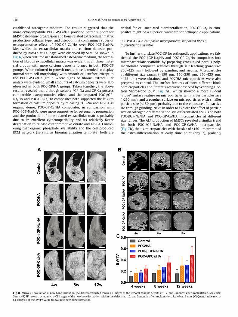
Fig. 8. Micro-CT evaluation of new bone formation. (A) 3D reconstructed micro-CT imag5 mm. (B) 3D reconstructed micro-CT images of the new bone formation within the defecCT analysis of the BV/TV value to evaluate new bone formation.
critical for cell-mediated biomineralization, POC-GP-Ca/HA com-posites might be a superior candidate for orthopedic applications.
3.5. POC-GP/HA composite microparticles supported hMSCsdifferentiation in vitro
To further translate POC-GP for orthopedic applications, we fab-ricated the POC-bGP-Na/HA and POC-GP-Ca/HA composites intomicroparticulate scaffolds by preparing crosslinked porous poly-mer/60%HA composite scaffolds through salt leaching (pore size:250–425 mm), followed by grinding and sieving. Microparticlesat different size ranges (<150 mm; 150–250 mm; 250–425 mm;>425 mm) were obtained and POC/HA microparticles were alsoprepared as control. The surface features of three different kindsof microparticles at different sizes were observed by Scanning Elec-tron Microscope (SEM; Fig. 7A), which showed a more evident‘‘ridge” surface feature on microparticles with larger particles size(>250 mm), and a rougher surface on microparticles with smallerparticle size (<150 mm), probably due to the exposure of bioactiveHA through grinding. Next, in order to explore the effect of particlesize on osteogenic differentiation, we differentiated hMSCs on bothPOC-bGP-Na/HA and POC-GP-Ca/HA microparticles at differentsize ranges. The ALP production of hMSCs revealed a similar trendfor both POC-bGP-Na/HA and POC-GP-Ca/HA microparticles(Fig. 7B), that is, microparticles with the size of <150 mm promotedthe osteo-differentiation at early time point (day 7), probably
es of the femoral condyle defects at 1, 2, and 3 months after implantation. Scale bar:ts at 1, 2, and 3 months after implantation. Scale bar: 1 mm. (C) Quantitative micro-
Y. He et al. / Acta Biomaterialia 93 (2019) 180–191 189
because of the more exposed bioactive HA; while larger size ofmicroparticles were shown to favor ALP production when the dif-ferentiation proceeded to middle stages (day 14), probably due tothe rough surface feature facilitating cell adhesion and 3D interac-tion for differentiation [18]. Eventually, at late stage (day 21),hMSCs cultured on microparticles with different sizes all producedALP to a similar extent. Therefore, a mixture of microparticles ofdifferent sizes were used in the following in vivo evaluation of theircapability to support bone regeneration.
3.6. POC-GP/HA microparticles promoted bone regeneration in femoralcondyle defects
Based on the results from in vitro studies, we further investi-gated the in vivo osteopromotion functionality of both POC-bGP-Na/HA and POC-GP-Ca/HA microparticle scaffolds in a rabbitfemoral condyle defect, a standardized and reproducible defectmodel without the need for fixation that has been successfullyapplied to evaluate the efficacy of particulate bone grafts in bonerepair [18,35]. POC/HA microparticles were also implanted asmaterial control, while defects without any implants served asnegative control. At 4, 8 and 12 weeks after implantation, micro-CT analysis was performed to evaluate the new bone formationwithin defects. The 3D reconstructed images (Fig. 8A) of the defectsclearly showed new bone ingrowth in all three scaffold groupsoccurring from the defect edge towards the defect center, resultingin a gradual decrease of defect size. The bone ingrowth speed in allthree scaffold groups was much higher than that in the controlgroup without any microparticles implanted, although new boneingrowth was also observed in the control group from 4 to 12weeks. 3D reconstructed images of new bone formation withindefects also support the above conclusion (Fig. 8B), displaying sig-nificantly less density signal in the Control group than that in thethree materials groups at each time point. Moreover, both Fig. 8Aand B showed an enhanced new bone formation in the POC-GP-
Fig. 9. Masson staining images of the newly formed bone within the defects implanted wpost-implantation.
Ca/HA group, as compared with that in POC-bGP-Na/HA and POC/HA group. Compared with the previously developed CBPB/HA,iCMBA/HA and BPLP-PSer, which were also citrate-based poly-mer/HA composites, POC-GP-Ca/HA showed excellent osteointe-gration ability and reached a complete new born ingrowthsurrounding implants in a relatively short time (8 weeks)[15,16,18]. Quantitative micro-CT analysis data (Fig. 8C) furtherconfirmed a significantly higher BV/TV value in the POC-GP-Ca/HA group as compared to other control groups, especially at 12weeks. These in vivo results supported the superior performanceof POC-GP-Ca/HA microparticles in promoting new bone forma-tion, which is consistent with the in vitro differentiation results(Fig. 5).
Histological analysis of the newly formed tissue within thedefects were further performed. In the representative Massonstaining images of three different microparticles at 1 month ofimplantation (Fig. 9), blue-stained immature bone growing fromthe defect edge towards its center through the microparticle scaf-folds, with intimate interaction between new bone and materialswith fibrous connective tissue in the defect center was observedin all material groups. There is no separation, such as fibrous cap-sule between the implants and host bone tissue, demonstrating theexcellent osteoconductivity and osteointegration of all citrate-based composite microparticles. At 2-month post implantation,more blue-stained bone was found to deposit directly onto themicroparticles, while red-stained mature bone started to be visiblearound the POC-GP-Ca/HA implants, suggesting that the POC-GP-Ca/HA might favor bone maturation compared with other controlgroups. H&E staining images at 1 month (Fig. 10A & B) further con-firmed a close contact between the new bone and both POC-bGP-Na/HA and POC-GP-Ca/HA microparticles. The connective tissuein POC-GP-Ca/HA group (Fig. 10D) that intertwined with micropar-ticles in the defect center and close to the bone growth frontseemed to be denser than that in the POC-bGP-Na/HA group evenafter 2 months (Fig. 10C). Direct bone deposition with osteoblasts
ith POC/HA, POC-bGPNa/HA and POC-GPCa/HA at (A) 1 month, and (B-C) 2 months
Fig. 10. H&E staining images of tissues surrounding microparticle implants. (A) POC-bGP-Na/HA at 1 month; and (B) POC-GP-Ca/HA at 1 month (scale bar = 500 mm). (C) POC-bGP-Na/HA at 2 months and (D) POC-GP-Ca/HA at 1 month (scale bar = 100 mm).
190 Y. He et al. / Acta Biomaterialia 93 (2019) 180–191
lined up around the microparticles and with visible trapped osteo-cytes were observed in the POC-GP-Ca/HA group at 1 month and inthe POC-GP-Na/HA group at 2 months. Together with the micro-CTanalysis, these results consistently suggested that POC-GP-Ca/HAmicroparticles possessed excellent osteoconductivity and pro-moted bone regeneration in the femoral condyle defects, servingas a better candidate than POC-bGP-Na/HA for orthopedicapplications.
4. Conclusions
In conclusion, in the present study we developed a new class ofosteopromotive bioactive biodegradable materials, POC-bGP-Naand POC-GP-Ca with widely tunable mechanical properties anddegradation. POC-GPs could be synthesized by a simple and cost-effective polycondensation method. Within the formulationsinvestigated, POC-GP-Ca/HA composites demonstrated betterbioactivity in promoting osteopromotive differentiation of hMSCsin vitro and further improving bone regeneration in rabbit femoralcondyle defects. The development of POC-GP expands the reper-toire of the well-recognized citrate-based biomaterials to meetthe ever-increasing needs for functional biomaterials in tissueengineering and other biomedical applications.
Acknowledgements
This work was supported in part by National Institutes of HealthAwards (CA182670, EB024829, and AR072731, to J.Y.), andNational Natural Science Foundation of China (Nos. 81460173,81860326 to L.G. and L.L.), and the Department of Science andTechnology of Yunnan Province of China (Nos. 2017FF117(-062),2018FE001(-165) to L.G.).
Conflict of interest
Dr. Yang and The Pennsylvania State University have a financialinterest in Acuitive Technologies, Inc. These interests have beenreviewed by the University’s Institutional and Individual Conflictof Interest Committees and are currently being managed by theUniversity.
References
[1] C. Laurencin, Y. Khan, S.F. El-Amin, Bone graft substitutes, Expert Rev. Med.Devices 3 (1) (2006) 49–57.
[2] V. Campana, G. Milano, E. Pagano, M. Barba, C. Cicione, G. Salonna, W. Lattanzi,G. Logroscino, Bone substitutes in orthopaedic surgery: from basic science toclinical practice, J. Mater. Sci.: Mater. Med. 25 (10) (2014) 2445–2461.
[3] J. Song, V. Malathong, C.R. Bertozzi, Mineralization of synthetic polymerscaffolds: a bottom-up approach for the development of artificial bone, J. Am.Chem. Soc. 127 (10) (2005) 3366–3378.
[4] S. Bhumiratana, W.L. Grayson, A. Castaneda, D.N. Rockwood, E.S. Gil, D.L.Kaplan, G. Vunjak-Novakovic, Nucleation and growth of mineralized bonematrix on silk-hydroxyapatite composite scaffolds, Biomaterials 32 (11) (2011)2812–2820.
[5] B.M. Chesnutt, A.M. Viano, Y. Yuan, Y. Yang, T. Guda, M.R. Appleford, J.L. Ong,W.O. Haggard, J.D. Bumgardner, Design and characterization of a novelchitosan/nanocrystalline calcium phosphate composite scaffold for boneregeneration, J. Biomed. Mater. Res. A 88 (2) (2009) 491–502.
[6] S.L. Liang, W.D. Cook, G.A. Thouas, Q.Z. Chen, The mechanical characteristicsand in vitro biocompatibility of poly(glycerol sebacate)-bioglass elastomericcomposites, Biomaterials 31 (33) (2010) 8516–8529.
[7] H. Liu, T.J. Webster, Mechanical properties of dispersed ceramic nanoparticlesin polymer composites for orthopedic applications, Int. J. Nanomed. 5 (2010)299–313.
[8] R.L. Hartles, Citrate in mineralized tissues, Adv. Oral Biol. 1 (1964) 225–253.[9] Y.Y. Hu, X.P. Liu, X. Ma, A. Rawal, T. Prozorov, M. Akinc, S.K. Mallapragada, K.
Schmidt-Rohr, Biomimetic self-assembling copolymer�hydroxyapatitenanocomposites with the nanocrystal size controlled by citrate, Chem.Mater. 23 (9) (2011) 2481–2490.
[10] C. Ma, E. Gerhard, D. Lu, J. Yang, Citrate chemistry and biology for biomaterialsdesign, Biomaterials 178 (2018) 383–400.
[11] R.T. Tran, J. Yang, G.A. Ameer, Citrate-based biomaterials and their applicationsin regenerative engineering, Ann. Rev. Mater. Res. 45 (2015) 277–310.
Y. He et al. / Acta Biomaterialia 93 (2019) 180–191 191
[12] E.J. Chung, P. Kodali, W. Laskin, J.L. Koh, G.A. Ameer, Long-term in vivoresponse to citric acid-based nanocomposites for orthopaedic tissueengineering, J. Mater. Sci. Mater. Med. 22 (9) (2011) 2131–2138.
[13] Y. Guo, R.T. Tran, D. Xie, Y. Wang, D.Y. Nguyen, E. Gerhard, J. Guo, J. Tang, Z.Zhang, X. Bai, J. Yang, Citrate-based biphasic scaffolds for the repair of largesegmental bone defects, J. Biomed. Mater. Res. A 103 (2) (2015) 772–781.
[14] D. Sun, Y. Chen, R.T. Tran, S. Xu, D. Xie, C. Jia, Y. Wang, Y. Guo, Z. Zhang, J. Guo, J.Yang, D. Jin, X. Bai, Citric acid-based hydroxyapatite composite scaffoldsenhance calvarial regeneration, Sci. Rep. 4 (2014) 6912-–6922.
[15] R.T. Tran, L. Wang, C. Zhang, M. Huang, W. Tang, C. Zhang, Z. Zhang, D. Jin, B.Banik, J.L. Brown, Z. Xie, X. Bai, J. Yang, Synthesis and characterization ofbiomimetic citrate-based biodegradable composites, J. Biomed. Mater. Res. A102 (8) (2014) 2521–2532.
[16] D. Xie, J. Guo, M. Mehdizadeh, R.T. Tran, R. Chen, D. Sun, G. Qian, D. Jin, X. Bai, J.Yang, Development of injectable citrate-based bioadhesive bone implants, J.Mater. Chem. B, Mater. Biol. Med. 3 (3) (2015) 387–398.
[17] J. Tang, J. Guo, Z. Li, C. Yang, D. Xie, J. Chen, S. Li, S. Li, G.B. Kim, X. Bai, Z. Zhang,J. Yang, Fast degradable citrate-based bone scaffold promotes spinal fusion, J.Mater. Chem. B, Mater. Biol. Med. 3 (27) (2015) 5569–5576.
[18] C. Ma, X. Tian, J.P. Kim, D. Xie, X. Ao, D. Shan, Q. Lin, M.R. Hudock, X. Bai, J. Yang,Citrate-based materials fuel human stem cells by metabonegenic regulation,Proc. Natl. Acad. Sci. 115 (50) (2018) 11741–11750.
[19] S. Prasadh, R.C.W. Wong, Unraveling the mechanical strength of biomaterialsused as a bone scaffold in oral and maxillofacial defects, Oral Sci. Int. 15 (2)(2018) 48–55.
[20] J. Dey, H. Xu, J. Shen, P. Thevenot, S.R. Gondi, K.T. Nguyen, B.S. Sumerlin, L.Tang, J. Yang, Development of biodegradable crosslinked urethane-dopedpolyester elastomers, Biomaterials 29 (35) (2008) 4637–4649.
[21] J. Guo, Z. Xie, R.T. Tran, D. Xie, D. Jin, X. Bai, J. Yang, Click chemistry plays a dualrole in biodegradable polymer design, Adv. Mater. 26 (12) (2014) 1906–1911.
[22] S. Saravanan, S. Vimalraj, P. Thanikaivelan, S. Banudevi, G. Manivasagam, Areview on injectable chitosan/beta glycerophosphate hydrogels for bone tissueregeneration, Int. J. Biol. Macromol. 121 (2019) 38–54.
[24] J. Kucinska-Lipka, I. Gubanska, O. Korchynskyi, K. Malysheva, M. Kostrzewa, D.Włodarczyk, J. Karczewski, H. Janik, The influence of calcium glycerophosphate
(GPCa) modifier on physicochemical mechanical, and biological performanceof polyurethanes applicable as biomaterials for bone tissue scaffoldsfabrication, Polymers 9 (8) (2017) 329–350.
[25] S. Morochnik, Y. Zhu, C. Duan, M. Cai, R.R. Reid, T.C. He, J. Koh, I. Szleifer, G.A.Ameer, A thermoresponsive, citrate-based macromolecule for boneregenerative engineering, J. Biomed. Mater. Res. Part A 106 (6) (2018) 1743–1752.
[26] T. Holland, E. Bodde, V. Cuijpers, L. Baggett, Y. Tabata, A. Mikos, J. Jansen,Degradable hydrogel scaffolds for in vivo delivery of single and dual growthfactors in cartilage repair, Osteoarthritis Cartilage 15 (2) (2007) 187–197.
[27] L.A. Solchaga, J.S. Temenoff, J. Gao, A.G. Mikos, A.I. Caplan, V.M. Goldberg,Repair of osteochondral defects with hyaluronan-and polyester-basedscaffolds, Osteoarthritis Cartilage 13 (4) (2005) 297–309.
[28] Y. Liu, X.Z. Shu, G.D. Prestwich, Osteochondral defect repair with autologousbone marrow-derived mesenchymal stem cells in an injectable situ, cross-linked synthetic extracellular matrix, Tissue Eng. 12 (12) (2006) 3405–3417.
[29] M.I. Tejedor-Tejedor, M.A. Anderson, Protonation of phosphate on the surfaceof goethite as studied by CIR-FTIR and electrophoretic mobility, Langmuir 6(1990) 602–612.
[30] G.A. Holzapfel, Biomechanics of soft tissue, in: The Handbook of MaterialsBehavior Models 3, 2001, pp. 1049–1063.
[31] H.J. Sung, C. Meredith, C. Johnson, Z.S. Galis, The effect of scaffold degradationrate on three-dimensional cell growth and angiogenesis, Biomaterials 25 (26)(2004) 5735–5742.
[32] J. Hu, K. Peng, J. Guo, D. Shan, G.B. Kim, Q. Li, E. Gerhard, L. Zhu, W. Tu, W. Lv, M.A. Hickner, J. Yang, Click cross-linking-improved waterborne polymers forenvironment-friendly coatings and adhesives, ACS Appl. Mater. Interfaces 8(2016) 17499–117410.
[33] H. Qiu, J. Yang, P. Kodali, J. Koh, G.A. Ameer, A citric acid-based hydroxyapatitecomposite for orthopedic implants, Biomaterials 27 (34) (2006) 5845–5854.
[34] S.L. Goss, K.A. Lemons, J.E. Kerstetter, R.H. Bogner, Determination of calciumsalt solubility with changes in pH and PCO2, simulating varyinggastrointestinal environments, J. Pharm. Pharmacol. 59 (11) (2007) 1485–1492.
[35] M.J. Coathup, Q. Cai, C. Campion, T. Buckland, G.W. Blunn, The effect of particlesize on the osteointegration of injectable silicate-substituted calciumphosphate bone substitute materials, J. Biomed. Mater. Res. B Appl.Biomater. 101 (6) (2013) 902–910.