33
Diabetic Foot Infections Improving Outcomes (or why I’m not going into vascular!) John C. Lantis II, MD Assistant Professor of Surgery College of Physicians and Surgeons Columbia University
| Date post: | 17-Jul-2016 |
| Category: |
Documents |
| Upload: | siusiuwidyanto |
| View: | 18 times |
| Download: | 0 times |
Diabetic Foot InfectionsImproving Outcomes
(or why I’m not going into vascular!)John C. Lantis II, MD
Assistant Professor of SurgeryCollege of Physicians and Surgeons
Columbia University
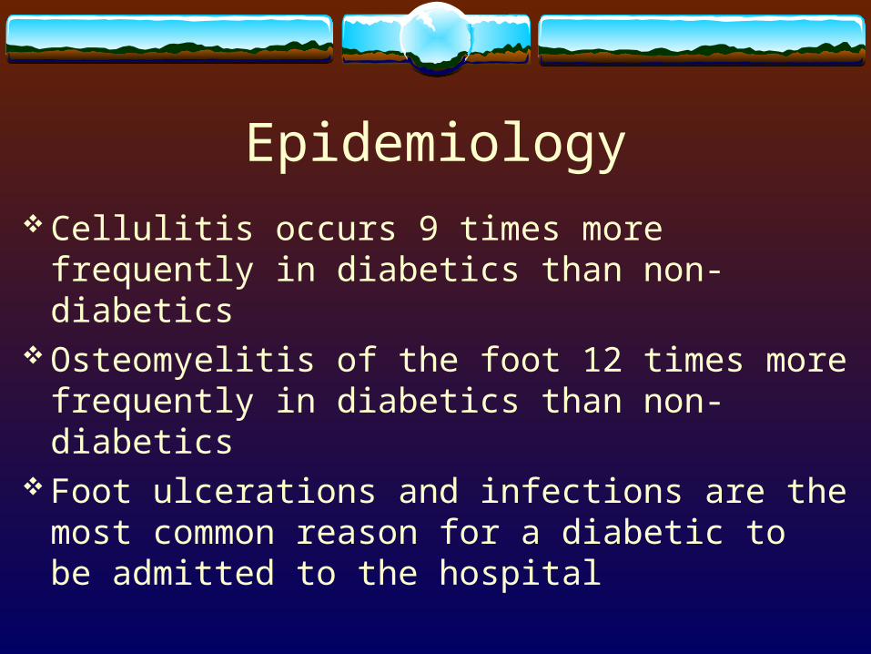
Epidemiology Cellulitis occurs 9 times more frequently in
diabetics than non-diabetics Osteomyelitis of the foot 12 times more frequently
in diabetics than non-diabetics Foot ulcerations and infections are the most
common reason for a diabetic to be admitted to the hospital

Epidemiology 25 % of diabetics will develop a foot ulcer 40-80% of these ulcers will become infected 25 % of these will become deep 50 % of patients with cellulitis will have another
episode within 2 years

Epidemiology(of amputation)
25-50 % of diabetic foot infections lead to minor amputations
10-40 % require major amputations 10-30 % of patients with a diabetic foot ulcer will
go on to amputation

Pathophysiology Metabolic derangement Faulty wound healing Neuropathy Angiopathy Mechanical stress Patient and provider neglect
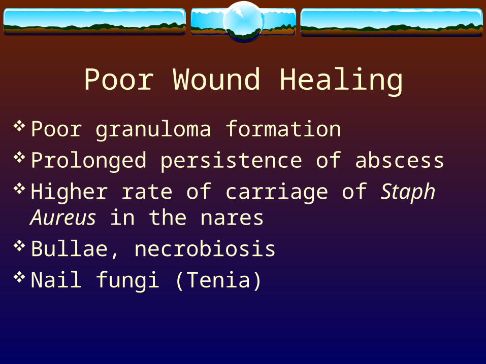
Poor Wound Healing Poor granuloma formation Prolonged persistence of abscess Higher rate of carriage of Staph Aureus in the
nares Bullae, necrobiosis Nail fungi (Tenia)
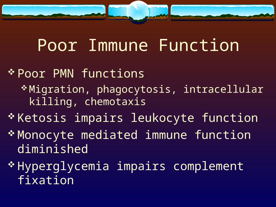
Poor Immune Function Poor PMN functions
Migration, phagocytosis, intracellular killing, chemotaxis
Ketosis impairs leukocyte function Monocyte mediated immune function diminished Hyperglycemia impairs complement fixation

Sensory Neuropathy Unaware of a foreign body
Pressure in shoesAbrasions in shoesTears or brakes in the skin

Motor Neuropathy Architectural deformities
Hammer or claw toeHigh plantar archSubluxation of metatarsals
Autonomic Neuropathy Anhidrosis
Dry, cracked skin Arterial to venous shunting Temperature regulation disorders

Angiopathy Can play a primary role
Microangiopathy +/- Certainly plays a primary role in healing
Pulsatile flow will augment healing

Foot Anatomy Compartments, low amount of soft tissue, tendon sheaths Deep plantar space
Medial, central and lateral Rigid fascial structures
Edema – rapidly elevates compartment pressures Ischemic necrosis Infections spread between compartments
Calcaneal convergence, direct perforation of the septae
Microbiology Infection – invasion of host tissue by pathogens,
which elicits a host inflammatory response (erythema, induration, pain or tenderness, warmth, loss of function)
Superficial-confined to skin supeficial to fascia Deep-invasion of fascia, muscle, tendon, joint or
bone

Microbiology Normal skin bacteria
Coag neg Staph, alpha-hemolytic strep, corynebacteriae Acute wound
Monomicrobial (Gram positive) Chronic wound
Polymicrobial (GNRs, Anaerobes, enterococcus, GPCs)

Wound Cultures Uninfected wound
If concerned about unique pathogen - MRSA Infected wound
Help tailor and constrain antibiotic therapyAntibiotic naïve wound – staph or strep aloneAntibiotic resistant organisms

Wound Cultures Deep space pus – most accurate Curretage or tissue scraping from the base of a
debrided ulcer gives the best information - next most accurate
Cotton swab across the surface is of little utility
Wound Cultures Staph Aureus – most important pathogen in
diabetic foot Serious infections are usually caused by 3 to 5
bacterial species GNR – Enterobacteriaciae – chronic or previously
treated wounds Pseudomonas – often in wounds treated with
hydrotherapy or wet dressings

Diagnosis Clinical presentation
Presence of purulence Pain, swelling, ulceration, sinus tract formation, crepitation Systemic infection (fever, rigors, vomitting, tachycardia, change
in mental status, malaise) Surprisingly uncommon
Metabolic disorder (hyperglycemia, ketosis, azotemia) Should be considered even when local signs are less severe
Clinical Presentation 60 years old 66 % male DM 15-20 years 66 % PVD 80 % loss of protective sensation 33 % have lesion for > 1 month 50% lack – fever, leukocytosis or elevated ESR
Evaluation Describe lesion and drainage Enumerate signs of infalmmation Define whether infection is present and cause Examine soft tissue for crepitus, sinus tract,
abscess Probe skin breaks with sterile metal probe and see
if skin can be reached
Evaluation Measure wound (? Photograph ?) Determine inflow Neurologic status? Sensation, motor, autonomic Cleanse and debride wound Culture the cleansed wound (curettage) Plain radiographs

Osteomyelitis 50-60 % complication in severe foot infections Where in the foot is the lesion? Vascular supply to the area Degree of systemic illness Two classifications systems
WaldvogelCleary and Mader

Osteomyelitis Larger (>2cm) Deeper (>3mm) ESR > 70 mm/hr If you can touch bone 90% correlation with osteo Xray – changes take 2 weeks to occur
Sensitivity 55 %, specificity 75%Focal osteopenia, cortical erosions, periosteal reaction

Osteomyelitis Bone (technitium Tc 99)
85% sensitive, 45% specific Leukocyte scans
85% sensitive, 75% specific MRI
Sensitivity > 90%, specificity > 80 %Can miss early changes, mis-read evolving neuropathic
osteoarthropathy

Osteomyelits Etiologic organisms
Staph aureus – 40% of infectionsStreptococci – 30%Staph epidermidis – 25%Enterobacteriaceae – 40%
Treatment Debridement
Minor- Remove all necrotic tissue including escharRemove all callusSharply saucerize the woundDebride boneRepeat visits are normal

Treatment Surgical
“Salvage the foot but not at the expense of the leg or the patient”
Early surgical debridement decreases LOS, improves foot salvage and decreases morbidity and mortality
All necrotic tissue and pus

Treatment Plantar abscess
Disappearance of the longitudinal arch and skin creasesFoot edemaCentral plantar infections – worse outcomesWide incision and drainage necessary

Treatment Antibiotics
Do not improve outcomes of non-infected lesions In PVD – therapeutic antibiotic levels are not achieved
in infected tissuesMild infection –Topical therapy
Peptide antibiotic Pexiganin acetate 1% cream nearly as effective as oral ofloxacin

Treatment Empiric antibiotic therapy
StaphStrepGNREnterococcusAnaerobes*Tailor to clinical progress
Treatment Prospective studies they all work and there really
isn’t a difference Cost is an issue

Antibiotic thoughts Mild (po) – Augmentin/Levofloxacin (+Clinda)
Bactrim/Flagyl Moderate (IV until stable then po)
Unasyn or other Gorilla-cillin Clinda & Levofloxacin
Severe (IV only) Imipenem Amp/Tobra/Clinda Vanco/Aztreonam/Flagyl

Antibiotic thoughts Duration of therapy
No good studiesOnce active infection resolved plus 2 daysOsteomyelitis
6 weeksCan use Flouoquinolones and clindamycin


