9 Diagnosis and Endoscopic Treatments of Rectal Varices Takahiro Sato, Katsu Yamazaki and Jun Akaike Department of Gastroenterology, Sapporo Kosei General Hospital, Sapporo Japan 1. Introduction Esophagogastric varices are considered to be the most common complication in patients with portal hypertension, while ectopic varices, that is, those outside of the esophagogastric region, are less common. Rectal varices represent portal systemic collaterals that are manifested as discrete dilated submucosal veins and constitute a pathway for portal venous flow between the superior rectal veins of the inferior mesenteric system and the middle inferior rectal veins of the iliac system. Rectal varices are an infrequent but potentially serious cause of hematochezia. Massive bleeding from rectal varices occurs rarely, with a frequency ranging from 0.5% to 3.6% (1-3). In this chapter, we describe the diagnostic modalities and endoscopic treatments for rectal varices in patients with portal hypertension. 2. Diagnosis of rectal varices Endoscopy is the principal method for diagnosis of rectal varices. Endoscopic ultrasonography (EUS) can detect the presence and number of rectal varices better than endoscopy (4). Recently, color Doppler ultrasonography has allowed us to detect fine small blood flow (5). Sato et al. have reported the usefulness of percutaneous color Doppler ultrasonography (CDUS) for the hemodynamic evaluation of rectal varices (6). Although endoscopic injection sclerotherapy (EIS) (7) and endoscopic band ligation (EBL) (8) for esophageal varices are well-established therapies, there is no standard treatment for rectal varices. In this article, we also review the therapeutic effects and complications of EIS versus EBL on rectal varices in patients with portal hypertension. Several diagnostic procedures have been performed to evaluate rectal varices, including endoscopy, magnetic resonance (MR) angiography, EUS. Endoscopy is the principal method for diagnosis of rectal varices and MR angiography is useful for evaluating the overall portosystemic collateral circulation. On the other hand, conventional EUS (7.5 or 12 MHz) reveals rectal varices as rounded, oval, or longitudinal echo-free structures in the submucosa and also shows perirectal veins outside the rectal wall (4,9,10). With endoscopic color Doppler ultrasonography (ECDUS), color flow images in blood vessels can be obtained, and ECDUS allows for more detailed observation of the hemodynamics of rectal varices than EUS (11). CDUS is a simpler, more non-invasive method than ECDUS and it enables us to detect slight blood flow and to evaluate the portal venous system. Nelson et al. concluded that CDUS was valuable for accurate determination of the direction of portal flow and patency of the vessel (12). Sato et al. have reported the www.intechopen.com
Transcript
9
Diagnosis and Endoscopic Treatments of Rectal Varices
Takahiro Sato, Katsu Yamazaki and Jun Akaike Department of Gastroenterology, Sapporo Kosei General Hospital, Sapporo
Japan
1. Introduction
Esophagogastric varices are considered to be the most common complication in patients with portal hypertension, while ectopic varices, that is, those outside of the esophagogastric region, are less common. Rectal varices represent portal systemic collaterals that are manifested as discrete dilated submucosal veins and constitute a pathway for portal venous flow between the superior rectal veins of the inferior mesenteric system and the middle inferior rectal veins of the iliac system. Rectal varices are an infrequent but potentially serious cause of hematochezia. Massive bleeding from rectal varices occurs rarely, with a frequency ranging from 0.5% to 3.6% (1-3). In this chapter, we describe the diagnostic modalities and endoscopic treatments for rectal varices in patients with portal hypertension.
2. Diagnosis of rectal varices
Endoscopy is the principal method for diagnosis of rectal varices. Endoscopic
ultrasonography (EUS) can detect the presence and number of rectal varices better than
endoscopy (4). Recently, color Doppler ultrasonography has allowed us to detect fine small
blood flow (5). Sato et al. have reported the usefulness of percutaneous color Doppler
ultrasonography (CDUS) for the hemodynamic evaluation of rectal varices (6).
Although endoscopic injection sclerotherapy (EIS) (7) and endoscopic band ligation (EBL) (8) for esophageal varices are well-established therapies, there is no standard treatment for rectal varices. In this article, we also review the therapeutic effects and complications of EIS versus EBL on rectal varices in patients with portal hypertension. Several diagnostic procedures have been performed to evaluate rectal varices, including endoscopy, magnetic resonance (MR) angiography, EUS. Endoscopy is the principal method for diagnosis of rectal varices and MR angiography is useful for evaluating the overall portosystemic collateral circulation. On the other hand, conventional EUS (7.5 or 12 MHz) reveals rectal varices as rounded, oval, or longitudinal echo-free structures in the submucosa and also shows perirectal veins outside the rectal wall (4,9,10). With endoscopic color Doppler ultrasonography (ECDUS), color flow images in blood vessels can be obtained, and ECDUS allows for more detailed observation of the hemodynamics of rectal varices than EUS (11). CDUS is a simpler, more non-invasive method than ECDUS and it enables us to detect slight blood flow and to evaluate the portal venous system. Nelson et al. concluded that CDUS was valuable for accurate determination of the direction of portal flow and patency of the vessel (12). Sato et al. have reported the
www.intechopen.com
Endoscopic Procedures in Colon and Rectum
146
usefulness of CDUS for the hemodynamic evaluation of rectal varices and compared velocities of rectal varices with CDUS and colonoscopic findings, and they concluded that CDUS was a useful noninvasive tool in the evaluation of portal hemodynamics, including the observation of blood flow in rectal varices (6).
2.1 Diagnosis of rectal varices via endoscopy
Endoscopy is the principal method for the diagnosis of rectal varices; it is a useful modality for diagnosing and observing rectal varices of a certain size and extent, and has a very sensitive predictive value for variceal hemorrhage. The endoscopic findings for rectal varices were evaluated according to the grading system outlined in‘The General Rules for
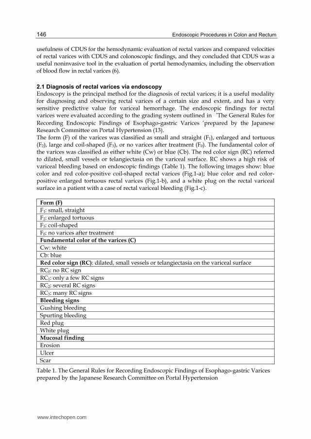
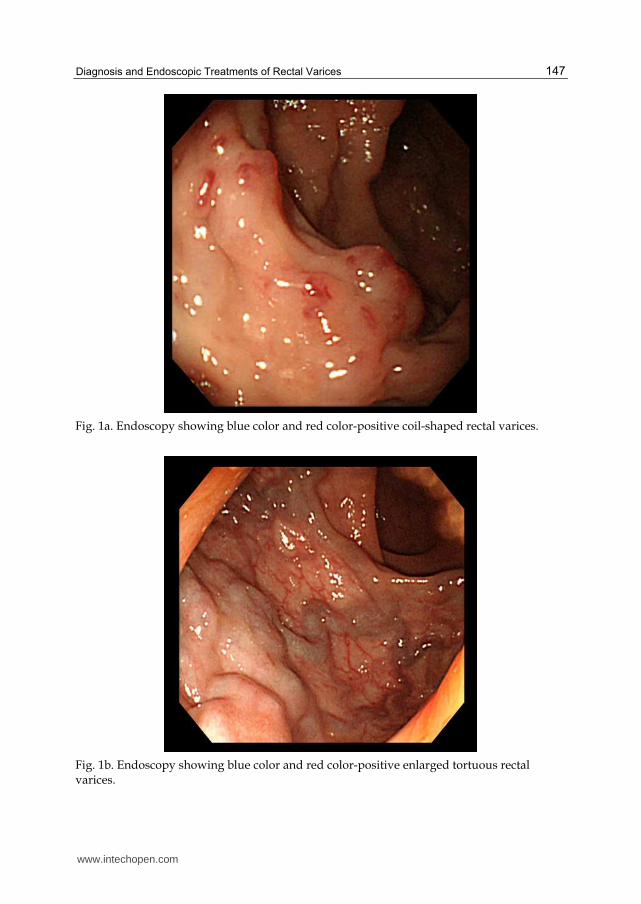
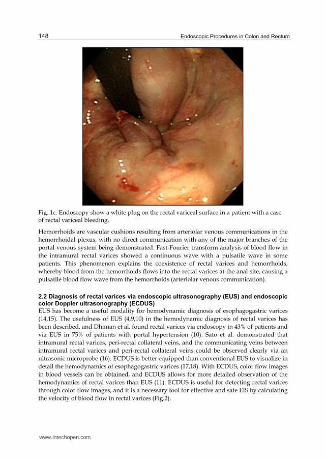
Recording Endoscopic Findings of Esophago-gastric Varices ’prepared by the Japanese Research Committee on Portal Hypertension (13). The form (F) of the varices was classified as small and straight (F1), enlarged and tortuous (F2), large and coil-shaped (F3), or no varices after treatment (F0). The fundamental color of the varices was classified as either white (Cw) or blue (Cb). The red color sign (RC) referred to dilated, small vessels or telangiectasia on the variceal surface. RC shows a high risk of variceal bleeding based on endoscopic findings (Table 1). The following images show: blue color and red color-positive coil-shaped rectal varices (Fig.1-a); blue color and red color-positive enlarged tortuous rectal varices (Fig.1-b), and a white plug on the rectal variceal surface in a patient with a case of rectal variceal bleeding (Fig.1-c).
Form (F)
F1: small, straight
F2: enlarged tortuous
F3: coil-shaped
F0: no varices after treatment
Fundamental color of the varices (C)
Cw: white
Cb: blue
Red color sign (RC): dilated, small vessels or telangiectasia on the variceal surface
RC0: no RC sign
RC1: only a few RC signs
RC2: several RC signs
RC3: many RC signs
Bleeding signs
Gushing bleeding
Spurting bleeding
Red plug
White plug
Mucosal finding
Erosion
Ulcer
Scar
Table 1. The General Rules for Recording Endoscopic Findings of Esophago-gastric Varices prepared by the Japanese Research Committee on Portal Hypertension
www.intechopen.com
Diagnosis and Endoscopic Treatments of Rectal Varices
147
Fig. 1a. Endoscopy showing blue color and red color-positive coil-shaped rectal varices.
Fig. 1b. Endoscopy showing blue color and red color-positive enlarged tortuous rectal varices.
www.intechopen.com
Endoscopic Procedures in Colon and Rectum
148
Fig. 1c. Endoscopy show a white plug on the rectal variceal surface in a patient with a case of rectal variceal bleeding.
Hemorrhoids are vascular cushions resulting from arteriolar venous communications in the
hemorrhoidal plexus, with no direct communication with any of the major branches of the
portal venous system being demonstrated. Fast-Fourier transform analysis of blood flow in
the intramural rectal varices showed a continuous wave with a pulsatile wave in some
patients. This phenomenon explains the coexistence of rectal varices and hemorrhoids,
whereby blood from the hemorrhoids flows into the rectal varices at the anal site, causing a
pulsatile blood flow wave from the hemorrhoids (arteriolar venous communication).
2.2 Diagnosis of rectal varices via endoscopic ultrasonography (EUS) and endoscopic color Doppler ultrasonography (ECDUS)
EUS has become a useful modality for hemodynamic diagnosis of esophagogastric varices
(14,15). The usefulness of EUS (4,9,10) in the hemodynamic diagnosis of rectal varices has
been described, and Dhiman et al. found rectal varices via endoscopy in 43% of patients and
via EUS in 75% of patients with portal hypertension (10). Sato et al. demonstrated that
intramural rectal varices, peri-rectal collateral veins, and the communicating veins between
intramural rectal varices and peri-rectal collateral veins could be observed clearly via an
ultrasonic microprobe (16). ECDUS is better equipped than conventional EUS to visualize in
detail the hemodynamics of esophagogastric varices (17,18). With ECDUS, color flow images
in blood vessels can be obtained, and ECDUS allows for more detailed observation of the
hemodynamics of rectal varices than EUS (11). ECDUS is useful for detecting rectal varices
through color flow images, and it is a necessary tool for effective and safe EIS by calculating
the velocity of blood flow in rectal varices (Fig.2).
www.intechopen.com
Diagnosis and Endoscopic Treatments of Rectal Varices
149
Fig. 2. Color flow images of rectal varices and inflowing vessel with endoscopic color Doppler ultrasonography
2.3 Diagnosis of rectal varices via percutaneous color Doppler ultrasonography (CDUS)
Recently, color Doppler ultrasonography has become widely accepted for the assessment of
the hemodynamics of abdominal vascular systems, but few color Doppler findings of
gastrointestinal varices have been reported. Komatsuda et al. reported the usefulness of
CDUS for the diagnosis of gastric and duodenal varices (19), and Sato et al. concluded that
CDUS was useful for evaluating the hemodynamics of gastric varices (20). CDUS cannot be
performed successfully without a suitable acoustic window. Impediments such as bowel
gas, body habitus, and cirrhosis limit the value of sonography for assessing the portal
venous system. In addition, with color Doppler sonography, it is difficult to observe the
collateral veins situated far from the probe due to the limitations of Doppler sensitivity. The
rectal wall was detected at the back area of the vagina in females or prostate in males by
sonography and rectal varices could be observed through the bladder filled with urine via
color Doppler ultrasonography (Fig.3).
www.intechopen.com
Endoscopic Procedures in Colon and Rectum
150
Fig. 3. Color flow images of rectal varices with color Doppler ultrasonography.
www.intechopen.com
Diagnosis and Endoscopic Treatments of Rectal Varices
151
Sato et al. compared the velocities of rectal varices with CDUS and colonoscopic findings (6).
The majority of the 44 cases underwent colonoscopy after diagnosis with color Doppler. In
this study, the mean velocity of the F2 type rectal varices was significantly higher than that
of the F1 type, and the mean velocity of the RC-positive varices was significantly higher
than that of RC-negative varices with color Doppler ultrasonography. These results suggest
that the measurement of velocity in rectal varices via color Doppler ultrasonography is
useful in diagnosing the grade of rectal varices. CDUS is a useful noninvasive tool in the
evaluation of portal hemodynamics, including the observation of blood flow in rectal
varices.
3. Endoscopic treatments of rectal varices
Rectal varices are considered to occur infrequently, however, several articles have reported that they occur with high frequency in patients with hepatic abnormalities (21-23). Hosking et al. reported that 44 % of 100 consecutive cirrhotic patients had anorectal varices (21). Other studies found that the prevalence of anorectal varices was 78% in 72 portal hypertensive patients (22) and 43% in 103 cirrhotic patients (23). Although EIS and EBL for esophageal varices are well-established therapies, there is no standard treatment for rectal varices. Various medical treatments have been used to control bleeding from rectal varices, but none of these is currently considered to be a standard method. Surgical approaches include portosystemic shunting, ligation, and under-running suturing (21). Some investigators have reported that interventional radiologic techniques such as transjugular intrahepatic portosystemic shunts were successfully employed for rectal variceal bleeding (24-26). Several cases of successful treatment of rectal varices with endoscopic treatments have been reported. Wang et al. first reported the usefulness of EIS in treating rectal varices and found it to be effective for controlling bleeding (27). EBL was introduced as a new method for treating esophageal varices, and it is reportedly both easier to perform and safer than EIS. Several cases of successful treatment of rectal varices using EBL have been reported (28-30).
3.1 EIS for rectal varices
We performed EIS in 21 of the 30 patients, who were successfully treated without
complications. EIS was performed using 5% ethanolamine oleate with iopamidol (5%EOI),
which was injected intermittently under fluoroscopy. The procedure was performed using a
flexible GI endoscope (GIF XQ200; Olympus Optical Co., Ltd., Tokyo, Japan) by a free-hand
method, using a 25-gauge injection needle. EIS was repeated every week until the
disappearance of all rectal varices and RC signs were confirmed by endoscopy. Fluoroscopic
observation with infusion of 5%EOI was performed to determine the extent of the varices,
taking care that 5%EOI did not flow into the systemic circulation. We decided the amount of
5%EOI on depiction of passageways (superior rectal vein) of rectal varices. After EIS,
colonoscopy revealed shrinkage of the rectal varices in all 21 patients, with no complications
reported.
It is necessary to evaluate the hemodynamics of the rectal varices before EIS to avoid severe
complications such as pulmonary embolism, and the sclerosant should be injected slowly
under fluoroscopy (Fig.4).
www.intechopen.com
Endoscopic Procedures in Colon and Rectum
152
Fig. 4. Fluoroscopic observation with infusion of 5%EOI was performed.
3.2 EBL for rectal varices
EBL was introduced as a new method for treating esophageal varices, and it is reportedly
both easier to perform and safer than EIS. Several cases of successful treatment of rectal
varices using EBL have been reported. Levine et al. treated rectal varices initially with EIS,
and 1 week later, EBL was performed on the remaining rectal varices (28). These
investigators described EBL as a safe and effective therapy for rectal varices (30).
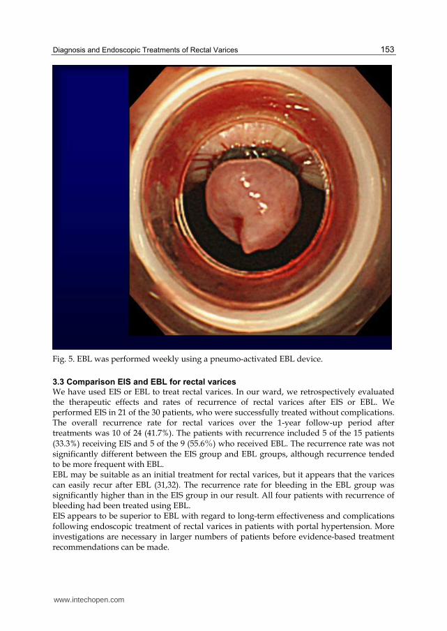
EBL was performed in 9 patients on our ward; it was performed weekly using a pneumo-
activated EBL device (Sumitomo Bakelite, Tokyo, Japan), and bands were placed on the
varices. An overtube was not used during EBL (Fig.5). After EBL, colonoscopy revealed
ulcers and improvement of the varices in the rectum of all 9 patients. Eight of the 9 patients
experienced no operative complications. However, colonoscopy revealed bleeding from
ulcers after EBL in 1 case, in whom endoscopic clipping was performed on the oozing
ulcers.
www.intechopen.com
Diagnosis and Endoscopic Treatments of Rectal Varices
153
Fig. 5. EBL was performed weekly using a pneumo-activated EBL device.
3.3 Comparison EIS and EBL for rectal varices
We have used EIS or EBL to treat rectal varices. In our ward, we retrospectively evaluated the therapeutic effects and rates of recurrence of rectal varices after EIS or EBL. We performed EIS in 21 of the 30 patients, who were successfully treated without complications. The overall recurrence rate for rectal varices over the 1-year follow-up period after treatments was 10 of 24 (41.7%). The patients with recurrence included 5 of the 15 patients
(33.3%) receiving EIS and 5 of the 9 (55.6%) who received EBL. The recurrence rate was not significantly different between the EIS group and EBL groups, although recurrence tended to be more frequent with EBL. EBL may be suitable as an initial treatment for rectal varices, but it appears that the varices can easily recur after EBL (31,32). The recurrence rate for bleeding in the EBL group was significantly higher than in the EIS group in our result. All four patients with recurrence of bleeding had been treated using EBL. EIS appears to be superior to EBL with regard to long-term effectiveness and complications following endoscopic treatment of rectal varices in patients with portal hypertension. More investigations are necessary in larger numbers of patients before evidence-based treatment recommendations can be made.
www.intechopen.com
Endoscopic Procedures in Colon and Rectum
154
4. Conclusion
Hemorrhage from rectal varices should be kept in mind in patients with portal hypertension presenting with lower gastrointestinal bleeding. It is difficult to determine the best treatment strategy for rectal varices because of inaccessibility, initial difficulty in diagnosis and subsequent difficulty in treatment.
5. Acknowledgment
The authors thank Dr Jouji Toyota, Dr Yoshiyasu Karino, Dr Takumi Ohmura of Sapporo Kosei Hospital, for their help with the manuscript.
6. References
[1] McCormack TT, Bailey HR, Simms JM, Johnson AG. (1984). Rectal varices are not piles.
Br J Surg 71:163.
[2] Johansen K, Bardin J, Orloff MJ. (1980). Massive bleeding from hemorrhoidal varices in
portal hypertension. JAMA 224:2084-5.
[3] Wilson SE, Stone RT, Christie JP, Passaro E. (1979). Massive lower gastrointestinal
bleeding from intestinal varices. Arch Surg 114:1158-61.
[4] Dhiman RK, Choudhuri G, Saraswat VA, et al. (1993). Endoscopic ultrasonographic
evaluation of the rectum in cirrhotic portal hypertension. Gastrointest Endosc 39:635-
40.
[5] Ueno N, Sasaki A, Tomiyama T, et al. (1997). Color Doppler ultrasonography in the
diagnosis of cavernous transformation of the portal vein. J Clin Ultrasound 25:227-
33.
[6] Sato T, Yamazaki K, Toyota J, Karino Y, Ohmura T, Akaike J. (2007). Diagnosis of rectal
varices via color Doppler ultrasonography. Am J Gastroenterol 102:2253-8.
[7] The Veterans Affairs Cooperative Variceal Sclerotherapy Group. (1991). Prophylactic
sclerotherapy for esophageal varices in men with alcoholic liver disease. N Engl J
InTech ChinaUnit 405, Office Block, Hotel Equatorial Shanghai No.65, Yan An Road (West), Shanghai, 200040, China
Phone: +86-21-62489820 Fax: +86-21-62489821
Endoscopic procedures in colon and rectum presents nine chapters which start with introductory ones likescreening by colonoscopy as the preparation and monitoring for this exam. In addition to these approachesthe book aims in the last four chapters to explain endoscopic diagnostic and therapeutic aspects in the colonand rectum. The description of each text is very comprehensive, instructive and easy to understand andpresents the most current practices on the topics described. This book is recommended for general andcolorectal surgeons as it presents guidelines for diagnosis and treatment which are very well established.
How to referenceIn order to correctly reference this scholarly work, feel free to copy and paste the following:
Takahiro Sato, Katsu Yamazaki and Jun Akaike (2011). Diagnosis and Endoscopic Treatments of RectalVarices, Endoscopic Procedures in Colon and Rectum, Prof. Jose Ribeiro Da Rocha (Ed.), ISBN: 978-953-307-677-5, InTech, Available from: http://www.intechopen.com/books/endoscopic-procedures-in-colon-and-rectum/diagnosis-and-endoscopic-treatments-of-rectal-varices


![Transanal endoscopic microsurgery for radical resection of ...Transanal endoscopic microsurgery in sigmoid cancer 1451 JBUON 2019; 24(4): 1451 large rectal polyps by TEM [11]. However,](https://static.documents.pub/doc/80x56/60f7e35c60455642d5494ef7/transanal-endoscopic-microsurgery-for-radical-resection-of-transanal-endoscopic.jpg)


