Page 1
5/4/2012
1
Diagnosis and Treatment of Ocular Surface Conditions:
Focus on Allergy and Conjunctivitis
C. Lisa Prokopich OD, MSc, FAAO
University of Waterloo, School of Optometry and Vision Science
COPE Course Code ID: 31665-SD Expires: 04/01/2014 Qualified Credit: 2 hours
Thank you also to Drs. Karpecki, Melton, Thomas, Bartlett
and Michaud for their contributions.
Learning Objectives
After completing this lesson, optometrists will be
able to:
Understand the epidemiology and etiology of allergy
and conjunctivitis
Diagnose allergic ocular surface conditions and
conjunctivitis
Manage and recommend treatment for these
conditions
Counsel patients for better self-management
Ocular Surface Conditions
Allergy
Epidemiology/Etiology
Diagnosis
Management/Treatment
Cases
Allergic Eye Disease
Seasonal allergic conjunctivitis (SAC)
*Perennial allergic conjunctivitis (PAC)
Giant papillary conjunctivitis (GPC)
With a potential of vision threat
Atopic keratoconjunctivitis (AKC)
Vernal keratoconjunctivitis (VKC)
* All type 1 hypersensitivity reactions
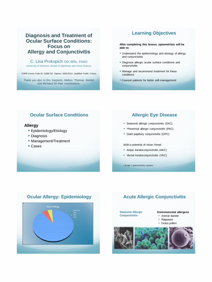
Ocular Allergy: Epidemiology
90% - 95%
Acute Allergic Conjunctivitis
Seasonal Allergic
Conjunctivitis
• Environmental allergens
Animal dander
Ragweed
Grass pollen
Page 2
5/4/2012
2
Perennial Allergic
Conjunctivitis•Milder than seasonal allergy
Associated with asthma
Year-round problem and indoors
High pollen counts
•70-80% allergic to dust mite
droppings: Mites are 10-24µ
10-20 waste pellets/day
1 gram dust = 240,000 droppings
After 5 years, 50% of pillow
weight is dust mite droppings
Allergic Conjunctivitis
Airborne allergen Contact allergen e.g.,
nail polish
Clinical Presentation:
Acute Allergic Conjunctivitis
Clinical Presentation
Symptoms:
Ocular itching
Burning
Tearing
Redness
Sensitivity to light
Grittiness/foreign-body sensation
Blurred vision
Signs of Acute Allergic
Conjunctivitis
Hyperemia/chemosis
of bulbar conjunctiva
Micro or macro
papillary changes
Follicular response
Possible eyelid
swelling
Classic Allergic Conjunctivitis
Presentation
With conjunctival chemosis, mild injection and tearing
SAC/PAC: Diagnosis
Hallmark symptom: ITCHING!
DDx vs. viral/bacterial conjunctivitis:
Personal/family Hx of atopic disorders
Pink/glossy conjunctiva
Quality of discharge
Stringy, ropy
Page 3
5/4/2012
3
Conjunctival Chemosis DDx
Adenovirus/EKC
Pre-auricular lymphadenopathy present
Pseudomembrane or SEIs could be present
Symptoms include red eye with FB sensation but
itching is not a typical symptom of adenovirus
Duration: 7-21 days
Treatment
To understand the underlying allergic
mechanisms
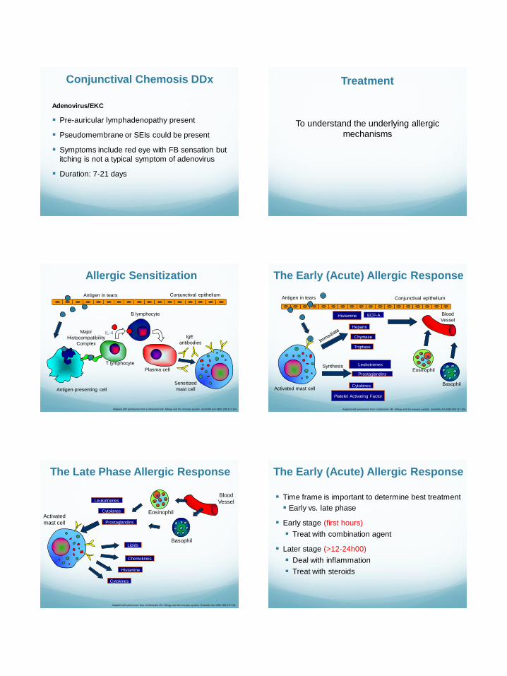
Allergic Sensitization
Antigen in tears Conjunctival epithelium
Antigen-presenting cell
B lymphocyte
IL-4IgE
antibodies
Sensitized
mast cell
Plasma cell
T lymphocyte
Major
Histocompatibility
Complex
Adapted with permission from Lichtenstein LM. Allergy and the immune system. Scientific Am 1993; 269:117-124.
The Early (Acute) Allergic Response
Antigen in tears Conjunctival epithelium
Activated mast cellCytokines
Synthesis
Platelet Activating Factor
Prostaglandins
Leukotrienes
Heparin
Chymase
Tryptase
Histamine ECF-A
Basophil
Eosinophil
Blood
Vessel
Adapted with permission from Lichtenstein LM. Allergy and the immune system. Scientific Am 1993;269:117-124.
The Late Phase Allergic Response
Adapted with permission from Lichtenstein LM. Allergy and the immune system. Scientific Am 1993; 269:117-124.
Activated
mast cell
Lipids
Cytokines
Chemokines
Histamine
Blood
Vessel
Basophil
EosinophilCytokines
Prostaglandins
Leukotrienes Time frame is important to determine best treatment
Early vs. late phase
Early stage (first hours)
Treat with combination agent
Later stage (>12-24h00)
Deal with inflammation
Treat with steroids
The Early (Acute) Allergic Response
Page 4
5/4/2012
4
Symptom severity
“Affecting daily activities or lifestyle”
i.e., can’t work
Once symptoms are under control can go to
combination agents longer term
Treatment Based on SymptomsAssessed by the Eye Allergy Patient Impact Questionnaire
N = 124
Pe
rce
nt
of
Pa
tie
nts
(%
)
Going Outdoors Reading Driving Concentratingon Daily Tasks
Sleeping Putting on/Wearing Make-up
70%73%
61%58%
45%
55%
Lorenz, et al. J Outcomes Res 2003; 7:21.
Impact on Daily Activities
Mild will self-treat
More than 50% of patients will try OTC antihistamines before visiting an eye doctor
OTC antihistamines/decongestants –vasoconstrictors
e.g. Visine®, Clear Eyes®, or Naphcon A®
Rebound hyperemia
SAC Treatment
LEVEL 1 Non pharmacological - Allergen avoidance
- Cold compresses
- Artificial tears
Pharmacological - Oral antihistamine drugs
If symptoms other than ocular
If no improvement,
add level 2 treatments
LEVEL 2 Non pharmacological - Unpreserved artificial tears (q2-4h00)
Pharmacological
- EARLY PHASE (< 24h00) -Combo drugs (antihistamine/ mast cell
stabilizers)
- LATE PHASE (> 24h00) -Ester-based steroids (loteprednol 0.2%)
-In conjunction with combo drugs as needed
If no improvement,
add level 3 treatments
LEVEL 3 Non pharmacological -Abundant lubrication-Mucolytic agents (acetylcysteine)
Pharmacological -Switch loteprednol 0.2% to 0.5% -Consult with an allergist
© Dr Langis Michaud, o.d. M.Sc.FAAO
Treatment Algorithm:Ocular Allergy (Seasonal/Perennial)
Palliative
Intensive lubrication (unpreserved if >q.i.d.) Cold compresses
Pharmaceutical Mast cell stabilizers
Topical antihistamines (with or w/o decongestant)
Combination agents (mast cell stabilizers + antihistamines)
Topical steroids Oral medications
SAC/PAC: Treatment
Counsel patients
Re: chronic nature of the condition
To practice avoidance
Stay indoors during peak pollen days
Minimize ocular exposure
Wash hair before sleeping
Lower ceiling fans
Wash linens, etc.
SAC/PAC: Management
Page 5
5/4/2012
5
RefreshTears®
TEARS Naturale FREE®
Bion Tears®
THERA TEARS®
iDrop®
Preservative-Free Artificial Tears
Mast cell stabilizers
Lodoxamide tromethamine ophthalmic solution 0.1% (Alomide®)
Sodium cromoglycate (Opticrom®)
Delay for clinical action
Pre-seasonal application
Limited clinical effects
Soparkar, et al. Arch Ophthalmol 1990; 108:520-4.
SAC/PAC: Topical
Topical antihistamine (with or w/o decongestant)
Visine®, Murine® – not recommended
Inconvenient dosing (q.i.d.)
Rebound hyperemia w/ long-term use
Levocabastine eye drops
H1 receptor antagonist (Livostin®)
Prescribed by GPs
SAC/PAC: Topical
Combination agents
Provide rapid relief
1st drop applied
No need to load dose in the system to become effective
Long-term management
Convenient dosing (die to b.i.d.)
Ketotifen 0.025% (Zaditor® [Rx], Alaway® *, Refresh® Eye Itch Relief*)
Olopatadine hydrochloride 0.1% (Patanol®)
*available in US only
SAC/PAC: Topical
Topical ester steroids (Loteprednol etabonate 0.2% – Alrex®, loteprednol etabonate 0.5% w/v – Lotemax™)
Initial dosing Moderate cases: q.i.d. x 2-3 wks Severe cases: can be increased to q2 hrs for the first 2-
3 days Schedule follow-up at 3-4 weeks to check IOP and
therapeutic response
No adverse effects reported with up to 4,000 doses over 36 months*
IOP risk: 1-2%
Use
In non-compliant patients In young male patients with asthma In conjunction with olopatadine – Patanol®
* Ilyas, et al. Eye Contact Lens 2004; 30:10-13.
SAC/PAC: Steroids
Loteprednol Ester steroids
Prednisolone Ketone steroids
Fluorometholone
Dexamethasone
Betamethasone
Ester vs. Ketone Steroids
Page 6
5/4/2012
6
Ester Steroids are inactivated by naturally occurring
esterases
Fewer side effects, better safety profile*
No rebound effect
Ketone Steroids are not inactivated and have
propensity to remain in anterior chamber post-
breakdown as active metabolites
Benefits/risk of use e.g., cost
Switching from other steroids to ester steroids
* Ilyas, et al. Eye Contact Lens 2004; 30:10-13.
Ester vs. Ketone Steroids
Significant systemic involvement
Rhinitis, itchy throat, cough, sinus congestion
Add oral medications to topical regimen
Diphenhydramine hydrochloride (e.g., Benadryl®)
Take before bed
Consider consult with allergist depending on
severity, duration and recurrence rates
SAC Treatment
Allergy Could Be a Systemic Problem
Allergic
Sinusitis
P.O. Claritin®, Allegra®, Reactine®, Aerius®, Benadryl® q.d.
If sinus congestion is present:
P.O. Claritin®-D 24-hr or Allegra®-D 24-hr – q.d.
Contains pseudoephedrine
Avoid in patients with hypertension
Choose carefully in patients with dry eye
SAC Oral
Not often prescribed with ocular signs/symptoms
present only
Maintain or increase preservative-free artificial tears
Can exacerbate the ocular condition
ORAL: Non-Sedating
Does Not Mean Non-Drying!
Consider nasal inhalers
Antihistamine
Astelin ®b.i.d., Dristan ®, Otrivin®
Steroid
Beconase®, Flonase®, Vancenase®, Nasonex® b.i.d.
Cromolyn sodium OTC
Poor systemic absorption – few side effects
SAC Treatment
Page 7
5/4/2012
7
Although rare, risks still exist for steroid inhalers:
IOP rise
Conjunctivitis
Glaucoma
PSC formation
SAC Treatment
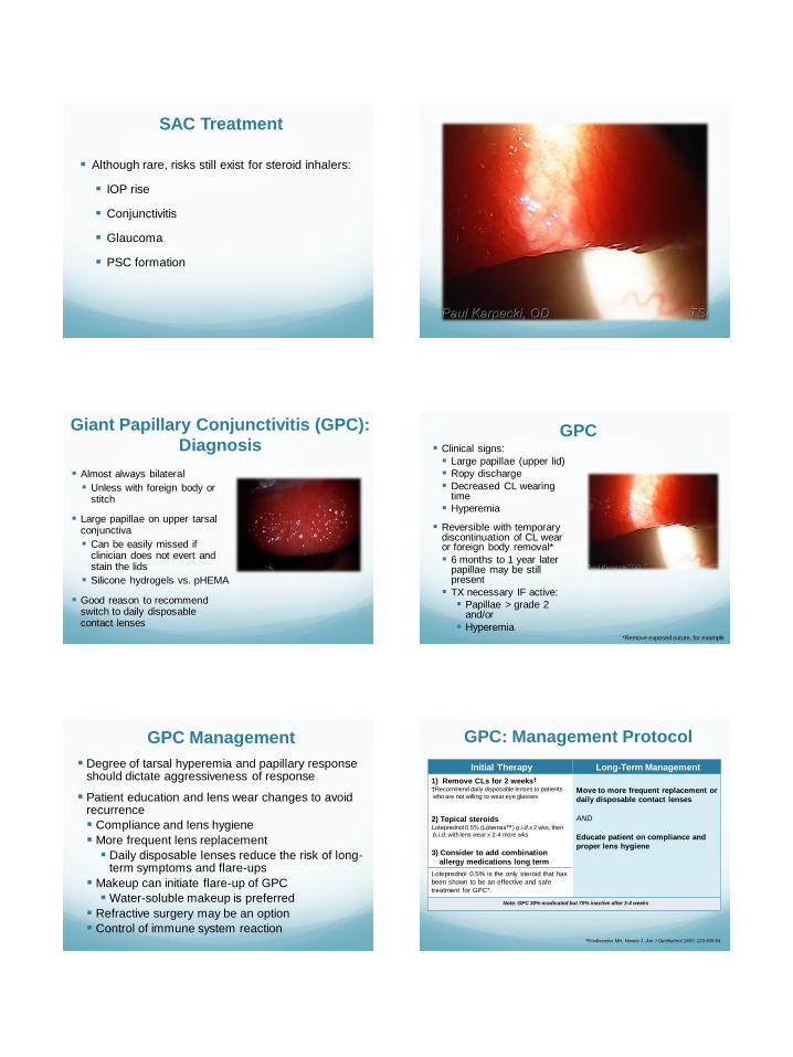
Giant Papillary Conjunctivitis (GPC):
Diagnosis
Almost always bilateral
Unless with foreign body or stitch
Large papillae on upper tarsal conjunctiva
Can be easily missed if clinician does not evert and stain the lids
Silicone hydrogels vs. pHEMA
Good reason to recommend switch to daily disposable contact lenses
Clinical signs:
Large papillae (upper lid)
Ropy discharge
Decreased CL wearing time
Hyperemia
Reversible with temporary discontinuation of CL wear or foreign body removal*
6 months to 1 year later papillae may be still present
TX necessary IF active:
Papillae > grade 2 and/or
Hyperemia *Remove exposed suture, for example
GPC
Degree of tarsal hyperemia and papillary response should dictate aggressiveness of response
Patient education and lens wear changes to avoid recurrence
Compliance and lens hygiene
More frequent lens replacement
Daily disposable lenses reduce the risk of long-term symptoms and flare-ups
Makeup can initiate flare-up of GPC
Water-soluble makeup is preferred
Refractive surgery may be an option
Control of immune system reaction
GPC Management GPC: Management Protocol
*Friedlaender MH, Howes J. Am J Ophthalmol 1997; 123:455-64.
Initial Therapy Long-Term Management
1) Remove CLs for 2 weeks†
†Recommend daily disposable lenses to patients
who are not willing to wear eye glasses
2) Topical steroidsLoteprednol 0.5% (Lotemax™) q.i.d.x 2 wks, then b.i.d. with lens wear x 2-4 more wks
3) Consider to add combination
allergy medications long term
Move to more frequent replacement or
daily disposable contact lenses
AND
Educate patient on compliance and
proper lens hygiene
Loteprednol 0.5% is the only steroid that has
been shown to be an effective and safe
treatment for GPC*.
Note: GPC 30% eradicated but 70% inactive after 3-4 weeks
Page 8
5/4/2012
8
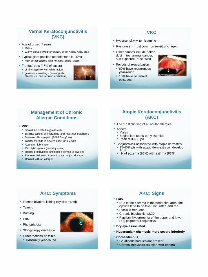
Age of onset: 7 years Males
Warm climate (Mediterranean, West Africa, Asia, etc.)
Typical giant papillae (cobblestone in 20%) May be associated with keratitis, shield ulcers
Trantas’ dots (17% of cases) Limbal papillae with white apical
gelatinous swellings (eosinophils,fibroblasts, and necrotic epithelium)
Vernal Keratoconjunctivitis
(VKC) Hypersensitivity to histamine
Rye grass = most common sensitizing agent
Other causes include pollen,dust mites, animal dander,sun exposure, dust, wind
Periods of exacerbation
60% have recurrencesyear-round
16% have perennialepisodes
VKC
Management of Chronic
Allergic Conditions
VKC Should be treated aggressively
1st line: topical antihistamine and mast cell stabilizers
Systemic AH + aspirin (0.5-1.0 mg/day)
Topical steroids in severe case for 1-2 wks
Abundant lubrication
Mucolytic agents (acetylcysteine)
Topical prophylactic antibiotic if cornea is involved
Frequent follow-up to monitor and adjust dosage
Consult with an allergist
The most blinding of all ocular allergies
Affects Males Begins late teens-early twenties Peak at 30-50 y.o.
Conjunctivitis associated with atopic dermatitis 15-40% pts with atopic dermatitis will develop
AKC Hx of eczema (95%) with asthma (87%)
Atopic Keratoconjunctivitis
(AKC)
Intense bilateral itching (eyelids +conj)
Tearing
Burning
FBS
Photophobia
Stringy, ropy discharge
Exacerbations possible
Habitually year-round
AKC: Symptoms Lids
Due to the eczema in the periorbital area, the eyelids tend to be thick, indurated and red
Ptosis is frequent
Chronic blepharitis, MGD
Papillary hypertrophia of the upper and lower(++) palpebral conjunctiva
Dry eye associated
Hyperemia + chemosis more severe inferiorly
Cornea/limbus
Gelatinous nodules are present
Corneal neovascularization with edema
AKC: Signs
Page 9
5/4/2012
9
Management of Chronic
Allergic Conditions
AKC
Topical mast cell stabilizers
Topical steroids
Immunosuppressive medication (cyclosporine A)
Topical prophylactic antibiotic if cornea is
involved
Therapy adjustment needed on a 2wk basis
Surgery for severe damages to the cornea
Consult with an allergist
Recommend steroid cream e.g., polymyxin B
sulphate neomycin sulphate dexamethasone
0.05% (Maxitrol® ophthalmic ointment)
Management of Contact
Dermatitis
Comorbidities are common
Consider drying effects of oral medications
Patients who suffer from dry eye allow the
allergens to stay on the ocular surface longer
Management of Dry Eye and Allergy
SOME CLINICAL PEARLS…
Avoid eye rubbing
Mechanical mast cell
degranulation
Refrigerate drops
Soothing and effective
Allergic Pearls
Environmental management
Pillows
Ceiling fans etc.
Wash sheets more often in allergy season
Shower before sleeping
Allergic Pearls
Page 10
5/4/2012
10
Contact lens use?
Depends on:
Severity and contributing factors
Evert lid:
Papillae
Hyperemia
Recommend daily disposable lenses or hiatus
Use preservative-free contact lens solutione.g., H2O2
Re-wet lenses during the day
Allergic Pearls
When treating w/topical steroids e.g., loteprednol, schedule a F/U exam within4 weeks
Educate patient to call office if red painful eye
Confirm compliance, efficacy, symptom relief
Evaluate IOP and educate about long-term management
Steroids in CL wearers:
Use b.i.d. (before and after lens wear)
Allergic Pearls
Generic Guidelines
What can I hope for?
How often should I bring the patient back in? For allergy re-checks: 1-4 wks depending on severity
For dry eye: 3-4 weeks
Provincial jurisdiction/regulation
Cost of treatment Consider substitution if cost is prohibitive
Determine drug plan coverage
Practice Management
Conjunctivitis
One of the most common reasons for acute eye-related primary care visits
15% of all pediatric referrals to Wills Eye Institute
1-in-8 pediatric visits are for pink eye
*A non-specific term for inflammation of the conjunctiva
Conjunctivitis*
Diverse range of etiologies:
Allergic
Viral
Bacterial
Chlamydial
Nonspecific / Toxic / CLARE
Associated with lid and dermatologic problems
Conjunctivitis
Page 11
5/4/2012
11
Elements of an Effective
Physical Exam
Examine the periorbital skin closely and note any
lesions on face or scalp
Palpate for preauricular and submandibular lymph
nodes (viral)
Ask about upper respiratory infection (URI)
Redness
Clear watery discharge
FB sensation or pain
Photophobia
Decreased vision possible
Symptoms of
Viral Conjunctivitis
Most commonly caused by adenovirus (>60%)
Pharyngoconjunctival fever (PCF)
Associated with URI
Epidemic keratoconjunctivitis (EKC)
Hemorrhagic conjunctivitis
Other implicated viruses
Herpes simplex (HSV)
Epstein-Barr
Viral Conjunctivitis Conjunctival Chemosis
Related to Adenovirus
Viral Conjunctivitis:
Epidemic Keratoconjunctivitis (EKC)Children or adults
Acute red eye and watery discharge
Begins in one eye and spreads to fellow eye within a few days
Preauricular submandibular lymph node swelling on the ipsilateral side
After one week without treatment will see SEIs
Highly contagious
Hx of recurrent contact lens intolerance Young female On and off C.L. for several weeks Moderate redness
Slight decrease in vision – most other forms of conjunctivitis do not affect vision
Periorbital edema
Small conjunctival vesicular hemorrhages
May be present without infiltrates
Key Diagnostic Indicators of EKC
Page 12
5/4/2012
12
Rapid Pathogen Screening
(RPS) Adeno Detector
Rapid Pathogen Screening (RPS) Adeno Detector facilitates
diagnosis (available in US)
$12-$13 per test
Will ship to Canada
Management of Viral EKC
Consider severity of presentation
Early/mild: Supportive therapy alone
Recommend daily disposable lenses
More symptomatic suppress inflammation with
steroids and temporarily cease contact lens wear
If sub-epithelial infiltrates and reduced V/A
Steroids q.i.d. x 1 month (or longer), then taper
slowly
May have rebound – less likely with
loteprednol
Tapering
With ester steroids may not be necessary
With non-ester steroids – duration depends
on severity but is required
Povidone-iodine
Broad-spectrum microbicide
Indicated for “irrigation of the ocular surface”
EKC Treatment
“Off label” use: Tx adenoviral keratoconjunctivitis Anesthetize with proparacaine
Instill 1 or 2 drops of NSAID
Instill several drops Betadine® 5% in eye(s), close eye(s)
Swab or rub excess over eyelid margin
After 1 minute, irrigate with sterile saline
Instill 1 or 2 drops of NSAID
Rx Lotemax™ q.i.d. x 4 days
No reports in clinical trials of adverse reactions
Avoid use if patient is allergic to iodine
Regimen courtesy of Randall Thomas OD and Ron Melton OD
Povidone Iodine
Off-label Applications and
Standard of Care
Standard of care
A diagnostic and treatment process that a clinician
should follow for a certain type of patient, illness,
or clinical circumstance based on:
What an average prudent doctor would do in
that given situation
Evidence-based medicine and research
Previous precedents
Pharyngoconjunctival Fever (PCF)
Usually in children
Almost alwaysunilateral
Accompanied by
Mild sore throator URI
Low fever, somemalaise
Adenopathy in severe cases
Courtesy of Randall Thomas OD and Ron Melton OD
Page 13
5/4/2012
13
Self-limiting
Usually resolves within 2 weeks without treatment
Support and educate family
Supportive therapy
Cool compresses
Artificial tears
In more severe cases
Low-dose ester steroid to reduce inflammation
First, rule out lid involvement/signs of HSV
Antibiotic/steroid combination if corneal involvement
Courtesy of Randall Thomas OD and Ron Melton OD
Management of PCF
Herpes simplex (HSV) conjunctivitis
Usually presents with lid involvement first
First exposure early in life
Clearest signs of HSV conjunctivitis
Unilateral, rarely bilateral, involvement with watery discharge AND
Ulceration of the lid margin and/or
Vesicles on the face or around the eyes
Herpetic Conjunctivitis
Be suspicious of herpes zoster in patients >50 yrs. with nonspecific pain in 1 eye
Have patient seen quickly, won’t usually get dendrites (although may have pseudodendrites in rare circumstances)
Iritis is more common in HZO than corneal involvement
Tell the patient to report any lesions on the eyelids, skin or scalp
< 5% of all patients
Zoster
Herpes simplex – primarily infectious
With lid/dermatologic involvement: oral antiviral
therapy (acyclovir, valacyclovir, famciclovir)
In children over 6 years dosing per adults; ask
pediatrician in children under 6 years of age
and in low-weight children
Caution: topical steroids will worsen HSV
Management of
Herpetic Conjunctivitis
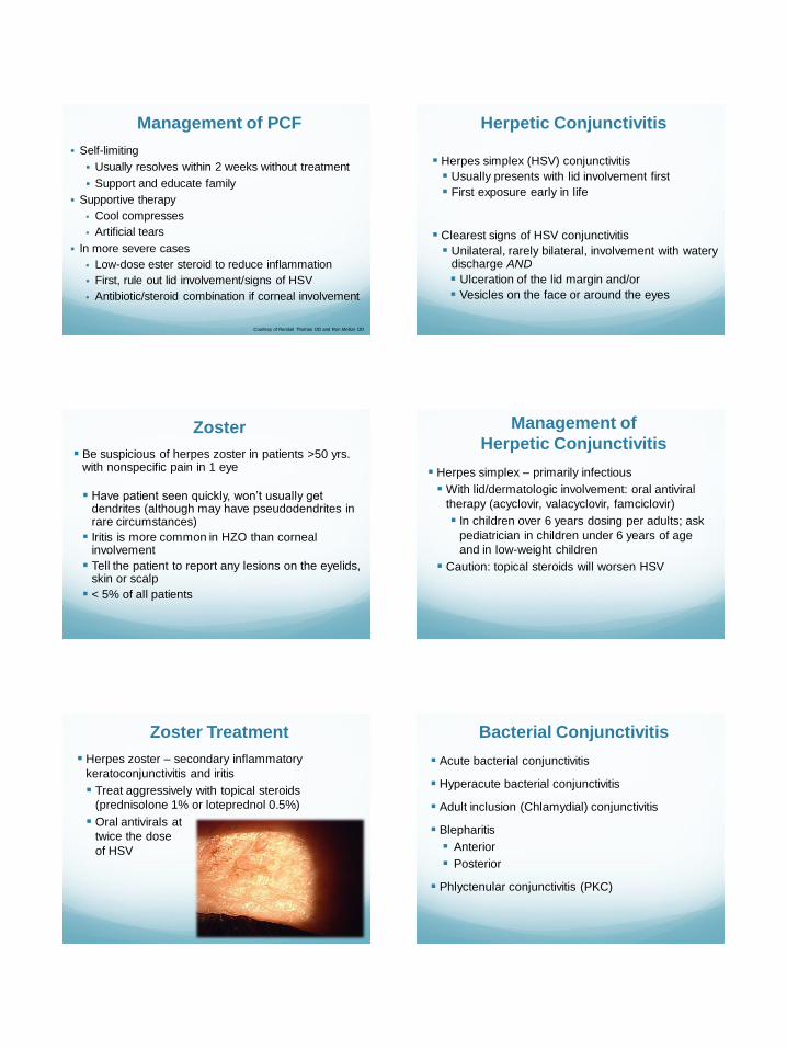
Herpes zoster – secondary inflammatory
keratoconjunctivitis and iritis
Treat aggressively with topical steroids
(prednisolone 1% or loteprednol 0.5%)
Oral antivirals at
twice the dose
of HSV
78
Zoster Treatment
Acute bacterial conjunctivitis
Hyperacute bacterial conjunctivitis
Adult inclusion (Chlamydial) conjunctivitis
Blepharitis
Anterior
Posterior
Phlyctenular conjunctivitis (PKC)
Bacterial Conjunctivitis
Page 14
5/4/2012
14
Occurs in 1 of 8 children every year1
Most cases of conjunctivitis are acute bacterial
conjunctivitis2
≈1% of all consultations in primary care2
Most common causative pathogens3
Haemophilus influenzae*
Streptococcus pneumoniae*
Staphylococcus aureus**
Staphylococcus epidermidis**
* More common in children **More common in adults
1. Rose PW, et al. Lancet 2005; 366:37-43.
2.. Hovding G. Acta Ophthalmol 2008; 86:5-17.
3. Kowalski RP, Dhaliwal DK. Expert Rev Anti Infect Ther 2005; 3:131-139.
Acute Bacterial Conjunctivitis
Meaty red eye
Discharge ordebris in tearfilm
Bacterial Conjunctivitis
Presentation
How to Effectively Manage
Childhood Conjunctivitis
Rule out trauma
Less likely to respond
More difficult to diagnose
It can alter the management plan
i.e., involve a pediatrician
Increased risk for gram-positive infection, such as
MRSA or Streptococcal cellulitis
Algorithm:
Conjunctivitis Tool Box
Ideal Profile for Treatment of
Bacterial Conjunctivitis
Broad spectrum
Potent activity against prevalent pathogens
Bactericidal
Low propensity for resistance development
Low incidence of adverse events (AEs)
Convenient dosing
Long dwell time at site of infection (ocular surface)
Local treatment for a local disease
Treatment with a
Broad Spectrum Antibiotic
Many choices
Best to use the strongest product – dead bugs don’t
mutate
Page 15
5/4/2012
15
Fluoroquinolones
Use right tool at right time
More economical
Time away from work/school decreased
Decrease chance of resistance
Evidence-based standard of care
Besifloxacin is a New Chemical Entity Fluoroquinolone (FQ)
Unique combination of substituents at C7 and C8 positions of FQ core structure
Besifloxacin mode of action is consistent with newer FQs (inhibition of DNA gyrase and topoisomerase IV)
F
N N
O
COOH
NH2
HCl
Cl
DuraSite® Technology
Proprietary mucoadhesive
delivery system1
Polymer composed of
Polycarbophil
Edetate disodium dihydrate
Sodium chloride2
May prevent tearing out
medication in children
1. DuraSite is a trademark of InSite Vision Incorporated, Alameda, CA.
2. Besivance Product Monograph, Bausch & Lomb Canada, October 23, 2009.
Besivance™ Indication
Similar to Other FQs
BESIVANCE™ (besifloxacin ophthalmic suspension) 0.6% w/v
is indicated for the treatment of patients one year of age and
older with bacterial conjunctivitis caused by susceptible strains
of the following organisms:
Aerobic, Gram-Positive
CDC coryneform group G
Staphylococcus aureus
Staphylococcus epidermidis
Streptococcus mitis
Streptococcus oralis
Streptococcus pneumoniae
Besivance Product Monograph, Bausch & Lomb Canada, October 23, 2009.
Aerobic, Gram-Negative
Haemophilus influenzae
BESIVANCE™ is a 7-day course
of therapy for bacterial
conjunctivitis.1
Dosing is 1 drop in the affected
eye(s) 3 times a day for 7 days.
Besivance Product Monograph, Bausch & Lomb Canada, October 23, 2009.
Besivance™ Dosage
Besifloxacin binds to and inhibits two enzymes that are essential for maintaining bacterial DNA in the proper conformation.
DNA gyrase
Topoisomerase IV
relaxed DNA supercoiled DNA
catenated DNA decatenated DNA
Besifloxacin
Besifloxacin Mechanism
of Action
Page 16
5/4/2012
16
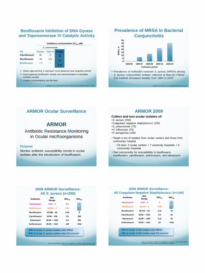
Besifloxacin Inhibition of DNA Gyrase
and Topoisomerase IV Catalytic Activity
Cambau E, et al. J Antimicrob Chemother. Advanced access published January 15, 2009.
Ratios approaching 1 represent more balanced dual targeting activity
Dual targeting besifloxacin activity also demonstrated in cleavable
complex assays
Lowest concentrations are the best
Inhibitory concentration (IC50, µM)
S. pneumoniae
FQ Gyrase Topo IV
Ciprofloxacin 40 5
Moxifloxacin 10 2.5
Besifloxacin 2.5 1
Ratio
8
4
2.5
Prevalence of MRSA In Bacterial
Conjunctivitis
Prevalence of methicillin-resistant S. aureus (MRSA) among
S. aureus conjunctivitis isolates collected at Bascom Palmer
Eye Institute increased steadily from 1994 to 20031
1994-95 1996-97 1998-99 2000-01 2002-03
Collection period
0
5
10
15
20
25
30
35
MR
SA
, %
Adapted from Cavuoto K, et al. Ophthalmology 2008; 115:51-56.
ARMOR
Antibiotic Resistance Monitoring
in Ocular micRoorganisms
Purpose
Monitor antibiotic susceptibility trends in ocular
isolates after the introduction of besifloxacin
ARMOR Ocular Surveillance ARMOR 2009Collect and test ocular isolates of:S. aureus (200)
Coagulase negative staphylococci (150)
S. pneumoniae (75)
H. influenzae (75)
P. aeruginosa (150)
Target a mix of isolates from ocular centers and those from
community hospital
19 sites: 3 ocular centers + 7 university hospitals + 9
community hospitals
Test concurrently for susceptibility to besifloxacin,
moxifloxacin, ciprofloxacin, azithromycin, and tobramycin
2009 ARMOR Surveillance:
All S. aureus (n=200)
AntibioticMIC
RangeMIC50 MIC90
Vancomycin 0.25 – 2 0.5 1
Besifloxacin ≤0.008 – 4 0.03 1
Moxifloxacin ≤0.008 – 64 0.06 8
Ciprofloxacin ≤0.06 – 256 0.5 256
Tobramycin ≤0.06 – >256 0.5 256
Azithromycin ≤0.25 – >512 128 >512
Haas, et al. Presented at ARVO, Fort Lauderdale, FL, May 2-6, 2010. Abstract # D965,
% resistance based on oxacillin and ciprofloxacin breakpoints.
39% of ocular S. aureus isolates were MRSA
38% of ocular S. aureus isolates were FQ-resistant
2009 ARMOR Surveillance:
All Coagulase-Negative Staphlylococci (n=144)
AntibioticMIC
RangeMIC50 MIC90
Vancomycin 0.12 – 2 1 2
Besifloxacin ≤0.015 – 8 0.06 2
Moxifloxacin ≤0.015 – 64 0.12 16
Ciprofloxacin ≤0.06 – >512 0.5 64
Tobramycin ≤0.06 – >256 0.12 32
Azithromycin ≤0.25 – >512 64 >512
53% of ocular CoNS isolates were MRSA
43% of ocular CoNS isolates were FQ-resistant
Haas et al. Presented at ARVO, Fort Lauderdale, FL, May 2-6, 2010. Abstract #D965,
% resistance based on oxacillin and ciprofloxacin breakpoints.
Page 17
5/4/2012
17
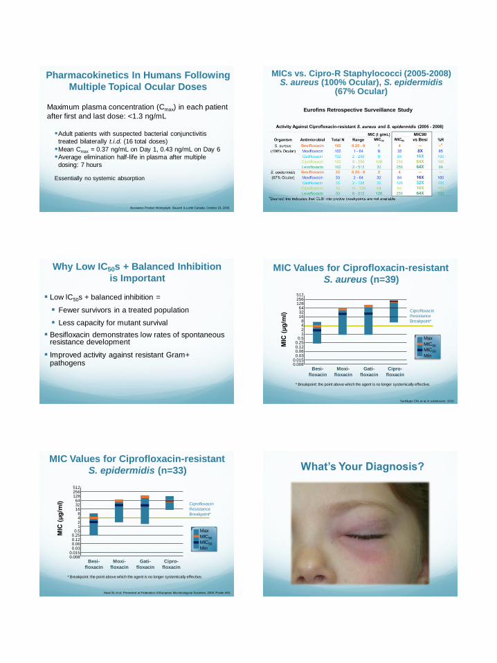
Pharmacokinetics In Humans Following
Multiple Topical Ocular Doses
Maximum plasma concentration (Cmax) in each patient
after first and last dose: <1.3 ng/mL
Adult patients with suspected bacterial conjunctivitis
treated bilaterally t.i.d. (16 total doses)
Mean Cmax = 0.37 ng/mL on Day 1, 0.43 ng/mL on Day 6
Average elimination half-life in plasma after multiple
dosing: 7 hours
Essentially no systemic absorption
Besivance Product Monograph, Bausch & Lomb Canada, October 23, 2009.
MICs vs. Cipro-R Staphylococci (2005-2008)S. aureus (100% Ocular), S. epidermidis
(67% Ocular)
Eurofins Retrospective Surveillance Study
Why Low IC50s + Balanced Inhibition
is Important
Low lC50s + balanced inhibition =
Fewer survivors in a treated population
Less capacity for mutant survival
Besifloxacin demonstrates low rates of spontaneous resistance development
Improved activity against resistant Gram+ pathogens
MIC Values for Ciprofloxacin-resistant
S. aureus (n=39)
Ciprofloxacin
Resistance
Breakpoint*
512256128
643216
8421
0.50.250.120.060.03
0.0150.008
MIC
(μ
g/m
l)
Sanfilippo CM, et al. In submission. 2010.
* Breakpoint: the point above which the agent is no longer systemically effective.
Besi-
floxacin
Moxi-
floxacin
Gati-
floxacin
Cipro-
floxacin
Max
MIC90
MIC50
Min
MIC Values for Ciprofloxacin-resistant
S. epidermidis (n=33)
Haas W, et al. Presented at Federation of European Microbiological Societies. 2009. Poster #53.
Ciprofloxacin
Resistance
Breakpoint*
512256128
643216
8421
0.50.250.120.060.03
0.0150.008
MIC
(μ
g/m
l)
* Breakpoint: the point above which the agent is no longer systemically effective.
Besi-
floxacin
Moxi-
floxacin
Gati-
floxacin
Cipro-
floxacin
Max
MIC90
MIC50
Min
What’s Your Diagnosis?
Page 18
5/4/2012
18
One of the most common complications associated with acute bacterial conjunctivitis
Examine skin and adnexa around the orbit for a discrete reddish sheen
Patients often have ethmoidal or maxillary sinus involvement, which results in orbital tenderness
Preseptal CellulitisWhat’s Your Diagnosis?
When to Refer to a Pediatrician /
Pediatric Ophthalmologist
Fever or general malaise
Purchase a tympanic or forehead thermometer
Acute earache or ear infection
Approximately one-third of all childhood cases are otitis-conjunctivitis syndrome
A notable red sheen around the eyelids
Preseptal cellulitis or cellulitis
Significant purulent rhinorrhea or an upper respiratory infection associated with any fussiness or sleeplessness
Systemic involvement in children presenting with
conjunctivitis is necessary to rule out and refer to a
pediatrician
Preseptal cellulitis
Cellulitis
Otitis media
URI
Ocular Infection Pearls
Environmental triggers e.g.,:
Ozone
Cold
Dry air
Perfumes
Non-Allergic Conjunctivitis Phlyctenular Conjunctivitis
Usually secondary to staphylococcal blepharitis
In adults, associated w/rosacea, dry eye
In zones of poverty: associated with tuberculosis
Presentation
Scratchy, FB sensation
Sectoral injection, raised bump on conj
No discharge
Page 19
5/4/2012
19
Management of Phlyctenular
Conjunctivitis
Staphylococcal
Combination antibiotic/steroid q 2-4 hrs for 1-2 days,
then q.i.d. for 7-10 days
Lid therapy
Warm compresses and eyelid scrubs
Doxycycline 50 mg b.i.d. for 1 month then q d 1
month
Long-term prevention in recurrent cases
Tubercular – if no signs of Staph, consider tuberculosis
Co-manage with patient’s doctor
Bacterial Keratitis
26 y.o. Caucasian male
“Painful eye,” “light sensitivity” and “eye
is red”
Long-standing contact lens wearer
Began this morning – acute onset
Case S.P. History
2+/3- conjunctival injection
Slight lid edema
Pupils normal
Cornea –small peripheral infiltrate, SPK over
infiltrate
AC grade 2 cell and flare
Examination DDx KERATITIS Infectious
(If one item is +, lesion is
suspected infectious)
Non infectious (sterile)
(If all items apply, lesion is
suspected sterile)
SYMPTOMS
- Pain ++ to +++ Discomfort
- Hyperemia /redness ++ to +++ x 360 deg Sectoral injection, limited
- Photophobia ++ to +++ None to trace
-Visual acuity Reduced Normal except if many infiltrates in the visual axis
SIGNS
Number Single More than one
Position Pupillary area, mid-periphery Mid-periphery to periphery
Size > 1.5 mm < 1.5 mm
Edges Not well defined Well defined
WBCs (edema) surrounding the lesion > ¼ cornea Limited to the lesion area
Colour White to yellowish White to greyish
Shape Concave with large epithelial defect Convex with small epithelial defect
Corneal staining = or > infiltrate < infiltrate, negative staining
Corneal edema Striae and folds None
Endothelium Precipitates None
Lids Superior lid ptosis Normal
Anterior chamber + reaction (cells and flare) Non active
© Dr Langis Michaud, o.d. M.Sc.FAAO
Page 20
5/4/2012
20
Acute onset
Pain
Photophobia
Discharge – mucopurulent
Decreased vision
Redness
Excessive tearing, lid edema, blepharospasm
Symptoms
Conjunctival hyperemia and ciliary flush
Lid edema
Tear film debris – thick & cells present
Epithelial defect
Grayish-white stromal infiltrate
AC reaction
From few cells to hypopyon
Signs
Bacterial Keratitis Treatment
Loading dose first –
q 15 minutes x 1-2 hours
Never taper antibiotics beyond therapeutic dosing
Cycloplegic drops for pain
Fortified medication?
At night:
Tobramycin ointment in suspected gram negative
Polymyxin B sulfate – gramicidin –
e.g., Polysporin® ointment in all others
Treatment
Practice Management:
Bacterial Keratitis
When to culture:
1,2,3 Rule:
1 mm from visual axis
2 infiltrates (or more)
3 mm or greater in size
Nosocomial infections
Immuno-compromised patient
Post-surgical
Page 21
5/4/2012
21
Check for lymph adenopathy
Pre-auricular or submandibularpea-shaped node may indicate viral or AIC
Look at tear film closely under high magnification
A murky tear film can indicate a bacterial conjunctivitis discharge
Ocular Infection Pearls
Ocular allergies and conjunctivitis
Two most common reasons for visits to a pediatrician
Accounting for over 20% of all visits
DDx of allergies and appropriate tx = critical
DDx infectious conjunctivitis vs. keratitis is key
To optimize results and to prevent spread or comorbidity
Make a confident diagnosis and be aggressive in treatment
Summary