Diagnosis of Autism Spectrum Disorders Using Regional and Interregional Morphological Features Chong-Yaw Wee, Li Wang, Feng Shi, Pew-Thian Yap, and Dinggang Shen* Image Display, Enhancement, and Analysis (IDEA) Laboratory, Biomedical Research Imaging Center (BRIC), Department of Radiology, University of North Carolina at Chapel Hill, Chapel Hill, North Carolina r r Abstract: This article describes a novel approach to identify autism spectrum disorder (ASD) utilizing regional and interregional morphological patterns extracted from structural magnetic resonance images. Two types of features are extracted to characterize the morphological patterns: (1) Regional features, which includes the cortical thickness, volumes of cortical gray matter, and cortical-associated white matter regions, and several subcortical structures extracted from different regions-of-interest (ROIs); (2) Interregional features, which convey the morphological change pattern between pairs of ROIs. We demonstrate that the integration of regional and interregional features via multi-kernel learn- ing technique can significantly improve the classification performance of ASD, compared with using either regional or interregional features alone. Specifically, the proposed framework achieves an accu- racy of 96.27% and an area of 0.9952 under the receiver operating characteristic curve, indicating excel- lent diagnostic power and generalizability. The best performance is achieved when both feature types are weighted approximately equal, indicating complementary between these two feature types. Regions that contributed the most to classification are in line with those reported in the previous stud- ies, particularly the subcortical structures that are highly associated with human emotional modulation and memory formation. The autistic brains demonstrate a significant rightward asymmetry pattern particularly in the auditory language areas. These findings are in agreement with the fact that ASD is a behavioral- and language-related neurodevelopmental disorder. By concurrent consideration of both regional and interregional features, the current work presents an effective means for better characteri- zation of neurobiological underpinnings of ASD that facilitates its identification from typically devel- oping children. Hum Brain Mapp 00:000–000, 2013. V C 2013 Wiley Periodicals, Inc. Key words: autism spectrum disorders (ASD); magnetic resonance imaging (MRI); regional features; interregional features; multiple-kernel learning (MKL); limbic system; rightward asymmetry r r INTRODUCTION Autism spectrum disorder (ASD) is a highly heterogene- ous, behaviorally defined neurodevelopmental disorder with multiple causes and courses. It is associated with sev- eral comorbid disorders, including intellectual impairment, seizures, and anxiety [Amaral et al., 2008; Ecker et al., 2010a]. Based on the latest report released by the Centers for Disease Control and Prevention, it is estimated that 1 in 88 American children was affected by some forms of ASD, a 78% increase compared to a decade ago, with boys outnumbering girls by a ratio of 5:1 [Centers for Disease Contract grant sponsor: National Institute of Health; Contract grant numbers: EB006733, EB008374, EB009634, MH100217, AG041721, AG042599 *Correspondence to: Dinggang Shen, Department of Radiology and BRIC, University of North Carolina at Chapel Hill, Chapel Hill, NC 27599. E-mail: [email protected]Received for publication 31 October 2012; Revised 9 August 2013; Accepted 16 September 2013. DOI 10.1002/hbm.22411 Published online 00 Month 2013 in Wiley Online Library (wileyonlinelibrary.com). r Human Brain Mapping 00:00–00 (2013) r V C 2013 Wiley Periodicals, Inc.
Transcript
Diagnosis of Autism Spectrum Disorders UsingRegional and Interregional Morphological Features
Chong-Yaw Wee Li Wang Feng Shi Pew-Thian Yap and Dinggang Shen
Image Display Enhancement and Analysis (IDEA) Laboratory Biomedical Research ImagingCenter (BRIC) Department of Radiology University of North Carolina at Chapel Hill Chapel
Hill North Carolina
r r
Abstract This article describes a novel approach to identify autism spectrum disorder (ASD) utilizingregional and interregional morphological patterns extracted from structural magnetic resonanceimages Two types of features are extracted to characterize the morphological patterns (1) Regionalfeatures which includes the cortical thickness volumes of cortical gray matter and cortical-associatedwhite matter regions and several subcortical structures extracted from different regions-of-interest(ROIs) (2) Interregional features which convey the morphological change pattern between pairs ofROIs We demonstrate that the integration of regional and interregional features via multi-kernel learn-ing technique can significantly improve the classification performance of ASD compared with usingeither regional or interregional features alone Specifically the proposed framework achieves an accu-racy of 9627 and an area of 09952 under the receiver operating characteristic curve indicating excel-lent diagnostic power and generalizability The best performance is achieved when both feature typesare weighted approximately equal indicating complementary between these two feature typesRegions that contributed the most to classification are in line with those reported in the previous stud-ies particularly the subcortical structures that are highly associated with human emotional modulationand memory formation The autistic brains demonstrate a significant rightward asymmetry patternparticularly in the auditory language areas These findings are in agreement with the fact that ASD isa behavioral- and language-related neurodevelopmental disorder By concurrent consideration of bothregional and interregional features the current work presents an effective means for better characteri-zation of neurobiological underpinnings of ASD that facilitates its identification from typically devel-oping children Hum Brain Mapp 00000ndash000 2013 VC 2013 Wiley Periodicals Inc
Key words autism spectrum disorders (ASD) magnetic resonance imaging (MRI) regional featuresinterregional features multiple-kernel learning (MKL) limbic system rightward asymmetry
r r
INTRODUCTION
Autism spectrum disorder (ASD) is a highly heterogene-ous behaviorally defined neurodevelopmental disorderwith multiple causes and courses It is associated with sev-eral comorbid disorders including intellectual impairmentseizures and anxiety [Amaral et al 2008 Ecker et al2010a] Based on the latest report released by the Centersfor Disease Control and Prevention it is estimated that 1in 88 American children was affected by some forms ofASD a 78 increase compared to a decade ago with boysoutnumbering girls by a ratio of 51 [Centers for Disease
Contract grant sponsor National Institute of Health Contractgrant numbers EB006733 EB008374 EB009634 MH100217AG041721 AG042599
Correspondence to Dinggang Shen Department of Radiologyand BRIC University of North Carolina at Chapel Hill ChapelHill NC 27599 E-mail dgshenmeduncedu
Received for publication 31 October 2012 Revised 9 August 2013Accepted 16 September 2013
DOI 101002hbm22411Published online 00 Month 2013 in Wiley Online Library(wileyonlinelibrarycom)
r Human Brain Mapping 0000ndash00 (2013) r
VC 2013 Wiley Periodicals Inc
Control and Prevention 2012] Although most obvioussigns and symptoms of ASD tend to emerge in the first 3years of life most children are only diagnosed betweenages 4 and 5 when the brain is more mature with lessplasticity Some children with ASD may become depressedor experience behavioral problems during adolescencePeople with ASD usually require continue services andsupports as they get older although many of them areable to work and live independently or within a support-ive environment
ASD is characterized in varying degrees by difficultiesin (1) verbal and nonverbal communication (2) socialinteraction and (3) repetitive and ritualized behaviors[Gillberg 1993 Kanner 1943 Wing 1997] Since thebehavioral phenotype of ASD is well known the diagnosisof ASD to date relies entirely on the history symptomsand signs of the disorder Even the latest diagnostic instru-ments that are proposed in the new editions of the Diag-nostic and Statistical Manual (DSM-5) of the AmericaPsychiatry Association [American Psychiatric Association2013] and the International Classification of Diseases (ICD-11) of the World Health Organization [Lord and Jones2012] are also solely behavioral based The etiology andpathogenesis of ASD remain elusive although some brainregions and neural systems have been suggested to beassociated with the disorder [Amaral et al 2008 Toalet al 2005] An important drawback of solely behavioral-based diagnostic tools is that many of these behavioralphenotypes are associated with numerous other psycho-logical and psychiatric disorders [Geschwind and Levitt2007 Guilmatre et al 2009] such as Fragile X syndrome(which causes mental retardation) tuberous sclerosis epi-leptic seizures Tourette syndrome learning disabilitiesand attention deficit disorder It has been reported thatabout 20ndash30 of children with ASD will develop epilepsyby the time they reach adulthood [Centers for DiseaseControl and Prevention 2012] Moreover retrospectiveaccounts of past symptoms rely heavily on an informantbeing both reliable and available [Ecker et al 2010b] Inaddition problems arise from the fact that some adultsbeing able to modulate symptoms via copying strategiesdeveloped over their life spans [Ecker et al 2010b Pers-son 2000] Therefore it is assumed that combining biologi-cal information with behavioral measurements canprovide additional objectivity for ASD diagnosis
Although group comparisons using whole-brain mass-univariate analyses such as voxel-based morphometry(VBM) predominantly point toward anomalies in the fron-tal and parietal lobes the limbic system the basal gangliaand the cerebellum [McAlonan et al 2005 Rojas et al2006 Waiter et al 2004] there are currently no clinicalbiomarkers for the ASD diagnosis or for the prediction oftreatment response [Anderson et al 2011 Calderoni et al2012 Ecker et al 2010ab Uddin et al 2011] Despite itsexploratory power whole brain mass-univariate analysishas only moderate statistical power due to the need formultiple comparison corrections in limiting false positives
More important VBM-type approaches are unable to pro-vide subject-specific information that is crucial for person-alized diagnosis
Machine learning-based techniques have recently beenapplied to train classifiers such as support vectormachines (SVM) which can reliably distinguish differentclinical groups at an individual subject level These techni-ques have been successfully applied to classify variousdiseases including mild cognitive impairment [Wee et al2011 2012ab in press] Alzheimerrsquos disease [Davatzikoset al 2008 Kloppel et al 2008 Magnin et al 2008 Zhanget al 2011] Parkinson disease [Haller et al in press Panet al 2012] depressive illness [Costafreda et al 2009Gong et al 2011] psychosis [Koutsouleris et al 2009] etcOf the different imaging modalities used structural mag-netic resonance imaging (MRI) is the most commonly useddue to its faster acquisition speed Because of its ability todetect the anatomical brain abnormalities structural MRIis a viable alternative for objective ASD diagnosis Besidesusing MRI other neuroimaging techniques such as diffu-sion tensor imaging (DTI) [Ingalhalikar et al 2011 Langeet al 2010] and electrophysiology techniques such as elec-troencephalogram (EEG) [Ahmadlou et al 2012 Boslet al 2011 Duffy and Als 2012] and magnetoencephalog-raphy (MEG) [Tsiaras et al 2011] have been utilized forASD identification with relatively high diagnosis accura-cies A multimodal classifier fusing information from DTIand MEG has also been shown to achieve better perform-ance when compared with using any single modality[Ingalhalikar et al 2012]
In this study we investigate the effectiveness of neuroa-natomical information derived from T1-weighted struc-tural MRI scans for ASD classification Features that areextracted from the T1-weighted images for ASD classifica-tion include both the regional and interregional (correla-tive) features derived from cortical and subcorticalregions-of-interest (ROIs) The inclusion of interregionalfeatures in addition to the commonly used regional fea-tures allows us to take into account the coherence of inter-regional structural changes for greater sensitivity to thepathologies that are associated with the disorder Wehypothesize that these two types of information are com-plementary to each other and their integration is better inrevealing neuropathological underpinnings of disorderwhich helps in facilitating the diagnosis accuracy and effi-cacy In order for a more appropriate integration of infor-mation we utilize a multi-kernel learning (MKL) strategyto determine the optimal proportion between two differentfeature types A hybrid feature selection approach is uti-lized to reduce feature dimension and also to select thecombination of pertinent features that are most favorableto ASD diagnosis
The rest of the article is organized as follows SectionMethod and Materials provides information on the imagedataset and postprocessing pipeline This is followed by adescription on how the regional and interregional featuresare extracted and fused via multi-kernel learning
r Wee et al r
r 2 r
technique for better classification performance The pro-posed ASD classification framework is then evaluated inSection Experimental Results Interesting findings andtheir relationships with neurobiological underpinnings ofASD are discussed extensively in Section Discussion Sec-tion Conclusion concludes this article
METHOD AND MATERIALS
Subjects Characterization and Diagnosis
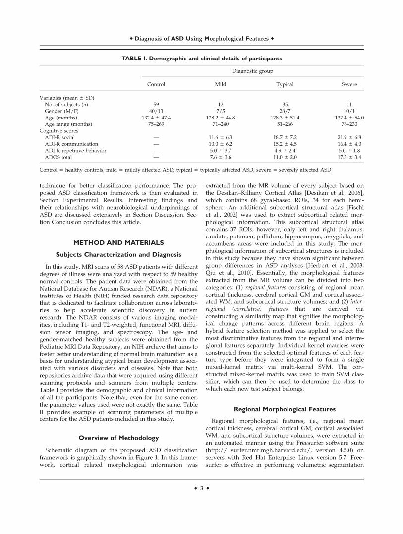
In this study MRI scans of 58 ASD patients with differentdegrees of illness were analyzed with respect to 59 healthynormal controls The patient data were obtained from theNational Database for Autism Research (NDAR) a NationalInstitutes of Health (NIH) funded research data repositorythat is dedicated to facilitate collaboration across laborato-ries to help accelerate scientific discovery in autismresearch The NDAR consists of various imaging modal-ities including T1- and T2-weighted functional MRI diffu-sion tensor imaging and spectroscopy The age- andgender-matched healthy subjects were obtained from thePediatric MRI Data Repository an NIH archive that aims tofoster better understanding of normal brain maturation as abasis for understanding atypical brain development associ-ated with various disorders and diseases Note that bothrepositories archive data that were acquired using differentscanning protocols and scanners from multiple centersTable I provides the demographic and clinical informationof all the participants Note that even for the same centerthe parameter values used were not exactly the same TableII provides example of scanning parameters of multiplecenters for the ASD patients included in this study
Overview of Methodology
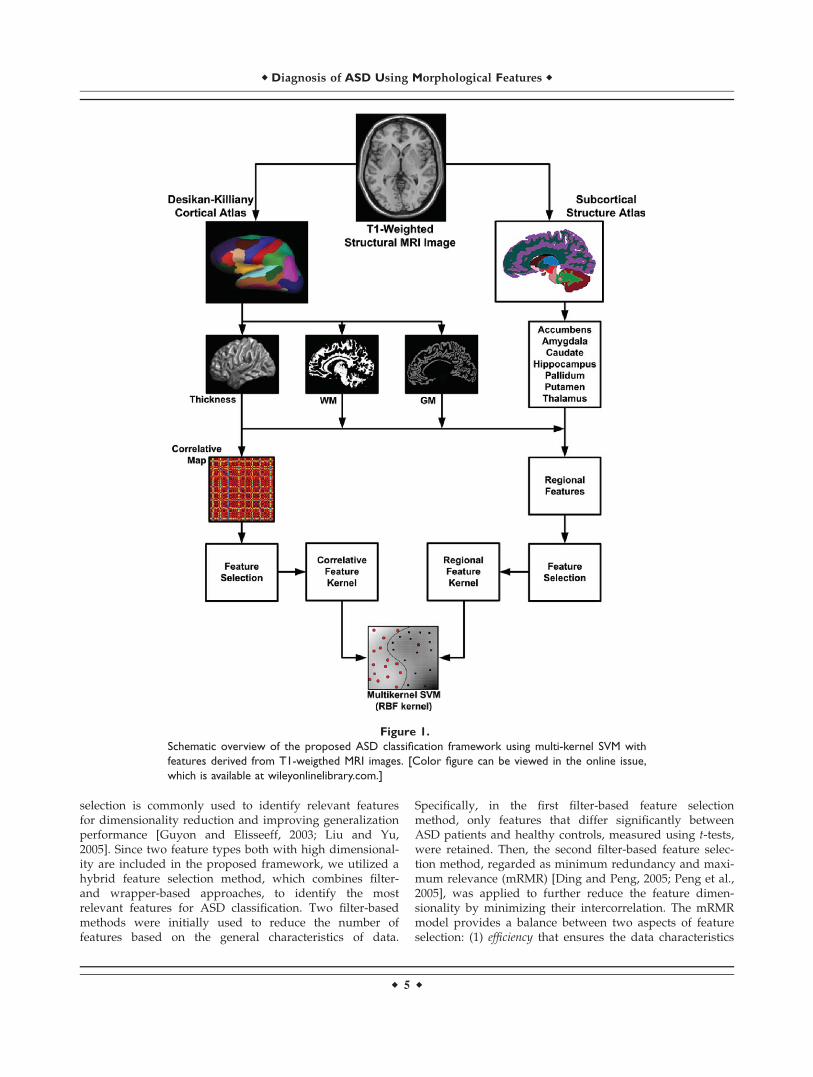
Schematic diagram of the proposed ASD classificationframework is graphically shown in Figure 1 In this frame-work cortical related morphological information was
extracted from the MR volume of every subject based onthe DesikanndashKilliany Cortical Atlas [Desikan et al 2006]which contains 68 gyral-based ROIs 34 for each hemi-sphere An additional subcortical structural atlas [Fischlet al 2002] was used to extract subcortical related mor-phological information This subcortical structural atlascontains 37 ROIs however only left and right thalamuscaudate putamen pallidum hippocampus amygdala andaccumbens areas were included in this study The mor-phological information of subcortical structures is includedin this study because they have shown significant betweengroup differences in ASD analyses [Herbert et al 2003Qiu et al 2010] Essentially the morphological featuresextracted from the MR volume can be divided into twocategories (1) regional features consisting of regional meancortical thickness cerebral cortical GM and cortical associ-ated WM and subcortical structure volumes and (2) inter-regional (correlative) features that are derived viaconstructing a similarity map that signifies the morpholog-ical change patterns across different brain regions Ahybrid feature selection method was applied to select themost discriminative features from the regional and interre-gional features separately Individual kernel matrices wereconstructed from the selected optimal features of each fea-ture type before they were integrated to form a singlemixed-kernel matrix via multi-kernel SVM The con-structed mixed-kernel matrix was used to train SVM clas-sifier which can then be used to determine the class towhich each new test subject belongs
Regional Morphological Features
Regional morphological features ie regional meancortical thickness cerebral cortical GM cortical associatedWM and subcortical structure volumes were extracted inan automated manner using the Freesurfer software suite(http surfernmrmghharvardedu version 450) onservers with Red Hat Enterprise Linux version 57 Free-surfer is effective in performing volumetric segmentation
TABLE I Demographic and clinical details of participants
Control 5 healthy controls mild 5 mildly affected ASD typical 5 typically affected ASD severe 5 severely affected ASD
r Diagnosis of ASD Using Morphological Features r
r 3 r
and cortical surface reconstruction [Desikan et al 2006Fischl and Dale 2000 Fischl et al 2002 1999ab] Bothintensity and continuity information from the entire threedimensional MR volumes are used in the segmentation anddeformation procedures to produce representations of corti-cal thickness [Dale et al 1999 Fischl et al 1999a] Corticalthickness measurement of this package has been thoroughlyvalidated against histological analysis [Rosas et al 2002]and manual measurements [Kuperberg et al 2003 Salatet al 2004] and demonstrated a good test-retest reliabilityacross different scanners and field strengths [Dickersonet al 2008 Han et al 2006] The regional mean corticalthickness feature for each ROI defined in the DesikanndashKill-iany cortical atlas [Desikan et al 2006] was normalizedwith respect to the standard deviation
The cerebral cortical GM and cortical associated WMvolumes were extracted using the same cortical atlas Vol-umes of seven subcortical structures ie thalamus cau-date putamen pallidum hippocampus amygdala andaccumbens areas were extracted from both hemispheresbased on the ROIs defined in the subcortical structuralatlas [Fischl et al 2002] The corticl and subcortical vol-umes of each subject were normalized with respect to theintracranial volume to minimize the effects of intersubjectvariation [Whitwell et al 2001]
Interregional Morphological Features
Connection abnormalities have been suggested to beassociated with a wide range of neurological and psychiat-ric brain disorders eg Alzheimerrsquos disease Parkinson
disease and schizophrenia Brain communication effi-ciency and capacity are affected in a different manner ineach disease [Bosboom et al 2009 He et al 2008 Lynallet al 2010 Stam et al 2007 van den Heuvel et al 2010Zalesky et al 2011] A study based on FDG-PET imagingsuggested that the brain of ASD children is damaged in alarge-scale network level in addition to abnormality inisolated regions [Lee et al 2011] In view of this wehypothesize that interregional morphological informationwhich conveys higher order information of disease pathol-ogy can be integrated with regional information toimprove the diagnosis accuracy of ASD
In this study interregional features were computedbetween pairs of ROIs using their mean cortical thicknessA 68 3 68 correlative matrix map was constructed withevery element representing the relative change pattern ofregional mean cortical thicknesses between a pair of ROIsThe constructed correlative map is symmetric with onesalong its diagonal Because of this symmetrical propertyonly the upper triangular of the correlative map was usedFor each subject all elements of the upper triangular partof the correlative map were concatenated to form a longfeature vector Details of extracting the interregional fea-tures can be found in Wee et al [in press]
Feature Selection
Neuroimage classification using high-dimensional datais a challenging task Because of the possible presence ofirrelevant or redundant features learning models tend tooverfit data and become less generalizable Feature
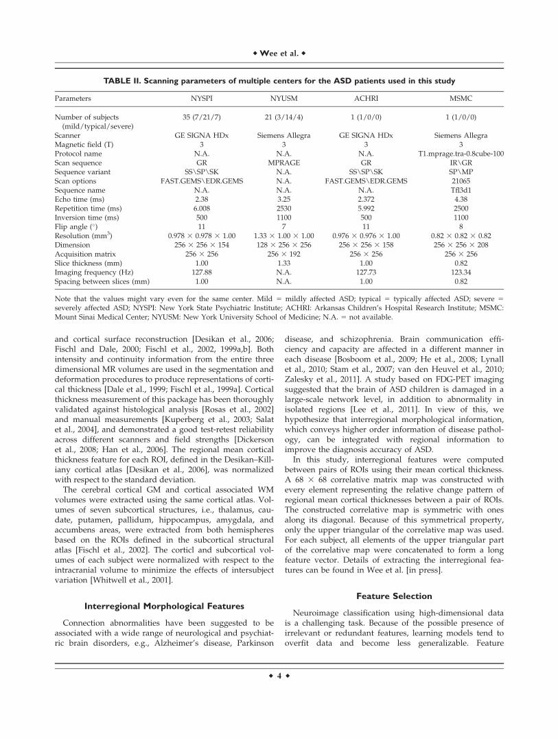
TABLE II Scanning parameters of multiple centers for the ASD patients used in this study
Parameters NYSPI NYUSM ACHRI MSMC
Number of subjects(mildtypicalsevere)
35 (7217) 21 (3144) 1 (100) 1 (100)
Scanner GE SIGNA HDx Siemens Allegra GE SIGNA HDx Siemens AllegraMagnetic field (T) 3 3 3 3Protocol name NA NA NA T1mpragetra-08cube-100Scan sequence GR MPRAGE GR IRGRSequence variant SSSPSK NA SSSPSK SPMPScan options FASTGEMSEDRGEMS NA FASTGEMSEDRGEMS 21065Sequence name NA NA NA Tfl3d1Echo time (ms) 238 325 2372 438Repetition time (ms) 6008 2530 5992 2500Inversion time (ms) 500 1100 500 1100Flip angle () 11 7 11 8Resolution (mm3) 0978 3 0978 3 100 133 3 100 3 100 0976 3 0976 3 100 082 3 082 3 082Dimension 256 3 256 3 154 128 3 256 3 256 256 3 256 3 158 256 3 256 3 208Acquisition matrix 256 3 256 256 3 192 256 3 256 256 3 256Slice thickness (mm) 100 133 100 082Imaging frequency (Hz) 12788 NA 12773 12334Spacing between slices (mm) 100 NA 100 082
Note that the values might vary even for the same center Mild 5 mildly affected ASD typical 5 typically affected ASD severe 5
severely affected ASD NYSPI New York State Psychiatric Institute ACHRI Arkansas Childrenrsquos Hospital Research Institute MSMCMount Sinai Medical Center NYUSM New York University School of Medicine NA 5 not available
r Wee et al r
r 4 r
selection is commonly used to identify relevant featuresfor dimensionality reduction and improving generalizationperformance [Guyon and Elisseeff 2003 Liu and Yu2005] Since two feature types both with high dimensional-ity are included in the proposed framework we utilized ahybrid feature selection method which combines filter-and wrapper-based approaches to identify the mostrelevant features for ASD classification Two filter-basedmethods were initially used to reduce the number offeatures based on the general characteristics of data
Specifically in the first filter-based feature selectionmethod only features that differ significantly betweenASD patients and healthy controls measured using t-testswere retained Then the second filter-based feature selec-tion method regarded as minimum redundancy and maxi-mum relevance (mRMR) [Ding and Peng 2005 Peng et al2005] was applied to further reduce the feature dimen-sionality by minimizing their intercorrelation The mRMRmodel provides a balance between two aspects of featureselection (1) efficiency that ensures the data characteristics
Figure 1
Schematic overview of the proposed ASD classification framework using multi-kernel SVM with
features derived from T1-weigthed MRI images [Color figure can be viewed in the online issue
which is available at wileyonlinelibrarycom]
r Diagnosis of ASD Using Morphological Features r
r 5 r
can be represented with a minimal number of features and(2) broadness that ensures the selected feature subset canmaximally represent the original space covered by the entiredataset Specifically mRMR minimizes the total relevance ofeach featurendashfeature pairs to achieve minimum redundancycondition while simultaneously maximizes the totalrelevance of each featurendashclass pairs to achieve maximumrelevance condition Finally support vector machine basedrecursive feature elimination (SVM-RFE) [Guyon et al 2004Rakotomamonjy 2003] an effective wrapper-based modelwas utilized to determine a subset of features that optimizesthe performance of the SVM classifier The basic principle ofSVM-RFE is to remove features to make the classificationerror smallest Note that hybrid feature selection was per-formed separately on each feature type (ie regional andinterregional features) as shown in Figure 1
Features Integration
To provide a more comprehensive way of characterizingthe whole feature space we utilized an MKL-basedapproach to combine the selected regional and interregionalfeatures with appropriate weighting This approach enablesthe utilization of different types of kernels or feature typesconcurrently in one single classifier instance Specifically inthis study a kernel matrix was first constructed for eachindividual feature type based on radial basic function (RBF)before they were integrated via a multi-kernel SVM to forma mixed-kernel matrix with appropriate weighting factorbm 0 [Wee et al 2012b] The optimal SVM model andunbiased estimation of the generalization performancewere obtained via a nested crossvalidation scheme
Statistical Measures and Performance Evaluation
In addition to classification accuracy (ACC) and areaunder receiver operating characteristic curve (AUC) otherstatistical measures were also been adopted to evaluatethe diagnostic power of compared methods more compre-hensively including Balanced ACcuracy (BAC) Youdenrsquosindex (Y) and F-score (F) [Sokolova et al 2006 Wee et al2012b] F-score provides a composite measure that favorsalgorithms with higher sensitivity while challenges thosewith higher specificity Youdenrsquos index evaluates the abil-ity of an algorithm to avoid failure by equally weightingits performance on positive and negative samples
In order to provide a more conservative evaluation onclassification performance a twofold crossvalidation wasadopted in our study to access the discriminative power ofeach compared method Specifically at the beginning ofexperiment the whole dataset was randomly partitionedinto two sets one for training and one for testing withsimilar number of subjects from each class in each set Thetraining set was used to construct the optimal SVM modelbased on the nested crossvalidation approach Then theconstructed optimal SVM model was used to identify the
ASD patients from healthy controls from the testing setThe same training and testing procedures were repeatedby swapping the training and testing sets The crossvalida-tion classification performance was determined by averag-ing the statistical measures over the twofold Thisprocedure was repeated for 100 times to obtain the finalmean classification performance The superiority of theproposed method with respect to other compared methodswas evaluated via a paired t-test on the mean classificationaccuracy over these 100 repetitions
EXPERIMENTAL RESULTS
ASD Classification Performance
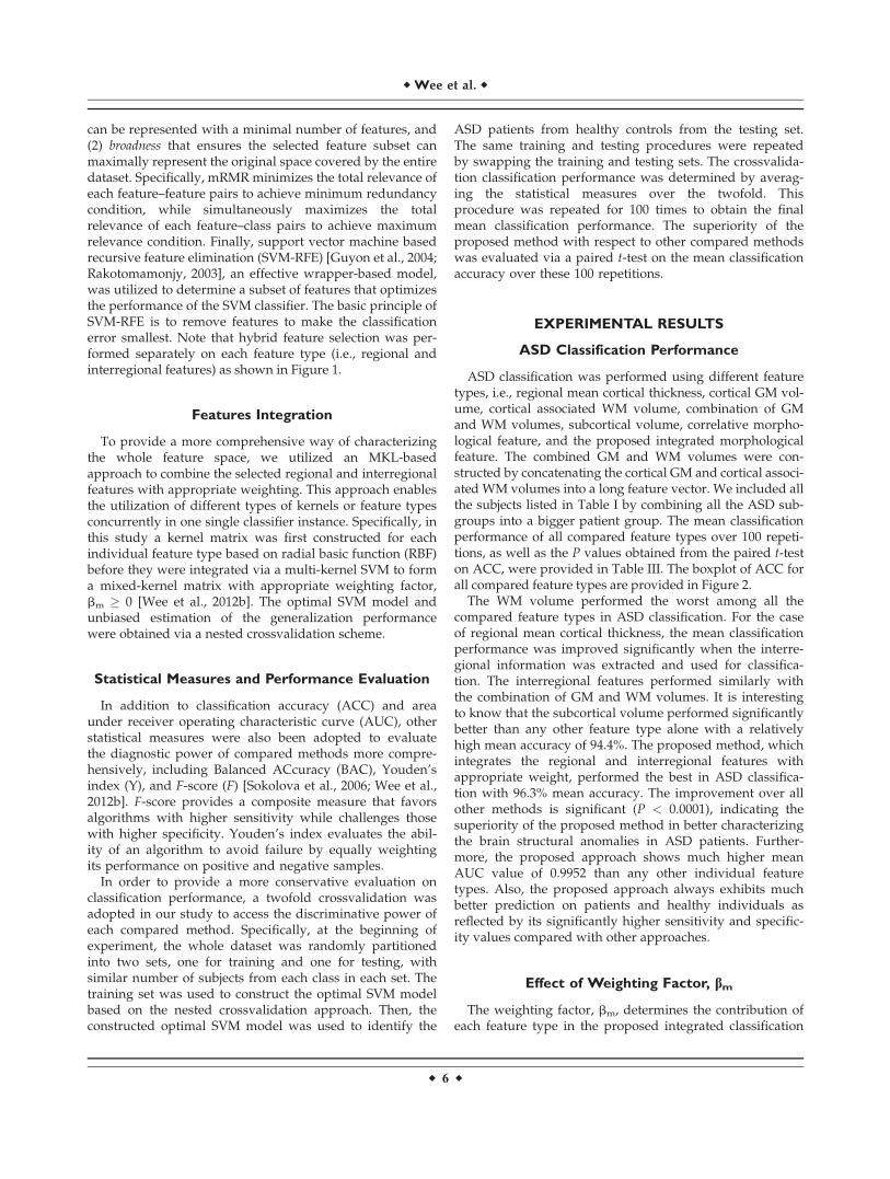
ASD classification was performed using different featuretypes ie regional mean cortical thickness cortical GM vol-ume cortical associated WM volume combination of GMand WM volumes subcortical volume correlative morpho-logical feature and the proposed integrated morphologicalfeature The combined GM and WM volumes were con-structed by concatenating the cortical GM and cortical associ-ated WM volumes into a long feature vector We included allthe subjects listed in Table I by combining all the ASD sub-groups into a bigger patient group The mean classificationperformance of all compared feature types over 100 repeti-tions as well as the P values obtained from the paired t-teston ACC were provided in Table III The boxplot of ACC forall compared feature types are provided in Figure 2
The WM volume performed the worst among all thecompared feature types in ASD classification For the caseof regional mean cortical thickness the mean classificationperformance was improved significantly when the interre-gional information was extracted and used for classifica-tion The interregional features performed similarly withthe combination of GM and WM volumes It is interestingto know that the subcortical volume performed significantlybetter than any other feature type alone with a relativelyhigh mean accuracy of 944 The proposed method whichintegrates the regional and interregional features withappropriate weight performed the best in ASD classifica-tion with 963 mean accuracy The improvement over allother methods is significant (P lt 00001) indicating thesuperiority of the proposed method in better characterizingthe brain structural anomalies in ASD patients Further-more the proposed approach shows much higher meanAUC value of 09952 than any other individual featuretypes Also the proposed approach always exhibits muchbetter prediction on patients and healthy individuals asreflected by its significantly higher sensitivity and specific-ity values compared with other approaches
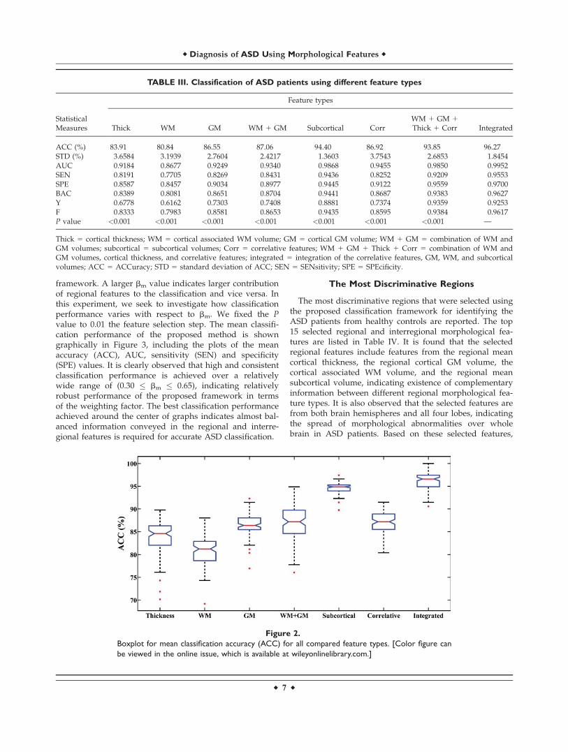
Effect of Weighting Factor bm
The weighting factor bm determines the contribution ofeach feature type in the proposed integrated classification
r Wee et al r
r 6 r
framework A larger bm value indicates larger contributionof regional features to the classification and vice versa Inthis experiment we seek to investigate how classificationperformance varies with respect to bm We fixed the Pvalue to 001 the feature selection step The mean classifi-cation performance of the proposed method is showngraphically in Figure 3 including the plots of the meanaccuracy (ACC) AUC sensitivity (SEN) and specificity(SPE) values It is clearly observed that high and consistentclassification performance is achieved over a relativelywide range of (030 bm 065) indicating relativelyrobust performance of the proposed framework in termsof the weighting factor The best classification performanceachieved around the center of graphs indicates almost bal-anced information conveyed in the regional and interre-gional features is required for accurate ASD classification
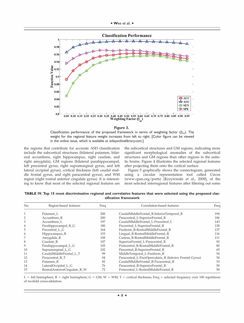
The Most Discriminative Regions
The most discriminative regions that were selected usingthe proposed classification framework for identifying theASD patients from healthy controls are reported The top15 selected regional and interregional morphological fea-tures are listed in Table IV It is found that the selectedregional features include features from the regional meancortical thickness the regional cortical GM volume thecortical associated WM volume and the regional meansubcortical volume indicating existence of complementaryinformation between different regional morphological fea-ture types It is also observed that the selected features arefrom both brain hemispheres and all four lobes indicatingthe spread of morphological abnormalities over wholebrain in ASD patients Based on these selected features
TABLE III Classification of ASD patients using different feature types
Thick 5 cortical thickness WM 5 cortical associated WM volume GM 5 cortical GM volume WM 1 GM 5 combination of WM andGM volumes subcortical 5 subcortical volumes Corr 5 correlative features WM 1 GM 1 Thick 1 Corr 5 combination of WM andGM volumes cortical thickness and correlative features integrated 5 integration of the correlative features GM WM and subcorticalvolumes ACC 5 ACCuracy STD 5 standard deviation of ACC SEN 5 SENsitivity SPE 5 SPEcificity
Figure 2
Boxplot for mean classification accuracy (ACC) for all compared feature types [Color figure can
be viewed in the online issue which is available at wileyonlinelibrarycom]
r Diagnosis of ASD Using Morphological Features r
r 7 r
the regions that contribute for accurate ASD classificationinclude the subcortical structures (bilateral putamen bilat-eral accumbens right hippocampus right caudate andright amygdala) GM regions (bilateral parahippocampalleft precentral gyrus right supramarginal gyrus and leftlateral occipital gyrus) cortical thickness (left caudal mid-dle frontal gyrus and right paracentral gyrus) and WMregion (right rostral anterior cingulate gyrus) It is interest-ing to know that most of the selected regional features are
the subcortical structures and GM regions indicating moresignificant morphological anomalies of the subcorticalstructures and GM regions than other regions in the autis-tic brains Figure 4 illustrates the selected regional featuresafter projecting them onto the cortical surface
Figure 5 graphically shows the connectogram generatedusing a circular representation tool called Circos(wwwcpanorgports) [Krzywinski et al 2009] of themost selected interregional features after filtering out some
Figure 3
Classification performance of the proposed framework in terms of weighting factor (bm) The
weight for the regional feature weight increases from left to right [Color figure can be viewed
in the online issue which is available at wileyonlinelibrarycom]
TABLE IV Top 15 most discriminative regional and correlative features that were selected using the proposed clas-
sification framework
No Region-based features Freq Correlation-based features Freq
L 5 left hemisphere R 5 right hemisphere G 5 GM W 5 WM T 5 cortical thickness Freq 5 selected frequency over 100 repetitionsof twofold crossvalidation
r Wee et al r
r 8 r
connections that are with low selection frequency It canbe clearly observed that pairs of regions (links in the con-nectogram) that contributed for accurate ASD classificationare not only restricted within the same hemisphere orsame lobe but across all hemispheres and lobes particu-larly between the left and right frontal lobes right frontaland left parietal lobes right and left temporal lobes rightfrontal and right temporal lobes right frontal and rightoccipital lobes and right frontal and right parietal lobes Itis interesting to observe that most of the selected interre-gional features are within the right hemisphere of thebrain as indicated by thicker and more frequent links con-necting between regions located in the right hemisphere asshown in Figure 5 In addition most of the within lobeinterregional features connect regions located in the bilat-eral frontal lobes It can also be observed that some of theregions associated with the selected interregional featuresare same as those associated with the selected regionalfeatures
DISCUSSION
Neuroimaging-based ASD classification frameworks pro-posed recently were essentially based on either volumetricinformation [Akshoomoff et al 2004 Uddin et al 2011]cortical surface information [Ecker et al 2010ab] or func-tional connectivity of GM region [Anderson et al 2011]The highest ASD classification accuracy achieved usingthese frameworks are slightly greater than 90 but withrelatively low sensitivity andor specificity values Fur-thermore the datasets used in these studies are relativelysmall with the total number of ASD patients and healthysubjects slightly more than 60 These datasets were alsoacquired in relatively well-controlled conditions using thesame scanner with the same acquisition protocols
In the present study the proposed method extracts inaddition to regional information interregional (or correla-tive) morphological information [Wee et al in press] and
Figure 4
The most discriminative regional features projected onto the cortical surface [Color figure can
be viewed in the online issue which is available at wileyonlinelibrarycom]
Figure 5
Connectogram of the most discriminative connections selected
from the correlative features Red color lines are intrahemi-
sphere links and gray color lines are interhemisphere links
Thickness of each line reflects its selection frequency eg a
thicker line indicates a higher selection frequency For the abbre-
viations of the regions please refer to Table V [Color figure can
be viewed in the online issue which is available at
wileyonlinelibrarycom]
r Diagnosis of ASD Using Morphological Features r
r 9 r
was evaluated using a multicenter ASD dataset Whilemost work in the literature has focused on measuring mor-phological changes in individual ROIs our study suggeststhat interregional cortical morphological changes acrossthe whole brain can be utilized to provide additionaldisease-related information that may facilitate the diagno-sis process on an individual level The interregional fea-ture extraction approach is essentially based on theassumption that the morphological alterations caused byASD pathological attacks are not restricted to certain brainareas but widely spread over the whole brain [Lee et al2011] and the relative changes between pairs of ROIsmight convey information that is associated with neurobio-logical underpinnings of ASD This approach addressesthe brain anomaly caused by diseases through the interre-gional morphological change patterns of entire brain Inour study it was found that this feature extraction methodyield performance that is comparable or even better thanmost of the individual regional features in discriminating
ASD patients from healthy controls except the subcorticalstructure volume To fully utilize information conveyed bydifferent feature types and hence further enhance the clas-sification performance we suggested to fusing both theregional and interregional features with appropriateweights via multi-kernel learning technique Promisingclassification results were obtained using the proposedfused (or integrated) regional and interregional features onthe NDAR dataset ACC 5 9627 AUC 5 09952 SEN 5
09553 and SPE 5 09700 Almost perfect AUC value wasachieved indicating excellent diagnostic power and gener-alizability of the proposed framework to future unseensubjects
To determine whether site protocol variance or individ-ual variance drives classification errors we have per-formed three additional experiments (1) classificationusing samples from each site individually (2) classificationbetween ASD samples of two largest sites and (3) permu-tation test by perturbing the clinical label of the samplesThe proposed method performs similarly when the classi-fication is performed on the samples from each site sepa-rately Specifically the classification accuracies for theNYSPI and NYUSM sites are 860 and 840 respec-tively For the experiment to distinguish ASD samplesfrom NYPSI and from NYUSM a near-random perform-ance with accuracy of 668 is achieved indicating thenondifferentiability of ASD samples from these two sitesFinally in the permutation test the proposed frameworkis statistically significance in the context of discriminativeanalysis compared with random classifiers at the 1 sig-nificance level computing using 10000 repetitions
The integration of regional and interregional morpholog-ical features provided the best classification results whenthe weighting factor between two feature types is withinthe range (030 bm 065) suggesting that the interre-gional features convey additional and complementaryASD-related information to the regional features Highand consistent classification performance over this wideweighting factor range demonstrates that the proposedapproach is relatively robust with respect to the parameterthat determines the contribution of each feature type Thisalso implies that the difficulty of determining the mostappropriate weighting factor for fusing the regional andinterregional features in our proposed approach can some-what be reduced
High accuracy predictive classification as reported inthis study is important from the clinical perspective asASD is still regarded by many as a functional mental dis-order lacking a robust neurological or structural basisThis view persists despite many reports of differences inbrain structure at a group level between autistic and nor-mal brains However group level differences provide lit-tle useful information for individual patients and therehas furthermore been marked heterogeneity of reportedgroup level abnormalities [Amaral et al 2008 Lainhart2006 Salmond et al 2007 Toal et al 2005] Conse-quently these findings have had limited impact on
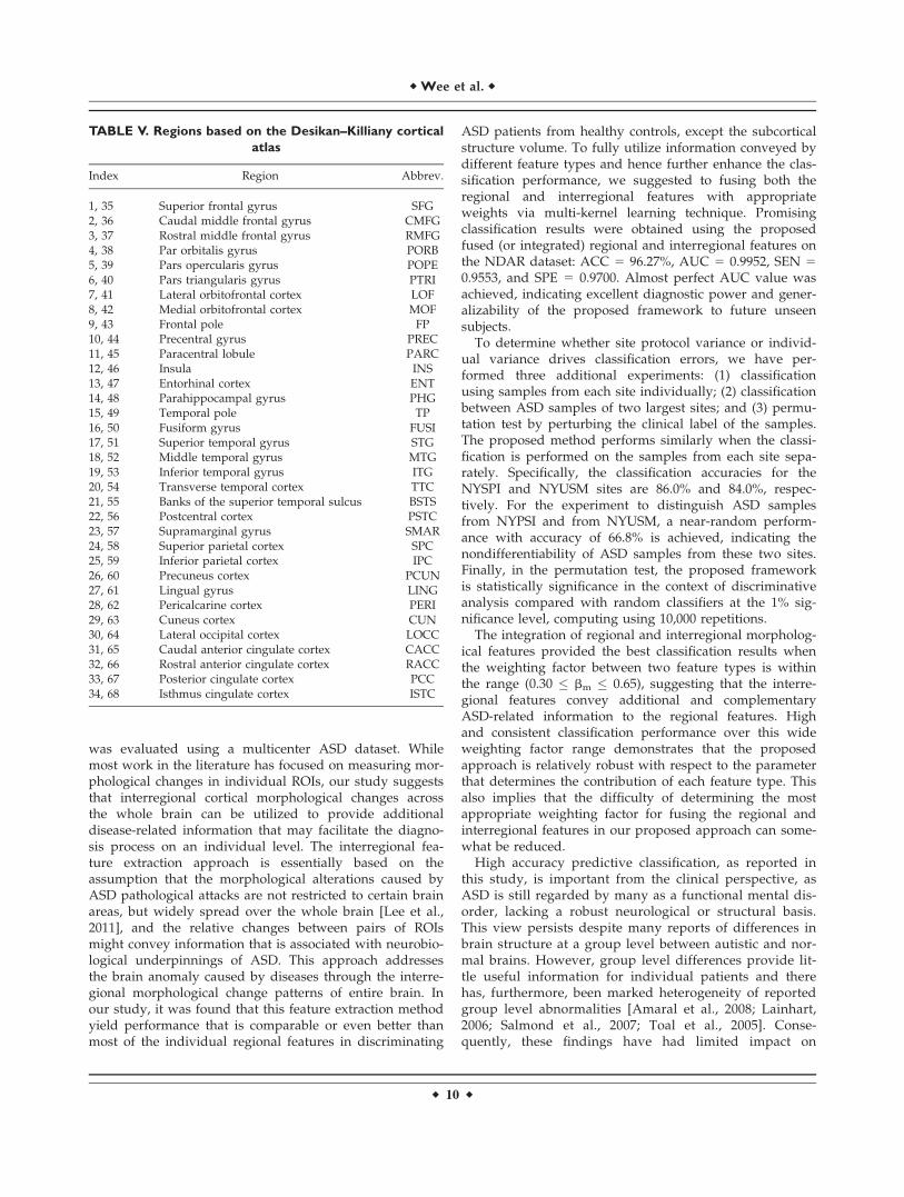
TABLE V Regions based on the DesikanndashKilliany cortical
clinical practice Development of a high accuracy predic-tive classification framework that can identify ASDpatients from healthy controls with results replicableacross scanners and subjects is therefore a significantadvance
The brain regions that are associated with the ASDpathology have already been extensively reported in previ-ous studies either based on between-group comparison[Hardan et al 2006 McAlonan et al 2005 Nordahl et al2007 Redcay and Courchesne 2008] or individual-levelclassification [Akshoomoff et al 2004 Anderson et al2011 Ecker et al 2010ab] Regions which are associatedwith the selected regional features are spread over thewhole brain not restricted to particular brain hemispheresor lobes These regions have been widely reported in theliterature to be associated with ASD including bilateralputamen [Haznedar et al 2006 Langen et al 2009 Qiuet al 2010 Toal et al 2009] bilateral accumbens [Dichteret al 2012 Langen et al 2009] right hippocampus [Ayl-ward et al 1999 Baron-Cohen 2004 Sparks et al 2002]right caudate [Langen et al 2007 2009 Qiu et al 2010Sears et al 1999] right amygdala [Ashwin et al 2007Aylward et al 1999 Baron-Cohen 2004 Baron-Cohenet al 2000 Sparks et al 2002] bilateral parahippocampal[Ecker et al 2010a Toal et al 2009] left precentral gyrus[Cauda et al 2011 Rojas et al 2006 Scheel et al 2011Toal et al 2009] right supramarginal gyrus [Bolling et al2011 Brieber et al 2007] left lateral occipital gyrus [Eckeret al 2010a Greene et al 2011] left caudal middle frontalgyrus [Baron-Cohen 2004 Rojas et al 2006 Schmitz et al2008] right paracentral gyrus [Cheng et al 2011 Scheelet al 2011 Toal et al 2009] and right rostral anterior cin-gulate gyrus [Ashwin et al 2007 Cauda et al 2011Cheng et al 2011 Toal et al 2009]
Some of the regions that are associated with the selectedregional features have also been detected in the selectedinterregional features including the left caudal middlefrontal gyrus right paracentral lobule and left precentralgyrus Regions that are associated only with the selectedinterregional features are frontal lobe (ie right caudalmiddle frontal gyrus [Ecker et al 2010a Gomot et al2006 Turner et al 2006] left paracentral lobule [Bollinget al 2011 Hyde et al 2010] bilateral superior frontalgyrus [Cheng et al 2010 Hyde et al 2010] right rostralmiddle frontal gyrus [Ecker et al 2010b] right precentralgyrus [Ecker et al 2010b Hyde et al 2010] and rightpars opercularis [Dapretto et al 2005 Nordahl et al 2007Yamasaki et al 2010]) temporal lobe (right inferior tem-poral gyrus [Abell et al 1999 Ecker et al 2010a Petersonet al 2006] left middle temporal gyrus [Abell et al 1999Ecker et al 2010a] and right fusiform gyrus [Ecker et al2010ab Hall et al 2003 Hyde et al 2010]) occipital lobe(right lingual gyrus [Cheng et al 2011 Ecker et al 2010b]and right cuneus cortex [Greene et al 2011 Luders et al2012]) and parietal lobe (bilateral postcentral gyrus [Eckeret al 2010b Hyde et al 2010]) The fact that our findingsare consistent with results reported in previous studies
demonstrates the efficacy of our proposed framework alsoin identifying disease-related biomarkers for ASD classifi-cation Furthermore the morphological change patternsbetween region pairs together with the regional featuresprovide crucial yet complementary information for betterunderstanding of the neurobiological underpinnings ofASD
It is interesting to observe that several components inthe limbic system have been selected in the proposedframework as important features for ASD classificationThe human limbic system is primarily responsible for reg-ulating the human emotions as well as the formation ofmemories indicating the capability of the proposed frame-work in reflecting the relationships between behavioralimpairments and structural abnormalities Amygdalacomponent of the limbic system that located in the medialtemporal lobe of the brain is the brain area that concernswith the autonomic responses associated with fear emo-tional responses sexual arousal and hormonal secretions[Amunts et al 2006] It has been strongly supported par-ticularly in functional neuroimaging analysis that amyg-dala plays a critical role in emotional processing withoutlateralization in terms of gender or valence [Sergerie et al2008] Specifically the amygdala parallelly involvesimplicit emotional learning and memory emotional modu-lation of memory emotional influences on attention andperception emotion and social behavior and emotion inhi-bition and regulation [Davis and Whalen 2001 Phelpsand LeDoux 2005] Based on a postmortem study thedysfunction of amygdala which may lead to changes inthe aggressive and emotional behavior might be explainedby the significant reduced number of neurons in this areaof the autistic brain when compared with age-matchedtypically developing children [Schulkin 2007 Schumannand Amaral 2006] In addition based on MRI studies theamygdala appears to undergo an abnormal pattern ofdevelopment that includes early enlargement and adecreased number of neurons in adulthood [Schumannet al 2004] Impairment of the amygdala usually results ininappropriate behavioral and emotional reactions thateventually lead to physical harm to the patients andothers In fact most of such patients need to be undersupervised care and cannot function alone in society [Leeet al 1988 1995]
Other components of the human limbic system thatwere selected by the proposed framework include the cin-gulate gyrus orbitofrontal cortex nucleus accumbens andparahippocampus The developmental dysfunction of theorbitofrontal-amygdala circuit of the brain is a critical fac-tor in the development of autism particularly for personswith autism in socio-emotional cognition and behavioralself-regulation [Bachevalier and Loveland 2006] A reviewof cases with early prefrontal damage by [Eslinger et al2004] suggested that early injury to the orbitofrontal cortexis associated with intractable deficits in the regulation ofemotions and social functioning Developmental trajecto-ries of the caudate nucleus putamen and nucleus
r Diagnosis of ASD Using Morphological Features r
r 11 r
accumbens differed between subjects with ASD andhealthy controls [Langen et al 2009] It was suggestedthat adults with ASD had a significantly smaller totalbrain WM volume lower fractional anisotropy of WMtracts connecting putamen to frontal cortical areas andhigher mean diffusivity of WM tracts connecting accum-bens to frontal cortex [Langen et al 2012] It has beenreported that women with ASD had a significantly smallerGM density than controls bilaterally in the parahippocam-pal gyrus medial and lateral orbitofrontal cortex basalganglia (lentiform nucleus and caudate nucleus) leftcuneus and right anterior cingulate [Craig et al 2007] AVBM study suggested that the parahippocampal gyruscontributes to understanding of the interactions betweenemotion and cognition and thus may be associated withreactivation of contextual fear memory that results inavoidance behavior [Ke et al 2008] The cingulate gyrus aconduit of messages to and from the inner limbic systemis involved with emotion formation and processing learn-ing memory executive function and respiratory controlRight anterior cingulate gyrus has been reported to experi-ence significant reduction in volume besides metabolicallyless active in the autistic patients than in the healthy con-trols [Haznedar et al 1997] Postmortem study of theautistic brains has shown a broad spectrum of abnormal-ities in the cerebellum and neocortex involving anteriorcingulate cortex [Simms et al 2009]
The selected interregional features show significantlymore abnormality in the right hemisphere than the lefthemisphere ie rightward asymmetry of the autisticbrain when compared with healthy controls a similarresult that was reported in [Herbert et al 2005] The autis-tic brain shows more pronounced rightward asymmetrythan healthy controls and this sizeable rightward asymme-try may be a consequence of the early abnormal braingrowth trajectories in ASD [Herbert et al 2005] Similarrightward asymmetry in ASD has also been reported infunctional study [Chiron et al 1995] It was also reportedrecently in a MRI study that ASD children with languageproblem demonstrated a rightward asymmetry in auditorylanguage cortex and the asymmetry increases with age[Gage et al 2009] This morphological pattern of autisticbrains has been detected by the proposed framework andthus facilitates the ASD classification
It was observed that the number of misclassified mildlyaffected patients is twice the number of misclassifiedseverely affected patients in 100 repetitions of twofoldcrossvalidation although the sample size is similar forboth subgroups This finding is in agreement with the factthat the difficulty of identifying the mildly affectedpatients decreases with the degree of illness Althoughdataset with highly imbalance gender ratio (male to femaleratio as 351) was used in this study only two femalepatients both are from the mildly affected subgroup aremisclassified in 100 repetitions of twofold crossvalidationThis demonstrates the robustness of our framework withrespect to gender
It has been reported recently that the autistic symptomscan be vastly improved if early medical andor behavioraltreatments are provided to the toddlers as young as 18months [Dawson et al 2012] Hence early diagnosis ofASD is tremendously important to improve quality of lifeof ASD patients when they achieve adulthood Since theage range of ASD subjects used in this study is between 4and 26 years old the proposed diagnosis framework hasonly been tested with subjects who are older than 4 yearsold Because of the importance of early diagnosis futureresearch will be directed to extending the current frame-work to the diagnosis of children prior to 4 years of age
The performance of the proposed ASD identificationmethod is limited by the accuracy of the segmentationresults given by Freesurfer Several recent publicationshave indicated that segmentation by Freesurfer in subcorti-cal regions is incorrect particularly for deep structuressuch as amygdala [Khan et al 2008 Klauschen et al2009 Morey et al 2009 2010 Zhong et al 2010] Subcorti-cal segmentation errors may result in the inability to cor-rectly capture the true biological differences betweenpatient and control groups and eventually influencing dis-ease identification accuracy Since precise measurement ofbiological differences is crucial for improving identificationaccuracy we expect that the performance of the proposedmethod can be further improved by utilizing better seg-mentation algorithms in the future
Artifacts caused by head motion can be a significantimpediment to acquiring the in vivo MRI scans of thehuman brain Motion artifacts adversely affect the abilityto accurately characterize the size shape and tissue prop-erties of brain structures [Brown et al 2010 Kupermanet al 2011] This issue is particularly severe for pediatricpatients who have difficulties remaining still in the scan-ner during data acquisition We have visually inspectedthe quality of all MRI scans from both groups and foundno significant motion artifact We believe that the investi-gators who contributed to these archives have performedcertain quality control procedures before uploading theirdatasets to the repositories
CONCLUSIONS
In this article a new classification framework has beenproposed to differentiate ASD patients from healthy con-trols using regional and interregional morphological fea-tures derived from T1-weighted MRI scans Thisframework fuses the regional and interregional (correla-tive) morphological features using multiple kernel learningfor accurate classification of ASD which is a highly hetero-geneous neurodevelopmental disorder Significantimprovement in classification performance demonstratesthe reliability and robustness of our proposed frameworkFurthermore the regions that were selected for accurateclassification are similar to those reported in the previousASD studies particularly components in the human limbic
r Wee et al r
r 12 r
system demonstrating the capability of our frameworkalso in determining the biologically meaningful anddisease-associated biomarkers Furthermore the observedrightward asymmetry anatomically anomalies pattern par-ticularly in auditory language cortex provides supportiveevidence that the ASD is a neurodevelopmental disorderthat is closely related to language problem The promisingresults obtained demonstrate the effectiveness of integrat-ing regional and interregional information for diagnosis ofhighly heterogeneous neurodevelopmental disorders suchas ASD While these findings in this study are intriguingthey are preliminary and require further study particularlyusing larger dataset
REFERENCES
Abell F Krams M Ashburner J Passingham R Friston KFrackowiak R Happe F Frith C Frith U (1999) The neuroan-atomy of autism A voxel-based whole brain analysis of struc-tural scans Neuroreport 101647ndash1651
Ahmadlou M Adeli H Adeli A (2012) Improved visibility graphfractality with application for the diagnosis of autism spectrumdisorder Phys A 3914720ndash4726
Akshoomoff N Lord C Lincoln AJ Courchesne RY Carper RATownsend J Courchesne E (2004) Outcome classification ofpreschool children with autism spectrum disorders using MRIbrain measures J Am Acad Child Adolesc Psychiatry 43349ndash357
American Psychiatric Association (2013) Diagnostic and StatisticalManual of Mental Disorders Fifth Edition (DSM-5) AmericanPsychiatric Association
Amunts K Kedo O Kindler M Pieperhoff P Mohlberg H ShahNJ Habel U Schneider F Zilles K (2006) Cytoarchitectonicmapping of the human amygdala hippocampal region andentorhinal cortex Intersubject variability and probability mapsAnat Embryol (Berl) 210343ndash352
Anderson JS Nielsen JA Froehlich AL DuBray MB Druzgal TJCariello AN Cooperrider JR Zielinski BA Ravichandran CFletcher PT Alexander AL Bigler ED Lange N Lainhart JE(2011) Functional connectivity magnetic resonance imagingclassification of autism Brain 1343742ndash3754
Ashwin C Baron-Cohen S Wheelwright S OrsquoRiordan MBullmore ET (2007) Differential activation of the amygdalaand the lsquosocial brainrsquo during fearful face-processing inAsperger syndrome Neuropsychologia 452ndash14
Aylward EH Minshew NJ Goldstein G Honeycutt NAAugustine AM Yates KO Barta PE Pearlson GD (1999) MRIvolumes of amygdala and hippocampus in nonndashmentallyretarded autistic adolescents and adults Neurology 532145ndash2150
Bachevalier J Loveland KA (2006) The orbitofrontalndashamygdalacircuit and self-regulation of socialndashemotional behavior inautism Neurosci Biobehav Rev 3097ndash117
Baron-Cohen S (2004) The cognitive neuroscience of autism JNeurol Neurosurg Psychiatry 75945ndash948
Baron-Cohen S Ring HA Bullmore ET Wheelwright S AshwinC Williams SC (2000) The amygdala theory of autism Neuro-sci Biobehav Rev 24355ndash364
Bolling DZ Pitskel NB Deen B Crowley MJ McPartland JCKaiser MD Vander Wyk BC Wu J Mayes LC Pelphrey KA(2011) Enhanced neural responses to rule violation in childrenwith autism A comparison to social exclusion Dev Cogn Neu-rosci 1280ndash294
Bosboom JL Stoffers D Wolters EC Stam CJ Berendse HW(2009) MEG resting state functional connectivity in Parkin-sonrsquos disease related dementia J Neural Transm 116193ndash202
Bosl W Tierney A Tager-Flusberg H Nelson C (2011) EEG com-plexity as a biomarker for autism spectrum disorder risk BMCMed 918
Brieber S Neufang S Bruning N Kamp-Becker I Remschmidt HHerpertz-Dahlmann B Fink GR Konrad K (2007) Structuralbrain abnormalities in adolescents with autism spectrum disor-der and patients with attention deficithyperactivity disorderJ Child Psychol Psychiatry 481251ndash1258
Brown TT Kuperman JM Erhart M White NS Roddey JCShankaranarayanan A Han ET Rettmann D Dale AM (2010)Prospective motion correction of high-resolution magnetic res-onance imaging data in children Neuroimage 53139ndash145
Calderoni S Retico A Biagi L Tancredi R Muratori F Tosetti M(2012) Female children with autism spectrum disorder Aninsight from mass-univariate and pattern classification analy-ses Neuroimage 591013ndash1022
Cauda F Geda E Sacco K DrsquoAgata F Duca S Geminiani GKeller R (2011) Grey matter abnormality in autism spectrumdisorder An activation likelihood estimation meta-analysisstudy J Neurol Neurosurg Psychiatry 821304ndash1313
Centers for Disease Control and Prevention (2012) Prevalence ofautism spectrum disordersmdashAutism and Developmental Dis-abilities Monitoring Network 14 sites United States 2008MMWR Surveill Summ 611ndash19
Cheng Y Chou K-H Fan Y-T Lin C-P (2011) ANS Aberrant neu-rodevelopment of the social cognition network in adolescentswith autism spectrum disorders PLoS One 6e18905
Cheng Y Chou KH Chen IY Fan YT Decety J Lin CP (2010)Atypical development of white matter microstructure in ado-lescents with autism spectrum disorders Neuroimage 50873ndash882
Chiron C Leboyer M Leon F Jambaque I Nuttin C Syrota A(1995) SPECT of the brain in childhood autism Evidence for alack of normal hemispheric asymmetry Dev Med Child Neu-rol 37849ndash860
Costafreda SG Chu C Ashburner J Fu CHY (2009) Prognosticand diagnostic potential of the structural neuroanatomy ofdepression PLoS One 4e6353
Craig MC Zaman SH Daly EM Cutter WJ Robertson DMHallahan B Toal F Reed S Ambikapathy A Brammer MMurphy CM Murphy DG (2007) Women with autistic-spectrum disorder Magnetic resonance imaging study of brainanatomy Br J Psychiatry 191224ndash228
Dale AM Fischl B Sereno MI (1999) Cortical surface-based analy-sis I Segmentation and surface reconstruction Neuroimage 9179ndash194
Dapretto M Davies MS Pfeifer JH Scott AA Sigman MBookheimer SY Iacoboni M (2005) Understanding emotions inothers Mirror neuron dysfunction in children with autismspectrum disorders Nat Neurosci 928ndash30
Davatzikos C Fan Y Wu X Shen D Resnick SM (2008) Detectionof prodromal Alzheimerrsquos disease via pattern classification ofmagnetic resonance imaging Neurobiol Aging 29514ndash523
r Diagnosis of ASD Using Morphological Features r
r 13 r
Davis M Whalen PJ (2001) The amygdala Vigilance and emotionMol Psychiatry 613ndash34
Dawson G Jones EJH Merkle K Venema K Lowy R Faja SKamara D Murias M Greenson J Winter J Smith M RogersSJ Webb SJ (2012) Early behavioral intervention is associatedwith normalized brain activity in young children with autismJ Am Acad Child Adolesc Psychiatry 511150ndash1159
Desikan RS Segonne F Fischl B Quinn BT Dickerson BC BlackerD Buckner RL Dale AM Maguire RP Hyman BT et al(2006) An automated labeling system for subdividing thehuman cerebral cortex on MRI scans into gyral based regionsof interest Neuroimage 31968ndash980
Dichter GS Felder JN Green SR Rittenberg AM Sasson NJBodfish JW (2012) Reward circuitry function in autism spec-trum disorders Soc Cogn Affect Neurosci 7160ndash172
Dickerson BC Fenstermacher E Salat DH Wolk DA Maguire RPDesikan R Pachecoc J Quinn BT der Kouwe AV Greve DNBlacker D Albert MS Killiany RJ Fischl B (2008) Detection ofcortical thickness correlates of cognitive performance Reliabil-ity across MRI scan sessions scanners and field strengthsNeuroimage 3910ndash18
Ding C Peng H (2005) Minimum redundancy feature selectionfrom microarray gene expression data J Bioinform ComputBiol 3185ndash205
Duffy FH Als H (2012) A stable pattern of EEG spectral coher-ence distinguishes children with autism from neuro-typicalcontrolsmdashA large case control study BMC Med 1064
Ecker C Marquand A Mourao-Miranda J Johnston P Daly EMBrammer MJ Maltezos S Murphy CM Robertson D WilliamsSC Murphy DG (2010a) Describing the brain in five dimen-sionsmdashMagnetic resonance imaging-assisted diagnosis ofautism spectrum disorder using a multiparameter classificationapproach J Neurosci 3010612ndash10623
Ecker C Rocha-Rego V Johnston P Mourao-Miranda JMarquand A Daly EM Brammer MJ Murphy C Murphy DGthe MRC AIMS Consortium (2010b) Investigating the predic-tive value of whole-brain structural MR scans in autism Apattern classification approach Neuroimage 4944ndash56
Eslinger PJ Flaherty-Craig CV Benton AL (2004) Developmentaloutcomes after early prefrontal cortex damage Brain Cogn 5584ndash103
Fischl B Dale AM (2000) Measuring the thickness of the humancerebral cortex from magnetic resonance images Proc NatlAcad Sci U S A 9711050ndash11055
Fischl B Sereno MI Dale AM (1999a) Cortical surface-based anal-ysis II Inflation flattening and a surface-based coordinatesystem Neuroimage 9195ndash207
Fischl B Sereno MI Tootell RBH Dale AM (1999b) High-resolu-tion intersubject averaging and a coordinate system for thecortical surface Hum Brain Mapp 8272ndash284
Fischl B Salat DH Busa E Albert M Dieterich M Haselgrove Cvan der Kouwe A Killiany R Kennedy D Klaveness SMontillo A Makris N Rosen B Dale AM (2002) Whole brainsegmentation Automated labeling of neuroanatomical struc-tures in the human brain Neuron 33341ndash355
Gage NM Juranek J Filipek PA Osann K Flodman P IsenbergAL Spence MA (2009) Rightward hemispheric asymmetries inauditory language cortex in children with autistic disorder AnMRI investigation J Neurodev Disord 1205ndash214
Gillberg C (1993) Autism and related behaviors J Intellect DisabilRes 37343ndash372
Gomot M Bernard FA Davis MH Belmonte MK Ashwin CBullmore ET Baron-Cohen S (2006) Change detection in chil-dren with autism An auditory event-related fMRI study Neu-roimage 29475ndash484
Gong Q Wu Q Scarpazza C Lui S Jia Z Marquand A Huang XMcGuire P Mechelli A (2011) Prognostic prediction of thera-peutic response in depression using high-field MR imagingNeuroimage 551497ndash1503
Greene DJ Colich N Iacoboni M Zaidel E Bookheimer SYDapretto M (2011) Atypical neural networks for socialorienting in autism spectrum disorders Neuroimage 56354ndash362
Guilmatre A Dubourg C Mosca A-L Legallic S Goldenberg ADrouin-Garraud V Layet V Rosier A Briault S Bonnet-Brilhault F Laumonnier F Odent S Le Vacon G Joly-Helas GDavid V Bendavid C Pinoit JM Henry C Impallomeni CGermano E Tortorella G Di Rosa G Barthelemy C Andres CFaivre L Frebourg T Saugier Veber P Campion D (2009)Recurrent rearrangements in synaptic and neurodevelopmentalgenes and shared biologic pathways in schizophrenia autismand mental retardation Arch Gen Psychiatry 66947ndash956
Guyon I Elisseeff A (2003) An introduction to variable and fea-ture selection J Mach Learn Res 31157ndash1182
Guyon I Weston J Barnhill S Vapnik V (2004) Gene selection forcancer classification using support vector machines MachineLearning 46389ndash422
Hall GBC Szechtman H Nahmias C (2003) Enhanced salienceand emotion recognition in autism A PET study Am J Psychi-atry 1601439ndash1441
Haller S Badoud S Nguyen D Garibotto V Lovblad KOBurkhard PR (2012) Individual detection of patients with Par-kinson disease using support vector machine analysis of diffu-sion tensor imaging data Initial results AJNR Am JNeuroradiol 332123ndash2128
Han X Jovicich J Salat D van der Kouwe A Quinn B Czanner SBusa E Pacheco J Albert M Killiany R Maguire P Rosas DMakris N Dale A Dickerson B Fischl B (2006) Reliability ofMRI-derived measurements of human cerebral cortical thick-ness The effects of field strength scanner upgrade and manu-facturer Neuroimage 32180ndash194
Hardan AY Muddasani S Vemulapalli M Keshavan MSMinshew NJ (2006) An MRI study of increased cortical thick-ness in autism Am J Psychiatry 1631290ndash1292
Haznedar MM Buchsbaum MS Metzger M Solimando ASpiegel-Cohen J Holander E (1997) Anterior cingulate gyrusvolume and glucose metabolism in autistic disorder Am J Psy-chiatry 1541047ndash1050
Haznedar MM Buchsbaum MS Hazlett EA LiCalzi EMCartwright C Hollander E (2006) Volumetric analysis andthree-dimensional glucose metabolic mapping of the striatumand thalamus in patients with autism spectrum disorders AmJ Psychiatry 1631252ndash1263
He Y Chen ZJ Evans AC (2008) Structural insights into aberranttopological patterns of large-scale cortical networks in Alzhei-merrsquos disease J Neurosci 284756ndash4766
Herbert MR Ziegler DA Deutsch CK OrsquoBrien LM Lange NBakardjiev A Hodgson J Adrien KT Steele S Makris NKennedy D Harris GJ Caviness VS Jr (2003) Dissociations ofcerebral cortex subcortical and cerebral white matter volumesin autistic boys Brain 1261182ndash1192
r Wee et al r
r 14 r
Herbert MR Ziegler DA Deutsch CK OrsquoBrien LM Kennedy DNFilipek PA Bakardjiev AI Hodgson J Takeoka M Makris NCaviness VS Jr (2005) Brain asymmetries in autism and devel-opmental language disorder A nested whole-brain analysisBrain 128213ndash226
Hyde KL Samson F Evans AC Mottron L (2010) Neuroanatomi-cal differences in brain areas implicated in perceptual andother core features of autism revealed by cortical thicknessanalysis and voxel-based morphometry Hum Brain Mapp 31556ndash566
Ingalhalikar M Parker D Bloy L Roberts TP Verma R (2011)Diffusion based abnormality markers of pathology Towardlearned diagnostic prediction of ASD Neuroimage 57918ndash927
Ingalhalikar M Parker WA Bloy L Roberts TP Verma R (2012)Using multiparametric data with missing features for learningpatterns of pathology In Med Image Comput Comput AssistInterv Nice France Springer pp 468ndash475
Kanner L (1943) Autistic disturbances of affective contact Nerv-ous Child 2217ndash250
Ke X Hong S Tang T Zou B Li H Hang Y Zhou Z Ruan Z LuZ Tao G et al (2008) Voxel-based morphometry study onbrain structure in children with high-functioning autism Neu-roreport 19921ndash925
Khan AR Wang L Beg MF (2008) FreeSurfer-initiated putamencadate and thalamus segmentation in MRI using large deforma-tion diffeomorphic metric mapping Neuroimage 41735ndash746
Klauschen F Goldman A Barra V Meyer-Lindenberg ALundervold A (2009) Evaluation of automated brain MRimage segmentation and volumetry methods Hum BrainMapp 301310ndash1327
Kloppel S Stonnington CM Chu C Draganski B Scahill RIRohrer JD Fox NC Jack CR Jr Ashburner J Frackowiak RSJ(2008) Automatic classification of MR scans in Alzheimerrsquosdisease Brain 131681ndash689
Koutsouleris N Meisenzahl EM Davatzikos C Bottlender R FrodlT Scheuerecker J Schmitt G Zetzsche T Decker P Reiser MMeurouller HJ Gaser C (2009) Use of neuroanatomical pattern clas-sification to identify subjects in at-risk mental states of psycho-sis and predict disease transition Arch Gen Psychiatry 66700ndash712
Krzywinski M Schein J Birol _I Connors J Gascoyne R HorsmanD Jones SJ Marra MA (2009) Circos An information aestheticfor comparative genomics Genome Res 191639ndash1645
Kuperberg GR Broome MR Mcguire PK David AS Eddy MOzawa F Goff D West C Williams SCR van der Kouwe AJWSalat DH Dale AM Fischl B (2003) Regionally localized thin-ning of the cerebral cortex in schizophrenia Arch Gen Psychia-try 60878ndash888
Kuperman JM Brown TT Ahmadi ME Erhart MJ White NSRoddey JC Shankaranarayanan A Han ET Rettmann D DaleAM (2011) Prospective motion correction improves diagnosticutility of pediatric MRI scans Pediatr Radiol 411578ndash1582
Lainhart JE (2006) Advances in autism neuroimaging research forthe clinician and geneticist Am J Med Genet C Semin MedGenet 142C33ndash39
Lange N Dubray MB Lee JE Froimowitz MP Froehlich AAdluru N Wright B Ravichandran C Fletcher PT Bigler EDAlexander AL Lainhart JE (2010) Atypical diffusion tensorhemispheric asymmetry in autism Autism Res 3350ndash358
Langen M Durston S Staal WG Palmen SJMC van Engeland H(2007) Caudate nucleus is enlarged in high-functioning medi-cation-naive subjects with autism Biol Psychiatry 62262ndash266
Langen M Schnack HG Nederveen H Bos D Lahuis BE deJonge MV van Engeland H Durston S (2009) Changes in thedevelopmental trajectories of striatum in autism Biol Psychia-try 66327ndash333
Langen M Leemans A Johnston P Ecker C Daly E Murphy CMDellrsquoacqua F Durston S AIMS Consortium Murphy DG(2012) Fronto-striatal circuitry and inhibitory control inautism Findings from diffusion tensor imaging tractographyCortex 48183ndash193
Lee GP Meador KJ Smith JR Loring DW Flanigin HF (1988)Preserved crossmodal association following bilateral amygda-lotomy in man Int J Neurosci 4047ndash55
Lee GP Reed MF Meador KJ Smith JR Loring DW (1995) Is theamygdala crucial for cross-modal association in humansNeuropsychology 9236ndash245
Lee H Lee DS Kang H Kim B-N Chung MK (2011) Sparse brainnetwork recovery under compressed sensing IEEE Trans MedImaging 301154ndash1165
Liu H Yu L (2005) Toward integrating feature selection algo-rithms for classification and clustering IEEE Trans Knowledgeand Data Engineering 17491ndash502
Lord C Jones RM (2012) Annual research review Re-thinking theclassification of autism spectrum disorders J Child PsycholPsychiatry 53490ndash509
Luders E Kurth F Mayer EA Toga AW Narr KL Gaser C(2012) The unique brain anatomy of meditation practitionersalterations in cortical gyrification Front Hum Neurosci 634
Lynall ME Bassett DS Kerwin R McKenna PJ Kitzbichler MMuller U Bullmore E (2010) Functional connectivity and brainnetworks in schizophrenia J Neurosci 309477ndash9487
Magnin B Mesrob L Kinkingnehun S Pelegrini-Issac M ColliotO Sarazin M Dubois B Lehericy S Benali H (2008)Support vector machine-based classification of Alzheimerrsquosdisease from whole-brain anatomical MRI Neuroradiology 5173ndash83
McAlonan GM Cheung V Cheung C Suckling J Lam GY TaiKS Yip L Murphy DGM Chua SE (2005) Mapping the brainin autism A voxel-based MRI study of volumetric differencesand intercorrelations in autism Brain 128268ndash276
Morey RA Petty CM Xu Y Hayes JP Wagner HRI Lewis DVLaBar KS Styner M McCarthy G (2009) A comparison ofautomated segmentation and manual tracing for quantifyinghippocampal and amygdala volumes Neuroimage 45855ndash866
Morey RA Selgrade ES Wagner HRI Huettel SA Wang LMcCarthy G (2010) Scanndashrescan reliability of subcortical brainvolumes derived from automated segmentation Hum BrainMapp 311751ndash1762
Nordahl CW Dierker D Mostafavi I Schumann CM Rivera SMAmaral DG Van Essen DC (2007) Cortical folding abnormal-ities in autism revealed by surface-based morphometry J Neu-rosci 2711725ndash11735
Pan S Iplikci S Warwick K Aziz TZ (2012) Parkinsonrsquos diseasetremor classificationmdashA comparison between support vectormachines and neural networks Expert Syst Appl 3910764ndash10771
Peng H Long F Ding C (2005) Feature selection based on mutualinformation Criteria of max-dependency max-relevance andmin-redundancy IEEE Trans Pattern Anal Mach Intell 271226ndash1285
Persson B (2000) Brief report A longitudinal study of quality oflife and independence among adult men with autism J AutismDev Disord 3061ndash66
r Diagnosis of ASD Using Morphological Features r
r 15 r
Peterson E Schmidt GL Tregellas JR Winterrowd E Kopelioff LHepburn S Reite M Rojas DC (2006) A voxel-based mor-phometry study of gray matter in parents of children withautism Neuroreport 171289ndash1292
Phelps EA LeDoux JE (2005) Contributions of the amygdala toemotion processing From animal models to human behaviorNeuron 48175ndash187
Qiu A Adler M Crocetti D Miller MI Mostofsky SH (2010) Basalganglia shapes predict social communication and motor dys-functions in boys with autism spectrum disorder J Am AcadChild Adolesc Psychiatry 49539ndash551 551e1ndashe4
Rakotomamonjy A (2003) Variable selection using SVM based cri-teria J Mach Learn Res 31357ndash1370
Redcay E Courchesne E (2008) Deviant functional magnetic reso-nance imaging patterns of brain activity to speech in 2-3-year-old children with autism spectrum disorder Biol Psychiatry64589ndash598
Rojas DC Peterson E Winterrowd E Reite ML Rogers SJTregellas JR (2006) Regional gray matter volumetric changesin autism associated with social and repetitive behavior symp-toms BMC Psychiatry 656
Rosas HD Liu AK Hersch S Glessner M Ferrante RJ Salat DHvan der Kouwe A Jenkins BG Dale AM Fischl B (2002)Regional and progressive thinning of the cortical ribbon inHuntingtonrsquos disease Neurology 58695ndash701
Salat DH Buckner RL Snyder AZ Greve DN Desikan RSR BusaE Morris JC Dale AM Fischl B (2004) Thinning of the cere-bral cortex in aging Cereb Cortex 14721ndash730
Salmond CH Vargha-Khadem F Gadian DG de Haan MBaldeweg T (2007) Heterogeneity in the patterns of neuralabnormality in autistic spectrum disorders Evidence from ERPand MRI Cortex 43686ndash699
Scheel C Rotarska-Jagiela A Schilbach L Lehnhardt FG Krug BVogeley K Tepest R (2011) Imaging derived cortical thicknessreduction in high-functioning autism Key regions and tempo-ral slope Neuroimage 58391ndash400
Schmitz N Rubia K van Amelsvoort T Daly E Smith A MurphyDG (2008) Neural correlates of reward in autism Br J Psychia-try 19219ndash24
Schulkin J (2007) Autism and the amygdala An endocrinehypothesis Brain Cogn 6587ndash99
Schumann CM Amaral DG (2006) Stereological analysis of amyg-dala neuron number in autism J Neurosci 267674ndash7679
Schumann CM Hamstra J Goodlin-Jones BL Lotspeich LJ KwonH Buonocore MH Lammers CR Reiss AL Amaral DG (2004)The amygdala is enlarged in children but not adolescents withautism the hippocampus is enlarged at all ages J Neurosci 246392ndash6401
Sears LL Vest C Mohamed S Bailey J Ranson BJ Piven J (1999)An MRI study of the basal ganglia in autism Prog Neuropsy-chopharmacol Biol Psychiatry 23613ndash624
Sergerie K Chochol C Armony JL (2008) The role of the amyg-dala in emotional processing A quantitative meta-analysis offunctional neuroimaging studies Neurosci Biobehav Rev 32811ndash830
Simms ML Kemper TL Timbie CM Bauman ML Blatt GJ (2009)The anterior cingulate cortex in autism Heterogeneity of quali-tative and quantitative cytoarchitectonic features suggests pos-sible subgroups Acta Neuropathol 118673ndash684
Sokolova M Japkowicz N Szpakowicz S (2006) Beyond accuracyF-Score and ROC A family of discriminant measures for per-
formance evaluation In Advances in Artificial Intelligence (AI2006) pp 1015ndash1021
Sparks BF Friedman SD Shaw DW Aylward EH Echelard DArtru AA Maravilla KR Giedd JN Munson J Dawson GDager SR (2002) Brain structural abnormalities in youngchildren with autism spectrum disorder Neurology 59184ndash192
Stam CJ Jones BF Nolte G Breakspear M Scheltens P (2007)Small-world networks and functional connectivity in Alzhei-merrsquos disease Cereb Cortex 1792ndash99
Toal F Murphy DGM Murphy KC (2005) Autistic-spectrum dis-orders Lessons from neuroimaging Br J Psychiatry 187395ndash397
Toal F Bloemen OJ Deeley Q Tunstall N Daly EM Page LBrammer MJ Murphy KC Murphy DG (2009) Psychosis andautism Magnetic resonance imaging study of brain anatomyBr J Psychiatry 194418ndash425
Tsiaras V Simos PG Rezaie R Sheth BR Garyfallidis E CastilloEM Papanicolaou AC (2011) Extracting biomarkers of autismfrom MEG resting-state functional connectivity networksComput Biol Med 411166ndash1177
Turner KC Frost L Linsenbardt D McIlroy JR Meurouller R-A(2006) Atypically diffuse functional connectivity betweencaudate nuclei and cerebral cortex in autism Behav BrainFunct 234
Uddin LQ Menon V Young CB Ryali S Chen T Khouzam AMinshew NJ Harde AY (2011) Multivariate searchlightclassification of structural magnetic resonance imaging inchildren and adolescents with autism Biol Psychiatry 70833ndash841
van den Heuvel MP Mandl RC Stam CJ Kahn RS Hulshoff PolHE (2010) Aberrant frontal and temperal network structure inschizophrenia A graph theoretical analysis J Neurosci 3015915ndash15926
Waiter GD Williams JHG Murray AD Gilchrist A Perrett DIWhiten A (2004) A voxel-based investigation of brain structurein male adolescents with autistic spectrum disorder Neuro-image 22619ndash625
Wee C-Y Yap P-T Li W Denny K Browndyke JN Potter GGWelsh-Bohmer KA Wang L Shen D (2011) Enriched whitematter connectivity networks for accurate identification of MCIpatients Neuroimage 541812ndash1822
Wee C-Y Yap P-T Denny K Browndyke JN Potter GG Welsh-Bohmer KA Wang L Shen D (2012a) Resting-state multi-spec-trum functional connectivity networks for identification ofMCI patients PLoS One 7e37828
Wee C-Y Yap P-T Zhang D Denny K Browndyke JN Potter GGWelsh-Bohmer KA Wang L Shen D (2012b) Identification ofMCI individuals using structural and functional connectivitynetworks Neuroimage 592045ndash2056
Wee C-Y Yap P-T Shen D the Alzheimerrsquos Disease Neuroimag-ing Initiative Predictive of Alzheimerrsquos disease and mild cog-nitive impairment using baseline cortical morphologicalabnormality patterns Hum Brain Mapp (in press)
Whitwell JL Crum WR Watt HC Fox NC (2001) Normalizationof cerebral volumes by use of intracranial volume Implicationsfor longitudinal quantitative MR imaging Am J Neuroradiol221483ndash1489
Wing L (1997) The autistic spectrum Lancet 3501761ndash1766
Yamasaki S Yamasue H Abe O Suga M Yamada H InoueH Kuwabara H Kawakubo Y Yahata N Aoki S Kano YKato N Kasai K (2010) Reduced gray matter volume of
r Wee et al r
r 16 r
pars opercularis is associated with impaired social commu-nication in high-functioning autism spectrum disordersBiol Psychiatry 681141ndash1147
Zalesky A Fornito A Seal ML Cocchi L Westin CFBullmore ET Egan GF Pantelis C (2011) Disrupted axo-nal fiber connectivity in schizophrenia Biol Psychiatry 6980ndash89
Zhang D Wang Y Zhou L Yuan H Shen D the Alzheimers DiseaseNeuroimaging Initiative (2011) Multimodal classification of Alz-heimerrsquos disease and mild cognitive impairment Neuroimage 55856ndash867
Zhong J Phua YL Qiu A (2010) Quantitative evaluation ofLDDMM FreeSurfer and CARET for cortical surface mappingNeuroimage 52131ndash141
r Diagnosis of ASD Using Morphological Features r
r 17 r
Control and Prevention 2012] Although most obvioussigns and symptoms of ASD tend to emerge in the first 3years of life most children are only diagnosed betweenages 4 and 5 when the brain is more mature with lessplasticity Some children with ASD may become depressedor experience behavioral problems during adolescencePeople with ASD usually require continue services andsupports as they get older although many of them areable to work and live independently or within a support-ive environment
ASD is characterized in varying degrees by difficultiesin (1) verbal and nonverbal communication (2) socialinteraction and (3) repetitive and ritualized behaviors[Gillberg 1993 Kanner 1943 Wing 1997] Since thebehavioral phenotype of ASD is well known the diagnosisof ASD to date relies entirely on the history symptomsand signs of the disorder Even the latest diagnostic instru-ments that are proposed in the new editions of the Diag-nostic and Statistical Manual (DSM-5) of the AmericaPsychiatry Association [American Psychiatric Association2013] and the International Classification of Diseases (ICD-11) of the World Health Organization [Lord and Jones2012] are also solely behavioral based The etiology andpathogenesis of ASD remain elusive although some brainregions and neural systems have been suggested to beassociated with the disorder [Amaral et al 2008 Toalet al 2005] An important drawback of solely behavioral-based diagnostic tools is that many of these behavioralphenotypes are associated with numerous other psycho-logical and psychiatric disorders [Geschwind and Levitt2007 Guilmatre et al 2009] such as Fragile X syndrome(which causes mental retardation) tuberous sclerosis epi-leptic seizures Tourette syndrome learning disabilitiesand attention deficit disorder It has been reported thatabout 20ndash30 of children with ASD will develop epilepsyby the time they reach adulthood [Centers for DiseaseControl and Prevention 2012] Moreover retrospectiveaccounts of past symptoms rely heavily on an informantbeing both reliable and available [Ecker et al 2010b] Inaddition problems arise from the fact that some adultsbeing able to modulate symptoms via copying strategiesdeveloped over their life spans [Ecker et al 2010b Pers-son 2000] Therefore it is assumed that combining biologi-cal information with behavioral measurements canprovide additional objectivity for ASD diagnosis
Although group comparisons using whole-brain mass-univariate analyses such as voxel-based morphometry(VBM) predominantly point toward anomalies in the fron-tal and parietal lobes the limbic system the basal gangliaand the cerebellum [McAlonan et al 2005 Rojas et al2006 Waiter et al 2004] there are currently no clinicalbiomarkers for the ASD diagnosis or for the prediction oftreatment response [Anderson et al 2011 Calderoni et al2012 Ecker et al 2010ab Uddin et al 2011] Despite itsexploratory power whole brain mass-univariate analysishas only moderate statistical power due to the need formultiple comparison corrections in limiting false positives
More important VBM-type approaches are unable to pro-vide subject-specific information that is crucial for person-alized diagnosis
Machine learning-based techniques have recently beenapplied to train classifiers such as support vectormachines (SVM) which can reliably distinguish differentclinical groups at an individual subject level These techni-ques have been successfully applied to classify variousdiseases including mild cognitive impairment [Wee et al2011 2012ab in press] Alzheimerrsquos disease [Davatzikoset al 2008 Kloppel et al 2008 Magnin et al 2008 Zhanget al 2011] Parkinson disease [Haller et al in press Panet al 2012] depressive illness [Costafreda et al 2009Gong et al 2011] psychosis [Koutsouleris et al 2009] etcOf the different imaging modalities used structural mag-netic resonance imaging (MRI) is the most commonly useddue to its faster acquisition speed Because of its ability todetect the anatomical brain abnormalities structural MRIis a viable alternative for objective ASD diagnosis Besidesusing MRI other neuroimaging techniques such as diffu-sion tensor imaging (DTI) [Ingalhalikar et al 2011 Langeet al 2010] and electrophysiology techniques such as elec-troencephalogram (EEG) [Ahmadlou et al 2012 Boslet al 2011 Duffy and Als 2012] and magnetoencephalog-raphy (MEG) [Tsiaras et al 2011] have been utilized forASD identification with relatively high diagnosis accura-cies A multimodal classifier fusing information from DTIand MEG has also been shown to achieve better perform-ance when compared with using any single modality[Ingalhalikar et al 2012]
In this study we investigate the effectiveness of neuroa-natomical information derived from T1-weighted struc-tural MRI scans for ASD classification Features that areextracted from the T1-weighted images for ASD classifica-tion include both the regional and interregional (correla-tive) features derived from cortical and subcorticalregions-of-interest (ROIs) The inclusion of interregionalfeatures in addition to the commonly used regional fea-tures allows us to take into account the coherence of inter-regional structural changes for greater sensitivity to thepathologies that are associated with the disorder Wehypothesize that these two types of information are com-plementary to each other and their integration is better inrevealing neuropathological underpinnings of disorderwhich helps in facilitating the diagnosis accuracy and effi-cacy In order for a more appropriate integration of infor-mation we utilize a multi-kernel learning (MKL) strategyto determine the optimal proportion between two differentfeature types A hybrid feature selection approach is uti-lized to reduce feature dimension and also to select thecombination of pertinent features that are most favorableto ASD diagnosis
The rest of the article is organized as follows SectionMethod and Materials provides information on the imagedataset and postprocessing pipeline This is followed by adescription on how the regional and interregional featuresare extracted and fused via multi-kernel learning
r Wee et al r
r 2 r
technique for better classification performance The pro-posed ASD classification framework is then evaluated inSection Experimental Results Interesting findings andtheir relationships with neurobiological underpinnings ofASD are discussed extensively in Section Discussion Sec-tion Conclusion concludes this article
METHOD AND MATERIALS
Subjects Characterization and Diagnosis
In this study MRI scans of 58 ASD patients with differentdegrees of illness were analyzed with respect to 59 healthynormal controls The patient data were obtained from theNational Database for Autism Research (NDAR) a NationalInstitutes of Health (NIH) funded research data repositorythat is dedicated to facilitate collaboration across laborato-ries to help accelerate scientific discovery in autismresearch The NDAR consists of various imaging modal-ities including T1- and T2-weighted functional MRI diffu-sion tensor imaging and spectroscopy The age- andgender-matched healthy subjects were obtained from thePediatric MRI Data Repository an NIH archive that aims tofoster better understanding of normal brain maturation as abasis for understanding atypical brain development associ-ated with various disorders and diseases Note that bothrepositories archive data that were acquired using differentscanning protocols and scanners from multiple centersTable I provides the demographic and clinical informationof all the participants Note that even for the same centerthe parameter values used were not exactly the same TableII provides example of scanning parameters of multiplecenters for the ASD patients included in this study
Overview of Methodology
Schematic diagram of the proposed ASD classificationframework is graphically shown in Figure 1 In this frame-work cortical related morphological information was
extracted from the MR volume of every subject based onthe DesikanndashKilliany Cortical Atlas [Desikan et al 2006]which contains 68 gyral-based ROIs 34 for each hemi-sphere An additional subcortical structural atlas [Fischlet al 2002] was used to extract subcortical related mor-phological information This subcortical structural atlascontains 37 ROIs however only left and right thalamuscaudate putamen pallidum hippocampus amygdala andaccumbens areas were included in this study The mor-phological information of subcortical structures is includedin this study because they have shown significant betweengroup differences in ASD analyses [Herbert et al 2003Qiu et al 2010] Essentially the morphological featuresextracted from the MR volume can be divided into twocategories (1) regional features consisting of regional meancortical thickness cerebral cortical GM and cortical associ-ated WM and subcortical structure volumes and (2) inter-regional (correlative) features that are derived viaconstructing a similarity map that signifies the morpholog-ical change patterns across different brain regions Ahybrid feature selection method was applied to select themost discriminative features from the regional and interre-gional features separately Individual kernel matrices wereconstructed from the selected optimal features of each fea-ture type before they were integrated to form a singlemixed-kernel matrix via multi-kernel SVM The con-structed mixed-kernel matrix was used to train SVM clas-sifier which can then be used to determine the class towhich each new test subject belongs
Regional Morphological Features
Regional morphological features ie regional meancortical thickness cerebral cortical GM cortical associatedWM and subcortical structure volumes were extracted inan automated manner using the Freesurfer software suite(http surfernmrmghharvardedu version 450) onservers with Red Hat Enterprise Linux version 57 Free-surfer is effective in performing volumetric segmentation
TABLE I Demographic and clinical details of participants
Control 5 healthy controls mild 5 mildly affected ASD typical 5 typically affected ASD severe 5 severely affected ASD
r Diagnosis of ASD Using Morphological Features r
r 3 r
and cortical surface reconstruction [Desikan et al 2006Fischl and Dale 2000 Fischl et al 2002 1999ab] Bothintensity and continuity information from the entire threedimensional MR volumes are used in the segmentation anddeformation procedures to produce representations of corti-cal thickness [Dale et al 1999 Fischl et al 1999a] Corticalthickness measurement of this package has been thoroughlyvalidated against histological analysis [Rosas et al 2002]and manual measurements [Kuperberg et al 2003 Salatet al 2004] and demonstrated a good test-retest reliabilityacross different scanners and field strengths [Dickersonet al 2008 Han et al 2006] The regional mean corticalthickness feature for each ROI defined in the DesikanndashKill-iany cortical atlas [Desikan et al 2006] was normalizedwith respect to the standard deviation
The cerebral cortical GM and cortical associated WMvolumes were extracted using the same cortical atlas Vol-umes of seven subcortical structures ie thalamus cau-date putamen pallidum hippocampus amygdala andaccumbens areas were extracted from both hemispheresbased on the ROIs defined in the subcortical structuralatlas [Fischl et al 2002] The corticl and subcortical vol-umes of each subject were normalized with respect to theintracranial volume to minimize the effects of intersubjectvariation [Whitwell et al 2001]
Interregional Morphological Features
Connection abnormalities have been suggested to beassociated with a wide range of neurological and psychiat-ric brain disorders eg Alzheimerrsquos disease Parkinson
disease and schizophrenia Brain communication effi-ciency and capacity are affected in a different manner ineach disease [Bosboom et al 2009 He et al 2008 Lynallet al 2010 Stam et al 2007 van den Heuvel et al 2010Zalesky et al 2011] A study based on FDG-PET imagingsuggested that the brain of ASD children is damaged in alarge-scale network level in addition to abnormality inisolated regions [Lee et al 2011] In view of this wehypothesize that interregional morphological informationwhich conveys higher order information of disease pathol-ogy can be integrated with regional information toimprove the diagnosis accuracy of ASD
In this study interregional features were computedbetween pairs of ROIs using their mean cortical thicknessA 68 3 68 correlative matrix map was constructed withevery element representing the relative change pattern ofregional mean cortical thicknesses between a pair of ROIsThe constructed correlative map is symmetric with onesalong its diagonal Because of this symmetrical propertyonly the upper triangular of the correlative map was usedFor each subject all elements of the upper triangular partof the correlative map were concatenated to form a longfeature vector Details of extracting the interregional fea-tures can be found in Wee et al [in press]
Feature Selection
Neuroimage classification using high-dimensional datais a challenging task Because of the possible presence ofirrelevant or redundant features learning models tend tooverfit data and become less generalizable Feature
TABLE II Scanning parameters of multiple centers for the ASD patients used in this study
Parameters NYSPI NYUSM ACHRI MSMC
Number of subjects(mildtypicalsevere)
35 (7217) 21 (3144) 1 (100) 1 (100)
Scanner GE SIGNA HDx Siemens Allegra GE SIGNA HDx Siemens AllegraMagnetic field (T) 3 3 3 3Protocol name NA NA NA T1mpragetra-08cube-100Scan sequence GR MPRAGE GR IRGRSequence variant SSSPSK NA SSSPSK SPMPScan options FASTGEMSEDRGEMS NA FASTGEMSEDRGEMS 21065Sequence name NA NA NA Tfl3d1Echo time (ms) 238 325 2372 438Repetition time (ms) 6008 2530 5992 2500Inversion time (ms) 500 1100 500 1100Flip angle () 11 7 11 8Resolution (mm3) 0978 3 0978 3 100 133 3 100 3 100 0976 3 0976 3 100 082 3 082 3 082Dimension 256 3 256 3 154 128 3 256 3 256 256 3 256 3 158 256 3 256 3 208Acquisition matrix 256 3 256 256 3 192 256 3 256 256 3 256Slice thickness (mm) 100 133 100 082Imaging frequency (Hz) 12788 NA 12773 12334Spacing between slices (mm) 100 NA 100 082
Note that the values might vary even for the same center Mild 5 mildly affected ASD typical 5 typically affected ASD severe 5
severely affected ASD NYSPI New York State Psychiatric Institute ACHRI Arkansas Childrenrsquos Hospital Research Institute MSMCMount Sinai Medical Center NYUSM New York University School of Medicine NA 5 not available
r Wee et al r
r 4 r
selection is commonly used to identify relevant featuresfor dimensionality reduction and improving generalizationperformance [Guyon and Elisseeff 2003 Liu and Yu2005] Since two feature types both with high dimensional-ity are included in the proposed framework we utilized ahybrid feature selection method which combines filter-and wrapper-based approaches to identify the mostrelevant features for ASD classification Two filter-basedmethods were initially used to reduce the number offeatures based on the general characteristics of data
Specifically in the first filter-based feature selectionmethod only features that differ significantly betweenASD patients and healthy controls measured using t-testswere retained Then the second filter-based feature selec-tion method regarded as minimum redundancy and maxi-mum relevance (mRMR) [Ding and Peng 2005 Peng et al2005] was applied to further reduce the feature dimen-sionality by minimizing their intercorrelation The mRMRmodel provides a balance between two aspects of featureselection (1) efficiency that ensures the data characteristics
Figure 1
Schematic overview of the proposed ASD classification framework using multi-kernel SVM with
features derived from T1-weigthed MRI images [Color figure can be viewed in the online issue
which is available at wileyonlinelibrarycom]
r Diagnosis of ASD Using Morphological Features r
r 5 r
can be represented with a minimal number of features and(2) broadness that ensures the selected feature subset canmaximally represent the original space covered by the entiredataset Specifically mRMR minimizes the total relevance ofeach featurendashfeature pairs to achieve minimum redundancycondition while simultaneously maximizes the totalrelevance of each featurendashclass pairs to achieve maximumrelevance condition Finally support vector machine basedrecursive feature elimination (SVM-RFE) [Guyon et al 2004Rakotomamonjy 2003] an effective wrapper-based modelwas utilized to determine a subset of features that optimizesthe performance of the SVM classifier The basic principle ofSVM-RFE is to remove features to make the classificationerror smallest Note that hybrid feature selection was per-formed separately on each feature type (ie regional andinterregional features) as shown in Figure 1
Features Integration
To provide a more comprehensive way of characterizingthe whole feature space we utilized an MKL-basedapproach to combine the selected regional and interregionalfeatures with appropriate weighting This approach enablesthe utilization of different types of kernels or feature typesconcurrently in one single classifier instance Specifically inthis study a kernel matrix was first constructed for eachindividual feature type based on radial basic function (RBF)before they were integrated via a multi-kernel SVM to forma mixed-kernel matrix with appropriate weighting factorbm 0 [Wee et al 2012b] The optimal SVM model andunbiased estimation of the generalization performancewere obtained via a nested crossvalidation scheme
Statistical Measures and Performance Evaluation
In addition to classification accuracy (ACC) and areaunder receiver operating characteristic curve (AUC) otherstatistical measures were also been adopted to evaluatethe diagnostic power of compared methods more compre-hensively including Balanced ACcuracy (BAC) Youdenrsquosindex (Y) and F-score (F) [Sokolova et al 2006 Wee et al2012b] F-score provides a composite measure that favorsalgorithms with higher sensitivity while challenges thosewith higher specificity Youdenrsquos index evaluates the abil-ity of an algorithm to avoid failure by equally weightingits performance on positive and negative samples
In order to provide a more conservative evaluation onclassification performance a twofold crossvalidation wasadopted in our study to access the discriminative power ofeach compared method Specifically at the beginning ofexperiment the whole dataset was randomly partitionedinto two sets one for training and one for testing withsimilar number of subjects from each class in each set Thetraining set was used to construct the optimal SVM modelbased on the nested crossvalidation approach Then theconstructed optimal SVM model was used to identify the
ASD patients from healthy controls from the testing setThe same training and testing procedures were repeatedby swapping the training and testing sets The crossvalida-tion classification performance was determined by averag-ing the statistical measures over the twofold Thisprocedure was repeated for 100 times to obtain the finalmean classification performance The superiority of theproposed method with respect to other compared methodswas evaluated via a paired t-test on the mean classificationaccuracy over these 100 repetitions
EXPERIMENTAL RESULTS
ASD Classification Performance
ASD classification was performed using different featuretypes ie regional mean cortical thickness cortical GM vol-ume cortical associated WM volume combination of GMand WM volumes subcortical volume correlative morpho-logical feature and the proposed integrated morphologicalfeature The combined GM and WM volumes were con-structed by concatenating the cortical GM and cortical associ-ated WM volumes into a long feature vector We included allthe subjects listed in Table I by combining all the ASD sub-groups into a bigger patient group The mean classificationperformance of all compared feature types over 100 repeti-tions as well as the P values obtained from the paired t-teston ACC were provided in Table III The boxplot of ACC forall compared feature types are provided in Figure 2
The WM volume performed the worst among all thecompared feature types in ASD classification For the caseof regional mean cortical thickness the mean classificationperformance was improved significantly when the interre-gional information was extracted and used for classifica-tion The interregional features performed similarly withthe combination of GM and WM volumes It is interestingto know that the subcortical volume performed significantlybetter than any other feature type alone with a relativelyhigh mean accuracy of 944 The proposed method whichintegrates the regional and interregional features withappropriate weight performed the best in ASD classifica-tion with 963 mean accuracy The improvement over allother methods is significant (P lt 00001) indicating thesuperiority of the proposed method in better characterizingthe brain structural anomalies in ASD patients Further-more the proposed approach shows much higher meanAUC value of 09952 than any other individual featuretypes Also the proposed approach always exhibits muchbetter prediction on patients and healthy individuals asreflected by its significantly higher sensitivity and specific-ity values compared with other approaches
Effect of Weighting Factor bm
The weighting factor bm determines the contribution ofeach feature type in the proposed integrated classification
r Wee et al r
r 6 r
framework A larger bm value indicates larger contributionof regional features to the classification and vice versa Inthis experiment we seek to investigate how classificationperformance varies with respect to bm We fixed the Pvalue to 001 the feature selection step The mean classifi-cation performance of the proposed method is showngraphically in Figure 3 including the plots of the meanaccuracy (ACC) AUC sensitivity (SEN) and specificity(SPE) values It is clearly observed that high and consistentclassification performance is achieved over a relativelywide range of (030 bm 065) indicating relativelyrobust performance of the proposed framework in termsof the weighting factor The best classification performanceachieved around the center of graphs indicates almost bal-anced information conveyed in the regional and interre-gional features is required for accurate ASD classification
The Most Discriminative Regions
The most discriminative regions that were selected usingthe proposed classification framework for identifying theASD patients from healthy controls are reported The top15 selected regional and interregional morphological fea-tures are listed in Table IV It is found that the selectedregional features include features from the regional meancortical thickness the regional cortical GM volume thecortical associated WM volume and the regional meansubcortical volume indicating existence of complementaryinformation between different regional morphological fea-ture types It is also observed that the selected features arefrom both brain hemispheres and all four lobes indicatingthe spread of morphological abnormalities over wholebrain in ASD patients Based on these selected features
TABLE III Classification of ASD patients using different feature types
Thick 5 cortical thickness WM 5 cortical associated WM volume GM 5 cortical GM volume WM 1 GM 5 combination of WM andGM volumes subcortical 5 subcortical volumes Corr 5 correlative features WM 1 GM 1 Thick 1 Corr 5 combination of WM andGM volumes cortical thickness and correlative features integrated 5 integration of the correlative features GM WM and subcorticalvolumes ACC 5 ACCuracy STD 5 standard deviation of ACC SEN 5 SENsitivity SPE 5 SPEcificity
Figure 2
Boxplot for mean classification accuracy (ACC) for all compared feature types [Color figure can
be viewed in the online issue which is available at wileyonlinelibrarycom]
r Diagnosis of ASD Using Morphological Features r
r 7 r
the regions that contribute for accurate ASD classificationinclude the subcortical structures (bilateral putamen bilat-eral accumbens right hippocampus right caudate andright amygdala) GM regions (bilateral parahippocampalleft precentral gyrus right supramarginal gyrus and leftlateral occipital gyrus) cortical thickness (left caudal mid-dle frontal gyrus and right paracentral gyrus) and WMregion (right rostral anterior cingulate gyrus) It is interest-ing to know that most of the selected regional features are
the subcortical structures and GM regions indicating moresignificant morphological anomalies of the subcorticalstructures and GM regions than other regions in the autis-tic brains Figure 4 illustrates the selected regional featuresafter projecting them onto the cortical surface
Figure 5 graphically shows the connectogram generatedusing a circular representation tool called Circos(wwwcpanorgports) [Krzywinski et al 2009] of themost selected interregional features after filtering out some
Figure 3
Classification performance of the proposed framework in terms of weighting factor (bm) The
weight for the regional feature weight increases from left to right [Color figure can be viewed
in the online issue which is available at wileyonlinelibrarycom]
TABLE IV Top 15 most discriminative regional and correlative features that were selected using the proposed clas-
sification framework
No Region-based features Freq Correlation-based features Freq
L 5 left hemisphere R 5 right hemisphere G 5 GM W 5 WM T 5 cortical thickness Freq 5 selected frequency over 100 repetitionsof twofold crossvalidation
r Wee et al r
r 8 r
connections that are with low selection frequency It canbe clearly observed that pairs of regions (links in the con-nectogram) that contributed for accurate ASD classificationare not only restricted within the same hemisphere orsame lobe but across all hemispheres and lobes particu-larly between the left and right frontal lobes right frontaland left parietal lobes right and left temporal lobes rightfrontal and right temporal lobes right frontal and rightoccipital lobes and right frontal and right parietal lobes Itis interesting to observe that most of the selected interre-gional features are within the right hemisphere of thebrain as indicated by thicker and more frequent links con-necting between regions located in the right hemisphere asshown in Figure 5 In addition most of the within lobeinterregional features connect regions located in the bilat-eral frontal lobes It can also be observed that some of theregions associated with the selected interregional featuresare same as those associated with the selected regionalfeatures
DISCUSSION
Neuroimaging-based ASD classification frameworks pro-posed recently were essentially based on either volumetricinformation [Akshoomoff et al 2004 Uddin et al 2011]cortical surface information [Ecker et al 2010ab] or func-tional connectivity of GM region [Anderson et al 2011]The highest ASD classification accuracy achieved usingthese frameworks are slightly greater than 90 but withrelatively low sensitivity andor specificity values Fur-thermore the datasets used in these studies are relativelysmall with the total number of ASD patients and healthysubjects slightly more than 60 These datasets were alsoacquired in relatively well-controlled conditions using thesame scanner with the same acquisition protocols
In the present study the proposed method extracts inaddition to regional information interregional (or correla-tive) morphological information [Wee et al in press] and
Figure 4
The most discriminative regional features projected onto the cortical surface [Color figure can
be viewed in the online issue which is available at wileyonlinelibrarycom]
Figure 5
Connectogram of the most discriminative connections selected
from the correlative features Red color lines are intrahemi-
sphere links and gray color lines are interhemisphere links
Thickness of each line reflects its selection frequency eg a
thicker line indicates a higher selection frequency For the abbre-
viations of the regions please refer to Table V [Color figure can
be viewed in the online issue which is available at
wileyonlinelibrarycom]
r Diagnosis of ASD Using Morphological Features r
r 9 r
was evaluated using a multicenter ASD dataset Whilemost work in the literature has focused on measuring mor-phological changes in individual ROIs our study suggeststhat interregional cortical morphological changes acrossthe whole brain can be utilized to provide additionaldisease-related information that may facilitate the diagno-sis process on an individual level The interregional fea-ture extraction approach is essentially based on theassumption that the morphological alterations caused byASD pathological attacks are not restricted to certain brainareas but widely spread over the whole brain [Lee et al2011] and the relative changes between pairs of ROIsmight convey information that is associated with neurobio-logical underpinnings of ASD This approach addressesthe brain anomaly caused by diseases through the interre-gional morphological change patterns of entire brain Inour study it was found that this feature extraction methodyield performance that is comparable or even better thanmost of the individual regional features in discriminating
ASD patients from healthy controls except the subcorticalstructure volume To fully utilize information conveyed bydifferent feature types and hence further enhance the clas-sification performance we suggested to fusing both theregional and interregional features with appropriateweights via multi-kernel learning technique Promisingclassification results were obtained using the proposedfused (or integrated) regional and interregional features onthe NDAR dataset ACC 5 9627 AUC 5 09952 SEN 5
09553 and SPE 5 09700 Almost perfect AUC value wasachieved indicating excellent diagnostic power and gener-alizability of the proposed framework to future unseensubjects
To determine whether site protocol variance or individ-ual variance drives classification errors we have per-formed three additional experiments (1) classificationusing samples from each site individually (2) classificationbetween ASD samples of two largest sites and (3) permu-tation test by perturbing the clinical label of the samplesThe proposed method performs similarly when the classi-fication is performed on the samples from each site sepa-rately Specifically the classification accuracies for theNYSPI and NYUSM sites are 860 and 840 respec-tively For the experiment to distinguish ASD samplesfrom NYPSI and from NYUSM a near-random perform-ance with accuracy of 668 is achieved indicating thenondifferentiability of ASD samples from these two sitesFinally in the permutation test the proposed frameworkis statistically significance in the context of discriminativeanalysis compared with random classifiers at the 1 sig-nificance level computing using 10000 repetitions
The integration of regional and interregional morpholog-ical features provided the best classification results whenthe weighting factor between two feature types is withinthe range (030 bm 065) suggesting that the interre-gional features convey additional and complementaryASD-related information to the regional features Highand consistent classification performance over this wideweighting factor range demonstrates that the proposedapproach is relatively robust with respect to the parameterthat determines the contribution of each feature type Thisalso implies that the difficulty of determining the mostappropriate weighting factor for fusing the regional andinterregional features in our proposed approach can some-what be reduced
High accuracy predictive classification as reported inthis study is important from the clinical perspective asASD is still regarded by many as a functional mental dis-order lacking a robust neurological or structural basisThis view persists despite many reports of differences inbrain structure at a group level between autistic and nor-mal brains However group level differences provide lit-tle useful information for individual patients and therehas furthermore been marked heterogeneity of reportedgroup level abnormalities [Amaral et al 2008 Lainhart2006 Salmond et al 2007 Toal et al 2005] Conse-quently these findings have had limited impact on
TABLE V Regions based on the DesikanndashKilliany cortical
clinical practice Development of a high accuracy predic-tive classification framework that can identify ASDpatients from healthy controls with results replicableacross scanners and subjects is therefore a significantadvance
The brain regions that are associated with the ASDpathology have already been extensively reported in previ-ous studies either based on between-group comparison[Hardan et al 2006 McAlonan et al 2005 Nordahl et al2007 Redcay and Courchesne 2008] or individual-levelclassification [Akshoomoff et al 2004 Anderson et al2011 Ecker et al 2010ab] Regions which are associatedwith the selected regional features are spread over thewhole brain not restricted to particular brain hemispheresor lobes These regions have been widely reported in theliterature to be associated with ASD including bilateralputamen [Haznedar et al 2006 Langen et al 2009 Qiuet al 2010 Toal et al 2009] bilateral accumbens [Dichteret al 2012 Langen et al 2009] right hippocampus [Ayl-ward et al 1999 Baron-Cohen 2004 Sparks et al 2002]right caudate [Langen et al 2007 2009 Qiu et al 2010Sears et al 1999] right amygdala [Ashwin et al 2007Aylward et al 1999 Baron-Cohen 2004 Baron-Cohenet al 2000 Sparks et al 2002] bilateral parahippocampal[Ecker et al 2010a Toal et al 2009] left precentral gyrus[Cauda et al 2011 Rojas et al 2006 Scheel et al 2011Toal et al 2009] right supramarginal gyrus [Bolling et al2011 Brieber et al 2007] left lateral occipital gyrus [Eckeret al 2010a Greene et al 2011] left caudal middle frontalgyrus [Baron-Cohen 2004 Rojas et al 2006 Schmitz et al2008] right paracentral gyrus [Cheng et al 2011 Scheelet al 2011 Toal et al 2009] and right rostral anterior cin-gulate gyrus [Ashwin et al 2007 Cauda et al 2011Cheng et al 2011 Toal et al 2009]
Some of the regions that are associated with the selectedregional features have also been detected in the selectedinterregional features including the left caudal middlefrontal gyrus right paracentral lobule and left precentralgyrus Regions that are associated only with the selectedinterregional features are frontal lobe (ie right caudalmiddle frontal gyrus [Ecker et al 2010a Gomot et al2006 Turner et al 2006] left paracentral lobule [Bollinget al 2011 Hyde et al 2010] bilateral superior frontalgyrus [Cheng et al 2010 Hyde et al 2010] right rostralmiddle frontal gyrus [Ecker et al 2010b] right precentralgyrus [Ecker et al 2010b Hyde et al 2010] and rightpars opercularis [Dapretto et al 2005 Nordahl et al 2007Yamasaki et al 2010]) temporal lobe (right inferior tem-poral gyrus [Abell et al 1999 Ecker et al 2010a Petersonet al 2006] left middle temporal gyrus [Abell et al 1999Ecker et al 2010a] and right fusiform gyrus [Ecker et al2010ab Hall et al 2003 Hyde et al 2010]) occipital lobe(right lingual gyrus [Cheng et al 2011 Ecker et al 2010b]and right cuneus cortex [Greene et al 2011 Luders et al2012]) and parietal lobe (bilateral postcentral gyrus [Eckeret al 2010b Hyde et al 2010]) The fact that our findingsare consistent with results reported in previous studies
demonstrates the efficacy of our proposed framework alsoin identifying disease-related biomarkers for ASD classifi-cation Furthermore the morphological change patternsbetween region pairs together with the regional featuresprovide crucial yet complementary information for betterunderstanding of the neurobiological underpinnings ofASD
It is interesting to observe that several components inthe limbic system have been selected in the proposedframework as important features for ASD classificationThe human limbic system is primarily responsible for reg-ulating the human emotions as well as the formation ofmemories indicating the capability of the proposed frame-work in reflecting the relationships between behavioralimpairments and structural abnormalities Amygdalacomponent of the limbic system that located in the medialtemporal lobe of the brain is the brain area that concernswith the autonomic responses associated with fear emo-tional responses sexual arousal and hormonal secretions[Amunts et al 2006] It has been strongly supported par-ticularly in functional neuroimaging analysis that amyg-dala plays a critical role in emotional processing withoutlateralization in terms of gender or valence [Sergerie et al2008] Specifically the amygdala parallelly involvesimplicit emotional learning and memory emotional modu-lation of memory emotional influences on attention andperception emotion and social behavior and emotion inhi-bition and regulation [Davis and Whalen 2001 Phelpsand LeDoux 2005] Based on a postmortem study thedysfunction of amygdala which may lead to changes inthe aggressive and emotional behavior might be explainedby the significant reduced number of neurons in this areaof the autistic brain when compared with age-matchedtypically developing children [Schulkin 2007 Schumannand Amaral 2006] In addition based on MRI studies theamygdala appears to undergo an abnormal pattern ofdevelopment that includes early enlargement and adecreased number of neurons in adulthood [Schumannet al 2004] Impairment of the amygdala usually results ininappropriate behavioral and emotional reactions thateventually lead to physical harm to the patients andothers In fact most of such patients need to be undersupervised care and cannot function alone in society [Leeet al 1988 1995]
Other components of the human limbic system thatwere selected by the proposed framework include the cin-gulate gyrus orbitofrontal cortex nucleus accumbens andparahippocampus The developmental dysfunction of theorbitofrontal-amygdala circuit of the brain is a critical fac-tor in the development of autism particularly for personswith autism in socio-emotional cognition and behavioralself-regulation [Bachevalier and Loveland 2006] A reviewof cases with early prefrontal damage by [Eslinger et al2004] suggested that early injury to the orbitofrontal cortexis associated with intractable deficits in the regulation ofemotions and social functioning Developmental trajecto-ries of the caudate nucleus putamen and nucleus
r Diagnosis of ASD Using Morphological Features r
r 11 r
accumbens differed between subjects with ASD andhealthy controls [Langen et al 2009] It was suggestedthat adults with ASD had a significantly smaller totalbrain WM volume lower fractional anisotropy of WMtracts connecting putamen to frontal cortical areas andhigher mean diffusivity of WM tracts connecting accum-bens to frontal cortex [Langen et al 2012] It has beenreported that women with ASD had a significantly smallerGM density than controls bilaterally in the parahippocam-pal gyrus medial and lateral orbitofrontal cortex basalganglia (lentiform nucleus and caudate nucleus) leftcuneus and right anterior cingulate [Craig et al 2007] AVBM study suggested that the parahippocampal gyruscontributes to understanding of the interactions betweenemotion and cognition and thus may be associated withreactivation of contextual fear memory that results inavoidance behavior [Ke et al 2008] The cingulate gyrus aconduit of messages to and from the inner limbic systemis involved with emotion formation and processing learn-ing memory executive function and respiratory controlRight anterior cingulate gyrus has been reported to experi-ence significant reduction in volume besides metabolicallyless active in the autistic patients than in the healthy con-trols [Haznedar et al 1997] Postmortem study of theautistic brains has shown a broad spectrum of abnormal-ities in the cerebellum and neocortex involving anteriorcingulate cortex [Simms et al 2009]
The selected interregional features show significantlymore abnormality in the right hemisphere than the lefthemisphere ie rightward asymmetry of the autisticbrain when compared with healthy controls a similarresult that was reported in [Herbert et al 2005] The autis-tic brain shows more pronounced rightward asymmetrythan healthy controls and this sizeable rightward asymme-try may be a consequence of the early abnormal braingrowth trajectories in ASD [Herbert et al 2005] Similarrightward asymmetry in ASD has also been reported infunctional study [Chiron et al 1995] It was also reportedrecently in a MRI study that ASD children with languageproblem demonstrated a rightward asymmetry in auditorylanguage cortex and the asymmetry increases with age[Gage et al 2009] This morphological pattern of autisticbrains has been detected by the proposed framework andthus facilitates the ASD classification
It was observed that the number of misclassified mildlyaffected patients is twice the number of misclassifiedseverely affected patients in 100 repetitions of twofoldcrossvalidation although the sample size is similar forboth subgroups This finding is in agreement with the factthat the difficulty of identifying the mildly affectedpatients decreases with the degree of illness Althoughdataset with highly imbalance gender ratio (male to femaleratio as 351) was used in this study only two femalepatients both are from the mildly affected subgroup aremisclassified in 100 repetitions of twofold crossvalidationThis demonstrates the robustness of our framework withrespect to gender
It has been reported recently that the autistic symptomscan be vastly improved if early medical andor behavioraltreatments are provided to the toddlers as young as 18months [Dawson et al 2012] Hence early diagnosis ofASD is tremendously important to improve quality of lifeof ASD patients when they achieve adulthood Since theage range of ASD subjects used in this study is between 4and 26 years old the proposed diagnosis framework hasonly been tested with subjects who are older than 4 yearsold Because of the importance of early diagnosis futureresearch will be directed to extending the current frame-work to the diagnosis of children prior to 4 years of age
The performance of the proposed ASD identificationmethod is limited by the accuracy of the segmentationresults given by Freesurfer Several recent publicationshave indicated that segmentation by Freesurfer in subcorti-cal regions is incorrect particularly for deep structuressuch as amygdala [Khan et al 2008 Klauschen et al2009 Morey et al 2009 2010 Zhong et al 2010] Subcorti-cal segmentation errors may result in the inability to cor-rectly capture the true biological differences betweenpatient and control groups and eventually influencing dis-ease identification accuracy Since precise measurement ofbiological differences is crucial for improving identificationaccuracy we expect that the performance of the proposedmethod can be further improved by utilizing better seg-mentation algorithms in the future
Artifacts caused by head motion can be a significantimpediment to acquiring the in vivo MRI scans of thehuman brain Motion artifacts adversely affect the abilityto accurately characterize the size shape and tissue prop-erties of brain structures [Brown et al 2010 Kupermanet al 2011] This issue is particularly severe for pediatricpatients who have difficulties remaining still in the scan-ner during data acquisition We have visually inspectedthe quality of all MRI scans from both groups and foundno significant motion artifact We believe that the investi-gators who contributed to these archives have performedcertain quality control procedures before uploading theirdatasets to the repositories
CONCLUSIONS
In this article a new classification framework has beenproposed to differentiate ASD patients from healthy con-trols using regional and interregional morphological fea-tures derived from T1-weighted MRI scans Thisframework fuses the regional and interregional (correla-tive) morphological features using multiple kernel learningfor accurate classification of ASD which is a highly hetero-geneous neurodevelopmental disorder Significantimprovement in classification performance demonstratesthe reliability and robustness of our proposed frameworkFurthermore the regions that were selected for accurateclassification are similar to those reported in the previousASD studies particularly components in the human limbic
r Wee et al r
r 12 r
system demonstrating the capability of our frameworkalso in determining the biologically meaningful anddisease-associated biomarkers Furthermore the observedrightward asymmetry anatomically anomalies pattern par-ticularly in auditory language cortex provides supportiveevidence that the ASD is a neurodevelopmental disorderthat is closely related to language problem The promisingresults obtained demonstrate the effectiveness of integrat-ing regional and interregional information for diagnosis ofhighly heterogeneous neurodevelopmental disorders suchas ASD While these findings in this study are intriguingthey are preliminary and require further study particularlyusing larger dataset
REFERENCES
Abell F Krams M Ashburner J Passingham R Friston KFrackowiak R Happe F Frith C Frith U (1999) The neuroan-atomy of autism A voxel-based whole brain analysis of struc-tural scans Neuroreport 101647ndash1651
Ahmadlou M Adeli H Adeli A (2012) Improved visibility graphfractality with application for the diagnosis of autism spectrumdisorder Phys A 3914720ndash4726
Akshoomoff N Lord C Lincoln AJ Courchesne RY Carper RATownsend J Courchesne E (2004) Outcome classification ofpreschool children with autism spectrum disorders using MRIbrain measures J Am Acad Child Adolesc Psychiatry 43349ndash357
American Psychiatric Association (2013) Diagnostic and StatisticalManual of Mental Disorders Fifth Edition (DSM-5) AmericanPsychiatric Association
Amunts K Kedo O Kindler M Pieperhoff P Mohlberg H ShahNJ Habel U Schneider F Zilles K (2006) Cytoarchitectonicmapping of the human amygdala hippocampal region andentorhinal cortex Intersubject variability and probability mapsAnat Embryol (Berl) 210343ndash352
Anderson JS Nielsen JA Froehlich AL DuBray MB Druzgal TJCariello AN Cooperrider JR Zielinski BA Ravichandran CFletcher PT Alexander AL Bigler ED Lange N Lainhart JE(2011) Functional connectivity magnetic resonance imagingclassification of autism Brain 1343742ndash3754
Ashwin C Baron-Cohen S Wheelwright S OrsquoRiordan MBullmore ET (2007) Differential activation of the amygdalaand the lsquosocial brainrsquo during fearful face-processing inAsperger syndrome Neuropsychologia 452ndash14
Aylward EH Minshew NJ Goldstein G Honeycutt NAAugustine AM Yates KO Barta PE Pearlson GD (1999) MRIvolumes of amygdala and hippocampus in nonndashmentallyretarded autistic adolescents and adults Neurology 532145ndash2150
Bachevalier J Loveland KA (2006) The orbitofrontalndashamygdalacircuit and self-regulation of socialndashemotional behavior inautism Neurosci Biobehav Rev 3097ndash117
Baron-Cohen S (2004) The cognitive neuroscience of autism JNeurol Neurosurg Psychiatry 75945ndash948
Baron-Cohen S Ring HA Bullmore ET Wheelwright S AshwinC Williams SC (2000) The amygdala theory of autism Neuro-sci Biobehav Rev 24355ndash364
Bolling DZ Pitskel NB Deen B Crowley MJ McPartland JCKaiser MD Vander Wyk BC Wu J Mayes LC Pelphrey KA(2011) Enhanced neural responses to rule violation in childrenwith autism A comparison to social exclusion Dev Cogn Neu-rosci 1280ndash294
Bosboom JL Stoffers D Wolters EC Stam CJ Berendse HW(2009) MEG resting state functional connectivity in Parkin-sonrsquos disease related dementia J Neural Transm 116193ndash202
Bosl W Tierney A Tager-Flusberg H Nelson C (2011) EEG com-plexity as a biomarker for autism spectrum disorder risk BMCMed 918
Brieber S Neufang S Bruning N Kamp-Becker I Remschmidt HHerpertz-Dahlmann B Fink GR Konrad K (2007) Structuralbrain abnormalities in adolescents with autism spectrum disor-der and patients with attention deficithyperactivity disorderJ Child Psychol Psychiatry 481251ndash1258
Brown TT Kuperman JM Erhart M White NS Roddey JCShankaranarayanan A Han ET Rettmann D Dale AM (2010)Prospective motion correction of high-resolution magnetic res-onance imaging data in children Neuroimage 53139ndash145
Calderoni S Retico A Biagi L Tancredi R Muratori F Tosetti M(2012) Female children with autism spectrum disorder Aninsight from mass-univariate and pattern classification analy-ses Neuroimage 591013ndash1022
Cauda F Geda E Sacco K DrsquoAgata F Duca S Geminiani GKeller R (2011) Grey matter abnormality in autism spectrumdisorder An activation likelihood estimation meta-analysisstudy J Neurol Neurosurg Psychiatry 821304ndash1313
Centers for Disease Control and Prevention (2012) Prevalence ofautism spectrum disordersmdashAutism and Developmental Dis-abilities Monitoring Network 14 sites United States 2008MMWR Surveill Summ 611ndash19
Cheng Y Chou K-H Fan Y-T Lin C-P (2011) ANS Aberrant neu-rodevelopment of the social cognition network in adolescentswith autism spectrum disorders PLoS One 6e18905
Cheng Y Chou KH Chen IY Fan YT Decety J Lin CP (2010)Atypical development of white matter microstructure in ado-lescents with autism spectrum disorders Neuroimage 50873ndash882
Chiron C Leboyer M Leon F Jambaque I Nuttin C Syrota A(1995) SPECT of the brain in childhood autism Evidence for alack of normal hemispheric asymmetry Dev Med Child Neu-rol 37849ndash860
Costafreda SG Chu C Ashburner J Fu CHY (2009) Prognosticand diagnostic potential of the structural neuroanatomy ofdepression PLoS One 4e6353
Craig MC Zaman SH Daly EM Cutter WJ Robertson DMHallahan B Toal F Reed S Ambikapathy A Brammer MMurphy CM Murphy DG (2007) Women with autistic-spectrum disorder Magnetic resonance imaging study of brainanatomy Br J Psychiatry 191224ndash228
Dale AM Fischl B Sereno MI (1999) Cortical surface-based analy-sis I Segmentation and surface reconstruction Neuroimage 9179ndash194
Dapretto M Davies MS Pfeifer JH Scott AA Sigman MBookheimer SY Iacoboni M (2005) Understanding emotions inothers Mirror neuron dysfunction in children with autismspectrum disorders Nat Neurosci 928ndash30
Davatzikos C Fan Y Wu X Shen D Resnick SM (2008) Detectionof prodromal Alzheimerrsquos disease via pattern classification ofmagnetic resonance imaging Neurobiol Aging 29514ndash523
r Diagnosis of ASD Using Morphological Features r
r 13 r
Davis M Whalen PJ (2001) The amygdala Vigilance and emotionMol Psychiatry 613ndash34
Dawson G Jones EJH Merkle K Venema K Lowy R Faja SKamara D Murias M Greenson J Winter J Smith M RogersSJ Webb SJ (2012) Early behavioral intervention is associatedwith normalized brain activity in young children with autismJ Am Acad Child Adolesc Psychiatry 511150ndash1159
Desikan RS Segonne F Fischl B Quinn BT Dickerson BC BlackerD Buckner RL Dale AM Maguire RP Hyman BT et al(2006) An automated labeling system for subdividing thehuman cerebral cortex on MRI scans into gyral based regionsof interest Neuroimage 31968ndash980
Dichter GS Felder JN Green SR Rittenberg AM Sasson NJBodfish JW (2012) Reward circuitry function in autism spec-trum disorders Soc Cogn Affect Neurosci 7160ndash172
Dickerson BC Fenstermacher E Salat DH Wolk DA Maguire RPDesikan R Pachecoc J Quinn BT der Kouwe AV Greve DNBlacker D Albert MS Killiany RJ Fischl B (2008) Detection ofcortical thickness correlates of cognitive performance Reliabil-ity across MRI scan sessions scanners and field strengthsNeuroimage 3910ndash18
Ding C Peng H (2005) Minimum redundancy feature selectionfrom microarray gene expression data J Bioinform ComputBiol 3185ndash205
Duffy FH Als H (2012) A stable pattern of EEG spectral coher-ence distinguishes children with autism from neuro-typicalcontrolsmdashA large case control study BMC Med 1064
Ecker C Marquand A Mourao-Miranda J Johnston P Daly EMBrammer MJ Maltezos S Murphy CM Robertson D WilliamsSC Murphy DG (2010a) Describing the brain in five dimen-sionsmdashMagnetic resonance imaging-assisted diagnosis ofautism spectrum disorder using a multiparameter classificationapproach J Neurosci 3010612ndash10623
Ecker C Rocha-Rego V Johnston P Mourao-Miranda JMarquand A Daly EM Brammer MJ Murphy C Murphy DGthe MRC AIMS Consortium (2010b) Investigating the predic-tive value of whole-brain structural MR scans in autism Apattern classification approach Neuroimage 4944ndash56
Eslinger PJ Flaherty-Craig CV Benton AL (2004) Developmentaloutcomes after early prefrontal cortex damage Brain Cogn 5584ndash103
Fischl B Dale AM (2000) Measuring the thickness of the humancerebral cortex from magnetic resonance images Proc NatlAcad Sci U S A 9711050ndash11055
Fischl B Sereno MI Dale AM (1999a) Cortical surface-based anal-ysis II Inflation flattening and a surface-based coordinatesystem Neuroimage 9195ndash207
Fischl B Sereno MI Tootell RBH Dale AM (1999b) High-resolu-tion intersubject averaging and a coordinate system for thecortical surface Hum Brain Mapp 8272ndash284
Fischl B Salat DH Busa E Albert M Dieterich M Haselgrove Cvan der Kouwe A Killiany R Kennedy D Klaveness SMontillo A Makris N Rosen B Dale AM (2002) Whole brainsegmentation Automated labeling of neuroanatomical struc-tures in the human brain Neuron 33341ndash355
Gage NM Juranek J Filipek PA Osann K Flodman P IsenbergAL Spence MA (2009) Rightward hemispheric asymmetries inauditory language cortex in children with autistic disorder AnMRI investigation J Neurodev Disord 1205ndash214
Gillberg C (1993) Autism and related behaviors J Intellect DisabilRes 37343ndash372
Gomot M Bernard FA Davis MH Belmonte MK Ashwin CBullmore ET Baron-Cohen S (2006) Change detection in chil-dren with autism An auditory event-related fMRI study Neu-roimage 29475ndash484
Gong Q Wu Q Scarpazza C Lui S Jia Z Marquand A Huang XMcGuire P Mechelli A (2011) Prognostic prediction of thera-peutic response in depression using high-field MR imagingNeuroimage 551497ndash1503
Greene DJ Colich N Iacoboni M Zaidel E Bookheimer SYDapretto M (2011) Atypical neural networks for socialorienting in autism spectrum disorders Neuroimage 56354ndash362
Guilmatre A Dubourg C Mosca A-L Legallic S Goldenberg ADrouin-Garraud V Layet V Rosier A Briault S Bonnet-Brilhault F Laumonnier F Odent S Le Vacon G Joly-Helas GDavid V Bendavid C Pinoit JM Henry C Impallomeni CGermano E Tortorella G Di Rosa G Barthelemy C Andres CFaivre L Frebourg T Saugier Veber P Campion D (2009)Recurrent rearrangements in synaptic and neurodevelopmentalgenes and shared biologic pathways in schizophrenia autismand mental retardation Arch Gen Psychiatry 66947ndash956
Guyon I Elisseeff A (2003) An introduction to variable and fea-ture selection J Mach Learn Res 31157ndash1182
Guyon I Weston J Barnhill S Vapnik V (2004) Gene selection forcancer classification using support vector machines MachineLearning 46389ndash422
Hall GBC Szechtman H Nahmias C (2003) Enhanced salienceand emotion recognition in autism A PET study Am J Psychi-atry 1601439ndash1441
Haller S Badoud S Nguyen D Garibotto V Lovblad KOBurkhard PR (2012) Individual detection of patients with Par-kinson disease using support vector machine analysis of diffu-sion tensor imaging data Initial results AJNR Am JNeuroradiol 332123ndash2128
Han X Jovicich J Salat D van der Kouwe A Quinn B Czanner SBusa E Pacheco J Albert M Killiany R Maguire P Rosas DMakris N Dale A Dickerson B Fischl B (2006) Reliability ofMRI-derived measurements of human cerebral cortical thick-ness The effects of field strength scanner upgrade and manu-facturer Neuroimage 32180ndash194
Hardan AY Muddasani S Vemulapalli M Keshavan MSMinshew NJ (2006) An MRI study of increased cortical thick-ness in autism Am J Psychiatry 1631290ndash1292
Haznedar MM Buchsbaum MS Metzger M Solimando ASpiegel-Cohen J Holander E (1997) Anterior cingulate gyrusvolume and glucose metabolism in autistic disorder Am J Psy-chiatry 1541047ndash1050
Haznedar MM Buchsbaum MS Hazlett EA LiCalzi EMCartwright C Hollander E (2006) Volumetric analysis andthree-dimensional glucose metabolic mapping of the striatumand thalamus in patients with autism spectrum disorders AmJ Psychiatry 1631252ndash1263
He Y Chen ZJ Evans AC (2008) Structural insights into aberranttopological patterns of large-scale cortical networks in Alzhei-merrsquos disease J Neurosci 284756ndash4766
Herbert MR Ziegler DA Deutsch CK OrsquoBrien LM Lange NBakardjiev A Hodgson J Adrien KT Steele S Makris NKennedy D Harris GJ Caviness VS Jr (2003) Dissociations ofcerebral cortex subcortical and cerebral white matter volumesin autistic boys Brain 1261182ndash1192
r Wee et al r
r 14 r
Herbert MR Ziegler DA Deutsch CK OrsquoBrien LM Kennedy DNFilipek PA Bakardjiev AI Hodgson J Takeoka M Makris NCaviness VS Jr (2005) Brain asymmetries in autism and devel-opmental language disorder A nested whole-brain analysisBrain 128213ndash226
Hyde KL Samson F Evans AC Mottron L (2010) Neuroanatomi-cal differences in brain areas implicated in perceptual andother core features of autism revealed by cortical thicknessanalysis and voxel-based morphometry Hum Brain Mapp 31556ndash566
Ingalhalikar M Parker D Bloy L Roberts TP Verma R (2011)Diffusion based abnormality markers of pathology Towardlearned diagnostic prediction of ASD Neuroimage 57918ndash927
Ingalhalikar M Parker WA Bloy L Roberts TP Verma R (2012)Using multiparametric data with missing features for learningpatterns of pathology In Med Image Comput Comput AssistInterv Nice France Springer pp 468ndash475
Kanner L (1943) Autistic disturbances of affective contact Nerv-ous Child 2217ndash250
Ke X Hong S Tang T Zou B Li H Hang Y Zhou Z Ruan Z LuZ Tao G et al (2008) Voxel-based morphometry study onbrain structure in children with high-functioning autism Neu-roreport 19921ndash925
Khan AR Wang L Beg MF (2008) FreeSurfer-initiated putamencadate and thalamus segmentation in MRI using large deforma-tion diffeomorphic metric mapping Neuroimage 41735ndash746
Klauschen F Goldman A Barra V Meyer-Lindenberg ALundervold A (2009) Evaluation of automated brain MRimage segmentation and volumetry methods Hum BrainMapp 301310ndash1327
Kloppel S Stonnington CM Chu C Draganski B Scahill RIRohrer JD Fox NC Jack CR Jr Ashburner J Frackowiak RSJ(2008) Automatic classification of MR scans in Alzheimerrsquosdisease Brain 131681ndash689
Koutsouleris N Meisenzahl EM Davatzikos C Bottlender R FrodlT Scheuerecker J Schmitt G Zetzsche T Decker P Reiser MMeurouller HJ Gaser C (2009) Use of neuroanatomical pattern clas-sification to identify subjects in at-risk mental states of psycho-sis and predict disease transition Arch Gen Psychiatry 66700ndash712
Krzywinski M Schein J Birol _I Connors J Gascoyne R HorsmanD Jones SJ Marra MA (2009) Circos An information aestheticfor comparative genomics Genome Res 191639ndash1645
Kuperberg GR Broome MR Mcguire PK David AS Eddy MOzawa F Goff D West C Williams SCR van der Kouwe AJWSalat DH Dale AM Fischl B (2003) Regionally localized thin-ning of the cerebral cortex in schizophrenia Arch Gen Psychia-try 60878ndash888
Kuperman JM Brown TT Ahmadi ME Erhart MJ White NSRoddey JC Shankaranarayanan A Han ET Rettmann D DaleAM (2011) Prospective motion correction improves diagnosticutility of pediatric MRI scans Pediatr Radiol 411578ndash1582
Lainhart JE (2006) Advances in autism neuroimaging research forthe clinician and geneticist Am J Med Genet C Semin MedGenet 142C33ndash39
Lange N Dubray MB Lee JE Froimowitz MP Froehlich AAdluru N Wright B Ravichandran C Fletcher PT Bigler EDAlexander AL Lainhart JE (2010) Atypical diffusion tensorhemispheric asymmetry in autism Autism Res 3350ndash358
Langen M Durston S Staal WG Palmen SJMC van Engeland H(2007) Caudate nucleus is enlarged in high-functioning medi-cation-naive subjects with autism Biol Psychiatry 62262ndash266
Langen M Schnack HG Nederveen H Bos D Lahuis BE deJonge MV van Engeland H Durston S (2009) Changes in thedevelopmental trajectories of striatum in autism Biol Psychia-try 66327ndash333
Langen M Leemans A Johnston P Ecker C Daly E Murphy CMDellrsquoacqua F Durston S AIMS Consortium Murphy DG(2012) Fronto-striatal circuitry and inhibitory control inautism Findings from diffusion tensor imaging tractographyCortex 48183ndash193
Lee GP Meador KJ Smith JR Loring DW Flanigin HF (1988)Preserved crossmodal association following bilateral amygda-lotomy in man Int J Neurosci 4047ndash55
Lee GP Reed MF Meador KJ Smith JR Loring DW (1995) Is theamygdala crucial for cross-modal association in humansNeuropsychology 9236ndash245
Lee H Lee DS Kang H Kim B-N Chung MK (2011) Sparse brainnetwork recovery under compressed sensing IEEE Trans MedImaging 301154ndash1165
Liu H Yu L (2005) Toward integrating feature selection algo-rithms for classification and clustering IEEE Trans Knowledgeand Data Engineering 17491ndash502
Lord C Jones RM (2012) Annual research review Re-thinking theclassification of autism spectrum disorders J Child PsycholPsychiatry 53490ndash509
Luders E Kurth F Mayer EA Toga AW Narr KL Gaser C(2012) The unique brain anatomy of meditation practitionersalterations in cortical gyrification Front Hum Neurosci 634
Lynall ME Bassett DS Kerwin R McKenna PJ Kitzbichler MMuller U Bullmore E (2010) Functional connectivity and brainnetworks in schizophrenia J Neurosci 309477ndash9487
Magnin B Mesrob L Kinkingnehun S Pelegrini-Issac M ColliotO Sarazin M Dubois B Lehericy S Benali H (2008)Support vector machine-based classification of Alzheimerrsquosdisease from whole-brain anatomical MRI Neuroradiology 5173ndash83
McAlonan GM Cheung V Cheung C Suckling J Lam GY TaiKS Yip L Murphy DGM Chua SE (2005) Mapping the brainin autism A voxel-based MRI study of volumetric differencesand intercorrelations in autism Brain 128268ndash276
Morey RA Petty CM Xu Y Hayes JP Wagner HRI Lewis DVLaBar KS Styner M McCarthy G (2009) A comparison ofautomated segmentation and manual tracing for quantifyinghippocampal and amygdala volumes Neuroimage 45855ndash866
Morey RA Selgrade ES Wagner HRI Huettel SA Wang LMcCarthy G (2010) Scanndashrescan reliability of subcortical brainvolumes derived from automated segmentation Hum BrainMapp 311751ndash1762
Nordahl CW Dierker D Mostafavi I Schumann CM Rivera SMAmaral DG Van Essen DC (2007) Cortical folding abnormal-ities in autism revealed by surface-based morphometry J Neu-rosci 2711725ndash11735
Pan S Iplikci S Warwick K Aziz TZ (2012) Parkinsonrsquos diseasetremor classificationmdashA comparison between support vectormachines and neural networks Expert Syst Appl 3910764ndash10771
Peng H Long F Ding C (2005) Feature selection based on mutualinformation Criteria of max-dependency max-relevance andmin-redundancy IEEE Trans Pattern Anal Mach Intell 271226ndash1285
Persson B (2000) Brief report A longitudinal study of quality oflife and independence among adult men with autism J AutismDev Disord 3061ndash66
r Diagnosis of ASD Using Morphological Features r
r 15 r
Peterson E Schmidt GL Tregellas JR Winterrowd E Kopelioff LHepburn S Reite M Rojas DC (2006) A voxel-based mor-phometry study of gray matter in parents of children withautism Neuroreport 171289ndash1292
Phelps EA LeDoux JE (2005) Contributions of the amygdala toemotion processing From animal models to human behaviorNeuron 48175ndash187
Qiu A Adler M Crocetti D Miller MI Mostofsky SH (2010) Basalganglia shapes predict social communication and motor dys-functions in boys with autism spectrum disorder J Am AcadChild Adolesc Psychiatry 49539ndash551 551e1ndashe4
Rakotomamonjy A (2003) Variable selection using SVM based cri-teria J Mach Learn Res 31357ndash1370
Redcay E Courchesne E (2008) Deviant functional magnetic reso-nance imaging patterns of brain activity to speech in 2-3-year-old children with autism spectrum disorder Biol Psychiatry64589ndash598
Rojas DC Peterson E Winterrowd E Reite ML Rogers SJTregellas JR (2006) Regional gray matter volumetric changesin autism associated with social and repetitive behavior symp-toms BMC Psychiatry 656
Rosas HD Liu AK Hersch S Glessner M Ferrante RJ Salat DHvan der Kouwe A Jenkins BG Dale AM Fischl B (2002)Regional and progressive thinning of the cortical ribbon inHuntingtonrsquos disease Neurology 58695ndash701
Salat DH Buckner RL Snyder AZ Greve DN Desikan RSR BusaE Morris JC Dale AM Fischl B (2004) Thinning of the cere-bral cortex in aging Cereb Cortex 14721ndash730
Salmond CH Vargha-Khadem F Gadian DG de Haan MBaldeweg T (2007) Heterogeneity in the patterns of neuralabnormality in autistic spectrum disorders Evidence from ERPand MRI Cortex 43686ndash699
Scheel C Rotarska-Jagiela A Schilbach L Lehnhardt FG Krug BVogeley K Tepest R (2011) Imaging derived cortical thicknessreduction in high-functioning autism Key regions and tempo-ral slope Neuroimage 58391ndash400
Schmitz N Rubia K van Amelsvoort T Daly E Smith A MurphyDG (2008) Neural correlates of reward in autism Br J Psychia-try 19219ndash24
Schulkin J (2007) Autism and the amygdala An endocrinehypothesis Brain Cogn 6587ndash99
Schumann CM Amaral DG (2006) Stereological analysis of amyg-dala neuron number in autism J Neurosci 267674ndash7679
Schumann CM Hamstra J Goodlin-Jones BL Lotspeich LJ KwonH Buonocore MH Lammers CR Reiss AL Amaral DG (2004)The amygdala is enlarged in children but not adolescents withautism the hippocampus is enlarged at all ages J Neurosci 246392ndash6401
Sears LL Vest C Mohamed S Bailey J Ranson BJ Piven J (1999)An MRI study of the basal ganglia in autism Prog Neuropsy-chopharmacol Biol Psychiatry 23613ndash624
Sergerie K Chochol C Armony JL (2008) The role of the amyg-dala in emotional processing A quantitative meta-analysis offunctional neuroimaging studies Neurosci Biobehav Rev 32811ndash830
Simms ML Kemper TL Timbie CM Bauman ML Blatt GJ (2009)The anterior cingulate cortex in autism Heterogeneity of quali-tative and quantitative cytoarchitectonic features suggests pos-sible subgroups Acta Neuropathol 118673ndash684
Sokolova M Japkowicz N Szpakowicz S (2006) Beyond accuracyF-Score and ROC A family of discriminant measures for per-
formance evaluation In Advances in Artificial Intelligence (AI2006) pp 1015ndash1021
Sparks BF Friedman SD Shaw DW Aylward EH Echelard DArtru AA Maravilla KR Giedd JN Munson J Dawson GDager SR (2002) Brain structural abnormalities in youngchildren with autism spectrum disorder Neurology 59184ndash192
Stam CJ Jones BF Nolte G Breakspear M Scheltens P (2007)Small-world networks and functional connectivity in Alzhei-merrsquos disease Cereb Cortex 1792ndash99
Toal F Murphy DGM Murphy KC (2005) Autistic-spectrum dis-orders Lessons from neuroimaging Br J Psychiatry 187395ndash397
Toal F Bloemen OJ Deeley Q Tunstall N Daly EM Page LBrammer MJ Murphy KC Murphy DG (2009) Psychosis andautism Magnetic resonance imaging study of brain anatomyBr J Psychiatry 194418ndash425
Tsiaras V Simos PG Rezaie R Sheth BR Garyfallidis E CastilloEM Papanicolaou AC (2011) Extracting biomarkers of autismfrom MEG resting-state functional connectivity networksComput Biol Med 411166ndash1177
Turner KC Frost L Linsenbardt D McIlroy JR Meurouller R-A(2006) Atypically diffuse functional connectivity betweencaudate nuclei and cerebral cortex in autism Behav BrainFunct 234
Uddin LQ Menon V Young CB Ryali S Chen T Khouzam AMinshew NJ Harde AY (2011) Multivariate searchlightclassification of structural magnetic resonance imaging inchildren and adolescents with autism Biol Psychiatry 70833ndash841
van den Heuvel MP Mandl RC Stam CJ Kahn RS Hulshoff PolHE (2010) Aberrant frontal and temperal network structure inschizophrenia A graph theoretical analysis J Neurosci 3015915ndash15926
Waiter GD Williams JHG Murray AD Gilchrist A Perrett DIWhiten A (2004) A voxel-based investigation of brain structurein male adolescents with autistic spectrum disorder Neuro-image 22619ndash625
Wee C-Y Yap P-T Li W Denny K Browndyke JN Potter GGWelsh-Bohmer KA Wang L Shen D (2011) Enriched whitematter connectivity networks for accurate identification of MCIpatients Neuroimage 541812ndash1822
Wee C-Y Yap P-T Denny K Browndyke JN Potter GG Welsh-Bohmer KA Wang L Shen D (2012a) Resting-state multi-spec-trum functional connectivity networks for identification ofMCI patients PLoS One 7e37828
Wee C-Y Yap P-T Zhang D Denny K Browndyke JN Potter GGWelsh-Bohmer KA Wang L Shen D (2012b) Identification ofMCI individuals using structural and functional connectivitynetworks Neuroimage 592045ndash2056
Wee C-Y Yap P-T Shen D the Alzheimerrsquos Disease Neuroimag-ing Initiative Predictive of Alzheimerrsquos disease and mild cog-nitive impairment using baseline cortical morphologicalabnormality patterns Hum Brain Mapp (in press)
Whitwell JL Crum WR Watt HC Fox NC (2001) Normalizationof cerebral volumes by use of intracranial volume Implicationsfor longitudinal quantitative MR imaging Am J Neuroradiol221483ndash1489
Wing L (1997) The autistic spectrum Lancet 3501761ndash1766
Yamasaki S Yamasue H Abe O Suga M Yamada H InoueH Kuwabara H Kawakubo Y Yahata N Aoki S Kano YKato N Kasai K (2010) Reduced gray matter volume of
r Wee et al r
r 16 r
pars opercularis is associated with impaired social commu-nication in high-functioning autism spectrum disordersBiol Psychiatry 681141ndash1147
Zalesky A Fornito A Seal ML Cocchi L Westin CFBullmore ET Egan GF Pantelis C (2011) Disrupted axo-nal fiber connectivity in schizophrenia Biol Psychiatry 6980ndash89
Zhang D Wang Y Zhou L Yuan H Shen D the Alzheimers DiseaseNeuroimaging Initiative (2011) Multimodal classification of Alz-heimerrsquos disease and mild cognitive impairment Neuroimage 55856ndash867
Zhong J Phua YL Qiu A (2010) Quantitative evaluation ofLDDMM FreeSurfer and CARET for cortical surface mappingNeuroimage 52131ndash141
r Diagnosis of ASD Using Morphological Features r
r 17 r
pars opercularis is associated with impaired social commu-nication in high-functioning autism spectrum disordersBiol Psychiatry 681141ndash1147
Zalesky A Fornito A Seal ML Cocchi L Westin CFBullmore ET Egan GF Pantelis C (2011) Disrupted axo-nal fiber connectivity in schizophrenia Biol Psychiatry 6980ndash89
Zhang D Wang Y Zhou L Yuan H Shen D the Alzheimers DiseaseNeuroimaging Initiative (2011) Multimodal classification of Alz-heimerrsquos disease and mild cognitive impairment Neuroimage 55856ndash867
Zhong J Phua YL Qiu A (2010) Quantitative evaluation ofLDDMM FreeSurfer and CARET for cortical surface mappingNeuroimage 52131ndash141