Diagnosis of Tensilon-Negative Ocular MyastheniaGravis By Daily Selfie
Elan L. Guterman, MD, James V. Botelho, AB, Jonathan C. Horton, MD, PhD
Abstract: The initial symptoms of myasthenia gravis areusually ptosis and diplopia. The diagnosis is often confirmedby testing for anti-acetylcholine receptor antibodies or byobserving the effects of intravenous edrophonium (Tensilon)injection. However, these standard tests may be negative inpatients with isolated ocular findings. We present the case ofan 83-year-old woman with negative serologic and Tensilontesting. She was asked to photograph herself daily. The
resulting sequence of daily selfies captured striking fluctua-tions in her ocular alignment and ptosis. Daily selfies may bea useful strategy for confirming the clinical diagnosis ofocular myasthenia gravis.
A n 83-year-old woman, living alone, was evaluated fora 6-month history of intermittent ptosis and diplopia.
On initial examination, her extraocular eye movements werefull. However, there was ptosis of her right upper eyelid and20 prism-diopters of right exotropia. The ptosis becamemore pronounced after prolonged upgaze. Magnetic reso-nance imaging of the brain, acetylcholine receptor antibodylevels, and a Tensilon test were negative. On examination 2weeks later, her ptosis seemed slightly improved but theright exotropia was still present.
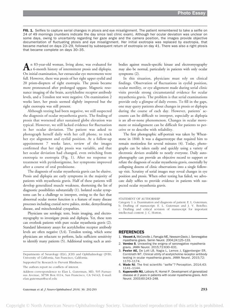
Although testing had been negative, we still suspectedthe diagnosis of ocular myasthenia gravis. The finding ofptosis that worsened after sustained globe elevation wastypical. However, we still lacked evidence for fluctuationin her ocular deviation. The patient was asked tophotograph herself daily with her cell phone, to trackher eye alignment and eyelid position. At a follow-upappointment 7 weeks later, review of the imagesconfirmed that her right ptosis was variable, and thather ocular deviation had changed, even switching fromexotropia to esotropia (Fig. 1). After no response totreatment with pyridostigmine, her symptoms improvedafter a course of oral prednisone.
The diagnosis of ocular myasthenia gravis can be elusive.Ptosis and diplopia are early symptoms in the majority ofpatients with myasthenia gravis. Half of these patients willdevelop generalized muscle weakness, shortening the list ofdiagnostic possibilities substantially (1). Isolated ocular symp-toms can be a challenge to interpret, owing to the fact thatabnormal ocular motor function is a feature of many diseaseprocesses including cranial nerve palsies, stroke, demyelinatingdisease, and mitochondrial cytopathies.
Physicians use serologic tests, brain imaging, and electro-myography to investigate ptosis and diplopia. Yet, these testscan overlook patients with pure ocular myasthenia gravis (2).Standard laboratory assays for acetylcholine receptor antibodylevels are often negative (3,4). Tensilon testing, which somephysicians are reluctant to perform, lacks sufficient sensitivityto identify many patients (5). Additional testing such as anti-
bodies against muscle-specific kinase and electromyographymay also be normal, particularly in patients with only ocularsymptoms (2).
In this situation, physicians must rely on clinicalfindings. Observation of fluctuations in eyelid position,ocular motility, or eye alignment made during serial clinicvisits provide strong circumstantial evidence for ocularmyasthenia gravis. The problem is that serial examinationsprovide only a glimpse of daily events. To fill in the gaps,one may query patients about changes in ptosis or diplopiaduring the course of each day. However, patients’ ac-counts can be difficult to interpret, especially as diplopiais an all-or-none phenomenon. Changes in ocular move-ment or misalignment can be difficult for patients to per-ceive or to describe with reliability.
The first photographic self-portrait was taken by Wheat-stone in 1840. It was a daguerreotype that required him toremain motionless for several minutes (4). Today, photo-graphs can be taken easily and quickly using a variety ofelectronic devices available to nearly everyone. Daily auto-photography can provide an objective record to support orrefute the diagnosis of ocular myasthenia gravis, essentially bycollapsing dozens of clinic observations into a single follow-up visit. Scrutiny of serial images may reveal changes in eyeposition and ptosis. When other testing has failed, we advo-cate daily selfies to provide evidence in patients with sus-pected ocular myasthenia gravis.
STATEMENT OF AUTHORSHIPCategory 1: a. Examination and diagnosis of patient: E. L. Guterman;b. Drafting of manuscript: E. L. Guterman and J. V. Botelho;c. Drafting and critical revision of manuscript for importantintellectual content: J. C. Horton.
REFERENCES1. Vincent A,McConville J, Farrugia ME, Newsom-Davis J. Seronegative
myasthenia gravis. Semin Neurol. 2004;24:125–133.2. Vernino S. Unraveling the enigma of seronegative myasthenia
gravis. JAMA Neurol. 2015;72:630–631.3. Peeler AE, De Lott LB, Nagia L, Lemos J, Eggenberger ER,
Cornblath WT. Clinical utility of acetylcholine receptor antibodytesting in ocular myasthenia gravis. JAMA Neurol. 2015;72:1170–1174.
4. Wade NJ. The first scientific “selfie”? Perception. 2014;43:1141–1144.
5. Kupersmith MJ, Latkany R, Homel P. Development of generalizeddisease at 2 years in patients with ocular myasthenia gravis. ArchNeurol. 2003;60:243–248.
Departments of Neurology (ELG, JCH) and Ophthalmology (JVB),University of California, San Francisco, California.
Supported by Research to Prevent Blindness.
The authors report no conflicts of interest.
Address correspondence to Elan L. Guterman, MD, 505 Parnas-sus Avenue, M798 Box 0114, San Francisco, CA 94143; E-mail:[email protected]
FIG. 1. Selfies to capture serial changes in ptosis and eye misalignment. The patient remembered to take a selfie on24 of 49 mornings (numbers indicate the day since last clinic exam). Although her ocular deviation was unclear onsome days, owing to uncertainty regarding her gaze angle and the camera position, the images provide objectivedocumentation of fluctuating ptosis and eye misalignment. Her initial exotropia was replaced by esotropia, thatbecame marked on days 23–29, followed by subsequent return of exotropia on day 41. There was also a right ptosisthat became complete on days 30–35.
Guterman et al: J Neuro-Ophthalmol 2016; 36: 292-293 293