REVIEW ARTICLE
Diagnostic and prognostic significance oftransient ischemic dilation (TID) in myocardialperfusion imaging: A systematic review andmeta-analysis
Mohamed Alama, MBBCH, MSc, MRCP (UK),a,e Christopher Labos, MDCM,
MSc,b Handel Emery, MBBS, DM, MRCP (UK),c Robert M. Iwanochko, MD,a,e
Michael Freeman, MD,d,e Mansoor Husain, MD,a,e,f and Douglas S. Lee, MD,
PhDa,b,e,f
a Division of Cardiology, Peter Munk Cardiac Center and the Joint Department of Medical
Imaging, Toronto, Canadab Institute for Clinical Evaluative Sciences, Toronto, Canadac University of the West Indies, Kingston, Jamaicad St. Michael’s Hospital, Toronto, Canadae Department of Medicine, University of Toronto, Toronto, Canadaf Ted Rogers Centre for Heart Research, Toronto, Canada
Received Dec 21, 2016; accepted Jun 6, 2017
doi:10.1007/s12350-017-1040-7
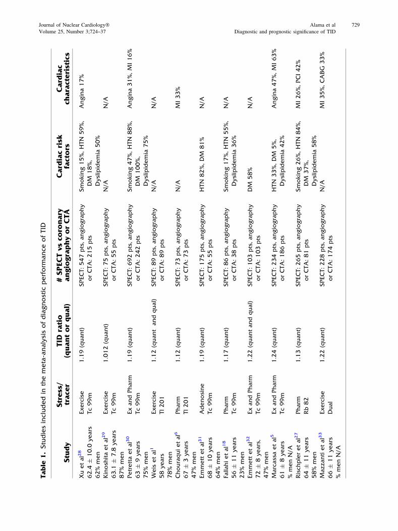
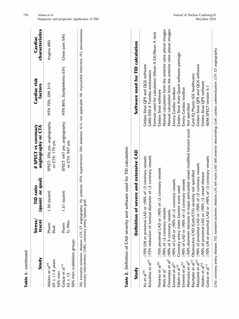
Background. Transient ischemic dilatation (TID) of the left ventricle is a potential marker ofhighriskobstructivecoronaryarterydiseaseonstressmyocardialperfusion imaging (MPI).There is,however, interstudy variation in the diagnostic performance of TID for identification of severe andextensive coronary disease anatomy, and varied prognostic implications in the published literature.
Methods. We searchedMEDLINE, EMBASE, andCOCHRANE databases for studies whereTID was compared with invasive or CT coronary angiography for evaluation of coronary arterystenosis. Two reviewers independently evaluated andabstracted data fromeach study.Abivariaterandom effects model was used to derive pooled sensitivities and specificities, in order to accountfor correlation between TID in MPI and anatomic disease severity.
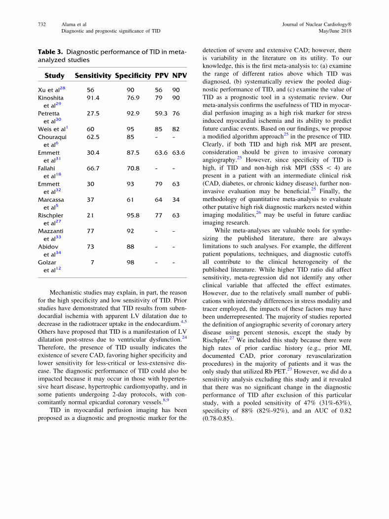
Results. A total of 525 articles were reviewed, of which 51 met inclusion criteria. Thirty-onestudies contributed to the analysis, representing a total of 2037 patients in the diagnostic meta-analysis and 9003 patients in the review of prognosis. The ratio above which TID was deemedpresent ranged from 1.13 to 1.38. Pooled sensitivity was 44% (95% CI 30%-60%) and specificitywas 88% (95% CI 83%-92%) for the detection of extensive or severe anatomic coronary arterydisease. Analysis of outcome data demonstrated increased cardiac event rates in patients with TIDand an abnormal MPI. In otherwise normal perfusion, TID is an indicator of poor prognosis inpatients with diabetes and/or a history of coronary disease.
Conclusions. Among patients undergoing MPI, the presence of TID is specific for thedetection of extensive or severe coronary artery disease. (J Nucl Cardiol 2018;25:724–37.)
Electronic supplementary material The online version of this
article (doi:10.1007/s12350-017-1040-7) contains supplementary
material, which is available to authorized users.
The authors of this article have provided a PowerPoint file, available
for download at SpringerLink, which summarises the contents of the
paper and is free for re-use at meetings and presentations. Search for
the article DOI on SpringerLink.com.
JNC thanks Erick Alexanderson MD, Carlos Guitar MD, and Diego
Vences MD, UNAM, Mexico, for providing the Spanish abstract;
Haipeng Tang MS, Zhixin Jiang MD, and Weihua Zhou PhD, for
providing the Chinese abstract; and Jean-Luc Urbain, MD, PhD,
CPE, Past President CANM, Chief Nuclear Medicine, Lebanon
VAMC, PA, for providing the French abstract.
Reprint requests: Douglas S. Lee, MD, PhD, Institute for Clinical
Evaluative Sciences, University of Toronto, 2075 Bayview Avenue,
Room G-106, Toronto, ON M4N 3M5, Canada; [email protected]/$34.00
Copyright © 2017 The Author(s). This article is an open access
publication
724
Spanish AbstractAntecedentes. La Dilatación Isquémica Transitoria (TID) del ventrículo izquierdo es un mar-
cador potencial de enfermedad arterial coronaria obstructiva de alto riesgo en la fase de estrés delestudio de perfusión miocárdica (EPM). Existe, sin embargo, una variación entre los estudios en elrendimiento diagnóstico de la TID para identificar la severidad y extensión de la enfermedadcoronaria anatómica, así como diversas implicaciones pronosticas en la literatura publicadas.
Métodos. Buscamos en las bases de datos de MEDLINE, EMBASE y COCHRANE estudiosdonde el TID fue comparado con la angiografía coronaria invasiva o por TC (TomografíaComputada) para la evaluación de la estenosis arterial coronaria. Dos revisores evaluaron deforma independiente resumieron y evaluaron los datos de cada estudio. Se utilizo un modelo deefectos aleatorios bivariante para derivar las sensibilidades y especificidades combinadas, a fin deobtener la correlación entre la TID en el EPM y la severidad de la enfermedad anatómica.
Resultados. Se revisaron un total de 525 artículos, de los cuales 51 cumplieron el criterio deinclusión. Treinta y un estudios contribuyeron al análisis, representando un total de 2037pacientes para el meta-análisis diagnóstico y 9003 pacientes para la revisión del pronóstico. Laproporción por encima de la cual la TID se consideró presente osciló entre 1.13 y 1.38. Lasensibilidad combinada fue del 44% (IC del 95%: 30-60%) y la especificidad fue del 88% (IC del95%: 83-92%) para la detección de enfermedad coronaria anatómica extensa o grave. El análisisde los datos de resultado demostró un aumento de las tasas de eventos cardíacos en pacientes conTID y un EPM anormal. En la perfusión normal, la TID es un indicador de mal pronóstico enpacientes con diabetes y/o antecedentes de enfermedad coronaria.
Conclusiones. En pacientes que son llevados a un EPM, la presencia de TID es especifico parala detección de enfermedad arterial coronaria extensa o severa. (J Nucl Cardiol 2018;25:724–37.)
Chinese Abstract背景. 负荷心肌灌注显像 (MPI) 下, 左心室短暂缺血性扩张 (TID) 是高风险冠心病的一个
潜在指标。然而, 现有的文献对于 TID 诊断严重冠心病以及评价预后的报道不尽相同。方法. 我们利用 MEDLINE, EMBASE 和 COCHRANE数据库,检索了 TID 与冠脉造影或
CT 对比来评估冠状动脉狭窄的文献。两个评审者独立的从每个研究中评估和提取数据。为了
了解 TID 和冠状动脉狭窄之间的关联性, 用一个二元随机效应模型来获得合并的敏感性和特
异性。结果. 总归回顾了 525 篇文献,其中 51 篇符合入选标准。 31个研究有助于本文的分析,在
诊断性分析中共 2037 个病人, 预后分析中共 9003 病人。TID 定义为比值超过 1.13*1.38。此
时, 诊断严重冠心病的合并敏感性是 44% (95%CI; 30-60%), 特异性是 88% (95%CI; 83-92%)。在 MPI 异常的患者中, TID 能够增加心血管事件的发生率; 在 MPI 正常, 有糖尿病和/或冠心病的患者中, TID 也是一个独立的预后指标。
结论. 在 MPI 患者中, TID 是一个诊断严重冠心病以及评价预后的特异性指标。 (J NuclCardiol 2018;25:724–37.)
French AbstractContexte. La dilatation ischémique transitoire (DIT) du ventricule gauche mesurée
par scintigraphie de perfusion myocardique est un factor pronostique relativement importantdans l’évaluation des risques liés à la maladie coronarienne obstructive. Néanmoins les pro-tocoles d’évaluation de la DIT et la signification des résultats obtenus dans la maladiecoronarienne sévère sont assez variables dans les études publiées dans la littérature.
Méthodes. Dans cet article nous avons recherché dans les bases de données MEDLINE,EMBASE et COCHRANE les études où les résultats de la DIT ont été comparés à une angiographiecoronaire obtenue demanière invasive oupar tomodensitométrie. Deux experts ont extrait et évaluéindépendamment les données de chaque étude. Demanière à tenir compte de la corrélation entre laDITet la sévéritéde lamaladie coronaire uneanalyse bi-variée des effets aléatoires a étéutilisée pourdéterminer la sensibilité et la spécificité globales.
Résultats. Au total, 525 articles ont été examinés, dont 51 répondant aux critères d’inclusion.Trente et une études ont contribué à l’analyse, incluant un total de 2037 patients dans la méta-
Journal of Nuclear Cardiology� Alama et al 725
Volume 25, Number 3;724–37 Diagnostic and prognostic significance of TID
analyse diagnostique et 9003 patients dans l’examen du pronostic. Le rapport au-dessus duquel laDIT a été jugé anormal varie de 1,13 à 1,38. La sensibilité globale est de 44% (IC à 95% ; 30 à 60%)et la spécificité globale est de 88% (IC 95%; 83-92%) pour la détection d’une maladie coronaireétendue ou sévère. Une augmentation du nombre d’événements cardiaques est présente chez lespatients démontrant une DIT avec étude anormale de perfusion myocardique. Lorsque la per-fusion myocardique est normale, une DIT est également un facteur de mauvais pronostic chez lespatients atteints de diabète et/ou d’un antécédent de maladie coronarienne.
Conclusions. La présence d’une DIT obtenue par scintigraphie myocardique est spécifiquepour la détection de la maladie coronaire étendue ou sévère. (J Nucl Cardiol 2018;25:724–37.)
Key Words: SPECT · outcomes research · diagnostic and prognostic application
AbbreviationsCABG Coronary artery bypass graft
CAD Coronary artery disease
CI Confidence interval
CT Computed tomography
DM Diabetes mellitus
LVEF Left ventricular ejection fraction
MI Myocardial infarction
PCI Percutaneous coronary intervention
ROC Receiver operating characteristic
TID Transient ischemic dilation
INTRODUCTION
Myocardial perfusion Imaging (MPI) is an estab-
lished tool for the diagnosis and risk stratification of
patients with coronary artery disease (CAD) for over
three decades.1 MPI has excellent diagnostic and prog-
nostic accuracy and also provides good insight into
cardiac function through the interpretation of a variety
of perfusion and functional parameters.2,3 One of these
functional parameters is transient ischemic dilation
(TID), which has been validated both as a marker of
severe and extensive coronary artery disease and as a
predictor of cardiac outcomes in independent studies.1-3
To date, the pathophysiology of ischemic LV
dilatation remains unclear with the theory of subendo-
cardial ischemia gaining the widest acceptance.4,5
Others cite data supporting ischemia induced physical
LV dilation post stress.6,7 However, several studies have
demonstrated that ischemic LV dilatation may be
present in patients with normal perfusion and no
significant epicardial coronary disease; for example in
patients with hypertrophic cardiomyopathy,8 or in
patients with hypertensive heart disease and left ven-
tricular hypertrophy.9 Therefore the true diagnostic
accuracy of TID on MPI is debated and the optimal
threshold for its definition remains undefined.
In this study, we conducted a meta-analysis of the
diagnostic performance of the presence of TID, compared
to anatomical coronary artery assessment. We also con-
ducted a systematic reviewof theprognostic significance of
TID. Both components of our study included patients who
underwent either exercise or pharmacologic stress MPI.
METHODS
We employed a systematic search of the MEDLINE,
EMBASE, and COCHRANE databases. We searched for
English language studies, which examined the diagnostic and/
or prognostic accuracy of TID in myocardial perfusion imag-
ing. The search words used were (transient ischemic dilation,
transient ischaemic dilation, left ventricular dilation, transient
dilation, SPECT, single photon tomography, CT single photon,
myocardial perfusion imaging, and myocardial scintigraphy).
Two investigators (MA, HE) independently reviewed the
studies and extracted the relevant data including patient demo-
graphics, the radiotracer used, the stress modality, and findings
on coronary angiography. Where additional data were required
to complete the meta-analysis or discrepancies existed, attempts
were made to contact the original authors to obtain such
information. We excluded studies where: a) there was no
coronary arterial anatomic assessment (either invasive coronary
angiogram or coronary CT angiography) for comparison, or b)
therewas no clear documentation of themethod used to calculate
the ratio above which TID was diagnosed.
The studies identified for inclusion by the two investiga-
tors, and data extracted, were reviewed for eligibility and
accuracy by a third investigator (DSL). Methodological quality
regarding the risk of bias and concerns of applicability was
assessed using the Quality Assessment of Diagnostic Accuracy
Studies 2 (QUADAS 2) tool.10 QUADAS 2 is a tool used for
quality assessment of diagnostic accuracy studies included in
systematic reviews and meta-analyses, to assess the risk of bias
and applicability for use in systematic reviews. This tool
contains 4 domains: patient selection, index test, reference
standard, and flow and timing.
Statistical Analysis
Data were extracted to construct 2 9 2 tables, from which
the sensitivity and specificity of each study was calculated. The
sensitivity and specificity estimates were pooled using a
See related editorial, pp. 738–741
726 Alama et al Journal of Nuclear Cardiology�Diagnostic and prognostic significance of TID May/June 2018
bivariate random effects model, as recommended by the
Cochrane Diagnostic Test Accuracy Working Group.11
The bivariate model was then used to construct a
hierarchical summary receiver operator curve (ROC). A Pvalue\.05 was considered statistically significant. We did not
calculate an I2 statistic given that it is not an accepted method
of measuring heterogeneity between diagnostic studies.11
Univariate meta-regression was used to assess the significance
of key covariates that were likely to affect the diagnostic
accuracy of the test. Due to the small number of studies,
multivariate meta-regression was not performed as it was
likely to be underpowered to detect any differences.
All statistical analyses were conducted using STATA/SE,
version 12.0 (Stata Corp LP, College Station, Texas, USA).
RESULTS
Summary of Studies Examining TID
From the initial database search, we identified 525
citations of which 368 articles remained after removing
duplicates. After reviewing the titles and abstracts of
these records, 317 articles were excluded because they
were not relevant to the purpose of the study.
Of the remaining 51 articles, 20 studies were
excluded because: (a) there was no evaluation of
coronary anatomy (invasive coronary angiogram or CT
angiography) for diagnostic studies [n = 11], (b) they
included only patients with left ventricular dysfunction
and fixed perfusion defects for prognostic studies
[n = 1], (c) there was no clear documentation of the
method used to calculate the ratio above which TID was
diagnosed or only visual assessment of TID was
employed [n = 5], (d) incomplete data [n = 2], and
(e) duplicate data [n = 1] as shown in Figure 1 (Group
Z). These excluded studies are shown in Online Table A.
We included 31 studies, of which 23 evaluated TID from
a diagnostic perspective. Of these, 13 studies were
included in the quantitative meta-analysis (Figure 1,
Group A), and 10 studies were not quantitatively
synthesized because the patient-level data were only
reported in aggregate, and patients could not be sepa-
rated into TID positive or negative, or severe or non-
severe CAD categories (Group C). The quantitative
meta-analysis encompassed a total of 2037 patients in
the diagnostic evaluation and 9003 patients in the
prognostic evaluation (Figure 1). There were 8 studies
examining the prognostic significance of TID, which did
not report patient-level data and therefore were incor-
porated in a narrative synthesis (Group B).
Studies of Diagnostic Performance
Characteristics of the 13 studies included in the
quantitative meta-analysis are shown in Table 1, and the
10 non-quantitatively analyzed studies are shown in Online
Table B. The definition of severe and extensive CAD and
software used for TID evaluation are shown in Table 2.
Technetium-99was themost commonly utilized radiotracer
(8 studies), followed by Tl-201 (2 studies), dual isotope
scanning (2 studies), and Rb-82 (1 study). Coronary
angiography was performed in 73% of patients. The
modality of stress included exercise (4 studies), pharmaco-
logic stress (6 studies), and both exercise and pharmacologic
stress (3 studies).MethodologicalQualityAssessment using
the QUADAS 2 tool revealed that most of the studies
included in themeta-analysis demonstrated low risk for bias
or concern regarding applicability (Figure 2).
The ratio above which TID was diagnosed ranged
from1.13 to 1.38,with differences noted depending on the
tracer used. Using the published TID ratios (as defined by
the individual study authors), the sensitivity of TID for the
detection of extensive or severe CAD ranged from 21% to
62.5% (see Table 3). One study only showed very low
sensitivity (7%).12 Specificities were higher and demon-
strated less variability, ranging from 77% to 98%
(Table 3). Bivariate analysis of the 13 studies, which
had complete statistical data, revealed a pooled sensitivity
of 44% (95% confidence interval [CI] 30%-60%) and a
pooled specificity of 88% (95%CI 83%-92%) as shown in
Figure 3. The pooled area under the ROC curve was 0.82
(0.78-0.85) for all studies (Figure 4).
Subgroup Analyses for DiagnosticPerformance of TID
Subgroup analysis of the technetium studies
revealed a pooled sensitivity of 42% (95% CI 23%-
63%) and a pooled specificity of 86% (95% CI 78%-
92%), which was similar to the overall results above (see
Figure 5). Subgroup analyses also demonstrated that
studies using exercise as a stressor demonstrated a
significantly higher pooled area under the receiver
operating characteristic curve (AUC 0.92 vs 0.78,
P \ .001) for detection of severe CAD compared to
studies using pharmacological stressors (Figure 5). In
studies that determined the presence of TID qualitatively
(instead of quantitatively) the pooled sensitivity was
46% (95% CI 38%-54%) and pooled specificity was
88% (95% CI 79%-93%).
For studies using technetium, the TID range was
1.13-1.31. In these studies, the sensitivity ranged from
34% to 56%, while the specificity ranged from 77% to
98% for detection of severe CAD. Overall sensitivity
and specificity were similar for both exercise and
pharmacologic technetium studies. Based on univariate
meta-regression, the TID ratio cutoff used in various
studies had little effect on specificity (P = .99).
Journal of Nuclear Cardiology� Alama et al 727
Volume 25, Number 3;724–37 Diagnostic and prognostic significance of TID
However, higher TID ratios resulted in decreased
sensitivity (P\ .01).
Studies of Prognostic Performance
Studies evaluating TID as a marker of increased
cardiac events are summarized in Table 4. Across
studies, the annualized rates of cardiac death or MI
ranged from approximately 0.2% to 1% in those with no
TID, 2% to 5% in those with TID and normal perfusion,
and 5% to 6% among those with TID and ischemia,
CAD, or diabetes. De Winter reported that TID was a
significant predictor of all-cause mortality even after
multivariable adjustment for resting heart rate, beta-
adrenoreceptor antagonism, summed rest score, and
resting LV ejection fraction.13
There was heterogeneity of patients in the different
prognostic studies of TID. In a study of diabetic patients
with TID and ischemia by Petretta et al, the annualized
rate of cardiac death or non-fatal MI was 7.2% with
post-stress LVEF ≤45% and 5.4% when post-stress
LVEF was greater than 45%.14 In another study of
patients with TID, rates of fatal and non-fatal MI were
substantially increased in those who were not revascu-
larized, compared to those who underwent CABG
surgery or PCI.15 One study examined patients under-
going MPI prior to non-cardiac surgery and reported
high postoperative cardiac event rates: 58% in the
presence of TID, 19% with reversible perfusion defects
but no TID, and 2% in patients with normal scans. These
events were temporally accelerated, with the majority of
cardiac events occurring within 4 months
postoperatively.16
Special Consideration of TID in Patientswith Otherwise Normal Perfusion Scans
The study by Abidov et al demonstrated that TID is
an independent prognostic marker for cardiac events in
patients with either normal or near-normal MPI.17
Patients in the highest TID quartile (mean TID ratio of
1.35 ± 0.14) were older and diabetic. The prognostic
Figure 1. Search strategy and study selection.
728 Alama et al Journal of Nuclear Cardiology�Diagnostic and prognostic significance of TID May/June 2018
Table
1.Studiesincludedin
themeta-analysisofdiagnostic
perform
anceofTID
Stu
dy
Stress/
trace
rTID
ratio
(quantorqual)
#SPECTvsco
ronary
angiographyorCTA
Card
iacrisk
factors
Card
iac
characteristics
Xuetal28
62.4
±10.0
years
62%
men
Exercise
Tc99m
1.19(quant)
SPECT:547pts,angiography
orCTA:215pts
Smoking15%,HTN
59%,
DM
18%,
Dyslipidemia
50%
Angina17%
Kinosh
itaetal29
63.1
±7.8
years
87%
men
Exercise
Tc99m
1.012(quant)
SPECT:75pts,angiography
orCTA:55pts
N/A
N/A
Petrettaetal30
63±
9years
75%
men
ExandPharm
Tc99m
1.19(quant)
SPECT:692pts,angiography
orCTA:242pts
Smoking47%,HTN
88%,
DM
100%,
Dyslipidemia
75%
Angina31%,M
I16%
Weis
etal1
58years
78%
men
Exercise
Tl201
1.12(quant
andqual)
SPECT:89pts,angiography
orCTA:89pts
N/A
N/A
Chouraquietal6
67±
3years
47%
men
Pharm
Tl201
1.12(quant)
SPECT:73pts,angiography
orCTA:73pts
N/A
MI33%
Emmett
etal31
68±
10years
64%
men
Adenosine
Tc99m
1.19(quant)
SPECT:175pts,angiography
orCTA:55pts
HTN
82%,DM
81%
N/A
Falla
hietal18
56±
11years
23%
men
Pharm
Tc99m
1.17(quant)
SPECT:86pts,angiography
orCTA:38pts
Smoking17%,HTN
55%,
Dyslipidemia
36%
N/A
Emmett
etal32
72±
8years,
47%
men
ExandPharm
Tc99m
1.22(quantandqual)
SPECT:103pts,angiography
orCTA:103pts
DM
58%
N/A
Marcassaetal5
61±
8years
%menN/A
ExandPharm
Tc99m
1.24(quant)
SPECT:234pts,angiography
orCTA:186pts
HTN
33%,DM
5%,
Dyslipidemia
42%
Angina47%,M
I63%
Rischpleretal27
64±
11years
58%
men
Pharm
Rb82
1.13(quant)
SPECT:265pts,angiography
orCTA:81pts
Smoking26%,HTN
84%,
DM
37%,
Dyslipidemia
58%
MI26%,PCI42%
Mazzantietal33
66±
11years
%menN/A
Exercise
Dual
1.22(quant)
SPECT:228pts,angiography
orCTA:174pts
N/A
MI35%,CABG
33%
Journal of Nuclear Cardiology� Alama et al 729
Volume 25, Number 3;724–37 Diagnostic and prognostic significance of TID
Table
1.continued
Stu
dy
Stress/
trace
rTID
ratio
(quant
orqual)
#SPECTvsco
ronary
angiographyorCTA
Card
iacrisk
factors
Card
iac
characteristics
Abidovetal34
69±
11.4
years
54%
men
Pharm
Dual
1.36(quant)
SPECT:356pts,angiography
orCTA:179pts
HTN
70%,DM
31%
Angina48%
Golzaretal12
62±
13
50%
men(validationgroup)
Pharm
Tc99m
1.31(quant)
SPECT:647pts,angiography
orCTA:547pts
HTN
86%,Dyslipidemia
63%
Chest
pain
54%
TID
,transientischemic
dilation;CTA,CTangiography;Pts,patients;HTN,hypertension;DM,diabetes;
N/A,notapplicable;MI,myocardialinfarction;PCI,percutaneous
coronary
intervention;CABG,coronary
artery
bypass
graft
Table
2.DefinitionofCAD
severity
andso
ftware
use
dforTID
calculation
Stu
dy
Definitionofse
vere
andextensiveCAD
Software
use
dfo
rTID
calculation
Xuetal28
≥70%
LM
orproxim
alLAD
or≥9
0%
of≥2
coronary
vessels
Cedars
SinaiQPSandQGSso
ftware
Kinosh
itaetal29
≥75%
reductionofluminaldiameterof≥2
coronary
vessels
GM
S-5
50A
Tosh
ibaworkstation
Form
ula
use
dforcalculation(M
eanA
EX/M
eanA
rest)
Petrettaetal30
≥70%
proxim
alLAD
or≥9
0%
of≥2
coronary
vessels
Cedars
Sinaiso
ftware
Weis
etal1
≥90%
of≥2
coronary
vessels
Manualcalculationform
theanteriorview
planarim
ages
Chouraquietal6
≥90%
of3coronary
vessels
Manualcalculationform
theanteriorview
planarim
ages
Emmett
etal31
≥90%
ofLAD
or≥9
0%
of≥2
coronary
vessels
Emory
Cardiactoolbox
Falla
hietal18
Coronary
artery
indexGensiniscore
use
dCedars
SinaiAuto
Quantso
ftware
package
Emmett
etal32
≥90%
ofLAD
or≥9
0%
of≥2
coronary
vessels
Emory
Cardiactoolbox
Marcassaetal5
[50%
diameterstenosisin
majorpericardialvesselmodifiedGensiniscore
Notsp
ecified
Rischpleretal27
ObstructiveCAD
(Cath/C
TA)se
verity
notsp
ecified
Card
IQPhysio(G
Ehealthcare)
Mazzantietal33
≥90%
ofproxim
alLAD
or≥9
0%
of≥2
coronary
vessels
Cedars
SinaiQPSandQGSso
ftware
Abidovetal34
≥90%
ofproxim
alLAD
or≥9
0%
≥2vesseldisease
Cedars
SinaiQPSso
ftware
Golzaretal12
≥70%
LM
orproxim
alLAD
or≥9
0%
of≥2
coronary
vessels
4DM
-SPECTversion5.1
CAD,coronary
artery
disease
;TID
,transientischemic
dilation;LM,left
main;LAD,left
anteriordescending;Cath,cardiaccatheterization;CTA,CTangiography
730 Alama et al Journal of Nuclear Cardiology�Diagnostic and prognostic significance of TID May/June 2018
impact of TID with normal myocardial perfusion was
modified by the presence of CAD or diabetes,18 with an
increased risk of cardiac death or MI reported in these
patients.19 In a study of diabetes patients with normal
post-stress LVEF and no ischemia, the annual event rate
was 4.9% in those with TID and 0.2% in those without
TID (P\ .001).14
DISCUSSION
Our study showed that TID is a specific but not a
sensitive marker for detection of severe and extensive
CAD with a pooled sensitivity of 44% and pooled
specificity of 88%. In the analysis of subgroups, we
found that exercise stress resulted in a trend toward
higher sensitivity than pharmacologic stress, but speci-
ficity was similar. The prognostic studies demonstrated
consistently elevated risk when TID was present despite
somewhat different populations studied.
This risk was heightened in those with TID and
post-stress LVEF ≤ 45%, with rates of cardiac death or
MI exceeding 7%·year−1. Among patients with normal
perfusion scans, the presence of TID was associated
with increased risk primarily when patients had a history
of CAD or DM.
While there are many disparate studies examining
diagnostic test performance, few have utilized meta-
analytic approaches and summary receiver operating
characteristic curves to evaluate the performance of a
specific high risk marker such as the presence of TID.
Many systematic reviews and meta-analyses were con-
ducted to study the overall diagnostic and prognostic
role of different imaging modalities like stress echocar-
diography,20 cardiac PET,21 and coronary CT
angiography.22,23 However, meta-analyses of specific
components of a diagnostic test, such as TID are less
commonly encountered. Despite the importance that is
imparted to the presence of TID in nuclear cardiology, to
our knowledge, this is the first meta-analysis that studied
its quantitative diagnostic performance for detection of
severe CAD and prognostic performance for prediction
of cardiac outcomes.
0% 20% 40% 60% 80% 100%
PATIENT SELECTION
INDEX TEST
REFERENCE STANDARD
FLOW AND TIMING
Propor�on of studies with low, high or unclear RISK of BIAS
QU
ADAS
-2 D
omai
n
0% 20% 40% 60% 80% 100%
Propor�on of studies with low, high, or unclearCONCERNS regarding APPLICABILITY
LowHighUnclear
Figure 2. Quality assessment using QUADAS-2.
Journal of Nuclear Cardiology� Alama et al 731
Volume 25, Number 3;724–37 Diagnostic and prognostic significance of TID
Mechanistic studies may explain, in part, the reason
for the high specificity and low sensitivity of TID. Prior
studies have demonstrated that TID results from suben-
docardial ischemia with apparent LV dilatation due to
decrease in the radiotracer uptake in the endocardium.4,5
Others have proposed that TID is a manifestation of LV
dilatation post-stress due to ventricular dysfunction.24
Therefore, the presence of TID usually indicates the
existence of severe CAD, favoring higher specificity and
lower sensitivity for less-critical or less-extensive dis-
ease. The diagnostic performance of TID could also be
impacted because it may occur in those with hyperten-
sive heart disease, hypertrophic cardiomyopathy, and in
some patients undergoing 2-day protocols, with con-
comitantly normal epicardial coronary vessels.8,9
TID in myocardial perfusion imaging has been
proposed as a diagnostic and prognostic marker for the
detection of severe and extensive CAD; however, there
is variability in the literature on its utility. To our
knowledge, this is the first meta-analysis to: (a) examine
the range of different ratios above which TID was
diagnosed, (b) systematically review the pooled diag-
nostic performance of TID, and (c) examine the value of
TID as a prognostic tool in a systematic review. Our
meta-analysis confirms the usefulness of TID in myocar-
dial perfusion imaging as a high risk marker for stress
induced myocardial ischemia and its ability to predict
future cardiac events. Based on our findings, we propose
a modified algorithm approach25 in the presence of TID.
Clearly, if both TID and high risk MPI are present,
consideration should be given to invasive coronary
angiography.25 However, since specificity of TID is
high, if TID and non-high risk MPI (SSS \ 4) are
present in a patient with an intermediate clinical risk
(CAD, diabetes, or chronic kidney disease), further non-
invasive evaluation may be beneficial.25 Finally, the
methodology of quantitative meta-analysis to evaluate
other putative high risk diagnostic markers nested within
imaging modalities,26 may be useful in future cardiac
imaging research.
While meta-analyses are valuable tools for synthe-
sizing the published literature, there are always
limitations to such analyses. For example, the different
patient populations, techniques, and diagnostic cutoffs
all contribute to the clinical heterogeneity of the
published literature. While higher TID ratio did affect
sensitivity, meta-regression did not identify any other
clinical variable that affected the effect estimates.
However, due to the relatively small number of publi-
cations with interstudy differences in stress modality and
tracer employed, the impacts of these factors may have
been underrepresented. The majority of studies reported
the definition of angiographic severity of coronary artery
disease using percent stenosis, except the study by
Rischpler.27 We included this study because there were
high rates of prior cardiac history (e.g., prior MI,
documented CAD, prior coronary revascularization
procedures) in the majority of patients and it was the
only study that utilized Rb PET.27 However, we did do a
sensitivity analysis excluding this study and it revealed
that there was no significant change in the diagnostic
performance of TID after exclusion of this particular
study, with a pooled sensitivity of 47% (31%-63%),
specificity of 88% (82%-92%), and an AUC of 0.82
(0.78-0.85).
Table 3. Diagnostic performance of TID in meta-analyzed studies
Study Sensitivity Specificity PPV NPV
Xu et al28 56 90 56 90
Kinoshita
et al2991.4 76.9 79 90
Petretta
et al3027.5 92.9 59.3 76
Weis et al1 60 95 85 82
Chouraqui
et al662.5 85 – –
Emmett
et al3130.4 87.5 63.6 63.6
Fallahi
et al1866.7 70.8 – –
Emmett
et al3230 93 79 63
Marcassa
et al537 61 64 34
Rischpler
et al2721 95.8 77 63
Mazzanti
et al3377 92 – –
Abidov
et al3473 88 – –
Golzar
et al127 98 – –
732 Alama et al Journal of Nuclear Cardiology�Diagnostic and prognostic significance of TID May/June 2018
Table
4.Studiesincludedin
thesy
stematicreview
ofprognosis
Stu
dy
Demographicsandfo
llow-u
p
TID
ratio
(quantor
qual),n(%
)ofpts
withTID
Annualizedca
rdiacoutcomes
Abidovetal17
66.1
±12.8
years
50%
men
ExandPharm
Dual
Primary
studycohort:1
560pts
withnorm
alp
erfusion;
Secondary
cohort:2037pts
withnorm
alornear-
norm
alperfusion:
2.3
±0.7
years
1.21(quant)
n=
390(25%)
Outcome:cardiacdeath,M
I,orcoronary
revascularization
Norm
alperfusion(SSS=
0):
(a)TID
prese
nt:2.4%·year−
1;
(b)NoTID
(i.e.,lowest
TID
quartile):0.6%·year−
1
Norm
al/near-norm
alperfusion(SSS0-3
):
(a)TID
prese
nt:2.2%·year−
1;
(b)Other3TID
quartiles:
1.0%·year−
1
Petrettaetal14
64±
8years
67%
men
ExandPharm
Tc99m
672pts
withDM
2+
years
1.19(quant)
n=
97(14%)
Outcome:cardiacdeath
orM
I
Noischemia
+LVEF[
45%:
(a)TID
prese
nt:4.9%·year−
1
(b)NoTID
:0.21%·year−
1
Ischemia
prese
nt+
LVEF[
45%:
(a)TID
prese
nt:5.4%·year−
1
(b)NoTID
:1.9%·year−
1
Ischemia
+LVEF≤45%:
(a)TID
prese
nt:7.2%·year−
1
(b)NoTID
:3.8%·year−
1
Doukkyetal19
58±
12years
52%
men
ExandPharm
Tc99m
1236pts
withnorm
alperfusion
27±
9(clinicalevents)
37±
8months(m
ortality)
1.16(Ex)
1.22(Pharm
)
(quant)
n=
76(6%)
Outcome:cardiacdeath
orM
I
Overall:
(a)TID
prese
nt:2.4%·year−
1
(b)NoTID
:0.4%·year−
1
DM
orCAD:
(a)TID
prese
nt:5.9%·year−
1
(b)NoTID
:0.2%·year−
1
Uzzamanetal15
56±
9years
67%
men
ExandPharm
Tc99m
2689pts
inasingle-center
18±
4months
1.22(quant)
n=
189(7%)
Outcome:fatalM
I
(a)TID
+notrevascularized:3.3%·year−
1
(b)TID
+revascularized:1.6%·year−
1
(c)NoTID
:notreported
Outcome:non-fatalM
I
(a)TID
+notrevascularized:14.2%·year−
1
(b)TID
+revascularized:3.3%·year−
1
(c)NoTID
:notreported
Journal of Nuclear Cardiology� Alama et al 733
Volume 25, Number 3;724–37 Diagnostic and prognostic significance of TID
Table
4.continued
Stu
dy
Demographicsandfo
llow-u
p
TID
ratio
(quantor
qual),n(%
)ofpts
with
TID
Annualizedca
rdiacoutcomes
Letteetal16
61±
10years
55%
men
Pharm
Tl201
510totalpts;
Prognostic
cohort:187undergoing
non-cardiacsu
rgery
12months
1.15(quant)
n=
45(9%)
Outcome:cardiacdeath
orM
I
(a)TID
prese
nt:58%·year−
1
(b)Reversible
defect:19%·year−
1
(c)Norm
al:2%·year−
1
Letteetal35
62years
57%
men
Pharm
Tl201
753pts
16months
1.15(quant)
n=
41(5%)
Outcome:cardiacdeath
orM
I
(a)TID
prese
nt:47.4%·year−
1
(b)Reversible
defect:15.8%·year−
1
(c)Norm
al:2.0%·year−
1
Thomasetal36
65±
12years
62%
men
ExandPharm
Tc99m
andDual
1612pts
24±
7months
Qualitative
n=
371(23%)
Outcome:cardiacdeath
orM
I
Technetium:
(a)TID
prese
nt:6%·year−
1
(b)NoTID
:1.5%·year−
1
Dualisotope:
(a)TID
prese
nt:3.8%·year−
1
(b)NoTID
:0.5%·year−
1
Dewinteretal13
78(IQR:76-8
0)years
54%
men
ExandPharm
Tc99m
294pts
aged≥75years
old
26months
1.005(quant)
n=
147
Outcome:all-cause
death
(a)TID
prese
nt:9.7%·year−
1
(b)NoTID
:5.0%·year−
1
TID
,transientischemic
dilation;Ex,exercisestress;Pharm
,pharm
acologic
stress;Dual,dualiso
topestudy;MI,myocardialinfarction;SSS,su
mmedstress
score;DM,diabetes;
LVEF,
left
ventricularejectionfraction;CAD,coronary
artery
disease
;Pts,patients;IQ
R,interquartilerange
734 Alama et al Journal of Nuclear Cardiology�Diagnostic and prognostic significance of TID May/June 2018
NEW KNOWLEDGE GAINED
The presence of TID has a high pooled area under
the receiver operating characteristic curve for the
detection of severe and extensive CAD. While sensitiv-
ity is low, specificity of TID is high for the detection of
severe and extensive CAD.
The rate of cardiac death or MI is increased in those
with TID and normal perfusion, primarily amongst those
with DM, CAD, or ischemia. Rates of cardiac death or
MI appear to be increased further in those with reduced
LVEF.
CONCLUSION
In conclusion, in this meta-analysis, we found that
transient ischemic dilation during myocardial perfusion
imaging is a specific diagnostic marker of severe and
extensive coronary artery disease. Transient ischemic
dilation is an indicator of poor prognosis, and risks were
significantly elevated among those with evidence
Figure 3. Forest plot of included studies in diagnostic meta-analysis.
Figure 4. ROC curve for all studies.
Journal of Nuclear Cardiology� Alama et al 735
Volume 25, Number 3;724–37 Diagnostic and prognostic significance of TID
suggestive of coronary disease or reduced stress LV
ejection fraction. The presence of TID significantly
worsens prognosis even among diabetes patients with
normal perfusion. Therefore, TID should be considered
a high risk marker that may guide clinical management
in patients with suspected or known coronary artery
disease.
Acknowledgments
This research was supported by a Foundation Grant fromthe Canadian Institutes of Health Research (Grant # FDN148446). Dr Lee is supported by a mid-career investigatoraward from the Heart and Stroke Foundation and is the TedRogers Chair in Heart Function Outcomes, a joint Hospital-University Chair of the University Health Network and theUniversity of Toronto. The Institute for Clinical EvaluativeSciences (ICES) is supported in part by a grant from theOntario Ministry of Health and Long-Term Care. The opinions,results, and conclusions are those of the authors and noendorsement by the Ministry of Health and Long-Term Care orby the Institute for Clinical Evaluative Sciences is intended orshould be inferred.
Disclosures
The study authors have no financial conflicts of interest.
Note
This paper is dedicated to the memory of Dr. MichaelFreeman (Dec 29, 1948–Sept 3, 2017).
Open Access
This article is distributed under the terms of the CreativeCommons Attribution 4.0 International License (http://creativecommons.org/licenses/by/4.0/), which permits unrestricted use,distribution, and reproduction in any medium, provided you
give appropriate credit to the original author(s) and the source,provide a link to the Creative Commons license, and indicate ifchanges were made.
References
1. Weiss AT, Berman DS, Lew AS, Nielsen J, Potkin B, Swan HJ,
et al. Transient ischemic dilation of the left ventricle on stress
thallium-201 scintigraphy: A marker of severe and extensive
coronary artery disease. J Am Coll Cardiol 1987;9:752-9.
2. Iskandrian AS, Heo J, Nguyen T, Lyons E, Paugh E. Left ven-
tricular dilatation and pulmonary thallium uptake after single-
photon emission computer tomography using thallium-201 during
adenosine-induced coronary hyperemia. Am J Cardiol 1990;
66:807-11.
3. Bestetti A, Di Leo C, Alessi A, Triulzi A, Tagliabue L, Tarolo GL.
Post-stress end-systolic left ventricular dilation: A marker of
endocardial post-ischemic stunning. Nucl Med Commun 2001;
22:685-93.
4. Takeishi Y, Tono-oka I, Ikeda K, Komatani A, Tsuiki K, Yasui S.
Dilatation of the left ventricular cavity on dipyridamole thallium-
201 imaging: A new marker of triple-vessel disease. Am Heart J
1991;121:466-75.
5. Marcassa C, Galli M, Baroffio C, Campini R, Giannuzzi P.
Transient left ventricular dilation at quantitative stress-rest
sestamibi tomography: Clinical, electrocardiographic, and angio-
graphic correlates. J Nucl Cardiol 1999;6:397-405.
6. Chouraqui P, Rodrigues EA, Berman DS, Maddahi J. Significance
of dipyridamole-induced transient dilation of the left ventricle
during thallium-201 scintigraphy in suspected coronary artery
disease. Am J Cardiol 1990;66:689-94.
7. Kurata C, Wakabayashi Y, Shouda S, Mikami T, Tawarahara K.
Quantification of left ventricular size on exercise thallium-201
single-photon emission tomography. Eur J Nucl Med
1996;23:762-7.
8. Sugihara H, Shiga K, Umamoto I, Harada Y, Katahira T, Naka-
gawa T, et al. [Assessment of transient dilation of the left
ventricular cavity in patients with hypertrophic cardiomyopathy by
exercise thallium-201 scintigraphy]. Kaku Igaku 1990;27:1281-9.
Figure 5. ROC curves in different subgroups.
736 Alama et al Journal of Nuclear Cardiology�Diagnostic and prognostic significance of TID May/June 2018
9. Robinson VJ, Corley JH, Marks DS, Eberhardt LW, Eubig C,
Burke GJ, et al. Causes of transient dilatation of the left ventricle
during myocardial perfusion imaging. AJR Am J Roentgenol
2000;174:1349-52.
10. Whiting PF, Rutjes AW, Westwood ME, Mallett S, Deeks JJ,
Reitsma JB, et al. QUADAS-2: A revised tool for the quality
assessment of diagnostic accuracy studies. Ann Intern Med
2011;155:529-36.
11. Macaskill PGC, Deeks JJ, Harbord RM, Takwoingi Y. Cochrane
handbook for systematic reviews of diagnostic test accuracy. In:
Deeks JJB, Bossuyt PM, Gatsonis C (eds) The Cochrane Collab-
oration. 2010. http://srdta.cochrane.org/.
12. Golzar Y, Olusanya A, Pe N, Dua SG, Golzar J, Gidea C, et al. The
significance of automatically measured transient ischemic dilation
in identifying severe and extensive coronary artery disease in
regadenoson, single-isotope technetium-99m myocardial perfusion
SPECT. J Nucl Cardiol 2015;22:526-34.
13. De Winter O, Velghe A, Van de Veire N, De Bondt P, De Buyzere
M, Van De Wiele C, et al. Incremental prognostic value of com-
bined perfusion and function assessment during myocardial gated
SPECT in patients aged 75 years or older. J Nucl Cardiol
2005;12:662-70.
14. PetrettaM,AcampaW,Daniele S, PetrettaMP, PlaitanoM,Cuocolo
A. Transient ischemic dilation in patients with diabetes mellitus:
Prognostic value and effect on clinical outcome after coronary
revascularization. Circ Cardiovasc Imaging 2013;6:908-15.
15. uz Zaman M, Fatima N, Samad A, Ishaq M, Wali A, Rehman K,
et al. Predictive and prognostic values of transient ischemic
dilatation of left ventricular cavity for coronary artery disease and
impact of various managements on clinical outcome using tech-
netium-99m sestamibi gated myocardial perfusion imaging. Ann
Nucl Med 2011;25:566-70.
16. Lette J, Lapointe J, Waters D, Cerino M, Picard M, Gagnon A.
Transient left ventricular cavitary dilation during dipyridamole-
thallium imaging as an indicator of severe coronary artery disease.
Am J Cardiol 1990;66:1163-70.
17. Abidov A, Bax JJ, Hayes SW, Hachamovitch R, Cohen I, Gerlach
J, et al. Transient ischemic dilation ratio of the left ventricle is a
significant predictor of future cardiac events in patients with
otherwise normal myocardial perfusion SPECT. J Am Coll Cardiol
2003;42:1818-25.
18. Fallahi B, Beiki D, Fard-Esfahani A, Akbarpour S, Abolhassani A,
Kakhki VR, et al. The additive value of transient left ventricular
dilation using two-day dipyridamole 99mTc-MIBI SPET for
screening coronary artery disease in patients with otherwise nor-
mal myocardial perfusion: A comparison between diabetic and
non-diabetic cases. Hell J Nucl Med 2010;13:246-52.
19. Doukky R, Frogge N, Bayissa YA, Balakrishnan G, Skelton JM,
Confer K, et al. The prognostic value of transient ischemic
dilatation with otherwise normal SPECT myocardial perfusion
imaging: A cautionary note in patients with diabetes and coronary
artery disease. J Nucl Cardiol 2013;20:774-84.
20. Mahajan N, Polavaram L, Vankayala H, Ference B, Wang Y, Ager
J, et al. Diagnostic accuracy of myocardial perfusion imaging and
stress echocardiography for the diagnosis of left main and triple
vessel coronary artery disease: A comparative meta-analysis.
Heart 2010;96:956-66.
21. Parker MW, Iskandar A, Limone B, Perugini A, Kim H, Jones C,
et al. Diagnostic accuracy of cardiac positron emission tomogra-
phy versus single photon emission computed tomography for
coronary artery disease: A bivariate meta-analysis. Circ Cardio-
vasc Imaging 2012;5:700-7.
22. Sun Z, Ng KH. Diagnostic value of coronary CT angiography with
prospective ECG-gating in the diagnosis of coronary artery
disease: A systematic review and meta-analysis. Int J Cardiovasc
Imaging 2012;28:2109-19.
23. Pontone G, Andreini D, Bartorelli AL, Bertella E, Mushtaq S,
Annoni A, et al. Radiation dose and diagnostic accuracy of mul-
tidetector computed tomography for the detection of significant
coronary artery stenoses: A meta-analysis. Int J Cardiol
2012;160:155-64.
24. Van Tosh A, Hecht S, Berger M, Roberti R, Luna E, Horowitz SF.
Exercise echocardiographic correlates of transient dilatation of the
left ventricular cavity on exercise thallium-201 SPECT imaging.
Chest 1994;106:1725-9.
25. Bourque JM. Contemporary relevance of TID: Based on the
company it keeps. J Nucl Cardiol 2015;22:535-8.
26. Gupta A, Baradaran H, Al-Dasuqi K, Knight-Greenfield A,
Giambrone AE, Delgado D, et al. Gadolinium enhancement in
intracranial atherosclerotic plaque and ischemic stroke: A sys-
tematic review and meta-analysis. J Am Heart Assoc 2016;5:
e003816. doi:10.1161/JAHA.116.003816.
27. Rischpler C, Higuchi T, Fukushima K, Javadi MS, Merrill J,
Nekolla SG, et al. Transient ischemic dilation ratio in 82Rb PET
myocardial perfusion imaging: Normal values and significance as
a diagnostic and prognostic marker. J Nucl Med 2012;53:723-30.
28. Xu Y, Arsanjani R, Clond M, Hyun M, Lemley M Jr, Fish M, et al.
Transient ischemic dilation for coronary artery disease in quanti-
tative analysis of same-day sestamibi myocardial perfusion
SPECT. J Nucl Cardiol 2012;19:465-73.
29. Kinoshita N, Sugihara H, Adachi Y, Nakamura T, Azuma A,
Kohno Y, et al. Assessment of transient left ventricular dilatation
on rest and exercise on Tc-99m tetrofosmin myocardial SPECT.
Clin Nucl Med 2002;27:34-9.
30. Petretta M, Acampa W, Daniele S, Petretta MP, Nappi C, Assante
R, et al. Transient ischemic dilation in SPECT myocardial perfu-
sion imaging for prediction of severe coronary artery disease in
diabetic patients. J Nucl Cardiol 2013;20:45-52.
31. Emmett L, Van Gaal WJ, Magee M, Bass S, Ali O, Freedman SB,
et al. Prospective evaluation of the impact of diabetes and left
ventricular hypertrophy on the relationship between ischemia and
transient ischemic dilation of the left ventricle on single-day
adenosine Tc-99m myocardial perfusion imaging. J Nucl Cardiol
2008;15:638-43.
32. Emmett L, Magee M, Freedman SB, Van der Wall H, Bush V,
Trieu J, et al. The role of left ventricular hypertrophy and diabetes
in the presence of transient ischemic dilation of the left ventricle
on myocardial perfusion SPECT images. J Nucl Med 2005;
46:1596-601.
33. Mazzanti M, Germano G, Kiat H, Kavanagh PB, Alexanderson E,
Friedman JD, et al. Identification of severe and extensive coronary
artery disease by automatic measurement of transient ischemic
dilation of the left ventricle in dual-isotope myocardial perfusion
SPECT. J Am Coll Cardiol 1996;27:1612-20.
34. Abidov A, Bax JJ, Hayes SW, Cohen I, Nishina H, Yoda S, et al.
Integration of automatically measured transient ischemic dilation
ratio into interpretation of adenosine stress myocardial perfusion
SPECT for detection of severe and extensive CAD. J Nucl Med
2004;45:1999-2007.
35. Lette J, Bertrand C, Gossard D, Ruscito O, Cerino M, McNamara
D, et al. Long-term risk stratification with dipyridamole imaging.
Am Heart J 1995;129:880-6.
36. Thomas GS, Miyamoto MI, Morello AP 3rd, Majmundar H,
Thomas JJ, Sampson CH, et al. Technetium 99m sestamibi
myocardial perfusion imaging predicts clinical outcome in the
community outpatient setting. The Nuclear Utility in the Com-
munity (NUC) Study. J Am Coll Cardiol 2004;43:213-23.
Journal of Nuclear Cardiology� Alama et al 737
Volume 25, Number 3;724–37 Diagnostic and prognostic significance of TID