12 Diagnostic and Therapeutic Procedures in Pain Management Salahadin Abdi YiLi Zhou This chapter outlines the major diagnostic and therapeutic procedures performed at the Massachusetts General Hospital (MGH) Pain Center. We have found these procedures to be valuable and to have a good safety record. Many of these techniques were developed by Donald Todd, who founded the Pain Unit in 1948. They have been refined over the years and are now effective, reliable, and reasonably free of unwanted effects, and we continue to use them and teach them to our fellows and residents. The specific nerve blocks described involve the use of C-arm fluoroscopy, which reflects standard practice at MGH. I. GENERAL PRINCIPLES 1. Diagnostic versus therapeutic procedures Before starting a procedure, the clinician determines whether to perform a diagnostic or a therapeutic procedure and prepares the patients accordingly. Diagnostic blocks are utilized for the following reasons: To evaluate and compare the roles of the sympathetic and somatosensory nerves in maintaining pain (differential nerve blocks) To identify the particular nerve(s) that carry pain, or to alter neuromuscular function (selective nerve blocks) For example, we may want to know the answers to the following questions. Is there a significant sympathetic component to a painful limb syndrome? Which of several bulging vertebral discs is actually causing pain by its impingement on a nerve root? Are degenerating facet joints causing a low back pain syndrome? Or, which intercostal nerve(s) are causing pain in a patient with chest wall metastases? Diagnostic nerve blocks help to identify the source of pain and to structure a treatment plan. The treatment plan could involve medical treatment (e.g., emphasis on neuropathic pain medications), further nerve blocks (e.g., a series of sympathetic blocks with physical therapy), permanent neurolysis (e.g., radiofrequency lesioning of nerves in the facet joint, alcohol ablation of intercostal nerves or celiac plexus), or referral to a surgeon for surgical P.156 Página 1 de 40 Ovid: 04/03/05 http://65.54.170.250/cgi-bin/getmsg/DiagnosticandTherapeuticProceduresinPainManageme...
Transcript
12 Diagnostic and Therapeutic Procedures in Pain Management
Salahadin Abdi
YiLi Zhou This chapter outl ines the major diagnostic and therapeutic procedures performed at the Massachusetts General Hospital (MGH) Pain Center. We have found these procedures to be valuable and to have a good safety record. Many of these techniques were developed by Donald Todd, who founded the Pain Unit in 1948. They have been refined over the years and are now effective, rel iable, and reasonably free of unwanted effects, and we continue to use them and teach them to our fel lows and residents.
The specif ic nerve blocks described involve the use of C-arm fluoroscopy, which reflects standard practice at MGH.
I. GENERAL PRINCIPLES
1. Diagnostic versus therapeutic procedures Before start ing a procedure, the clinician determines whether to perform a diagnostic or a therapeutic procedure and prepares the patients accordingly. Diagnostic blocks are uti l ized for the fol lowing reasons:
To evaluate and compare the roles of the sympathetic and somatosensory nerves in maintaining pain (differential nerve blocks)
To identify the particular nerve(s) that carry pain, or to alter neuromuscular function (selective nerve blocks)
For example, we may want to know the answers to the following questions. Is there a signif icant sympathetic component to a painful l imb syndrome? Which of several bulging vertebral discs is actually causing pain by its impingement on a nerve root? Are degenerating facet joints causing a low back pain syndrome? Or, which intercostal nerve(s) are causing pain in a patient with chest wall metastases?
Diagnostic nerve blocks help to identify the source of pain and to structure a treatment plan. The treatment plan could involve medical treatment (e.g., emphasis on neuropathic pain medications), further nerve blocks (e.g., a series of sympathetic blocks with physical therapy), permanent neurolysis (e.g., radiofrequency lesioning of nerves in the facet joint, alcohol ablation of intercostal nerves or celiac plexus), or referral to a surgeon for surgical
In performing a diagnostic block for sympathetic pain, the cl inician must choose a site at which the anesthetic is unlikely to affect somatic nerves, as this would interfere with interpretation (e.g., stel late and lumbar sympathetic ganglion, versus intrapleural and epidural). In the case of somatic pain, very small, concentrated amounts of anesthetic are used at each site so that the blocks are localized to specif ic nerves. This degree of accuracy requires C-arm fluoroscopic guidance.
Before a diagnostic block is performed, the cl inician must be sure that the patient has sustained and reproducible pain and must document what activit ies or stimuli evoke the pain. These factors can then be reassessed after the block and compared to the pre-block state. Finally, the cl inician must confirm (by objective testing) that the block was actually accomplished.
The use of placebo agents (e.g., saline instead of local anesthetic) for diagnostic procedures is unethical and strongly discouraged. This practice is l ikely to be helpful only in identifying placebo responders, not in identifying those with “real” versus psychogenic pain. Furthermore, the practice may damage the patient–physician relationship, and it may reduce the placebo effect of future treatments (see Chapter 3).
2. Pre-block management Patients are requested to consume only l ight meals on the day of a procedure, and to take only clear l iquids for 4 hours prior to the procedure. Baseline vital signs (including pain scale) are obtained at arrival in the cl inic. If indicated, an 18- or 20-gauge intravenous (IV) catheter with Hep-Lock is placed. An IV is routinely placed in patients undergoing procedures that are associated with a risk of sympathectomy and hypotension (e.g., stellate ganglion block, lumbar sympathetic nerve block, cervical epidural steroid injection). The medical condit ion of the patient could also dictate that an IV should be placed (e.g., extreme anxiety, history of vasovagal syncope, signif icant cardiovascular disease). In general, premedication is avoided so that the baseline pain is not altered and so that patient cooperation is maintained.
The patient is posit ioned appropriately, and monitors are placed as needed. Decisions about the level of monitoring needed are made on an individual basis using criteria similar to those for IV placement. The usual monitors are noninvasive blood pressure, electrocardiogram (EKG), and pulse oximetry. Baseline verbal analog scores (VAS) and range of motion estimates are obtained before starting the procedure.
3. Choice of injectant
Local anesthetic For most diagnostic nerve or plexus blocks, a mixture of equal parts of 1% lidocaine and 0.5% bupivacaine is used (giving a f inal concentration of 0.5% lidocaine and 0.25% bupivacaine). This adequately blocks sympathetic and somatic nerves, and a suff icient volume can be used without toxicity. Lidocaine provides rapid onset of effect, whereas
bupivacaine provides a useful prolongation of the effect so that patients can make observations that may have diagnostic value. It is not necessary to use epinephrine-containing solutions in the pain cl inic sett ing. In fact, in emotionally labile patients [e.g., those with complex regional pain syndrome I (CRPS-I)], epinephrine may cause a panic attack and is best avoided.
Steroids The steroids currently used are depot preparat ions of methylprednisolone (Depo-Medrol) and tr iamcinolone (Aristocort, which may be irr itating, and Kenalog, which is less irr i tating but al lergenic). The doses generally range between 40 and 80 mg for epidural injection and between 20 and 40 mg for selective nerve root injection. We typically use 80 mg of tr iamcinolone for epidural injection and 20 mg for selective nerve root injection.
Neurolytic agents Alcohol (50% to 95%) and phenol (6% to 10%) are the two agents commonly used for neurolysis. Alcohol has been extensively used as a neurolytic agent because it is effective and easy to inject. It is used as the neurolytic agent of choice for injecting into the tr igeminal ganglion, celiac plexus, and lumbar sympathetic chain. Occasionally, neurit is with intense burning pain is seen after alcohol neurolysis. We inject local anesthetics prior to the neurolytic to localize the target nerve or plexus and to minimize the incidence of neurit is. The analgesic effect of phenol is almost equal to that of alcohol, but i t does not produce neurit is, which is its advantage. It is extremely viscous and diff icult to inject (needing a larger-bore needle and slow injection). Neither agent is isobaric in cerebrospinal f luid (CSF) (alcohol is hypobaric, phenol hyperbaric); therefore, patients need to be posit ioned appropriately according the baricity of the agent chosen. Neurolytic blocks have a delayed effect (beginning within 1 week) that generally lasts for up to 1 year.
4. Defining landmarks The needle insertion point is usually designated by measurement or by palpation, using x-ray (C-arm fluoroscopy) guidance. It is also helpful to use a “tunnel-view” technique to help define the correct needle angle, and to identify any bony obstructions to the passage of the needle (e.g., r ibs) as it is guided toward the target for injection. First, rest the point of a metal clamp (e.g., a Kelly clamp) over the target site as seen on the radiograph, then over the skin insertion site. The posit ion of the C-arm is then angled so that the t ip of the clamp rests at the skin insertion point as well as at the target point. The chosen point is marked on the skin by f irmly pressing the hub of a 15-gauge needle onto the skin for 30 to 60 seconds (the old fashioned way), or by using a special skin marker. The needle is then inserted through the skin mark in a direction parallel to the x-ray beam, toward the target point. Using this tunnel view technique, the needle should reach the target without hitt ing the bone or other important structures. There are now commercially available laser-guided C-arms that help define the correct needle trajectory and obviate the use of markings.
5. Post-block management Once the procedure is over, patients recover in the recovery room. When they meet standard recovery room discharge criteria, they are discharged home with an escort.
II. SPECIFIC BLOCKS
1. Epidural steroid injections The intended effect of steroids is to reduce the inflammation, swell ing, and scarring that arise as a consequence of disc extrusion and nerve pressure (or inflammation and scarring that occur elsewhere in the body, such as in tendonit is). Pathology studies have shown that steroids reduce the bulk of a scar by diminishing its hyaline portion while leaving the f ibrous skeleton intact.
Indications
Acute herniated or bulging disc
Herniated nucleus pulposus, with nerve root irri tat ion or compression
Spinal stenosis
Spondylolisthesis
Scoliosis
Chronic degenerative disc disease
Potential complications
Dural puncture, with possible total spinal block
Postdural puncture headache
Epidural hematoma
Epidural abscess; cutaneous infections; meningit is
Intrathecal steroid injection, with potential complications such as anterior spinal artery syndrome, arachnoidit is, meningit is, urinary retention, and conus medullaris syndrome, as well as a lack of effect
Spinal cord injury and paralysis
Block treatment protocol We usually offer three epidural injections at 4-week intervals, as needed to ascertain a
response. A steroid-free period of 6 months avoids possible l igamentous atrophy from steroid injections. The three-injection cycle of steroids may then be repeated if needed.
Patient position The patient is prone, with feet over the end of the table and a bunched-up pil low under the abdomen (between i l iac crests and costal margin) to reverse lumbar lordosis and for comfort. For the cervical approach, the pil low is placed under the chest (see later). Supply a small support under the head if i t is requested. The arms are relaxed over the sides of the table.
a) Lumbar translaminar approach
Technique
1. Locate the appropriate interspinous process space with the C-arm, using the t ip of a Kelly clamp as marker. Mark spot on skin with the hub of a 15-gauge needle.
2. Prepare skin with Betadine and drape widely.
3. Begin intra- and subcutaneous 1% lidocaine infi l tration slowly, and in small amount. Deep interspinous infi ltration to full length of a 1½-inch, 22-gauge needle.
4. Insert 3½-inch, 22-gauge spinal needle in the plane of the C-arm (usually a l i t t le cephalad) down to the l igament.
5. Attach a well- lubricated 10-mL syringe to the needle, with 4 mL of air or saline in it. Holding the syringe (not the needle) with one hand, and pressing f irmly with f inger or thumb of the other hand on plunger, maintain posit ive pressure constantly while rapidly oscil lating the plunger. Advance the needle slowly and steadily unti l a sudden loss of resistance is achieved (allow only a minimum of air to escape).
6. If depth of the needle (before the loss of resistance) seems inappropriate, withdraw needle 3 mm and repeat.
7. Inject 1 to 2 mL of dye (Omnipaque-240); confirm with f luoroscopy that the dye is spread inside the epidural space.
8. Inject 2 mL of 40 mg/mL triamcinolone with 0.5 mL to 2 mL of 0.5% bupivacaine, di luted with saline if desired.
9. Replace stylet in needle (to avoid tracking steroid through skin) and withdraw the needle.
10. With gauze pressure on needle hole, sl ide skin back and forth to stop bleeding, and close tract.
11. Apply adhesive bandage to be removed on reaching home.
12. Slowly sit the patient up; do not leave unattended unti l safely in wheelchair, as legs
1. Skin wheal is made about 1 cm lateral and 1 cm caudad from the spinous process.
2. Remember, there is no interspinous l igament to provide resistance to air or saline. Resistance is encountered only when the l igamentum flavum is reached.
3. Proceed as in midline approach.
c) Lumbar transforaminal approach This approach is useful i f symptoms are mainly radicular, and if previous midline injections have not helped. Dose is usually reduced to 40 mg triamcinolone. Dilution is not needed.
d) Caudal epidural injection This approach is useful i f the patient has had multiple laminectomies that have resulted in severe scarring and an alteration of the normal anatomy. A total volume of 20 to 30 mL is needed to f i l l the caudal canal and reach the L5-S1 junction. The standard sacral hiatus approach is used. Using a lateral view on fluoroscopy wil l ensure needle placement in the sacral canal (Fig. 1).
e) Posterior S1 foramen approach This approach is useful i f epidural space is not otherwise accessible, and especially when symptoms are confined to the S1 or S2 level. About 5 to 15 mL (total volume) is needed to reach the L5-S1 junction, a common site of disease.
f) Cervical epidural injection Cervical epidural is used for the treatment of neck and shoulder pain associated with cervical radiculopathy.
1. Place the patient in a prone posit ion with a pil low under the chest to straighten the neck and open the cervical interspinous space.
2. Cervical interspinous space is identif ied with f luoroscopy.
3. A 1½-inch, 25-gauge spinal needle is inserted cephalad, parallel to the cervical spinous process at the midline.
4. Loss-of-resistance technique is used as described for lumbar epidural steroid injection.
5. After negative aspiration, 0.5 to 1.0 mL of dye is injected to confirm correct needle
posit ion with anteroposterior (AP) and lateral views on fluoroscopy.
6. Inject 0.5 to 1.0 mL of solution including 20 to 40 mg of tr iamcinolone into epidural space without local anesthetics.
7. Use extreme caution to avoid injection into subarachnoid space and injury to cervical spine.
2. Central nerve blocks
(i) Epidural blocks (catheters) We do not routinely employ cervical epidural catheters at MGH; therefore, this section focuses on lumbar and thoracic epidural catheter techniques. Epidurally administered local anesthetics block both somatic and sympathetic nerves. However, a predominantly sympathetic block, continuous or intermittent, can be achieved using dilute local anesthetic solutions (e.g., 0.1% to 0.25% bupivacaine). Somatic blockade is achieved by increasing the local anesthetic concentration (e.g., to 0.25% to 0.5% bupivacaine). For prolonged use, tunneled epidural catheters are placed.
Indications
Pain syndromes result ing from peripheral vascular insuff iciency, including ischemic vasospastic pain
Sympathetically maintained pain, unilateral or bi lateral
Acute herpes zoster and postherpetic neuralgia
Acute thoracic or lumbar strain with radiculopathy
Regional pain syndromes result ing from malignancy
Dural puncture and possible postdural puncture headache
Subarachnoid injection with high spinal anesthesia
Intravascular injection (e.g., into epidural veins), with possible seizures
Broken epidural catheter
Potential neural damage
Epidural hematoma
Epidural abscess
Arachnoidit is
Catheter treatment management Pain relief is achieved using a continuous infusion of 0.1% bupivacaine, up to 10 mL/hour, sometimes with the addit ion of opioids and/or clonidine. Vital signs are closely monitored during and after the init ial bolus injection or infusion. If the catheter is to be uti l ized for continuous block, the patient is either admitted for several days or sent home with adequate instructions. Hospital admission may be desirable for intensive, in-house physical therapy. Diet and activity recommendations are based on the cl inical situation.
If the catheter is to be used for cancer pain relief, 0.1% bupivacaine solution should be tr ied init ial ly, but i f pain relief is inadequate, 0.25% bupivacaine, with or without opioids and/or clonidine, may be needed. Infusions of opioid without local anesthetic are used in cancer patients in whom local anesthetics produce undesirable effects. Vital signs are monitored as described earl ier. Activity and diet are as tolerated. If prolonged infusions are required, the catheter is tunneled subcutaneously at the t ime of placement.
a) Lumbar epidural catheters
Midline Approach
Technique
1. Patient l ies in the lateral posit ion, with maximal f lexion of the back and with knees to the abdomen.
2. Obese patients should sit bent forward, with knees to the abdomen, or legs resting on a stool. This helps identify the midline and may widen the posterior interspinous space.
3. The spinal cord terminates at L1 or L2 in the adult; therefore, the spinous process, intervertebral space, and midline are carefully palpated below this level. An appropriate interspace is located.
4. The overlying skin is prepared with antiseptic solution (such as alcohol or Betadine),
and a steri le, fenestrated drape is placed over the site.
5. A skin wheal is made with a ½-inch 25-gauge needle, using 1% lidocaine, and deeper infi l tration is performed with a 1½-inch 22-gauge needle.
6. A 17- or 18-gauge Tuohy needle is directed perpendicularly or sl ightly cephalad in the interspinous space, bevel point ing cephalad, and advanced to the l igamentum flavum.
7. The stylet is removed, and a syringe containing 3 to 4 mL of air or saline is attached
to the Tuohy needle. The needle is advanced, with rapid oscil lation of the plunger of the syringe, until there is a loss of resistance. Loss of resistance may also be detected with continuous pressure on the plunger, advancing unti l pressure is lost. We do not recommend the hangingdrop method, as we have noted a higher incidence of dural punctures.
8. Medication or saline can be injected into the Tuohy needle to distend the epidural space. The catheter is advanced through the needle, no more than 5 cm into the epidural space.
9. The Tuohy needle is removed while the catheter is maintained in posit ion. The distance between the catheter mark and the skin is measured to ensure that the catheter has not been moved during the needle removal.
10. The catheter is then secured to the skin with a transparent dressing and tested for intravascular or intrathecal placement with 3 mL of 2% lidocaine with epinephrine. Aspiration is also performed for blood or cerebrospinal f luid.
11. To decrease displacement of catheter t ip, i t is wise to bring the excess catheter around the f lank to the epigastrium, rather than up the back and over the shoulder, as in operative cases. This is because flexing of the spine tends to pull the catheter back from the epidural space.
12. The catheter is then ready for use, either with bolus or continuous infusions.
Paramedian approach
Technique
1. A skin wheal is made 1 cm lateral and 2 cm caudad to the interspinous space chosen.
2. A 17- or 18-gauge Tuohy needle is aimed from the lateral skin wheal to the top of the target interspace (i.e., the needle is aimed medially and slightly cephalad).
3. The needle is advanced to the l igamentum flavum, and an airor saline-fi l led syringe is attached.
4. After obtaining a loss of resistance, the catheter is threaded and the Tuohy needle is removed; after negative aspiration, the catheter is tested and secured, as earl ier.
Tunneling
Technique
1. Place epidural catheter as described before.
2. Anesthetize a subcutaneous track, horizontally across one side of the back.
3. Tunnel a long, 14-gauge, 5½-inch IV catheter through the track, start ing at the distal point and emerging in the same skin nick as the epidural catheter (Note: Do not puncture epidural catheter with IV needle.)
4. Remove needle from IV catheter and thread epidural catheter through IV catheter.
5. Remove IV catheter and secure epidural catheter at lateral skin exit wound, with transparent dressing.
6. The epidural catheter is tested (as before) and used for intermittent boluses or continuous infusion.
b) Thoracic epidural catheters This procedure is performed as for lumbar epidural catheters. The choice of catheter location may vary, but the technique varies only in that addit ional care is required in approaching the epidural space, as the distances are less than those for the lumbar approach. Further, the needle must be aimed in a more cephalad direction to traverse the sloping thoracic interspinous space. Management and complications are similar to those of the lumbar epidural catheters.
(ii) Selective nerve root blocks For the purpose of identifying a specif ic nerve root as a possible source of pain, only a small amount of local anesthetic (1 mL) is needed. This ensures that only the target nerve root is blocked and reduces the possibil i ty of epidural spread of the local anesthetic. The forcible injection of local anesthetic (particularly of large volumes) directly into a nerve may disrupt it . The needle itself can also produce nerve damage if it pierces the nerve repeatedly. Nerve damage produced in this way can ult imately cause neuropathic pain. Selective blockade of thoracic nerve roots is not described or used at MGH because of the high risk of pneumothorax associated with these blocks. Intercostal nerve blocks are used instead (see II, 5).
To determine the nerve root(s) involved in a pain syndrome. The surgeon can then proceed with more confidence in removing a disc, or in decompressing the root that carries the pain.
To inject local anesthetics and/or steroid at the site of a scarred or compressed root, when pain has not been relieved by other approaches (e.g., epidural injection). This is particularly useful in lumbar and cervical radicular pain syndromes.
a) Cervical paravertebral nerve blocks The cervical nerves leave the spine above their respective vertebrae, and the C1 root has no cutaneous sensory function. The C2 root emerges laterally, between the C1 and the C2 vertebrae, passes over the articular process of C1, and emerges between the posterior arches of C1 and C2. It is distributed mainly to the greater occipital nerve.
C2 nerve root block The C2 root is best blocked as it passes over the posterior surface of the superior art icular process of C2. Generally, i t is simpler to block the occipital nerve, i f that fulf i l ls the need. With C-arm fluoroscopy guidance, and the patient in prone posit ion, the needle is directed vertically down to the articular process, where a paresthesia may be obtained.
C3 through C6 nerve root blocks
Technique
1. Patient is posit ioned supine, f lat (no pil low), head in neutral posit ion or rotated away from side to be blocked. Anatomy is more symmetrical in neutral posit ion, but it is more diff icult to palpate the site and to guide the needle. With the head rotated (it may be necessary to elevate shoulder on padding), i t is easier to view the intervertebral foramen and to guide the needle into the foramen, but the anatomy of the neck becomes spiral (Fig. 2).
2. Using C-arm fluoroscopy, a skin mark is made over the posterior border of the intervertebral foramen image and slightly cephalad to it. Approaching the cervical nerve in its foramen from a posterior posit ion avoids encountering the cervical plexus, since this l ies lateral and anterior to the transverse processes.
3. A 2½-inch, 22-gauge spinal needle is advanced so as to encounter the superior surface of the groove in the transverse process that supports the nerve. The needle
is then deviated toward the intervertebral opening unti l a paresthesia is obtained (Fig. 3).
4. The C-arm is then rotated to the AP posit ion to observe the medial progress of needle. The needle point should not go beyond the cephalad-directed lip on the lateral border of the body of the vertebra, which marks lateral extent of epidural space. The needle point should also be at least halfway into the mass of the transverse process image to avoid spread of anesthetic to other roots (Fig. 4).
5. A single nerve root can be anesthetized with 1 to 2 mL of a mixture of equal parts of 1% lidocaine and 0.5% bupivacaine with 1:400,000 epinephrine. Inject 0.25 to 0.5 mL at a t ime.
C7 Nerve root block The C7 root l ies on the anterior surface of the f lattened transverse process of C7.
Technique
1. Posit ion the patient supine.
2. The needle is inserted lateral and superior to the transverse process and advanced to touch it .
3. The needle is then moved about on the process unti l a paresthesia is achieved.
4. The injection is performed as described before.
b) Lumbar paravertebral nerve blocks There are two principal techniques: direct the needle either caudad from the transverse process above the nerve or cephalad from the process below the nerve. Greater accuracy and an increased possibil i ty of spreading solution along or into the perineural sheath is achieved with the second approach; however, subarachnoid injection is more frequently a hazard. The second approach is described here.
When using this technique in the lumbar region (as opposed to the thoracic region), i t is so easy to enter and inject the epidural or intrathecal spaces that the diagnostic value of a single nerve root block is questionable. However, the technique is chosen for its greater success rate and accuracy, as it is easy to obtain a paresthesia when the needle encounters a nerve as it emerges just caudad to the pedicle of the vertebra.
L1 through L4 nerve root blocks
Technique
1. Patient is posit ioned prone, with pil low under belly, using C-arm (Fig. 5).
2. Visualize the caudad border of the desired vertebral pedicle and mark this target skin projection.
3. Needle is inserted 5 cm lateral to midline (4 to 7 cm, depending on body mass), so as to encounter caudad border of transverse process above the target nerve.
4. The needle is then advanced from the transverse process, caudad and medially toward the pedicle of the vertebra, which projects as an oval shadow on an AP view of the spine. Lateral C-arm view wil l show the depth of the needle in relation to intervertebral foramen (Fig. 6).
5. Inject 0.2 mL of contrast dye to confirm the correct needle placement.
6. Once optimal needle placement is confirmed, inject 1 to 2 mL of solution, in 0.25-mL increments. For therapeutic purposes, 40 mg of tr iamcinolone (Aristocort or Kenalog) is adequate.
L5 nerve root block Because of the close proximity of the L5 transverse process to the sacrum, it is often diff icult to maneuver the needle in this narrow space from an insertion point 5 cm lateral to the midline. Hence, a more lateral insertion point is usually more successful, so that the needle passes either caudad or anteriorly, to a posteriorly inclined transverse process.
Figure 5. Lumbar paravertebral block. Patient position (used also for lumbar sympathetic and lumbar epidural injections).
1. Patient is posit ioned prone, with a pil low under lower belly.
2. The skin entry site is well anesthetized.
3. A 3½-inch, 22-gauge spinal needle is inserted 8 cm lateral to the midline, just above the i l iac crest under C-arm fluoroscopy guidance.
4. It is advanced medially and somewhat caudad, to strike the L5 transverse process.
5. The needle is then redirected sl ightly caudad and anteriorly, to encounter the posterior surface of the lateral body of L5, just caudad to the pedicle and about 2 cm deeper.
6. The point is maneuvered as for other lumbar nerves, to achieve an optimal proximity to the nerve root. Dye (0.3 mL) is injected.
7. The posit ion and dye distribution are checked using a lateral C-arm view; 1 to 2 mL of local anesthetic, in 0.25-mL increments, is injected.
a) Sacral paravertebral nerve block For diagnostic sacral nerve root blocks, i t is necessary to inject at a site where each root is anatomically separated from adjacent roots. This site is the anterior sacral foramen, where the anterior primary division emerges from the sacral epidural space to join the lumbosacral plexus in the posterior pelvis (Fig. 7). If, on the other hand, the aim is to concentrate steroid near the affected sacral root (but not exclusively to one root), sacral epidural injections can be made as soon as the needle is through the posterior foramen. At S1, the epidural space is about 2 cm deep; at S2, 1½ cm deep; at S3, 1 cm; and at S4, 1/4 cm deep.
S1 nerve root block Scan the sacral area briefly with the C-arm at different angles (remember, the sacrum takes off posteriorly from the lumbar spine at about 45 degrees) to see if the posterior foramen can be made to overl ie the anterior foramen. Usually, only the anterior foramen is seen. Bowel shadows (gas or feces) may partial ly obscure landmarks, but attempts to clean out the bowel using enemas or cathartics are not helpful.
1. Patient is posit ioned prone with a pil low under lower belly, using C-arm fluoroscopy for visualization.
2. Using a 3½-inch, 22-gauge spinal needle, insert needle at the level of the L5 vertebra (C-arm in straight AP direction), just lateral to image of anterior S1 foramen.
3. The needle is advanced at 45 degrees caudally, to strike the posterior surface of the sacrum.
4. The needle is then moved about unti l it fal ls through the posterior foramen.
5. The posterior foramen is usually found somewhat cephalad and lateral to the superomedial border of the ell iptical image of the anterior foramen.
6. For epidural injections, the needle point is advanced 1 cm through the posterior foramen.
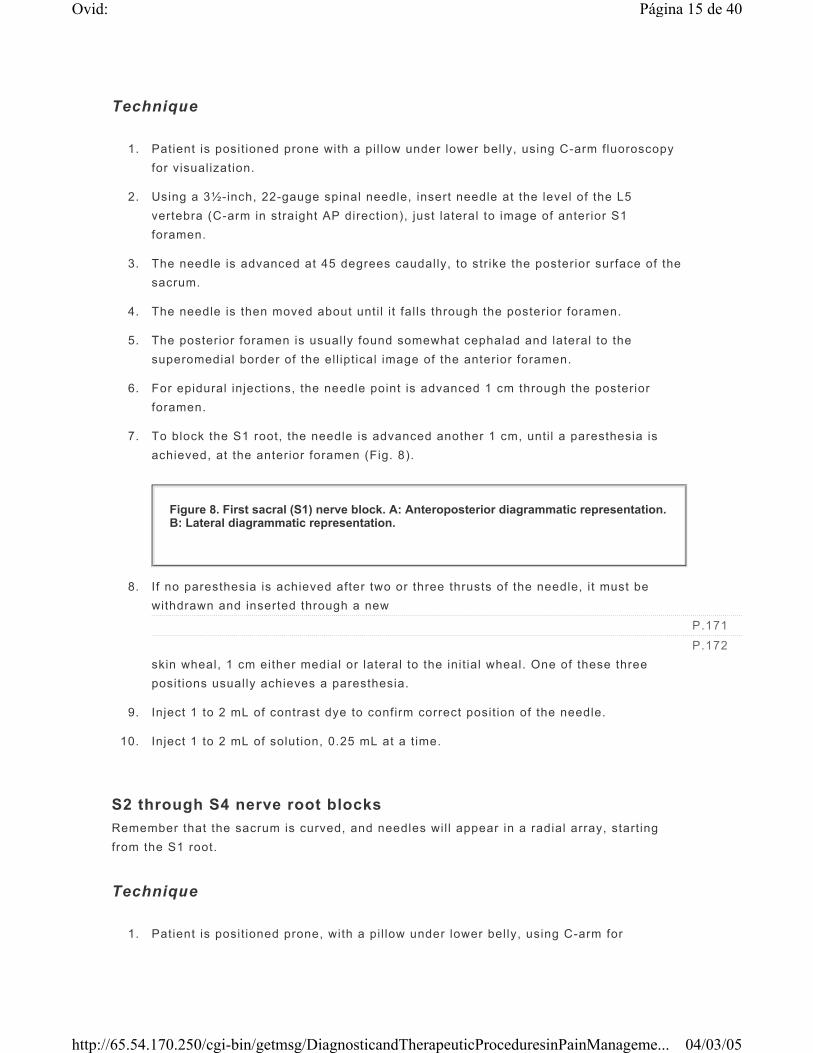
7. To block the S1 root, the needle is advanced another 1 cm, until a paresthesia is achieved, at the anterior foramen (Fig. 8).
8. If no paresthesia is achieved after two or three thrusts of the needle, i t must be withdrawn and inserted through a new skin wheal, 1 cm either medial or lateral to the init ial wheal. One of these three posit ions usually achieves a paresthesia.
9. Inject 1 to 2 mL of contrast dye to confirm correct posit ion of the needle.
10. Inject 1 to 2 mL of solution, 0.25 mL at a t ime.
S2 through S4 nerve root blocks Remember that the sacrum is curved, and needles wil l appear in a radial array, start ing from the S1 root.
Technique
1. Patient is posit ioned prone, with a pil low under lower belly, using C-arm for
2. Skin wheals are made slightly lateral to image of anterior foramen, and vertical to it .
3. As described earl ier, anterior foramina and paresthesias wil l be found about 1.5 cm, 1.0 cm, and 0.5 cm through the posterior foramina of S2, S3, and S4, respectively.
S5 nerve root block In most individuals, there is no S5 posterior or anterior foramen. This is not an accurate block, nor is accuracy often required. The S5 root can be anesthetized (without paresthesias) by passing a needle just inferior and lateral to the t ip of the sacrum (Fig. 9).
3. Sympathetic nerve blocks These techniques attempt to isolate the sympathetic from the somatic nerves. The fol lowing techniques are specif ically for use with C-arm fluoroscopy. Nonfluoroscopic techniques have been widely reported, but C-arm fluoroscopy increases the success rate of upper extremity sympathetic blocks from between 27% and 70% to about 90%.
Block treatment protocol In treating CRPS with sympathetic blocks, our practice is to give one block per week, for 3 weeks. If, in conjunction with physical therapy, the blocks allow the disease to improve, they are continued. If the duration of rel ief from the blocks is too short and symptoms fails to improve, a 1-week continuous block with in-hospital care is performed. If improvement is sustained after the week's hospital stay but symptoms recur, a second or even third attempt at hospital ized continuous catheter treatment is tr ied. If this fai ls, a surgical sympathectomy may be recommended. If a 4- to 6-week sympathetic ablation is desired, then aqueous phenol may be used. Spinal cord stimulation is a recent alternative treatment.
Confirmation of sympathetic blockade Confirmation is an essential element of both diagnostic and therapeutic blocks. Since there are no sensory measures of block eff icacy, other means are needed.
SKIN TEMPERATURE. This is simple to measure with a thermocouple, best placed on the pads of the f ingers or on the toe t ips, to record maximum temperature swings. This measurement applies, of course, only to blocks involving the upper or lower extremit ies.
No clear-cut means of assessing the completeness of sympathetic block of the trunk has been devised.
Temperature tests are best performed in a cool room (68° to 70°F) with patient fasting for more than 2 hours, with arms and legs exposed to air. This allows the baseline skin temperature to stabil ize at close to room temperature. Contralateral temperatures should be simultaneously measured for comparison. A complete sympathetic block should cause a rise in temperature from 72°F (22°C) to 93°F (34°C) in a patient with normal peripheral circulation. With vascular insufficiency (unusual in reflex sympathetic dystrophy), skin temperature does not fal l as low as 72° or rise as high as 93°. Other signs of sympathetic block should appear, such as f lushing and dry skin.
PSYCHOGALVANIC RESPONSE (PGR) OR GALVANIC SKIN RESPONSE. A more clear-cut endpoint is provided by this response. It is easily performed using a standard EKG as a galvanometer. Any two opposing leads (e.g., RA–LA as lead I, which is simple) are placed on palm and dorsum of hand, or on sole and dorsum of foot, depending on extremity involved. Other leads are attached at random for stabil i ty of recording (preferably not in EKG configuration to avoid EKG tracing). To “discharge” the sympathetic nerves, the stimulus of a single deep breath, sudden unexpected noise, or painful stimulus is used. If the sympathetic pathway is intact, a biphasic response occurs seconds later, returning to baseline in about 5 seconds (Fig. 10). If the sympathetic nerves are completely blocked, there is no response (f lat l ine). The response may not be present in very old people or diabetics with peripheral neuropathy. The electrophysiologic mechanism is not understood.
Evaluation of block efficacy: In sympathetically maintained pain, there should be immediate, complete pain relief, lasting hours to weeks. Partial relief is thought to indicate a sympathetic element to the pain. However, in some patients with a cl inical diagnosis of CRPS-I, there is no relief. The results of sympathetic blocks indicate whether blocking procedures would be useful in therapy.
(i) Stellate ganglion block
Indications
Diagnosis and therapy of sympathetically maintained pain (SMP)
Acute post-traumatic or postoperative vascular insufficiency of face, neck, or upper extremities
Potential complications
Transient nerve paralysis of the recurrent laryngeal nerve (hoarseness) or phrenic nerve (shortness of breath)
Pneumothorax
Hematoma
Subarachnoid or epidural anesthesia by injection into the dural sleeve of the cervical root
Seizures, as a result of intravascular inject ion of local anesthetic, including vertebral artery
Brachial plexus blockade
Prolongation of block Using a stellate ganglion catheter, the stellate ganglion block can be prolonged. Occasionally, this is an advantage for patients who have fai led to respond to repeat blocks and physical therapy because it facil itates aggressive in-patient physical therapy. Intrapleural catheter treatment can also provide prolonged sympathetic blockade to the upper extremity. However, these treatments require in-patient admission, and they are rarely offered today because of restrictions placed on the treatments by insurers.
Technique
1. Posit ion the patient supine with the neck extended and a pil low under the shoulders. Neck extension makes the cervical spine more superficial and easier to reach; and it draws the esophagus behind the trachea so it is less easily pierced by a left-sided approach.
2. Palpate the space between carotid pulsation and the lateral trachea, as low as possible in the neck.
3. Make a skin wheal over the medial edge of carotid pulsation at this level, usually at C6 or C7 over the transverse process, by C-arm fluoroscopy.
4. Direct a 2½-inch, 22-gauge spinal needle caudally and medially toward the junction
of the lateral portion of the bodies of C7 and T1. Figure 10 shows the level of the stellate ganglion.
5. When bone is encountered, check the posit ion (it should feel l ike the hard, f lat top of a table), withdraw the needle 1 mm, and inject 5 to 10 mL 0.25% bupivacaine or 0.5% lidocaine, or a mixture of the two.
6. The 10-mL solution wil l conveniently spread (as dye would show) from C1 to T4.
7. Because there is no anatomic guide to the depth of the appropriate fascial plane (Fig. 11 and Fig. 12), a certain percentage of blocks (about 10%) wil l be missed.
8. Because of the medial placement (3 to 5 mm medial to stellate ganglion), complications of vertebral artery injection, brachial plexus block, and pneumothorax are not common. Recurrent laryngeal nerve block does occur, especially i f the needle passes close to the trachea.
9. Horner's syndrome (ptosis, miosis, enophthalmos, often with anhidrosis and nasal congestion) is commonly produced, but this does not preclude the need for testing the upper extremity sympathetic nerve block. The appearance of Horner's syndrome does not ensure an adequate block of the upper extremity.
10. If only the cervical portion of the sympathetic nerves need to be blocked, then the procedure is more easily done at C6 or C5, but the C-arm should be employed for accuracy.
11. Patients who underwent a stellate ganglion block are given sips of water to detect
possible aspiration, prior to feeding. If hoarseness is present, oral intake should be avoided. Other potential complications should be sought prior to discharge.
(ii) Lumbar sympathetic blocks Because the principal spinal segmental sympathetic supply to the lower extremities comes mainly from L1, L2, and L3, it seems logical to place a needle at L2, relying on volume and diffusion of local anesthetics to cover the whole outflow.
Diagnosis and therapy of sympathetically maintained pain syndromes of the lower extremities
Evaluation of potential benefit of neurolytic sympathectomy
Acute peripheral vascular insuff iciency
Acute herpes zoster of the lower extremities
Some peripheral neuropathic pain syndromes of the lower extremities (sympathetically maintained pain syndromes)
Potential complications
Intravascular injection
Great vessel perforation and retroperitoneal hematoma
Puncture of abdominal viscera
Injection into ureters, kidneys, or peritoneal cavity
Inadvertent epidural, subarachnoid, or lumbar plexus injection
Transient backache and stiffness
Technique
1. Accurate placement of the needle requires f luoroscopy.
2. Posit ion the patient prone, with a pil low beneath the epigastrium.
3. Identify the lateral cephalad border of the body of the L2 vertebra (this point should be cephalad to the transverse process to avoid spinal nerves).
4. Mark skin projection.
5. Using f luoroscopy, make another mark 8 cm lateral to spinous process of L2, slightly cephalad to the f irst mark.
6. Check to see that the mark is over, or medial to, the twelfth rib. I f not, move medially.
7. Infi l trate l idocaine through the second skin mark down to the body of the vertebra.
8. A 20-gauge, 12.5-cm (5-inch) needle is inserted down to the f irst target (the lateral-cephalad portion of the body of L2).
9. Withdraw, redirect laterally, with needle bevel facing medially, and advance so that
the needle sl ides easily by the lateral surface of the vertebra (Fig. 13).
10. Rotate the C-arm to the lateral view, and advance the needle 1 to 2 mm anterior to the anterior margin of L2.
11. Inject a total volume of 20 to 30 mL 0.5% lidocaine and 0.25% bupivacaine.
Within minutes, f lushing and warming of foot should occur and skin temperature should rise. Sometimes, if the patient is very apprehensive and hyperreactive, the skin temperature does not r ise for 20 to 30 minutes, presumably because circulating epinephrine maintains vasoconstriction. Once patient relaxes in the recovery room, the foot usually warms up.
4. Visceral nerve blocks
(i) Celiac plexus block Because the celiac plexus is a network surrounding the celiac artery and the adjacent anterior aorta, the objective is to deposit solution anterior to the aorta. A variety of approaches are possible:
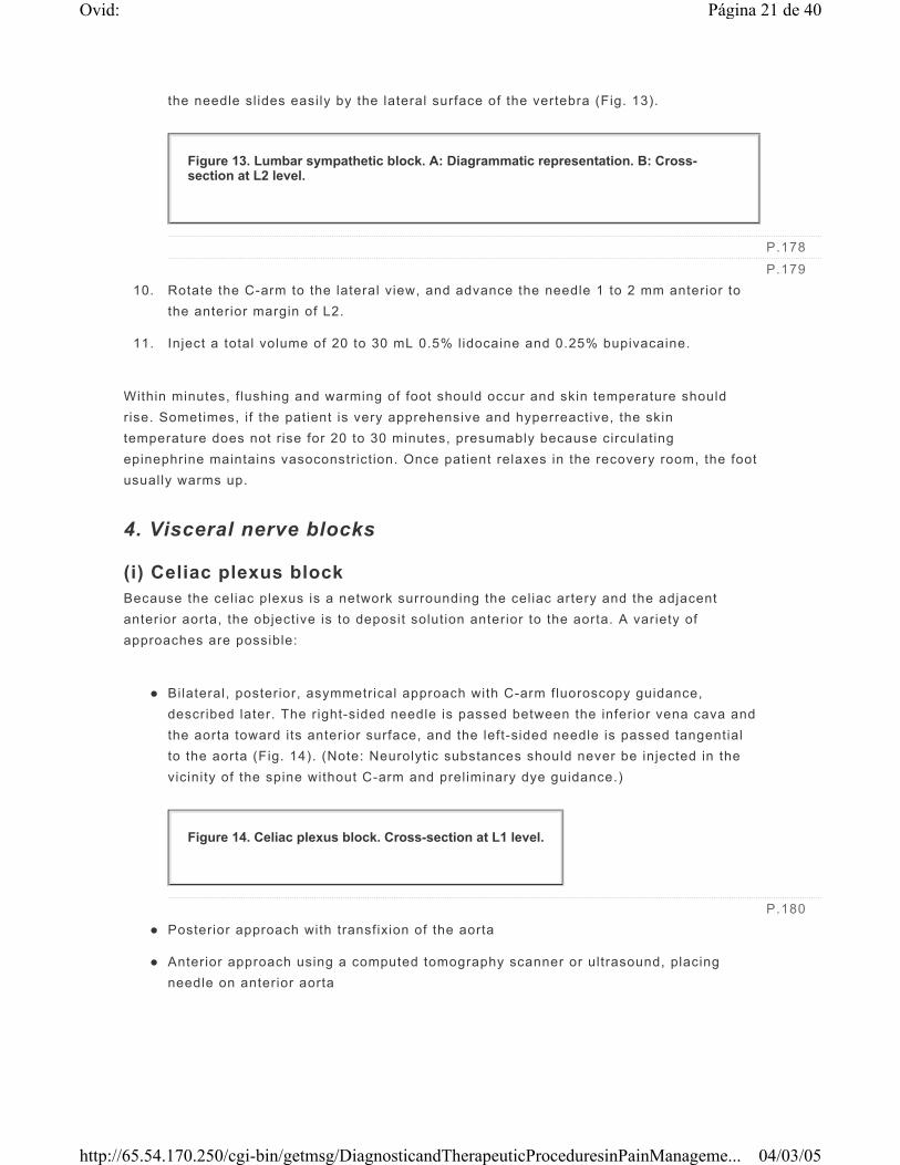
Bilateral, posterior, asymmetrical approach with C-arm fluoroscopy guidance, described later. The right-sided needle is passed between the inferior vena cava and the aorta toward its anterior surface, and the left-sided needle is passed tangential to the aorta (Fig. 14). (Note: Neurolytic substances should never be injected in the vicinity of the spine without C-arm and preliminary dye guidance.)
Posterior approach with transfixion of the aorta
Anterior approach using a computed tomography scanner or ultrasound, placing needle on anterior aorta
Treatment of pain for pancreatic cancer (and sometimes herpetic or gastric cancer)
Pain of chronic relapsing pancreatit is (the combination of local anesthetic and steroid is sometimes helpful)
Diagnosis and therapy of sympathetically mediated abdominal, retroperitoneal, or f lank pain
Potential complications
Intravascular injection
Great vessel perforation and retroperitoneal hematoma
Puncture of a viscus
Injection into kidneys, pancreas, peritoneal cavity, or l iver
Epidural, subarachnoid, or lumbar plexus injection, possibly with neurolytic agents
Acute abdominal and chest discomfort, lasting about 30 minutes
Orthostatic hypotension, as a result of profound sympathetic neural blockade, lasting for 48 hours or more, fol lowing a neurolytic injection
Thrombosis or pressure occlusion of the spinal branch of the aorta, with resultant paraplegia (extremely rare)
Post-block management Regular monitoring of vital signs following a celiac plexus block for up to 4 hours with a local anesthetic block, and 24 hours or longer with a neurolytic block, may be necessary. Evaluation for the other potential complications is also necessary.
a) Diagnostic block
Technique
1. Patient in prone posit ion, pi l low under epigastrium.
1. Mark the skin, as in lumbar sympathetic block, but at the L1 level ( instead of L2).
2. A 20-gauge, 15-cm stylet needle is passed to the upper lateral portion of the body of L1, deviated laterally and caudad about 2.5 cm anteriorly to the anterior surface of L1 vertebral body.
3. Proceed while aspirating carefully.
Left Side
1. A 20-gauge, 15-cm stylet needle is inserted to the back, 2 to 3 cm lateral to the spinous process of L1, aiming at the upper lateral margin of the L1 body, using C-arm fluoroscopy visualization.
2. The needle is carefully carried down to, and passed beyond, the lateral edge of the L1 vertebral body unti l a pulsating resistance is reached (the aorta) or the needle advances 2 cm anteriorly to L1.
3. After negative aspiration and a 2-mL test dose, 20 to 25 mL of 0.5% lidocaine or
0.25% bupivacaine (or a mixture of both) is injected on each side.
b) Neurolytic block
Technique
1. Prior to performing a neurolytic block, a diagnostic block should be done (a day before if the scheduling condit ion of the patient allows) to demonstrate pain relief. If this is only done just prior to the alcohol block, placebo effect and duration of rel ief cannot be evaluated.
2. Inject 10 mL of 1% lidocaine on either side.
3. This is fol lowed by 5 mL of water-soluble dye (Omnipaque, di luted 50% with saline). Ideally a sausage-like pattern around the aorta is produced, but layering either anterior or posterior to the aorta is satisfactory. If the dye streaks diagonally toward the diaphragm, it is in the crus of the diaphragm, and alcohol injection would be ineffective. If the dye fol lows posteriorly toward the intervertebral foramen, the needle needs to be reposit ioned to avoid alcohol contacting somatic nerves.
4. After a 20-minute wait to allow dispersal of l idocaine and dye (disappearance can be confirmed using f luoroscopy), 25 mL of 50% alcohol (absolute alcohol diluted with saline) is injected on each side.
5. Usually, 10 mL of l idocaine protects against the irr i tative effect of the alcohol, but the patient may experience a brief aching in the epigastrium or back.
(ii) Hypogastric plexus The superior hypogastric plexus l ies anterior to the L5 vertebra, innervating the organs in the pelvis and pelvic f loor, including the vagina, vulva, uterus, rectum, bladder, perineum, prostate.
Indications
Pelvic cancer pain
Nonmalignant pelvic pain
Complications
Hematoma
Infection
Impotence
Peripheral vascular occlusion
Technique
1. With patient in the prone posit ion, the L4-5 interspace is identif ied using f luoroscopy.
2. A skin wheel is made with local anesthetic 5 to 7 cm from the midline at the L4-5 interspace.
3. Insert a 7-inch, 22-gauge, short-beveled needle through the skin wheel, aiming at the anterolateral aspect of the bottom of the L5 vertebral body.
4. Guide the needle with f luoroscopy to avoid hitt ing the i l iac crest and the L5 transverse process.
5. The contralateral needle is inserted in the same way under f luoroscopic guidance.
6. Inject 2 to 4 mL of water-soluble contrast. In the AP view, the dye should be just anterior to the L5-S1 intervertebral space. In the lateral view, a smooth posterior contour, corresponding to the anterior psoas fascia, indicates proper needle posit ion.
7. For a diagnostic block, 5 to 10 mL of 0.25% bupivacaine is injected bilaterally after negative aspiration.
8. For neurolytic block, 10 mL of 10% phenol is used unilaterally or bi laterally. A diagnostic block should be performed before the neurolytic block.
(i) Trigeminal nerve blocks Trigeminal nerve blocks are commonly used for the treatment of tr igeminal neuralgia. At MGH, this procedure is performed by neurosurgeons. Under monitored sedation, the patient is placed in a supine posit ion with the neck extended. At a point 2.5 cm lateral to the corner of the mouth, the skin is prepared and draped in a standard steri le fashion. After anesthetizing the skin with 1% lidocaine, a 20-gauge, 13-cm Hinck needle is advanced through the anesthetized skin, in the direction of the f ixed pupil in a cephalad trajectory into the foramen ovale. At this point, there is often a free f low of CSF when the stylet is removed. After radiographic confirmation of needle posit ion, 0.1-mL aliquots of a preservative-free local anesthetic (1% lidocaine or 0.5% bupivacaine) or a neurolytic agent is injected. The patient is left in the supine posit ion if alcohol is to be injected. If phenol is chosen for the injection, the patient is moved into a sitt ing posit ion with the chin on the chest so that the solution gravitates around the maxil lary and mandibular divisions of the nerve and thus spares the ophthalmic division. A similar approach can be util ized to place radiofrequency and cryotherapy probes.
(ii) Occipital nerve block Occipital neuralgia results from stretching or entrapment of the occipital nerve. The nerve may become trapped in fascia overlying the posterior surface of C2 or in occipital l igamentous attachments.
Indication Occipital neuralgia as in either tension headaches or fol lowing injury (e.g., auto accidents with whiplash, fal ls, or work injuries)
Technique
1. Posit ion the patient sitt ing on a stool, elbows leaning on a table, and forehead in hands.
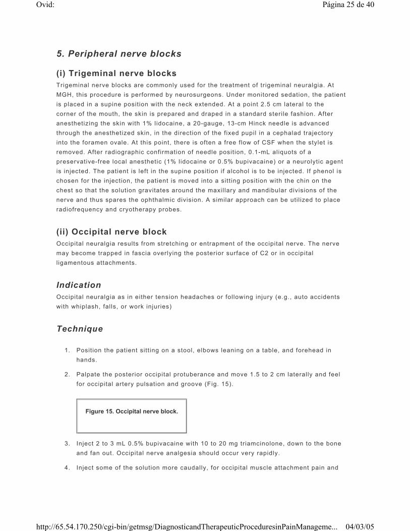
2. Palpate the posterior occipital protuberance and move 1.5 to 2 cm laterally and feel for occipital artery pulsation and groove (Fig. 15).
3. Inject 2 to 3 mL 0.5% bupivacaine with 10 to 20 mg triamcinolone, down to the bone and fan out. Occipital nerve analgesia should occur very rapidly.
4. Inject some of the solution more caudally, for occipital muscle attachment pain and
spasm, which often responds to steroid injections.
(iii) Suprascapular nerve block
Indications
Shoulder pain secondary to rotator cuff lesions, osteoarthrit is of the shoulder, or adhesive capsulit is (“frozen shoulder”)
Shoulder arthroscopy and other orthopedic manipulations of the shoulder
Potential complications
Pneumothorax
Infection
Intravascular injection
Seizure
Muscle atrophy
Technique
1. Posit ion the patient sitt ing.
2. A 22-gauge, 1½-inch needle is inserted 1 to 2 cm superior to the midpoint of the spine of the scapula and advanced toward the suprascapular notch unti l a paresthesia is el icited.
3. Inject 5 to 10 mL of local anesthetics with or without steroids.
4. Posit ion the hand ipsilateral to the block onto the contralateral shoulder: this can
move the scapula away from the posterior chest wall and reduce the risk of pneumothorax.
Post-thoracotomy pain or pain caused by percutaneous drainage tubes
Diagnostic or therapeutic blocks for abdominal pain or abdominal wall pain
Potential complications
Pneumothorax
Intravascular injection
Seizures
Infection
Bleeding
Technique
1. Posit ion the patient in a semilateral posit ion, with sites to be injected made prominent by a pil low under the opposite chest wall.
2. The injection site is the posterior axil lary l ine, to 5 to 7 cm lateral to the vertebral spinous processes. [The posterior division cannot be reached without injecting by the paravertebral approach because of the hazard of pneumothorax (see II, 2)]
3. Produce a skin wheal at the injection site. With a 25-gauge, ½-inch (or longer, if necessary) needle, enter vertically to the skin, and “walk” the needle to just below the rib and forward 2 mm. Inject 3 mL 0.5% bupivacaine with epinephrine at each rib (Fig. 16). (Note: Anesthetic is absorbed rapidly into the systemic circulation because of the vascularity of the injection site.)
4. Neurolytic intercostal block: f irst block the nerve proximally with 0.5% bupivacaine, then inject 2 to 3 mL 100% ethyl alcohol, lateral to the anesthetized site (alcohol injection is init ial ly very painful).
(vi) Lateral femoral cutaneous nerve block Lateral femoral cutaneous nerve pain is believed to be associated with obesity or
Figure 16. Intercostal nerve block. Patient in semilateral position.
pregnancy or the wearing of a t ight belt. It is thought to be caused by entrapment of the lateral femoral cutaneous nerve as it passes through the inguinal l igament. Neurolytic blocks are not recommended; however, surgical dissection may be considered.
Indication Meralgia paresthetica: burning pain, numbness and tingling in the anterolateral aspect of the thigh
Technique
1. Posit ion the patient supine.
2. Palpate the anterior superior i l iac spine, and insert a 1½-inch, 25- or 22-gauge
needle 2 cm medial and 2 cm caudal to it (Fig. 17).
3. Proceed through the fascia (feel a “pop”), and inject 10 mL of 0.5% bupivacaine with 30 mg triamcinolone fanwise, from the medial surface of the i l iac spine medially to beneath the insertion point.
Evaluation of block Analgesia of the upper two thirds of the anterolateral thigh should be produced.
(vii) Ilioinguinal nerve block
Indications
Postherniorrhaphy pain, which is usually caused by trauma to the genitofemoral nerve in the f loor of the inguinal canal
Diagnostic block, prior to surgical dissection or neurolytic block (Note: hazardous) (Fig. 17)
Testicular pain, with or without history of trauma or surgery
Technique
1. Posit ion the patient supine.
P.185
Figure 17. Lateral femoral cutaneous, ilioinguinal, and iliohypogastric nerve blocks.
2. Produce a skin wheal with local anesthetic, 2 cm medial to the anterior superior i l iac spine.
3. Infi l trate all layers of muscle toward the umbil icus with a 1½-inch, 22-gauge needle
and 10 to 20 mL of 0.5% bupivacaine, for a distance of 10 cm.
Evaluation of block Variable distribution of analgesia is noted in the medial thigh and groin, which should relieve the groin pain if the i l ioinguinal nerve is involved.
(viii) Genitofemoral nerve block
Indication Groin or testicular pain unrelieved by il ioinguinal block. (Lumbar sympathetic block is another reasonable treatment for testicular pain.)
Technique
1. Posit ion patient in supine posit ion.
2. Inject 5 mL of 0.5% bupivacaine around the spermatic cord, at the base of the scrotum.
Evaluation of block I f pain rel ief occurs but returns, the next option is either to repeat the block as frequently as required or to do a cryoneurolysis or even a rhizotomy of L1 and L2.
III. FACET JOINT BLOCKS Back pain may result from the effects of degenerative arthropathy (as a part of diffuse degenerative joint disease) (Fig. 18) and trauma such as whiplash injury on the zygapophysial joints. A facet joint block can be performed as a diagnostic aid for the orthopedic surgeon deciding whether to perform spinal fusion. Inject local anesthetic into a joint to determine if i t is the source of back pain. It may be worthwhile to inject steroid into the joints, although this usually provides relief for only up to 2 weeks. A facet joint block can also be used as a predictive tool, prior to denervating the joint with alcohol, or prior to radiofrequency lesioning of the joint nerve (the nerve of Luschka, the medial branch of posterior primary ramus) at the same level and one above.
a) Lumbar facet block Prior to beginning the block, the pain should be evaluated by putting the patient through a range of back motions. Extension characteristically produces facet pain.
Technique
1. Place the patient in a prone posit ion with the C-arm fluoroscopy in oblique posit ion.
2. Rotate the C-arm to get the best view into the joint: The “Scotty dog” x-ray image is characteristic—the vertically oriented joint image is behind the dogs ear.
3. Usually, an entry point 2 cm lateral to midline leads into the joint. However, entering directly over the joint image or even lateral to it may be required, depending on arthrit ic overgrowth of the joint.
4. A 22-gauge, 3½-inch spinal needle is carried with minimal local anesthetic to the edge of the joint. The needle is then “walked” in 1-mm steps medial or laterally, cephalad or caudad, unti l i t drops into the joint. A characteristic curve is seen at the distal end of the needle, showing that i t is fol lowing the joint, usually medially.
5. An intact joint wil l accept up to 1 mL of f luid to anesthetize the joint (0.5% bupivacaine). If the joint capsule is disrupted, i t wil l have a much greater capacity. Be careful not to inject more than 1 mL, preferably in 0.5-mL increments, or the f luid wil l spread to other innervating branches and cause a false-posit ive response.
6. Relief that outlasts the duration of the anesthetic (1 to 2 hours) suggests a nonspecif ic response.
b) Cervical facet block The reasons to use cervical facet blocks are similar to those for lumbar facet blocks, although the former are especially useful when abnormally moving facet joints are identif ied by previous continuous fluoroscopy. Whiplash injuries tend to disrupt facet motion.
Technique
1. An IV catheter is placed, vital signs are monitored, and fluoroscopy is employed.
2. The patient is posit ioned prone, with a pil low under the chest, the neck somewhat f lexed, and the forehead resting on an IV f luid bag for comfort.
3. With C-arm in the PA direction, enter at a point directly over the transverse process mass, usually 1.5 to 2.0 cm lateral to the midline, and about three vertebrae caudad to the desired level (see angle of joints).
4. With minimal local anesthetic, a 22-gauge, 3½-inch spinal needle is advanced at about a 30-degree angle with the skin unti l bone is encountered at the desired level.
5. The joint space is then visualized by placing the C-arm in lateral posit ion, and the needle is “walked” 1 mm at a t ime unti l it drops into joint. The final posit ion of the needle t ip is halfway through the joint.
6. The lateral-medial posit ion of the needle needs to be checked again with the C-arm in AP view. The needle point should be in the middle of the transverse process mass. (Slipping too far medially produces epidural or spinal anesthesia.)
7. Again, 1-mL 0.5% bupivacaine in 0.5-mL increments is suff icient to anesthetize the joint.
8. If the joint is the source of pain, motion of neck that was painful before the block should immediately disappear fol lowing the block.
c) Lumbar medial branch block Each lumbar facet joint is innervated by a medial branch of the posterior primary rami of the lumbar nerve at the same level and another medial branch from one level above. For example, the L4-L5 facet joint is innervated by the medial branches of the posterior primary rami of the L4 and L3 nerve roots. The L5-S1 facet joint, however, is innervated by the medial branch of the L4 posterior primary rami and the dorsal ramus of the L5 nerve root. It has been found that medial branch radiofrequency lesioning provides longer pain rel ief than intrafacet joint steroid injection. Thus it is more common now to do medial branch blocks (twice), fol lowed by radiofrequency lesioning if the patient's pain is signif icantly reduced by both medial branch blocks.
Technique
1. Place the patient in a prone posit ion.
2. The posit ion of the C-arm is adjusted in the PA view first, to square the endplate of the vertebral body at the level of the target nerve to be blocked. The C-arm is then rotated to an oblique view unti l the vertical l ine of the facet joint is at the middle of the endplate.
3. Lidocaine, 3 mL of 1%, is used to anesthetize the skin and the needle pathway, except for the area near the target point.
4. The target point of the block is just caudal to the most medial portion of the L2 through the L5 transverse processes for the L1 through the L4 medial branches,
respectively (i.e., high at the “Scotty dog's eye”). For the dorsal ramus of L5, the target point of the needle is at the junction of the sacral ala and the superior art icular process. The PA view is used for the block of the L5 dorsal ramus.
5. A 25-gauge, 90-mm spinal needle with a curve at the t ip is used. The point of needle insertion on the skin is selected above and lateral to the target point, usually just above the t ip of target transverse process. The needle is inserted in a caudal, ventral, and medial direction using a tunnel-view technique.
6. Insertion is terminated once the t ip of the needle strikes the bone of the target point. In the AP view, the needle t ip should be sl ightly medial to the lateral margin of the si lhouette of the superior art icular process.
7. Once the needle is in correct posit ion, 0.1 to 0.3 mL of contrast medium is injected to test that venous uptake does not occur. If i t does, the needle must be readjusted by 1 or 2 mm and the test repeated. If there is no venous uptake, 0.5 mL of 1% lidocaine is injected onto the target nerve.
d) Cervical medial branch block The C2-3 facet joint, the highest cervical facet joint, is innervated mainly by the third occipital nerve, which runs in an AP direction across the lateral surface of the C2-3 facet joint. The medial branches of the C3 and C4 dorsal rami innervate the C3-4 facet joint. Each of these medial branches runs anteroposteriorly, hugging the midpoint of the articular pi l lar. The rest of the cervical facet joints have innervations the similar to those of the C3-4 facet joint from the medial branches at their respective levels.
Potential complications
Infection
Injury and injection into carotid artery and vertebral arteries
Seizure
Damage to brachial plexus
Spinal cord injury
Epidural or spinal injection
Technique
1. The patient l ies in the lateral posit ion.
2. PA and lateral f luoroscopic views are used to identify the cervical art icular pi l lars
4. The target point for the block is sl ightly anterior to the midpoint of C2-3 facet joint in the lateral view and on the surface of the C2-3 facet joint in the PA view.
5. To block the deeper medial branches of C3 and the medial branches of C4 to C7, the end of the needle point is at the midpoint of the pyramid of the articular pil lars in the lateral view and on the lateral surface of the articular pi l lar in the PA view. The target point for the C7 medial branch block is high on the apex of the superior art icular process of C7.
6. Needle insertion should be carefully guided by f luoroscopy.
7. Once the needle reaches the target, and after negative aspiration, 0.5 mL of 1% lidocaine is injected.
IV. SACROILIAC JOINT BLOCK Although the sacroil iac (SI) joint is not a facet joint, i ts block technique is very similar to that of facet joint injection. The temptation to inject this joint is great because tenderness over the SI joint is a very common finding in patients with low back pain. In fact, this was the reason back pain used to be treated by SI fusion, before Mixter and Barr demonstrated that the herniated disc was a cause of sciatica in the early 1930s. As the joint is well innervated, it is no wonder that this can be the source of acute or chronic low back pain.
Indications
Since the cl inical presentation is very variable (pain in the gluteal area with or without radiation to posterior thigh or knee, or even down to the ankle), SI joint injection is used for the diagnosis and therapy of low back or sacral pain.
Potential complications
Infection
Penetration of pelvic viscus if needle traverses the joint (a remote possibil i ty)
Technique
1. As the joint region is easily palpable, its injection would seem simple. Actually, i t is not, and C-arm guidance is needed.
2. The patient should be in prone posit ion, with the C-arm rotated to the oblique posit ion unti l the images of the anterior and posterior joints overlap.
3. The insertion point is usually at the caudal t ip of the joint.
4. A 22-gauge, 3½-inch spinal needle is carried deeply into the joint. The needle t ip shows the same deviation it does in the lumbar facet under C-arm visualization (Fig. 19).
5. To anesthetize this joint, 2 to 3 cc of 0.5% bupivacaine should be adequate. Steroid is an option.
V. TRIGGER POINT INJECTIONS Trigger point injections are uti l ized to relieve pain associated with myofascial pain syndrome, which is characterized by spontaneous and evoked pain of affected muscles. It is commonly possible to palpate the band type of muscle spasms, also known as tr igger points, that tr igger pain. These points are usually very tender (also known as tender points). This syndrome can be either primary or secondary to underlying diseases such as facet arthropathy, sacroil iac arthropathy, and disc herniation. The reproduction of pain during injections into the muscle and the relief of pain post injection is the hallmark of this type of pain. Even though we routinely inject local anesthetics into the tr igger points, some physicians use just dry needling, or steroids with or without the local anesthetics; others use normal saline.
Technique Inject 1 to 2 mL of 0.25% bupivacaine, with or without 5 to 10 mg triamcinolone, into and around the tr igger point, using a 25-gauge, 1½-inch needle, and aspirating before injecting. Repeat as needed, and wherever required.
VI. ADDITIONAL DIAGNOSTIC AND THERAPEUTIC TECHNIQUES In addit ion to administering local anesthetics with or without steroids locally or regionally, anesthetic ( l idocaine) can be administered locally, or adrenergic blockers systemically.
a) Intravenous lidocaine injection Lidocaine is an amide local anesthetic and a sodium channel blocker that is given by controlled IV infusion to test the analgesic response and predict the possible eff icacy of
mexiletine (an oral sodium channel blocker). Although many posit ive responders demonstrate a good response to mexiletine, the true predictive value of this test is uncertain because it has not yet been shown that that negative responders do not respond to mexiletine.
Indications
Neuropathic pain syndromes, particularly with continuous or lancinating dysesthesias, (e.g., diabetic neuropathy, phantom limb pain, stump pain, and neuralgia)
Pretest management
1. The patient is asked to eat l ightly prior to the procedure, and to abstain for the 4 hours immediately preceding the procedure.
2. A baseline EKG and are obtained. Myocardial conduction abnormalit ies and abnormally elevated l iver function test results are contraindications to oral mexiletine.
1. Blood pressure, EKG, and oxygen saturation are monitored continuously.
2. Patient is supine on a bed, and an 18- or 20-gauge IV catheter is placed in an upper extremity.
3. Pre-block VAS pain rating is obtained. Pain must exist at the t ime of the test for the eff icacy of the infusion to be evaluated.
4. Lidocaine, 1 to 2 mg/kg (without epinephrine), is injected over 10 to 15 minutes (usually 100 mg for the average adult). If t inni tus, perioral numbness, metall ic oral taste, or dizziness is experienced, injection speed should be reduced, and then restarted with resolution of the symptoms.
Evaluation of procedure
VAS scores are obtained before, during, and after the infusion.
A 50% or greater reduction in pain suggests that a tr ial of oral mexiletine is worthwhile.
Specif icity of response can be tested by injecting 10 mL of normal saline prior to the IV l idocaine and then obtaining VAS scores.
After the infusion, the patient is observed for 30 minutes in the sitt ing posit ion and then allowed to ambulate. If the status is stable, the IV catheter is removed and the patient is discharged.
b) Intravenous phentolamine infusion Phentolamine is an alpha-1-adrenergic blocking agent, which is given IV as a test for sympathetically mediated pain.
Indications
Diagnosis, and occasionally therapy, of sympathetically maintained pain when sympathetic blocks are contraindicated (e.g., in anticoagulated patients, for infection of needle entry site)
Pre-block management
1. Patients are requested to eat l ightly up to 4 hours prior to the procedure.
2. They are evaluated for cardiac disease or other condit ions that may be affected by
Hypotension, mild to profound, possibly leading to hypoperfusion states
Dizziness, l ightheadedness
Reflex tachycardia
Syncope
Technique This procedure is performed at our institution according to the protocol described by Raja and colleagues.
1. With the patient supine, EKG, blood pressure, and oxygen saturation monitors are placed.
2. An 18- or 20-gauge IV catheter is placed.
3. Baseline pain levels are recorded (VAS scores).
4. A bolus of 500 mL of lactated Ringer's solution is administered via a continuous infusion. This helps counteract hypotension and can act as a placebo test.
5. Stimulus-independent pain evaluations (VAS scores), and stimulus-evoked pain evaluations (e.g., VAS, mechanical test, cold test) are performed and recorded.
6. Propranolol, 2 mg IV, is administered to counteract reflex tachycardia.
7. Phentolamine, 35 to 70 mg IV in 250 mL normal saline, is infused over 20 minutes, without the patient's direct knowledge of init iation of the infusion.
Evaluation of procedure: Pain testing and vital signs are evaluated for another half hour, prior to discharge. Relief of pain indicates that the pain was sympathetically maintained. If the pain is not relieved, either corroborative blocks or sympathetically independent pain may be considered, but the diagnosis is not definit ively excluded.
Post-block management Patients are observed for 30 minutes after the block, and somatosensory and pain evaluations are conducted. Patients are then allowed to sit and stand up, slowly, as tolerated. If they are stable, they are discharged; if not, they are observed for as long as
c) Intravenous regional sympathetic blocks (Bier blocks)
Background Bier blocks are done in an attempt to create a sympathetic block in an extremity (i.e., a peripheral block). Several medications can be used, including guanethidine, bretyl ium, labetalol, prazosin, clonidine, and reserpine. Intravenous guanethidine and reserpine are not readily available in the United States. At MGH, we use the mixed alpha and beta antagonist labetalol.
Indications
Therapeutic sympathetic blockade of an upper or lower extremity
Diagnostic blockade when patients refuse needle blocks or are on anticoagulants, or when needle blocks are contraindicated or have been unsuccessful
Pre-block management
1. Patients are instructed to abstain from food for up to 4 hours prior to the procedure.
3. Another 18- or 20-gauge IV catheter is placed in a nonaffected extremity, for IV access and prehydration.
4. The affected extremity is exsanguinated by elevating it and binding it t ightly with an elastic bandage (Esmarch bandage) from fingers to axil la.
5. A pneumatic cuff is applied proximally, at a pressure of 250 mm Hg.
6. Labetalol, 20 to 30 mg, with 100 mg of l idocaine, is made up to 20 mL of solution by adding normal saline, and it is injected into the affected upper extremity, OR.
7. Labetalol, 30 to 40 mg, with 200 mg of l idocaine, is made up to 35 mL with normal saline and is injected into the affected lower extremity.
8. The cuff 's pressure is maintained at 250 mm Hg for 20 min, then either deflated slowly or intermittently deflated and reinflated over 5 to 10 minutes, while monitoring vital signs closely.
Evaluation of procedure Baseline (prior to procedure) and postprocedure pain evaluations are performed, including obtaining a VAS score. A t ime course of the pain relief is noted, as relief can range from hours to months.
VII. CONCLUSION The blocks most commonly uti l ized in the MGH Pain Center are described. When used in carefully selected patients for specif ic indications, nerve blocking procedures are extremely helpful. Perhaps their greatest value is in (1) diagnosis, (2) facil i tating physical therapy in CRPS, (3) maintaining mobil i ty during episodes of acute back pain, and (4) end-of-l i fe pain management. It is not good practice to offer blocks to patients with no purpose other than short-term pain relief, or to give false hope of a cure. Patients wil l ult imately become disenchanted with this type of practice, as wil l health care insurers.
SELECTED READING
1. Bogduk N. Back pain, zygapophysial joint blocks and epidural steroids. In: Cousins MJ, Bridenbaugh PO, eds. Neural blockade in cl inical anesthesia and management of pain , 2nd ed. Philadelphia: Lippincott, 1988.
2. Bonica JJ. The management of pain , vols. 1 and 2, 2nd ed. Philadelphia: Lea & Febiger, 1990.
4. Moore DC. Regional block , 4th ed. Springfield, IL: Charles C. Thomas, 1965.
5. Raj PP, ed. Practical management of pain, 2nd ed. St. Louis: Mosby–Year Book, 1992.
6. RAJA et al. Systematic alpha-adrenergic blockade with phentolamine: a diagnostic test for sympathetically maintained pain. Anesthesiology 1991;74:691–698.
7. Wall PD, Melzack R, eds. Textbook of pain , 3rd ed. New York: Churchil l-Livingstone, 1994.
8. Mixter WJ, Barr JS. Rupture of intervertebral disc with involvement of the spinal cord. N Engl J Med 1934;211:210–215.
Copyright (c) 2000-2004 Ovid Technologies, Inc. Version: rel9.2.0, SourceID 1.9998.1.313