Abdominal pregnancies are a rare form of ectopic pregnancy, which presents a significant risk of maternal morbidity and mortality.We describe an unusual case of a late diagnosis of an abdominal pregnancy in the second trimester, which due to diagnosticchallenges, was not detected on 1st trimester and subsequent antenatal ultrasound scans (USS). The abdominal pregnancy waslater diagnosed at the repeat anomaly scan and confirmed with a pelvic MRI. This case of abdominal pregnancy is unique whencompared to other reported cases, as the fetus was initially enclosed within the amniotic sac with normal liquor volume. Bothtransvaginal and transabdominal scans appeared to demonstrate an intrauterine pregnancy. The diagnosis of abdominalpregnancy was only made possible following rupture of the amniotic sac, leading to anhydramnios, which resulted in therepositioning of the fetus to the upper maternal abdomen. This case represents the challenges faced by obstetricians indiagnosing, managing, and counselling a woman when faced with an abdominal pregnancy.
1. Case Report
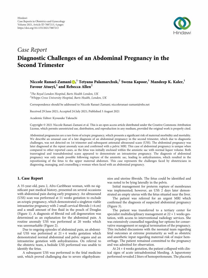
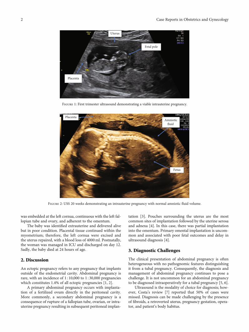
A 35-year-old, para 2, Afro-Caribbean woman, with no sig-nificant past medical history, presented on several occasionswith abdominal pain during early pregnancy. An ultrasound(USS) scan was performed at 11 weeks gestation to excludean ectopic pregnancy, which demonstrated a singleton viableintrauterine pregnancy with 2 small cervical fibroids (<4 cm)and a small amount of free fluid in the pouch of Douglas(Figure 1). A diagnosis of fibroid red cell degeneration wasdetermined as an explanation for the abdominal pain. Aroutine anomaly USS was performed at 20 weeks whichwas unremarkable (Figure 2).
Due to ongoing episodes of abdominal pain, an abdomi-nal USS was performed at 21 + 4 weeks gestation whichdemonstrated normal abdominal and pelvic organs and anintrauterine gestation with anhydramnios. On referral tothe obstetric team, a bedside USS performed was unable toidentify the fetus.
A subsequent USS was performed in the fetal medicineunit, which proved challenging due to severe oligohydram-
nios and uterine fibroids. The fetus could be identified andwas noted to be lying laterally in the pelvis.
Initial management for preterm rupture of membraneswas implemented; however, an USS 2 days later demon-strated an empty uterus with the fetus situated near the liver.
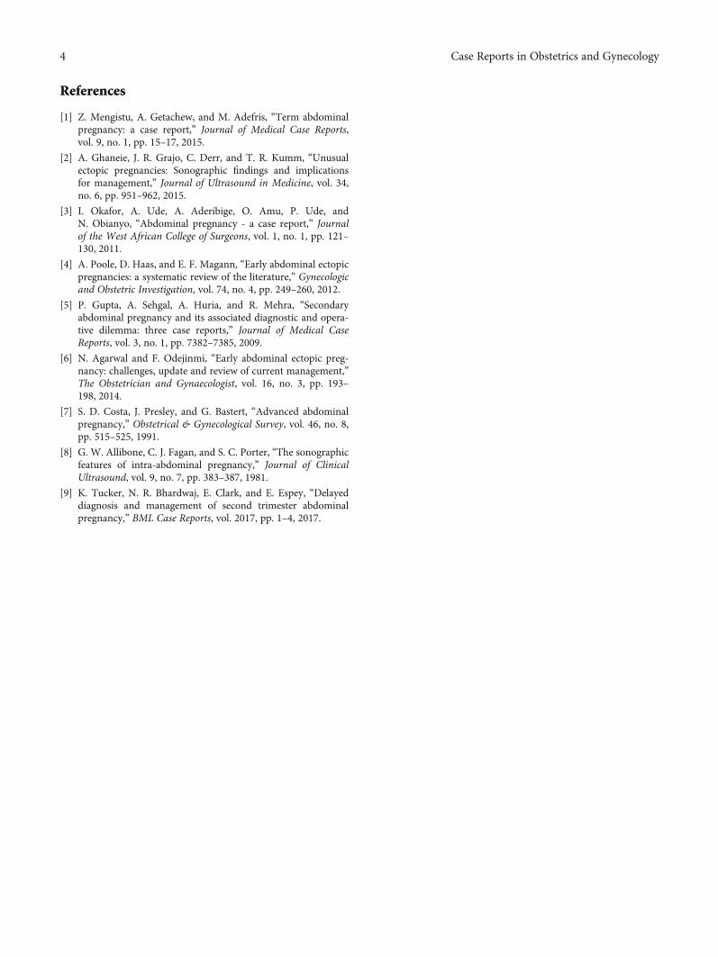
The patient was referred for an urgent MRI whichconfirmed the diagnosis of suspected abdominal pregnancy(Figure 3).
The patient was transferred to a tertiary center forspecialist multidisciplinary management at 22 + 1 weeks ges-tation, with access to interventional radiology services. Shewas extensively counselled regarding her options for conser-vative management or surgical termination of the pregnancy.This included discussions with the neonatal team regardingfetal outcomes at extreme prematurity as well as obstetricand anesthetic input regarding maternal risk of major hem-orrhage. The patient remained committed to the pregnancyand was admitted for observation.
At 24 + 4 weeks gestation, the patient collapsed with clin-ical signs of acute intraabdominal bleeding. A laparotomyperformed revealed 2 liters of hemoperitoneum. The placenta
HindawiCase Reports in Obstetrics and GynecologyVolume 2021, Article ID 7887213, 4 pageshttps://doi.org/10.1155/2021/7887213
was embedded at the left cornua, continuous with the left fal-lopian tube and ovary, and adherent to the omentum.
The baby was identified extrauterine and delivered alivebut in poor condition. Placental tissue continued within themyometrium; therefore, the left cornua were excised andthe uterus repaired, with a blood loss of 4000ml. Postnatally,the woman was managed in ICU and discharged on day 12.Sadly, the baby died at 24 hours of age.
2. Discussion
An ectopic pregnancy refers to any pregnancy that implantsoutside of the endometrial cavity. Abdominal pregnancy israre, with an incidence of 1 : 10,000 to 1 : 30,000 pregnancieswhich constitutes 1.4% of all ectopic pregnancies [1, 2].
A primary abdominal pregnancy occurs with implanta-tion of a fertilized ovum directly in the peritoneal cavity.More commonly, a secondary abdominal pregnancy is aconsequence of rupture of a fallopian tube, ovarian, or intra-uterine pregnancy resulting in subsequent peritoneal implan-
tation [3]. Pouches surrounding the uterus are the mostcommon sites of implantation followed by the uterine serosaand adnexa [4]. In this case, there was partial implantationinto the omentum. Primary omental implantation is uncom-mon and associated with poor fetal outcomes and delay inultrasound diagnosis [4].
3. Diagnostic Challenges
The clinical presentation of abdominal pregnancy is oftenheterogeneous with no pathognomic features distinguishingit from a tubal pregnancy. Consequently, the diagnosis andmanagement of abdominal pregnancy continues to pose achallenge. It is not uncommon for an abdominal pregnancyto be diagnosed intraoperatively for a tubal pregnancy [5, 6].
Ultrasound is the modality of choice for diagnosis; how-ever, Costa’s review [7] reported that 50% of cases weremissed. Diagnosis can be made challenging by the presenceof fibroids, a retroverted uterus, pregnancy gestation, opera-tor, and patient’s body habitus.
Fetal pole
Uterus
Placenta
Figure 1: First trimester ultrasound demonstrating a viable intrauterine pregnancy.
Fetus
Amnioticfluid
Placentaa
Figure 2: USS 20 weeks demonstrating an intrauterine pregnancy with normal amniotic fluid volume.
2 Case Reports in Obstetrics and Gynecology
Diagnosis was challenging in our case due to the presenceof fibroids, anhydramnios, and false reassurance from multi-ple previous scans reporting an intrauterine pregnancy. Thedilemma of whether this was a primary or secondary abdom-inal pregnancy remains as all previous USS demonstrated anintrauterine pregnancy. Her anomaly scan showed a normalvolume of amniotic fluid with the placental site seen clearly atthe fundus (Figure 2).
Although, on review of the 1st trimester USS images, theuterus, bladder, cervix, and cul-de-sac cannot be identifiedin one image. Furthermore, following referral to fetal medi-cine, with anhydramnios, there was no free fluid in the pelvisto suggest a uterine rupture leading to a secondary abdominalpregnancy. Interestingly, the patient was hemodynamicallystable with minimal abdominal pain.
Ultrasound features to aid diagnosis of abdominal preg-nancy include demonstration of a fetus in a gestational sacoutside the uterus or the depiction of an abdominal or pelvicmass identifiable as the uterus separate from the uterus, fail-ure to see a uterine wall between the fetus and bladder, andrecognition of close approximation of the fetus to the mater-nal abdominal wall and localization of the placenta outsidethe uterine cavity [8]. The difficulty visualizing the fetusand placenta on USS led to an MRI scan being performedwhich confirmed the diagnosis of abdominal pregnancy andthe appearances of possible uterine rupture at the fundus.
4. Decision-Making Challenges
Given the significant risk to maternal health (maternal mor-tality 0.5-20%) [2] and the limited literature to supportpositive fetal outcomes, counselling patients can be challeng-ing and additionally pose several ethical considerations.
Counselling should be individually tailored and influencedby gestation, site of implantation, and maternal morbidity.Advancing gestation increases maternal risk but has thepotential for encouraging fetal outcomes.
The option of surgical termination of pregnancy may beunacceptable to some patients as in our case, and therefore,significant maternal risk is accepted.
A patient’s decision may also be influenced by parity asthere is the potential risk of a hysterectomy. Additionally,the absence of major fetal abnormalities is likely to be aprerequisite for most patients. Previous case studies of fetaloutcomes may be inaccurate due to the significant advancesin neonatal care over the last decade.
5. Management
Once an abdominal pregnancy is suspected on USS, thisshould be confirmed with an MRI. Patients should bemanaged in a tertiary center with general surgery, vascularsurgery, interventional radiology, and advanced neonatalsupport facilities. Cross-matched blood should be readilyavailable in the event of emergency delivery and preparationsmade in anticipation of a major obstetric hemorrhage [9].
A surgical plan must be implemented in advance accord-ing to the site of implantation and its proximity to major vis-cera and blood vessels. Timing of delivery is also importantand should be delayed if possible until fetal lung maturity isreached.
6. Conclusion
Abdominal pregnancy is a rare form of ectopic pregnancywith significant maternal and fetal risks. They can often bechallenging to diagnose on ultrasound and therefore identi-fied late. Individualized counselling is crucial in order toenable patients to make informed decisions regardingcontinuation or ending the pregnancy. Fetal and maternaloutcomes depend on gestation, implantation site, and medi-cal facilities. Advances in antenatal ultrasound and neonatalfacilities are likely to improve outcomes.
Consent
Written consent obtained from the patient.
Conflicts of Interest
The authors declare that they have no conflicts of interest.
Authors’ Contributions
NR, TP, SK, FA, and RA wrote the article; it was thenapproved, reviewed, and edited by MKK and all authors.
Acknowledgments
The authors are thankful to Mr. Rehan Khan and Mr.Matthew Hogg.
Fetus
Figure 3: Abdominal MRI showing the fetus outside the uterus onthe right side of the abdomen with the fetal head adjacent to the liverand gallbladder. The placenta is seen outside the uterus in theabdomen superior to the uterus.
3Case Reports in Obstetrics and Gynecology
References
[1] Z. Mengistu, A. Getachew, and M. Adefris, “Term abdominalpregnancy: a case report,” Journal of Medical Case Reports,vol. 9, no. 1, pp. 15–17, 2015.
[2] A. Ghaneie, J. R. Grajo, C. Derr, and T. R. Kumm, “Unusualectopic pregnancies: Sonographic findings and implicationsfor management,” Journal of Ultrasound in Medicine, vol. 34,no. 6, pp. 951–962, 2015.
[3] I. Okafor, A. Ude, A. Aderibige, O. Amu, P. Ude, andN. Obianyo, “Abdominal pregnancy - a case report,” Journalof the West African College of Surgeons, vol. 1, no. 1, pp. 121–130, 2011.
[4] A. Poole, D. Haas, and E. F. Magann, “Early abdominal ectopicpregnancies: a systematic review of the literature,” Gynecologicand Obstetric Investigation, vol. 74, no. 4, pp. 249–260, 2012.
[5] P. Gupta, A. Sehgal, A. Huria, and R. Mehra, “Secondaryabdominal pregnancy and its associated diagnostic and opera-tive dilemma: three case reports,” Journal of Medical CaseReports, vol. 3, no. 1, pp. 7382–7385, 2009.
[6] N. Agarwal and F. Odejinmi, “Early abdominal ectopic preg-nancy: challenges, update and review of current management,”The Obstetrician and Gynaecologist, vol. 16, no. 3, pp. 193–198, 2014.
[7] S. D. Costa, J. Presley, and G. Bastert, “Advanced abdominalpregnancy,” Obstetrical & Gynecological Survey, vol. 46, no. 8,pp. 515–525, 1991.
[8] G. W. Allibone, C. J. Fagan, and S. C. Porter, “The sonographicfeatures of intra-abdominal pregnancy,” Journal of ClinicalUltrasound, vol. 9, no. 7, pp. 383–387, 1981.
[9] K. Tucker, N. R. Bhardwaj, E. Clark, and E. Espey, “Delayeddiagnosis and management of second trimester abdominalpregnancy,” BML Case Reports, vol. 2017, pp. 1–4, 2017.


![Management Dilemma in Case of Abdominal Pregnancy: A Case … · 2017. 8. 15. · A. Singh et al. 900 proximately 1% of all ectopic pregnancies [1]. The incidence of abdominal pregnancy](https://static.documents.pub/doc/80x56/60909f86b358e108bd7a9bce/management-dilemma-in-case-of-abdominal-pregnancy-a-case-2017-8-15-a-singh.jpg)


