Dietary fat increases solid tumor growth andmetastasis of 4T1 murine mammary carcinomacells and mortality in obesity-resistant BALB/c miceEun Ji Kim2, Mi-Ran Choi2, Heesook Park1, Minhee Kim1, Ji Eun Hong2, Jae-Yong Lee2,3, Hyang Sook Chun4,Ki Won Lee5 and Jung Han Yoon Park1,2*
Abstract
Introduction: High-fat diets (HFDs) are known to cause obesity and are associated with breast cancer progressionand metastasis. Because obesity is associated with breast cancer progression, it is important to determine whetherdietary fat per se stimulates breast cancer progression in the absence of obesity. This study investigated whether anHFD increases breast cancer growth and metastasis, as well as mortality, in obesity-resistant BALB/c mice.
Methods: The 4-week-old, female BALB/c mice were fed HFD (60% kcal fat) or control diet (CD, 10% kcal fat) for16 weeks. Subsequently, 4T1 mammary carcinoma cells were injected into the inguinal mammary fat pads of micefed continuously on their respective diets. Cell-cycle progression, angiogenesis, and immune cells in tumor tissues,proteases and adhesion molecules in the lungs, and serum cytokine levels were analyzed withimmunohistochemistry, Western blotting, and enzyme-linked immunosorbent assay (ELISA). In vitro studies werealso conducted to evaluate the effects of cytokines on 4T1 cell viability, migration, and adhesion.
Results: Spleen and gonadal fat-pad weights, tumor weight, the number and volume of tumor nodules in thelung and liver, and tumor-associated mortality were increased in the HFD group, with only slight increases inenergy intake and body weight. HF feeding increased macrophage infiltration into adipose tissues, the number oflipid vacuoles and the expression of cyclin-dependent kinase (CDK)2, cyclin D1, cyclin A, Ki67, CD31, CD45, andCD68 in the tumor tissues, and elevated serum levels of complement fragment 5a (C5a), interleukin (IL)-16,macrophage colony-stimulating factor (M-CSF), soluble intercellular adhesion molecule (sICAM)-1, tissue inhibitorsof metalloproteinase (TIMP)-1, leptin, and triggering receptor expressed on myeloid cells (TREM)-1. Protein levels ofthe urokinase-type plasminogen activator, ICAM-1, and vascular cell adhesion molecule-1 were increased, butplasminogen activator inhibitor-1 levels were decreased in the lungs of the HFD group. In vitro assays using 4T1cells showed that sICAM-1 increased viability; TREM-1, TIMP-1, M-CSF, and sICAM-1 increased migration; and C5a,sICAM-1, IL-16, M-CSF, TIMP-1, and TREM-1 increased adhesion.
Conclusions: Dietary fat increases mammary tumor growth and metastasis, thereby increasing mortality in obesity-resistant mice.
IntroductionBreast cancer is a leading cause of cancer-associatedmortality in women in the United States [1], and theincidence is increasing in the developing world. Themajority of breast cancer-related death results from
uncontrolled metastatic disease. Although in 10% to15% of cases, breast cancer spreads to other parts of thebody within 3 years of initial diagnosis, metastasis tendsto recur later, 10 years or more after the detection ofthe primary tumor [2]. However, current therapiesincluding surgery, hormone therapy, chemotherapy,radiation therapy, and selective combinations thereof arenot completely effective in the treatment of metastaticbreast cancer [3]. Thus, it is important to find safe and
* Correspondence: [email protected] of Food Science and Nutrition, College of Natural Sciences,Hallym University, 39 Hallymdaehak-gil, Chuncheon, 200-702, KoreaFull list of author information is available at the end of the article
Kim et al. Breast Cancer Research 2011, 13:R78http://breast-cancer-research.com/content/13/4/R78
effective lifestyle modifications, including dietary habits,for decreasing breast cancer development andmetastasis.Epidemiologic studies indicate that consuming a high-
fat (regardless of fat type) diet may lead to an increasedrisk of invasive breast cancer in postmenopausal women[4]. High-fat diets are known to induce obesity inhumans and rodents [5,6], and obese women have anincreased risk of developing postmenopausal breast can-cer [7,8]. Additionally, a high body mass index (BMI) isassociated with poor prognosis in breast cancer patients[9,10]. Thus, it is clearly important to understand themolecular basis for the association of high-fat diet and/orobesity with breast cancer development and mortality.In 1991 Rose et al. [11] reported that a diet containing
23% as opposed to 5% (wt/wt) corn oil (rich in theomega-6 fatty acid linoleic acid) increased the tumorgrowth rate and lung-metastasis incidence of MDA-MB-435 human breast cancer cells injected into the mam-mary fat pads of athymic nude mice. To evaluate theeffects of a high-fat diet on cancer development andprogression and the underlying mechanisms thereof, weused the 4T1 orthotopic model, in which 4T1 mammarycarcinoma cells are injected into the mammary fat padsof immune-competent BALB/c mice. The 4T1 cellswere derived from the mammary tumors of BALB/cmice lacking protein expression of the estrogen receptora [12,13]. When injected into the mammary fat pads ofsyngeneic BALB/c mice, 4T1 cells grow into solidtumors that metastasize to the lung, liver, lymph nodes,and brain, while the primary tumor grows in situ[14,15]. The 4T1 orthotopic model closely mimics theprogressive forms of estrogen-insensitive human meta-static breast cancer [16]. Additionally, BALB/c mice areobesity resistant, and high-fat diet (HFD) consumptionhas little effect on body weight [17].In this study we demonstrated that the prolonged con-
sumption of an HFD without any reduction in theintake of protein, minerals, vitamins, and fiber has littleeffect on energy intake and body weight but doesincrease breast cancer growth and metastasis, as well asmortality in BALB/c mice.
Materials and methodsMaterialsReagents were purchased from the following suppliers:3-[4,5-dimethylthiazol-2-yl]-2,5-diphenyltetrazolium bro-mide (MTT), Bouin solution, and anti-b-actin antibodyfrom Sigma (St. Louis, MO, USA); antibodies againstCD45, matrix metalloproteinase (MMP)-9, tissue inhibi-tor of matrix metalloproteinase (TIMP)-2, intercellularadhesion molecule (ICAM)-1, and vascular cell adhesionmolecule (VCAM)-1, and enzyme-linked immunosor-bent assay (ELISA) kits for soluble ICAM-1, macrophage
colony stimulating factor (M-CSF), TIMP-1, leptin, andtriggering receptor expressed on myeloid cells (TREM)-1 from R and D Systems (Minneapolis, MN, USA);ELISA kits for complement fragment 5a (C5a) and inter-leukin (IL)-16 from USCN Life Science and TechnologyCompany (Missouri City, TX, USA); antibodies againsturokinase-type plasminogen activator (uPA) from Cal-biochem (La Jolla, CA, USA); antibodies against E2F1,proliferating cell nuclear antigen (PCNA), p27, cyclin-dependent kinase (CDK)2, CDK4, cyclin A, cyclin D1,CD31, and vascular endothelial growth factor (VEGF),plasminogen activator inhibitor (PAI)-1 from Santa CruzBiotechnology (Santa Cruz, CA, USA); antibodies againstKi67 and F4/80 from Abcam (Cambridge, MA, USA);horseradish peroxidase-conjugated anti-rabbit, anti-mouse, and anti-goat IgG from Amersham Biosciences(Arlington Heights, IL, USA); an antibody against CD68from ABBIOTEC (San Diego, CA, USA); and ImmobilonWestern Chemiluminescent HRP Substrate and adhe-sion assay kit from Millipore Corporation (Billerica,MA, USA). If not otherwise noted, all other materialswere purchased from Sigma-Aldrich Co.
4T1 cell culture4T1 murine mammary carcinoma cells were acquiredfrom the American Type Culture Collection (Manassas,MA, USA) and maintained in Dulbecco’s ModifiedEagle’s Medium (DMEM) containing 100 ml/L of fetalbovine serum (FBS) with 100,000 U/L of penicillin and100 mg/L of streptomycin in a humidified atmosphereof 5% CO2 in air.
AnimalsThree-week-old female BALB/c mice were purchasedfrom Orient Bio Inc. (Seongnam, Korea) and housed atthe animal research facility of Hallym University. Micewere acclimatized to the laboratory conditions and pro-vided free access to a standard nonpurified rodent diet(Superfeed Co., Wonju, Korea) and water. The mice wereacclimated for 1 week before use and maintainedthroughout the study in a controlled environment: 24 ±2°C, 50 ± 10% relative humidity, and a 12-hour light/darkcycle. To determine the survival rate of animals, micewere killed when they reached moribund conditions, asdescribed by Toth et al. [18]. All experiments were con-ducted in accordance with the protocols approved by theAnimal Care and Use Committee of the Hallym Univer-sity, Korea (Ethical approval number: Hallym 2009-122).
DietsThe mice were randomly divided into two groups; thecontrol and high-fat groups. The purified diets used in thisstudy were purchased from Research Diets, Inc. (NewBrunswick, NJ, USA). The control diet (CD; No. D12450B)
Kim et al. Breast Cancer Research 2011, 13:R78http://breast-cancer-research.com/content/13/4/R78
Page 2 of 13
contained 10% of kcal from fat, and the HFDs (No.D12451 and D12452) contained 45% and 60% of kcal fromfat. The lard content was 4.4, 39.4, and 54.4 kcal% in the10, 45, and 60 kcal% diets, respectively. The three dietscontained identical quantities of protein, cellulose, soybeanoil, vitamins, and minerals per kilocalorie (Additional file1). Fresh diet was freely provided each day, and daily feedintake was monitored throughout the study.
In vivo mammary cancer orthograft modelTwelve or 16 weeks after initiating feeding, 4T1 cells (5× 10⁴ cells suspended in 0.1 ml Matrigel(BD Bios-ciences, San Jose, CA, USA) were injected into the ingu-inal mammary fat pads of the mice. The mice continuedon their respective diets. In experiment I, the mice weremaintained for 42 days after the 4T1 cell injection tomonitor mammary cancer-related death, and theKaplan-Meier curve was plotted for two dietary groupsassociated with mouse survival. In experiment II, 25days after the 4T1 cell injections, the mice were killedwith carbon dioxide asphyxiation, and the tumors,lungs, livers, kidneys, gonadal fat pads, and spleens wereexcised from the mice and weighed. The sera were pre-pared for ELISA.Tumor volumes were measured with a set of calipers
and calculated by using the following formula: 0.52 ×long diameter × short diameter2 [19]. The tumors andgonadal fat pads were formalin fixed and paraffinembedded for immunohistochemistry (IHC) or homoge-nized to prepare the tissue lysates [20] for Western blotanalysis. The lungs and livers were fixed in Bouin solu-tion or homogenized to prepare the tissue lysates [20].Metastatic nodules in the lungs and livers were counted,and the total tumor volumes were estimated asdescribed previously [21,22].
ImmunohistochemistryParaffin-embedded tumor tissues were sectioned, depar-affinized, rehydrated, incubated in 3% H2O2, andblocked with 5% BSA, as previously described [23]. IHCwas conducted with the indicated antibodies, biotiny-lated rabbit anti-mouse IgG, streptavidin-horseradishperoxidase, 1,3 -diaminobenzidine (DAB) tetrahy-drochloride, and hematoxylin, as described previously[23]. Randomly chosen fields were photographed at×200 magnification, and immuno-positive cells andstaining intensities were quantified with a Carl ZeissAxioImager microscope and Image M1 Software (CarlZeiss, Jena, Germany).
Western blot analysisWestern blot analyses were conducted as described pre-viously [24]. Signals were detected through enhancedchemiluminescence by using Immobilon Western
Chemiluminescent HRP Substrate (Millipore Corpora-tion). The relative abundance of each band was quanti-fied by using the Bio-profile Bio-1D application (Vilber-Lourmat, Marine la Vallee, France), and the expressionlevels were normalized to b-actin.
Mouse cytokine antibody arrayPooled sera from each group were applied to a pro-teome profiler antibody array kit (R&D Systems) to eval-uate cytokine expression, in accordance with themanufacturer’s instructions. The relative abundance ofeach protein was quantified by using the Bio-profile Bio-1D application (Vilber-Lourmat), and the expressionlevels were normalized to the control protein.
Enzyme-linked immunosorbent assayThe levels of C5a, sICAM-1, IL-16, M-CSF, TIMP-1,leptin, and TREM-1 in sera were estimated by using therelevant ELISA kits according to the manufacturers’instructions.
Cell-viability assay4T1 cells were plated in 24-well plates at 5 × 10⁴ cells/well in DMEM supplemented with 100 ml/L FBS. Oneday later, the monolayers were serum deprived withDMEM supplemented with 10 ml/L charcoal-strippedFBS (serum-deprivation medium) for 24 hours, and thecells were incubated in serum-deprivation medium inthe absence or presence of various cytokines. Viable cellnumbers were estimated with an MTT assay, asdescribed previously [25].
In vitro migration assayThe cell-migration assay was conducted as describedpreviously [26]. Serum-deprived cells were plated ontothe filter in 6.5-mm transwell inserts in 24-well plates at5 × 104 cells/filter and treated for 16 hours with variouscytokines in serum-deprivation medium. Migrated cellswere stained with hematoxylin and eosin (H&E), andthen counted under a light microscope in eight rando-mized fields.
Adhesion assay4T1 cells were plated in human collagen type I-coatedCytoMatrix Cell Adhesion Strips at 1 × 10⁵ cells. Cellswere incubated in DMEM containing 10 ml/L of char-coal-stripped FBS with various cytokines at 37°C for 45minutes. Cells were stained with crystal violet, and thecell-bound stains were quantified by measuring theabsorbance at 570 nm, as described previously [26].
Statistical analysisThe data were expressed as the mean ± SEM. The sig-nificance of the difference between groups was evaluated
Kim et al. Breast Cancer Research 2011, 13:R78http://breast-cancer-research.com/content/13/4/R78
Page 3 of 13
with the Student t test, by using SAS for Windows ver-sion 9.1 (SAS Institute). Differences were considered sig-nificant at P < 0.05. The Kaplan-Meier curves wereanalyzed with a Log-Rank test to assess the significanceof the differences in the survival rates of the mice.
ResultsProlonged high-fat diet consumption increases mammarycancer-related mortality in BALB/c mice injected with 4T1cellsThe first study determined mortality rates in 4T1 cell-injected mice fed HFD (45%). Relative to the mice fedon the CD (10% kcal as fats), the survival rate was lowerin the mice fed HFD (P < 0.001; Figure 1a). In the sub-sequent study, the mice were fed on a 60% kcal-diet orthe CD for 16 weeks and then injected with 4T1 cells.Again, survival rates were reduced in the mice fed onthe HFD (60% kcal) as compared with controls (P <0.001, Figure 1b).
Long-term consumption of a 60% kcal-fat diet increasessolid tumor growth of 4T1 cells in BALB/c miceBody weights were slightly higher in the mice fed on the60% kcal-fat diet than in controls from 1 week onwardafter initiating experimental diets (Figure 1c). Final bodyweights were 24.7 ± 0.3 g and 26.2 ± 0.5 g in the con-trol and high-fat groups. Energy intakes were onlyslightly higher in the high-fat group (Figure 1d). Spleenand gonadal fat-pad weights were significantly higher inthe HFD-fed mice. However, the weights of the liver,lung, and kidney were unaffected by prolonged feedingon the HFD (Table 1). The subcutaneous fat, mesentericfat, and retroperitoneal fat were almost undetectable tothe naked eye in the BALB/c mice fed on either the CDor HFD. The wet weight of the primary solid tumorswas increased by 22.4% (P < 0.0047), and tumor volume,increased by 28.0% (P < 0.0022) in HFD-fed mice com-pared with controls (Figure 1e, f).
Long-term consumption of a 60% kcal-fat diet increasescancer cell proliferation, angiogenesis, and infiltration ofimmune cells in 4T1 tumors in BALB/c miceIHC staining revealed that the HFD-fed mice had signif-icantly increased expression of Ki67 (P < 0.0084), CDK2(P < 0.0152), cyclin D1 (P < 0.0381), and cyclin A (P <0.0284) relative to controls (Figure 2a, b). Western blot-ting showed that the expression levels of PCNA andE2F1 were increased by 72.0% (P < 0.017) and 90.0% (P< 0.05), respectively, in the tumor tissues of HFD-fedmice relative to controls. In contrast, p27 (Kip1) expres-sion was reduced significantly in the HFD-fed mice (Fig-ure 2c).IHC staining showed that the expressions of CD31,
VEGF, CD68, and CD45 were markedly increased in the
tumors of the HFD-fed mice compared with controls(Figure 3a,b). H&E staining showed that the number oflipid vacuoles increased in the tumor tissues of theHFD-fed mice (Figure 3a, b). The number of F4/80+cells was significantly increased in the gonadal fat padsof mice fed on the HFD (Figure 3c).
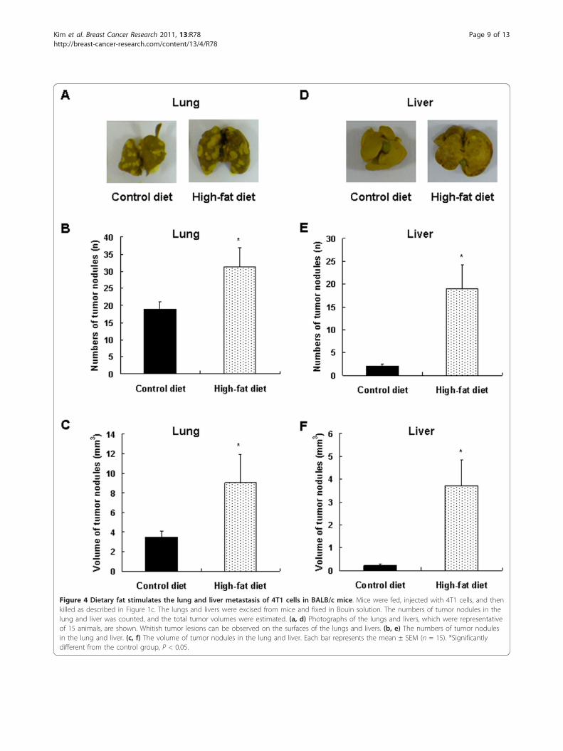
Prolonged consumption of a 60% kcal-fat diet increaseslung and liver metastasis of 4T1 cells in BALB/c miceTumor nodules grew in all mice in both groups, but inHFD-fed mice, the number and volume of tumornodules on the lung increased by 65.2% (P < 0.0298)and 159.9% (P < 0.05), respectively, as compared withthe mice fed on the CD (Figure 4a-c). In the liver, theincidence of tumor nodules was 35.7% and 66.7% in theCD group and HFD groups, respectively; and the num-ber of nodules was 844% (P < 0.0159) greater in theHFD-fed mice. Additionally, long-term consumption ofthe HFD resulted in a 1,568% (P < 0.0188) increase inthe volume of tumor nodules in the liver as comparedwith controls (Figure 4d-f).
A 60% kcal-fat diet alters the expression of proteinsinvolved in metastasis in the lungs of BALB/c miceinjected with 4T1 cellsWestern blot analysis of lung tissue lysates revealed thatHFD increased the protein levels of uPA, ICAM-1, andVCAM-1 in the lungs by 98.0% (P < 0.0089), 89.0% (P <0.008), and 63.0% (P < 0.0069), respectively, as com-pared with control mice, whereas a significant (P < 0.05)reduction of PAI-1 expression was noted in the micefed HFD (Figure 5).
A 60% kcal-fat diet increases serum levels of C5a, sICAM-1, IL-16, M-CSF, TIMP-1, leptin, and TREM-1 in BALB/cmice injected with 4T1 cellsBecause a variety of cytokines are known to affect theprocesses of breast cancer growth and metastasis(reviewed in [27]), the serum cytokines levels were esti-mated by using a mouse cytokine antibody array kit.Among the 40 cytokines measurable with the mousecytokine array kit, seven cytokines (C5a, sICAM-1, IL-16, G-CSF, M-CSF, TIMP-1, and TREM-1) weredetected in the sera of mice. The levels of C5a, sICAM-1, IL-16, M-CSF, TIMP-1, and TREM-1 were increasedin the sera of the HFD-fed mice relative to those of thecontrols, whereas the level of granulocyte colony-stimu-lating factor (G-CSF) was not altered (data not shown).We subsequently confirmed the results with ELISA andnoted changes similar to those observed in the antibodyarray. HFD increased serum levels of C5a, sICAM-1, IL-16, M-CSF, TIMP-1, and TREM-1 by 31.8% (P < 0.05),31.8% (P < 0.0002), 26.0% (P < 0.002), 61.0% (P <0.0001), 40.9% (P < 0.0032), and 41.3% (P < 0.002),
Kim et al. Breast Cancer Research 2011, 13:R78http://breast-cancer-research.com/content/13/4/R78
Page 4 of 13
Figure 1 A high-fat diet reduces the survival rate and increases solid tumor growth in BALB/c mice injected with 4T1 cells. (a) Four-week-old, female BALB/c mice were fed on a high-fat diet in which 45% of the kilocalories were provided as fats or a control diet in which 10%of kilocalories were provided as fats for 12 weeks. Twelve weeks after initiating feeding, 4T1 cells (5 × 10⁴ cells suspended in 0.1 ml gelatinousprotein (Matrigel) were injected into the inguinal mammary fat pads of the mice. The mice were fed continuously on the same diets. Survivalrates were monitored after the 4T1 cell injection (0 day). (b) Four-week-old, female BALB/c mice were fed on a high-fat diet in which 60% of thekilocalories were provided as fats or the control diet (10% kcal fat) for 16 weeks. Sixteen weeks after the initiation of feeding, 4T1 cells wereinjected, and mice were continuously fed on the same diet. (c-f) Mice were fed on the diets and injected with 4T1 cells, as described in b,except that they were killed 25 days after the 4T1 cells injection. (c) Body weights of mice were measured every week. Each point of bodyweight represents the mean ± SEM (n = 30). (d) Energy intake was calculated on the basis of 16.12 kJ/g in the control diet and 21.93 kJ/g in thehigh-fat diet. Each point of energy intake represents the mean ± SEM (n = 6). *Significantly different from the control group, P < 0.05. (e) Thetumors were excised from mice and weighed. (f) The tumor volume was measured by using calipers and calculated with the formula: 0.52 ×long diameter × short diameter2. Each bar represents the mean ± SEM (n = 30). *Significantly different from the control group, P < 0.05.
Kim et al. Breast Cancer Research 2011, 13:R78http://breast-cancer-research.com/content/13/4/R78
Page 5 of 13
respectively, relative to control mice. HFD also increasedthe serum levels of leptin by 36.0% (P < 0.0013) relativeto controls (Table 2).
Various cytokines elevated by HFD increased the viability,adhesion, and migration of 4T1 cells in vitroWe next conducted in vitro assays to determine theeffects of the cytokines that were elevated in vivo byHFD on the viability, adhesion, and migration of 4T1cells. The concentrations of cytokines used in theseassays were the mean concentrations detected in thesera of the HFD-fed mice (Table 2). Among these cyto-kines, only sICAM-1 enhanced the viability of 4T1 cells(Figure 6a). The capacity of the 4T1 cells to adhere tostrips coated with human collagen type I was increasedby C5a, sICAM-1, IL-16, M-CSF, TIMP-1, and TREM-1(Figure 6b). Additionally, TREM-1, TIMP-1, M-CSF,and sICAM-1 significantly increased the migration of4T1 cells (Figure 6c).
DiscussionThe principal objective of this study was to determinewhether dietary fat increases mammary tumor growthand metastasis as well as mammary cancer-associatedmortality in obesity-resistant BALB/c mice. BALB/cmice fed on the 60% kcal-fat diet consumed less food,such that their intake of protein, vitamins, minerals,fiber, and kilocalories was only slightly increased rela-tive to that of the control mice. Epidemiologic evi-dence indicates that the consumption of fiber,vegetable, and micronutrients is associated withreduced mortality in postmenopausal women diag-nosed with breast cancer [28]. Other previous findingsalso suggested the possible benefits of a low-fat/high-vegetable diet on disease-free survival (reviewed in[29]). The present results clearly show that increaseddietary fat intake, without decreasing other nutrients(except for carbohydrates) and with little effect onenergy intake and weight, increases mammary cancer
growth, angiogenesis, metastasis, and mortality ofBALB/c mice with implanted tumors.Xu et al. [30] demonstrated that a variety of inflam-
matory and macrophage-specific genes are upregulateddramatically in white adipose tissues of C57BL/6J micefed a 60% kcal diet for 16 weeks. Their control andhigh-fat diets were probably similar to those used in thisstudy because they were supplied by the same manufac-turer. Thus, in the current study, before the injection of4T1 mammary cancer cells, obesity-resistant BALB/cmice were fed a 60% kcal-fat diet for 16 weeks to inducechronic low-grade inflammation. We noted that thebody weights were only slightly increased in the BALB/cmice fed on the HFD. At the end of the experiment, thechange in body weight due to the long-term high-fatfeeding was measured at only 4% after correction fordifferences in tumor and spleen weights. At autopsy, wewere unable to detect visible adipose tissues except inthe gonadal fat pad, the weight of which was increasedin the HFD-fed mice. These results contrast markedlyfrom those reported by Xu et al. [30], who demon-strated that HFD markedly increased the body weightsof C57BL/6J mice. This discrepancy between the resultswas anticipated, however, because C57BL/6 mice areobesity susceptible, and BALB/c mice are obesity resis-tant [17]. Macrophage accumulation was noted in theadipose tissues of C57BL/6J mice fed on an HFD [31].In this study, we noted that F4/80+ macrophage infiltra-tion in the gonadal fat pad and the serum levels of cyto-kines (C5a, sICAM-1, IL-16, M-CSF, TIMP-1, andTREM-1) were elevated in HFD-fed mice (Table 2).These results indicate that dietary fat induces a low-grade inflammation in the absence of obesity, and asmall increase in adipose tissue may have contributed tothe induction of inflammation.Leptin is a cytokine-like protein secreted from adipose
tissue [32], and circulating leptin levels are positively asso-ciated with body weight and/or body fat [33,34]. Leptinand leptin receptor are involved in the development ofnormal mammary glands and in the progression of breastcancer [35-37]. In this study, we found that the serumlevels of leptin were elevated in HFD-fed mice without adiscernible change in body weight, indicating that a smallincrease in fat mass increases serum leptin levels, and theelevated leptin may have contributed to the stimulation ofmammary cancer progression in these mice.The increased expression of CD31 and VEGF in
tumor tissues (Figure 3a) indicates that tumor angiogen-esis increased in the HFD-fed mice. Recent evidenceindicates that in addition to the interactions of cancercells and endothelial cells, inflammatory cells also playan important role in the formation of the blood vesselsthat nourish a growing tumor (reviewed in [38]).Tumor-associated immune cells, including macrophages,
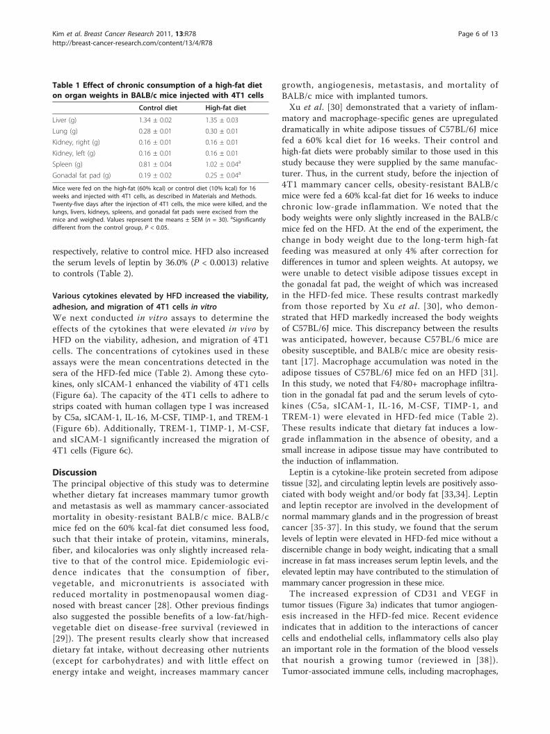
Table 1 Effect of chronic consumption of a high-fat dieton organ weights in BALB/c mice injected with 4T1 cells
Control diet High-fat diet
Liver (g) 1.34 ± 0.02 1.35 ± 0.03
Lung (g) 0.28 ± 0.01 0.30 ± 0.01
Kidney, right (g) 0.16 ± 0.01 0.16 ± 0.01
Kidney, left (g) 0.16 ± 0.01 0.16 ± 0.01
Spleen (g) 0.81 ± 0.04 1.02 ± 0.04a
Gonadal fat pad (g) 0.19 ± 0.02 0.25 ± 0.04a
Mice were fed on the high-fat (60% kcal) or control diet (10% kcal) for 16weeks and injected with 4T1 cells, as described in Materials and Methods.Twenty-five days after the injection of 4T1 cells, the mice were killed, and thelungs, livers, kidneys, spleens, and gonadal fat pads were excised from themice and weighed. Values represent the means ± SEM (n = 30). aSignificantlydifferent from the control group, P < 0.05.
Kim et al. Breast Cancer Research 2011, 13:R78http://breast-cancer-research.com/content/13/4/R78
Page 6 of 13
granulocytes, and mast cells, have been shown to stimu-late tumor angiogenesis (reviewed in [39]). Additionally,a significant positive correlation between the degree ofangiogenesis and the number of CD68+ cells has beendemonstrated in human breast carcinoma [40]. Thenumber of CD45+ and CD68+ cells was markedly
increased in the tumor tissues of HFD-fed mice (Figure3a), indicating increased infiltration of these immunecells into tumor tissues, which possibly increased tumorangiogenesis.In this study, we noted that blood levels of various
cytokines were increased in the HFD-fed mice (Table 2).
Figure 2 Dietary fat stimulates cell-cycle progression in 4T1 tumors in BALB/c mice. Mice were fed, injected with 4T1 cells, and then killedas described in Figure 1c. (a) Tumor sections were stained with antibody raised against Ki67, cyclin-dependent kinase (CDK)2, CDK4, cyclin D1, orcyclin A, and then with 1,3 -diaminobenzidine (DAB) and counterstained with hematoxylin (n = 15). Representative images of theimmunohistochemical analysis are shown. (b) The Ki67-, CDK2-, CDK4-, cyclin D1-, and cyclin A-positive cells were counted. Each bar representsthe means ± SEM (n = 15). (c) Tumor lysates were analyzed with Western blotting with the indicated antibodies. Photographs ofchemiluminescent detection of the blots are shown. The relative abundance of each band to its own b-actin was quantified, and the controllevels were at 100%. The adjusted mean ± SEM is shown above each blot. *Significantly different from the control group, P < 0.05. PCNA,proliferating cell nuclear antigen.
Kim et al. Breast Cancer Research 2011, 13:R78http://breast-cancer-research.com/content/13/4/R78
Page 7 of 13
Figure 3 Dietary fat stimulates angiogenesis and immune cell infiltration in 4T1 tumors and macrophage infiltration in the gonadalfat pad in BALB/c mice. Mice were fed, injected with 4T1 cells, and then killed as described in Figure 1c. (a) Tumor sections were stained withantibody raised against CD31, vascular endothelial growth factor (VEGF), CD45, or CD68, and then with 1,3 -diaminobenzidine (DAB), andcounterstained with hematoxylin, except that the last one was stained with hematoxylin and eosin (H&E) to visualize lipid vacuoles.Representative images of the immuno-histochemical analysis and H&E staining are shown, n = 15. (b) The staining intensity of CD31 and VEGFwas quantified. CD45+ and CD68+ cells and the number of lipid vacuoles were counted. Each bar represents the mean ± SEM (n = 15). (c)Gonadal fat-pad sections were stained with an F4/80 antibody, and F4/80+ cells were counted, n = 15. Each bar represents the mean ± SEM (n= 15).*Significantly different from the control group, P < 0.05.
Kim et al. Breast Cancer Research 2011, 13:R78http://breast-cancer-research.com/content/13/4/R78
Page 8 of 13
Figure 4 Dietary fat stimulates the lung and liver metastasis of 4T1 cells in BALB/c mice. Mice were fed, injected with 4T1 cells, and thenkilled as described in Figure 1c. The lungs and livers were excised from mice and fixed in Bouin solution. The numbers of tumor nodules in thelung and liver was counted, and the total tumor volumes were estimated. (a, d) Photographs of the lungs and livers, which were representativeof 15 animals, are shown. Whitish tumor lesions can be observed on the surfaces of the lungs and livers. (b, e) The numbers of tumor nodulesin the lung and liver. (c, f) The volume of tumor nodules in the lung and liver. Each bar represents the mean ± SEM (n = 15). *Significantlydifferent from the control group, P < 0.05.
Kim et al. Breast Cancer Research 2011, 13:R78http://breast-cancer-research.com/content/13/4/R78
Page 9 of 13
Our in vitro studies demonstrated that some of thesecytokines stimulated the growth, migration, and adhe-sion of 4T1 mammary cancer cells (Figure 6). Theseresults indicate that the increased cytokine levels mayhave contributed to changes in the expression of metas-tasis-related proteins (uPA, ICAM-1, and VCAM-1),thereby increasing metastasis. Future studies are neededto determine which types of cells secrete these cytokinesin tumors, adipose tissues, the lungs, and/or the liver.It was previously reported that adipocytes promote
breast carcinoma cell growth in collagen gels by thecancer-stromal interaction [41]. Additionally, when co-injected with SUM159PT mammary adenocarcinomacells in athymic nude mice, fully differentiated 3T3-L1adipocytes stimulate tumor growth and lung metastasis[42]. We observed increased numbers of lipid vacuolesin the tumor tissues of HFD-fed mice (Figure 3a). Alongwith the tumor cells, these adipocytes may participate in
the recruitment of immune cells into the tumor. Thecrosstalk between tumor cells, adipocytes, and immunecells within the tumors of the HFD-fed mice (Figure 3a)may have produced a broad variety of growth factors,cytokines, and chemokines, resulting in changes in theexpression of proteins involved in the stimulation ofcell-cycle progression of tumor cells (Ki67, PCNA,CDK2, CDK4, cyclin A, and cyclin D) and angiogenesis(VEGF) (Figure 2a-c, 3a). Additionally, the increase innew blood vessels may have supplied nutrients andgrowth factors for the stimulation of cell-cycle progres-sion in addition to the stimulation of metastasis. In thisstudy, the direct mechanism by which HFD feedinginduces the expression of metastasis-regulating proteins,cyclins, and CDKs in the tumor tissues was not fullyelucidated.In this study, we noted that the weights of spleen were
increased in the HFD-fed mice (Table 1), which wasaccompanied by increases in the serum levels of C5a,IL-16, sICAM-1, M-CSF, TIMP-1, TREM-1, and leptin(Table 2). Splenomegaly was invariably observed in 4T1-tumor-bearing mice [43-45], and correlated stronglywith increased extramedullary hematogenesis andmetastasis in the spleen and circulating levels of neutro-phils and leukocytes [46]. It has been reported that 4T1tumor growth is associated with increased splenomegalyor splenomegaly-associated inflammation, and varioustumor-derived cytokines, such as G-CSF, GM-CSF, andIFN-g, may be responsible for the splenomegaly in miceinjected with 4T1 cells [44]. Future studies are neededto determine whether the increases in C5a, IL-16,sICAM-1, M-CSF, TIMP-1, TREM-1, and/or leptin areresponsible for the increased splenic mass in HFD-fedmice.
Figure 5 Dietary fat alters the protein levels of urokinase-typeplasminogen activator (uPA), plasminogen activator inhibitor(PAI)-1, intercellular adhesion molecule (ICAM)-1, and vascularcell adhesion molecule (VCAM)-1 in the lungs of BALB/c miceinjected with 4T1 cells. Mice were fed, injected with 4T1 cells, andthen killed as described in Figure 1c. Lung lysates were analyzedwith Western blotting with the indicated antibodies. Photographs ofchemiluminescent detection of the blots are shown. The relativeabundance of each band to its own b-actin was quantified, and thecontrol levels were set at 100%. The adjusted mean ± SEM is shownabove each blot. *Significantly different from the control group, P <0.05. MMP, matrix metalloproteinase; TIMP, tissue inhibitor of matrixmetalloproteinase.
Table 2 Effect of prolonged consumption of a high-fatdiet on the levels of various cytokines in the sera ofBALB/c mice injected with 4T1 cells
Control diet High-fat diet
C5a (ng/ml) 22.0 ± 2.7 29.0 ± 3.1a
sICAM-1 (ng/ml) 494.2 ± 22.6 651.5 ± 29.4a
IL-16 (ng/ml) 150.8 ± 8.0 190.0 ± 8.4a
M-CSF (pg/ml) 714.1 ± 29.3 1,149.5 ± 35.6a
TIMP-1 (ng/ml) 4.4 ± 0.4 6.2 ± 0.4a
TREM-1 (pg/ml) 684.7 ± 17.8 967.8 ± 21.6a
Leptin (ng/ml) 138.0 ± 0.04 187.8 ± 11.3a
Mice were fed on the high-fat (60% kcal) or control diet (10% kcal) for 16weeks and injected with 4T1 cells, as described in Materials and Methods. At25 days after the 4T1 cells injection, blood samples were collected from themice, and the sera were prepared. The serum levels of complement fragment5a (C5a), soluble intercellular adhesion molecule (sICAM)-1, interleukin (IL)-16,macrophage colony-stimulating factor (M-CSF), tissue inhibitor of matrixmetalloproteinase (TIMP)-1, triggering receptor expressed on myeloid cells(TREM)-1, and leptin were measured with the appropriate ELISA kits. Valuesrepresent the mean ± SEM (n = 30). aSignificantly different from the controlgroup, P < 0.05.
Kim et al. Breast Cancer Research 2011, 13:R78http://breast-cancer-research.com/content/13/4/R78
Page 10 of 13
Figure 6 Effect of various cytokines on proliferation (a), adhesion (b), and migration (c) of 4T1 cells in vitro. (a) 4T1 cells were plated in24-well plates at 5 × 10⁴ cells/well in DMEM supplemented with 100 ml/L FBS. One day later, the monolayers were serum deprived with DMEMsupplemented with 10 ml/L charcoal-stripped FBS (serum-deprivation medium) for 24 h. After serum deprivation, the cells were incubated inserum-deprivation medium in the absence or presence of 29 ng/ml complement fragment 5a (C5a), 652 ng/ml soluble intercellular adhesionmolecule (sICAM)-1, 190 ng/ml interleukin (IL)-16, 1,150 pg/ml macrophage colony-stimulating factor (M-CSF), 6.2 ng/ml tissue inhibitor of matrixmetalloproteinase (TIMP)-1, and 970 pg/ml triggering receptor expressed on myeloid cells (TREM)-1 for 24 hours. Viable cell numbers wereestimated. Each bar represents the mean ± SEM (n = 3). Means without a common letter differ, P < 0.05. (b) Cells were plated in humancollagen type I-coated CytoMatrix Cell Adhesion Strips and incubated with or without various cytokines (the identical concentrations of cytokinesused in Figure 6a) in DMEM containing 10 ml/L charcoal-stripped FBS at 37°C for 45 min. The cells were stained with crystal violet, and the cell-bound stains were quantified by determining the absorbance at 570 nm. Each bar represents the mean ± SEM (n = 7). Means without acommon letter differ, P < 0.05. (c) Cells were serum deprived in DMEM supplemented with 10 ml/L charcoal-stripped FBS for 24 hours. Serum-deprived cells were plated onto the filter in 6.5-mm transwell inserts in 24-well plates at 5 × 10⁴ cells/filter. Before the plating of the cells, thelower side of the transwell filter was precoated with type IV collagen. The lower chamber of the well was filled with DMEM containing 100 ml/Lgelatinase-free charcoal-stripped FBS with or without various cytokines (the identical concentrations of cytokines used in Figure 6a). The cellswere incubated for 16 hours. Migrated cells were stained with hematoxylin and eosin. Each bar represents the mean ± SEM (n = 3). Meanswithout a common letter differ, P < 0.05.
Kim et al. Breast Cancer Research 2011, 13:R78http://breast-cancer-research.com/content/13/4/R78
Page 11 of 13
ConclusionsThis study clearly demonstrated that dietary fatincreases mammary cancer growth, metastasis, andmammary cancer-associated mortality in obesity-resis-tant mice. We also demonstrated that dietary fatincreases the expression of proteins (Ki67, CDKs, andcyclins) involved in the regulation of cell-cycle progres-sion as well as increases in the expression of CD31,VEGF, CD68, and CD45 in tumor tissues, thereby indi-cating that increases in immune cell infiltration andangiogenesis stimulate cell-cycle progression and themetastasis of tumor cells. Additionally, increases in pro-tease (uPA) and adhesion molecules (ICAM-1, VACM)may contribute to lung metastasis in HFD-fed mice.Furthermore, increases in the serum levels of cytokinesmay stimulate mammary cancer metastasis in HFD-fedmice. Our results suggest that dietary fat may increasebreast cancer progression, even in individuals whomaintain a healthy body weight, and that replacing diet-ary fat with carbohydrates may delay the progression ofbreast cancer, thereby reducing breast cancer-associatedmortality.
AcknowledgementsThis study was supported by the Mid-career ResearcherProgram (grant number 2010-0006923) and the SRCprogram (Center for Food & Nutritional Genomics:grant number 2010-0001886) of the National ResearchFoundation (NRF) of Korea, funded by the Ministry ofEducation, Science, and Technology.
Author details1Department of Food Science and Nutrition, College of Natural Sciences,Hallym University, 39 Hallymdaehak-gil, Chuncheon, 200-702, Korea. 2Centerfor Efficacy Assessment and Development of Functional Foods and Drugs,Hallym University, 39 Hallymdaehak-gil, Chuncheon, 200-702, Korea.3Department of Biochemistry, College of Medicine, Hallym University, 39Hallymdaehak-gil, Chuncheon, 200-702, Korea. 4Korea Food ResearchInstitute, 516 Baekhyun-dong, Bundang-gu, Sungnam, 463-746, Korea.5Department of Agricultural Biotechnology and Center for Agricultural
Biomaterials, Seoul National University, 1 Gwanak-ro, Gwanak-gu, Seoul, 151-921, Korea.
Authors’ contributionsMRC, HP, MK, and JEH carried out the majority of animal studies, includingevaluation of food consumption, tumor volumes, mortality, Western blotting,immunohistochemistry, ELISA, and metastasis. JYL, HSC, and KWL wereresponsible for conception of the project, oversight of experiments, andtraining of certain participants. EJK and JHYP were responsible forconception of the project, oversight of experiments, and training of certainparticipants, and they drafted the manuscript. All authors read and approvedthe final manuscript.
Competing interestsThe authors declare that they have no competing interests.
Received: 17 January 2011 Revised: 19 June 2011Accepted: 11 August 2011 Published: 11 August 2011
References1. Jemal A, Siegel R, Xu J, Ward E: Cancer statistics, 2010. CA Cancer J Clin
2010, 60:277-300.2. Helman S, Harris JR: Diseases of Breast Philadelphia: Lippincott Williams &
Wilkins; 2000.3. Ali SM, Harvey HA, Lipton A: Metastatic breast cancer: overview of
treatment. Clin Orthop Relat Res 2003, 415(Suppl):S132-S137.4. Thiebaut AC, Kipnis V, Chang SC, Subar AF, Thompson FE, Rosenberg PS,
Hollenbeck AR, Leitzmann M, Schatzkin A: Dietary fat and postmenopausalinvasive breast cancer in the National Institutes of Health-AARP Diet andHealth Study cohort. J Natl Cancer Inst 2007, 99:451-462.
5. Buettner R, Scholmerich J, Bollheimer LC: High-fat diets: modeling themetabolic disorders of human obesity in rodents. Obesity (Silver Spring,Md) 2007, 15:798-808.
6. Lin S, Thomas TC, Storlien LH, Huang XF: Development of high fat diet-induced obesity and leptin resistance in C57Bl/6J mice. Int J Obes RelatMetab Disord 2000, 24:639-646.
7. Carmichael AR: Obesity and prognosis of breast cancer. Obes Rev 2006,7:333-340.
8. Li Z, Bowerman S, Heber D: Health ramifications of the obesity epidemic.Surg Clin North Am 2005, 85:681-701.
9. Whiteman MK, Hillis SD, Curtis KM, McDonald JA, Wingo PA,Marchbanks PA: Body mass and mortality after breast cancer diagnosis.Cancer Epidemiol Biomarkers Prev 2005, 14:2009-2014.
10. Dawood S, Broglio K, Gonzalez-Angulo AM, Kau SW, Islam R,Hortobagyi GN, Cristofanilli M: Prognostic value of body mass index inlocally advanced breast cancer. Clin Cancer Res 2008, 14:1718-1725.
11. Rose DP, Connolly JM, Meschter CL: Effect of dietary fat on human breastcancer growth and lung metastasis in nude mice. J Natl Cancer Inst 1991,83:1491-1495.
12. Ford CE, Ekstrom EJ, Andersson T: Wnt-5a signaling restores tamoxifensensitivity in estrogen receptor-negative breast cancer cells. Proc NatlAcad Sci USA 2009, 106:3919-3924.
13. Banka CL, Lund CV, Nguyen MT, Pakchoian AJ, Mueller BM, Eliceiri BP:Estrogen induces lung metastasis through a host compartment-specificresponse. Cancer Res 2006, 66:3667-3672.
14. Pulaski BA, Ostrand-Rosenberg S: Reduction of established spontaneousmammary carcinoma metastases following immunotherapy with majorhistocompatibility complex class II and B7.1 cell-based tumor vaccines.Cancer Res 1998, 58:1486-1493.
15. Aslakson CJ, Miller FR: Selective events in the metastatic process definedby analysis of the sequential dissemination of subpopulations of amouse mammary tumor. Cancer Res 1992, 52:1399-1405.
16. Heppner GH, Miller FR, Shekhar PM: Nontransgenic models of breastcancer. Breast Cancer Res 2000, 2:331-334.
17. Olson LK, Tan Y, Zhao Y, Aupperlee MD, Haslam SZ: Pubertal exposure tohigh fat diet causes mouse strain-dependent alterations in mammarygland development and estrogen responsiveness. Int J Obesity 2010,34:1415-1426.
18. Toth LA: Defining the moribund condition as an experimental endpointfor animal research. ILAR J 2000, 41:72-79.
Kim et al. Breast Cancer Research 2011, 13:R78http://breast-cancer-research.com/content/13/4/R78
19. Sauter BV, Martinet O, Zhang WJ, Mandeli J, Woo SL: Adenovirus-mediatedgene transfer of endostatin in vivo results in high level of transgeneexpression and inhibition of tumor growth and metastases. Proc NatlAcad Sci USA 2000, 97:4802-4807.
20. Kim EJ, Shin M, Park H, Hong JE, Shin HK, Kim J, Kwon DY, Park JH: Oraladministration of 3,3’-diindolylmethane inhibits lung metastasis of 4T1murine mammary carcinoma cells in BALB/c mice. J Nutr 2009,139:2373-2379.
21. Welch DR, Neri A, Nicolson GL: Comparison of ‘spontaneous’ and‘experimental’ metastasis using rat 13762 mammary adenocarcinomametastatic cell clones. Invasion Metastasis 1983, 3:65-80.
22. Rose DP, Connolly JM: Influence of dietary fat intake on local recurrenceand progression of metastases arising from MDA-MB-435 human breastcancer cells in nude mice after excision of the primary tumor. NutrCancer 1992, 18:113-122.
23. Kim EJ, Hong JE, Eom SJ, Lee JY, Park JH: Oral administration of benzyl-isothiocyanate inhibits solid tumor growth and lung metastasis of 4T1murine mammary carcinoma cells in BALB/c mice. Breast Cancer Res Treat2011.
24. Cho HJ, Kim WK, Kim EJ, Jung KC, Park S, Lee HS, Tyner AL, Park JH:Conjugated linoleic acid inhibits cell proliferation and ErbB3 signaling inHT-29 human colon cell line. Am J Physiol Gastrointest Liver Physiol 2003,284:G996-G1005.
25. Denizot F, Lang R: Rapid colorimetric assay for cell growth and survival:modifications of the tetrazolium dye procedure giving improvedsensitivity and reliability. J Immunol Methods 1986, 89:271-277.
26. Kwon GT, Cho HJ, Chung WY, Park KK, Moon A, Park JH: Isoliquiritigenininhibits migration and invasion of prostate cancer cells: possiblemediation by decreased JNK/AP-1 signaling. J Nutr Biochem 2009,20:663-676.
27. Karnoub AE, Weinberg RA: Chemokine networks and breast cancermetastasis. Breast Dis 2006, 26:75-85.
28. McEligot AJ, Largent J, Ziogas A, Peel D, Anton-Culver H: Dietary fat, fiber,vegetable, and micronutrients are associated with overall survival inpostmenopausal women diagnosed with breast cancer. Nutr Cancer 2006,55:132-140.
29. Thomson CA, Thompson PA: Dietary patterns, risk and prognosis ofbreast cancer. Future Oncol 2009, 5:1257-1269.
30. Xu H, Barnes GT, Yang Q, Tan G, Yang D, Chou CJ, Sole J, Nichols A,Ross JS, Tartaglia LA, Chen H: Chronic inflammation in fat plays a crucialrole in the development of obesity-related insulin resistance. J Clin Invest2003, 112:1821-1830.
31. Weisberg SP, McCann D, Desai M, Rosenbaum M, Leibel RL, Ferrante AW Jr:Obesity is associated with macrophage accumulation in adipose tissue. JClin Invest 2003, 112:1796-1808.
32. Zhang Y, Proenca R, Maffei M, Barone M, Leopold L, Friedman JM:Positional cloning of the mouse obese gene and its human homologue.Nature 1994, 372:425-432.
33. McGregor GP, Desaga JF, Ehlenz K, Fischer A, Heese F, Hegele A, Lammer C,Peiser C, Lang RE: Radiommunological measurement of leptin in plasmaof obese and diabetic human subjects. Endocrinology 1996,137:1501-1504.
34. Sinha MK, Opentanova I, Ohannesian JP, Kolaczynski JW, Heiman ML, Hale J,Becker GW, Bowsher RR, Stephens TW, Caro JF: Evidence of free andbound leptin in human circulation. Studies in lean and obese subjectsand during short-term fasting. J Clin Invest 1996, 98:1277-1282.
35. Garofalo C, Koda M, Cascio S, Sulkowska M, Kanczuga-Koda L,Golaszewska J, Russo A, Sulkowski S, Surmacz E: Increased expression ofleptin and the leptin receptor as a marker of breast cancer progression:possible role of obesity-related stimuli. Clin Cancer Res 2006,12:1447-1453.
36. Hu X, Juneja SC, Maihle NJ, Cleary MP: Leptin: a growth factor in normaland malignant breast cells and for normal mammary glanddevelopment. J Natl Cancer Inst 2002, 94:1704-1711.
37. Cleary MP, Phillips FC, Getzin SC, Jacobson TL, Jacobson MK,Christensen TA, Juneja SC, Grande JP, Maihle NJ: Genetically obese MMTV-TGF-alpha/Lep(ob)Lep(ob) female mice do not develop mammarytumors. Breast Cancer Res Treat 2003, 77:205-215.
38. Albini A, Sporn MB: The tumour microenvironment as a target forchemoprevention. Nat Rev Cancer 2007, 7:139-147.
39. Albini A, Tosetti F, Benelli R, Noonan DM: Tumor inflammatoryangiogenesis and its chemoprevention. Cancer Res 2005, 65:10637-10641.
40. Lewis CE, Leek R, Harris A, McGee JO: Cytokine regulation of angiogenesisin breast cancer: the role of tumor-associated macrophages. J Leukoc Biol1995, 57:747-751.
41. Manabe Y, Toda S, Miyazaki K, Sugihara H: Mature adipocytes, but notpreadipocytes, promote the growth of breast carcinoma cells incollagen gel matrix culture through cancer-stromal cell interactions. JPathol 2003, 201:221-228.
42. Iyengar P, Combs TP, Shah SJ, Gouon-Evans V, Pollard JW, Albanese C,Flanagan L, Tenniswood MP, Guha C, Lisanti M, Pestell RG, Scherer PE:Adipocyte-secreted factors synergistically promote mammarytumorigenesis through induction of anti-apoptotic transcriptionalprograms and proto-oncogene stabilization. Oncogene 2003,22:6408-6423.
43. Walsh C, Tanjoni I, Uryu S, Tomar A, Nam JO, Luo H, Phillips A, Patel N,Kwok C, McMahon G, Stupack DG, Schlaepfer DD: Oral delivery of PND-1186 FAK inhibitor decreases tumor growth and spontaneous breast tolung metastasis in pre-clinical models. Cancer Biol Ther 2010, 9:778-790.
44. DuPre SA, Hunter KW: Murine mammary carcinoma 4T1 induces aleukemoid reaction with splenomegaly: association with tumor-derivedgrowth factors. Exp Mol Pathol 2007, 82:12-24.
45. Younos I, Donkor M, Hoke T, Dafferner A, Samson H, Westphal S,Talmadge J: Tumor- and organ-dependent infiltration by myeloid-derived suppressor cells. Int Immunopharmacol 2011, 11:816-826.
46. Tao K, Fang M, Alroy J, Sahagian GG: Imagable 4T1 model for the study oflate stage breast cancer. BMC Cancer 2008, 8:228.
doi:10.1186/bcr2927Cite this article as: Kim et al.: Dietary fat increases solid tumor growthand metastasis of 4T1 murine mammary carcinoma cells and mortality inobesity-resistant BALB/c mice. Breast Cancer Research 2011 13:R78.
Submit your next manuscript to BioMed Centraland take full advantage of:
• Convenient online submission
• Thorough peer review
• No space constraints or color figure charges
• Immediate publication on acceptance
• Inclusion in PubMed, CAS, Scopus and Google Scholar
• Research which is freely available for redistribution
Submit your manuscript at www.biomedcentral.com/submit
Kim et al. Breast Cancer Research 2011, 13:R78http://breast-cancer-research.com/content/13/4/R78