Di¡erent expression patterns for ubiquitous calpains and Capn3 splice variants in monkey ocular tissues Takeshi Nakajima a , Chiho Fukiage a;b , Mitsuyoshi Azuma a;b; *, Hong Ma b , Thomas R. Shearer b a Research Laboratory, Senju Pharmaceutical Co., Ltd., 1-5-4 Murotani, Nishiku, Kobe, 651-2241, Japan b Department of Oral Molecular Biology and Ophthalmology, Oregon Health Sciences University, Portland, OR 97201, USA Received 1 December 2000; received in revised form 8 March 2001; accepted 13 March 2001 Abstract The purpose of the present investigation was to compare the expression of ubiquitous and tissue-specific calpains in ocular tissues from the Macaca fascicularis monkey. Calpain isoforms in retina and corneal epithelium from adult M. fascicularis monkeys were characterized by RT-PCR, cDNA cloning and sequencing. Calpain isoform activities in ocular tissues were investigated by fractionation on DEAE- HPLC, immunoblotting, and casein zymography. Capn3 splice variants in the ocular tissues from rat, rabbit and monkey were compared after RT-PCR. RT-PCR analysis revealed that numerous splice variants of Capn3 were expressed in the epithelium from monkey cornea. The variants contained deletions or insertions in or around the IS1, IS2, and NS regions. The cDNAs for Capn3 variants were highly conserved, yet the expression patterns of the Capn3 isoforms were widely different among the mammalian species. In contrast, the expression patterns of ubiquitous calpains in ocular tissues were conserved among the mammalian species, and similarities between monkey and human cDNAs for Capn1 (W-calpain) and Capn2 (m-calpain) were 98 and 99%, respectively. These results suggested that differences in expression patterns of Capn3 variants might be related to the function of each variant in a particular tissue or species. ß 2001 Elsevier Science B.V. All rights reserved. Keywords : Proteinase; Calpain; Rat; Monkey; Calpain 3 isoform 1. Introduction Calpains (EC 3.4.22.17) are non-lysosomal, cysteine proteases with a neutral pH optimum and an absolute requirement of calcium for activation [1]. The superfamily of calpains includes the ubiquitous Capn1 and Capn2, and the tissue-speci¢c calpains Capn3/p94/calpain3 (muscle) [2], CLS4/nCL2 (stomach) [3], Capn9/nCL4 (digestive tract) [4], Lp82 and Lp85 (Capn3 splice variants ;lens) [5] and Rt88 (Capn3 splice variant; retina) [6]. Capn1 re- quires WM levels of calcium for in vitro activity, while Capn2 requires mM levels. Tissue-speci¢c Capn3 in solu- ble cell extracts is autolyzed in the presence of submicro- molar calcium levels, suggesting a requirement of calcium for activation [7]. In addition to calcium, calpain enzyme activity is regulated by an endogenous inhibitory protein, calpastatin, and by phospholipids [8]. Much evidence has suggested that the calpains may contribute both to phys- iological and pathological conditions [9]. For example, calpain may be involved in the pathogenesis of Alzheim- er’s disease [10]. Muscle-speci¢c calpain Capn3 is expressed almost exclu- sively in skeletal muscle, at least in the adult [11,12]. Capn3 has three unique insert regions: a novel sequence (NS) at the N-terminus, insert region No. 1 (IS1) in the protease domain, and insert region No. 2 (IS2) between domains III and IV. IS1 is the site of autolysis, while IS2 speci¢cally binds Capn3 to connectin (titin) in muscle [8]. Moreover, mutations in the Capn3 gene are responsible for limb girdle muscular dystrophy type 2A (LGMD2A) in man [13], possibly by disruption of a signaling pathway involving Capn3. At least 12 splice variants of Capn3 were recently de- scribed in rodents [15]. The best characterized variant at the protein level is lens-speci¢c calpain Lp82 [16^18]. Lp82 showed substitution of a new N-terminus termed AX1 by use of alternative e xon 1, and deletion of the IS1 and IS2 regions [5]. Retina-speci¢c variant Rt88 also showed sub- 0167-4781 / 01 / $ ^ see front matter ß 2001 Elsevier Science B.V. All rights reserved. PII:S0167-4781(01)00212-3 * Corresponding author, at address a. Fax: +81-78-997-1016; E-mail : [email protected]Biochimica et Biophysica Acta 1519 (2001) 55^64 www.bba-direct.com
Transcript
Di¡erent expression patterns for ubiquitous calpains and Capn3 splicevariants in monkey ocular tissues
Takeshi Nakajima a, Chiho Fukiage a;b, Mitsuyoshi Azuma a;b;*, Hong Ma b,Thomas R. Shearer b
a Research Laboratory, Senju Pharmaceutical Co., Ltd., 1-5-4 Murotani, Nishiku, Kobe, 651-2241, Japanb Department of Oral Molecular Biology and Ophthalmology, Oregon Health Sciences University, Portland, OR 97201, USA
Received 1 December 2000; received in revised form 8 March 2001; accepted 13 March 2001
Abstract
The purpose of the present investigation was to compare the expression of ubiquitous and tissue-specific calpains in ocular tissues fromthe Macaca fascicularis monkey. Calpain isoforms in retina and corneal epithelium from adult M. fascicularis monkeys were characterizedby RT-PCR, cDNA cloning and sequencing. Calpain isoform activities in ocular tissues were investigated by fractionation on DEAE-HPLC, immunoblotting, and casein zymography. Capn3 splice variants in the ocular tissues from rat, rabbit and monkey were comparedafter RT-PCR. RT-PCR analysis revealed that numerous splice variants of Capn3 were expressed in the epithelium from monkey cornea.The variants contained deletions or insertions in or around the IS1, IS2, and NS regions. The cDNAs for Capn3 variants were highlyconserved, yet the expression patterns of the Capn3 isoforms were widely different among the mammalian species. In contrast, theexpression patterns of ubiquitous calpains in ocular tissues were conserved among the mammalian species, and similarities between monkeyand human cDNAs for Capn1 (W-calpain) and Capn2 (m-calpain) were 98 and 99%, respectively. These results suggested that differences inexpression patterns of Capn3 variants might be related to the function of each variant in a particular tissue or species. ß 2001 ElsevierScience B.V. All rights reserved.
Calpains (EC 3.4.22.17) are non-lysosomal, cysteineproteases with a neutral pH optimum and an absoluterequirement of calcium for activation [1]. The superfamilyof calpains includes the ubiquitous Capn1 and Capn2, andthe tissue-speci¢c calpains Capn3/p94/calpain3 (muscle)[2], CLS4/nCL2 (stomach) [3], Capn9/nCL4 (digestivetract) [4], Lp82 and Lp85 (Capn3 splice variants ;lens) [5]and Rt88 (Capn3 splice variant; retina) [6]. Capn1 re-quires WM levels of calcium for in vitro activity, whileCapn2 requires mM levels. Tissue-speci¢c Capn3 in solu-ble cell extracts is autolyzed in the presence of submicro-molar calcium levels, suggesting a requirement of calciumfor activation [7]. In addition to calcium, calpain enzymeactivity is regulated by an endogenous inhibitory protein,calpastatin, and by phospholipids [8]. Much evidence has
suggested that the calpains may contribute both to phys-iological and pathological conditions [9]. For example,calpain may be involved in the pathogenesis of Alzheim-er's disease [10].
Muscle-speci¢c calpain Capn3 is expressed almost exclu-sively in skeletal muscle, at least in the adult [11,12].Capn3 has three unique insert regions: a novel sequence(NS) at the N-terminus, insert region No. 1 (IS1) in theprotease domain, and insert region No. 2 (IS2) betweendomains III and IV. IS1 is the site of autolysis, while IS2speci¢cally binds Capn3 to connectin (titin) in muscle [8].Moreover, mutations in the Capn3 gene are responsiblefor limb girdle muscular dystrophy type 2A (LGMD2A)in man [13], possibly by disruption of a signaling pathwayinvolving Capn3.
At least 12 splice variants of Capn3 were recently de-scribed in rodents [15]. The best characterized variant atthe protein level is lens-speci¢c calpain Lp82 [16^18]. Lp82showed substitution of a new N-terminus termed AX1 byuse of alternative exon 1, and deletion of the IS1 and IS2regions [5]. Retina-speci¢c variant Rt88 also showed sub-
0167-4781 / 01 / $ ^ see front matter ß 2001 Elsevier Science B.V. All rights reserved.PII: S 0 1 6 7 - 4 7 8 1 ( 0 1 ) 0 0 2 1 2 - 3
* Corresponding author, at address a. Fax: +81-78-997-1016;E-mail : [email protected]
BBAEXP 93526 29-5-01
Biochimica et Biophysica Acta 1519 (2001) 55^64
www.bba-direct.com
stitution of the AX1 N-terminus, but only the IS2 regionwas deleted [6]. Interestingly, the level of Rt88 mRNAincreased during retinal maturation, while levels of Lp82mRNA, protein and caseinolytic activity were decreasedduring lens maturation [16].
The types and amounts of Capn3 variants in a partic-ular tissue are thought to be related to speci¢c functions inthat tissue. However, the expression patterns of Capn3splice variants are complex. Patterns are in£uenced bytype of tissue, stage of tissue development, and species[14,15]. Expression patterns have been well delineated inrodents [15], but only limited data exist for human tissues[19]. In monkey, a useful animal subject for research stud-ies relevant to human ophthalmic pathology, e.g., glauco-ma [20], no cDNA cloning of calpain genes has been per-formed, and the reported data are only protein analysisfor the ubiquitous calpains in the brain, skeletal muscleand cardiac muscle [21^23]. Thus, the purposes of thepresent study were to: (1) provide cDNA sequences andexpression patterns for calpain isoforms in various oculartissues from monkey, and (2) compare these data to sim-ilar data from lower mammalian species.
2. Materials and methods
2.1. Human and animal tissues
Human eyes were obtained from Lions Eye Bank (Port-land). The research followed the tenets of the Declarationof Helsinki, and informed consent was obtained for alldonors. Animal eye tissues were obtained from rats(Sprague-Dawley), rabbits (Japanese white) and monkeys(Macaca fascicularis) of several ages. Experimental ani-mals were handled in accordance with the ARVO State-ment for the Use of Animals in Ophthalmic and VisionResearch and the Guiding Principles in the Care and Useof Animals (DHEW Publication, NIH 80-23).
2.2. Cloning of Capn1 and Capn2 from monkey retina
cDNAs were synthesized from monkey retina totalRNA isolated with the TRIzol Reagent (Life Technolo-gies) using SuperScript II reverse transcriptase (Life Tech-nologies). To obtain cDNAs for both known and novelcalpains, cDNAs were ampli¢ed using Platinum Taq DNApolymerase (Life Technologies) using degenerate senseprimer 5P-GC(ATGC) CT(ATGC) GG(ATGC) GA(CT)TG(CT) TGG CT-3P and antisense primer 5P-TC(ATGC)AC(CT) TG(ATGC) CCC CA(ATGC) GG(AG) TT-3Pwith the following PCR conditions: 94³C for 2 min, 40cycles of 94³C for 1 min, 50³C for 30 s, 72³C for 1.5 min,and 72³C for 7 min. The resulting 575 bp fragments weresubcloned with pCR II vector (Invitrogen) and sequenced.Since the sequences of retina cDNA fragments were sim-ilar to those from human Capn1 and Capn2, RT-PCR and
3P-RACE were performed to obtain the 5P- and 3P-ends ofthe cDNAs for monkey Capn1 and Capn2. These sequen-ces were used to generate primers for RT-PCR, and toperform subcloning and sequencing of full length cDNAsfor Capn1 and Capn2.
2.3. Cloning of Capn3 splice variants from monkey cornealepithelium
To obtain the cDNA for monkey Capn3 cDNA, totalRNA was extracted from the epithelium dissected frommonkey cornea. RT-PCR was performed with sense prim-er 5P-CAG CCA TCA TCA GCC GCA ATT TTC C-3Pcorresponding to nucleotides 131^155 of the cDNA fromhuman Capn3 (GenBank accession No. NM_000070) andantisense primer 5P-CCT CTG TGG TGT TCC CACCTT TCA G-3P corresponding to nucleotides 655^679,with the following PCR conditions: 94³C for 2 min, 40cycles of 94³C for 1 min, 50³C for 1 min, 72³C for 1.5 min,and 72³C for 7 min. The resulting 549 bp fragment wassubcloned and sequenced. Since the sequence of the cDNAfragment was similar to that from human Capn3, RT-PCR and 3P-RACE were performed to sequence the 5P-and 3P-ends. This allowed subcloning and sequencing ofmany potential colonies expressing full length cDNAs formonkey Capn3 (with negative results) and Capn3 splicevariants (positive results described below). RT-PCR todetermine the full length sequences of splice variantsused the sense primer 5P-TTT TTC TTC CAA AGCCAC TTG CCA-3P, and antisense primers 5P-CAG CCAAAC TTA CGA GTC GG-3P and 5P-CGA GTC GGACAG CCC AAA GTA ACC-3P. The PCR conditions were94³C for 2 min, followed by 35 cycles of 94³C for 15 s,63³C for 30 s and 68³C for 4 min, and ending with 68³Cfor 7 min. The resulting 3000 bp fragments were subclonedwith pCR II vector (Invitrogen) and sequenced.
2.4. Expression analysis of Capn3 (calpain 3) isoforms
Expression of Capn3 isoforms was measured in oculartissues and muscle by RT-PCR. cDNA was synthesizedfrom total RNA (1 Wg) using SuperScript II reverse tran-scriptase (Life Technologies). Random-primed cDNA (20Wl) was diluted with 60 Wl of 1UTE. PCR was performedin 20 mM Tris-HCl (pH 8.4), 50 mM KCl, 1 mM MgCl2,0.2 mM dNTP mixture (Life Technologies) and 0.2 WMeach primer (Table 1) with 1 Wl of cDNA solution. Plati-num Taq DNA polymerase (1.25 U; Life Technologies)was added in a ¢nal volume of 25 Wl. The cycling condi-tions were 94³C for 2 min, followed by adequate cycles at94³C for 30 s, annealing temperature (Table 1) for 30 sand 72³C for 1.5 min, and 72³C for 7 min.
2.5. Preparation of proteins
Samples of retina, cornea, lens, and muscle were ho-
BBAEXP 93526 29-5-01
T. Nakajima et al. / Biochimica et Biophysica Acta 1519 (2001) 55^6456
mogenized in bu¡er containing 20 mM Tris (pH 7.5),1 mM EGTA, 1 mM EDTA, 2 mM DTE. Soluble proteinswere obtained by centrifugation for 15 min at 14 000Ug.Protein concentrations were determined by the BCA Pro-tein assay (Pierce Chemical, USA) using bovine serumalbumin as standard.
2.6. Chromatography
The soluble proteins from monkey tissues were fraction-ated by HPLC using a 7.5 mmU7.5 mm DEAE 5-pw col-umn (Tosoh, Japan) with a linear 0.0^0.5 M NaCl gradientin bu¡er A containing 20 mM Tris (pH 7.5), 1 mM EGTA,1 mM EDTA and 2 mM DTE at 1 ml/min £ow rate.
2.7. Casein zymography
Column fractions were concentrated by centrifugal ul-tra¢ltration (Ultrafree-MC, Millipore, USA). Casein zy-mography was performed using the method of Raser etal. [24]. Ten percent acrylamide gels, copolymerized with0.1% casein (Tefco, Japan), were pre-run with bu¡er con-
taining 25 mM Tris (pH 8.3), 192 mM glycine, 1 mMEGTA, and 1 mM dithiothreitol (DTT) for 15 min at4³C. Each concentrated sample was then loaded andrun. After electrophoresis, the gels were incubated withslow shaking overnight at room temperature in 20 mMTris (pH 7.4) containing 2 mM CaCl2 and 10 mM dithio-threitol. Gels were stained with Coomassie brilliant blue,and bands of caseinolysis appeared white.
2.8. Immunoblot for Capn2
SDS-PAGE of total soluble proteins was performed ondiscontinuous, 10% gels (Tefco) using the glycine bu¡ersystem. Immunoblots for Capn2 were performed by elec-trotransferring proteins from SDS-PAGE gels to PVDFmembrane at 100 V (constant) for 80 min at ice-cold tem-perature using blotting bu¡er (25 mM Tris, 192 mM gly-cine, 0.1% SDS, 20% methanol). Rabbit polyclonal anti-body against Capn2 [25] was used at 1/1000 dilution, andimmunoreactivity was visualized with alkaline phospha-tase conjugated to anti-rabbit IgG secondary antibodyand BCIP/NBT (Bio-Rad).
Table 1Primer sequences for RT-PCR of Capn3 splice variants
Primer pair Primer sequence (5P-3P) PCR product size (bp) Annealing temperature for PCR (³C)
(Fig. 4)IS1-1 (all) CCA TGG AGG ACT TCA CAG GAG G 429 63IS1-2 (all) TCC AAG AGC CGT TCC ACT CCA C 285 (3exon 6)NS-1 (rat and monkey) CAG CCA TCA TCA GCC GCA ATT TTC C 373 55NS-2 (rat and monkey) CAG AAC TGG AAG TGG AAG ATC CCT G
NS-1 (rabbit) CAT CGG AGT GAA AGA GAA GAC ATT 345 55NS-2 (rabbit) CAG AAC TGG AAG TGG AAG ATC CCT G
AX1-1 (rat and rabbit) AAT GGA GAG GGC ACC GTC 374 55AX1-2 (rat and rabbit) AAG TGG AAG ATC CCT GCG TAG T
345 (3exons 15 and 16)IS2-3 (monkey) CTC CGG GTC TTC TCT GAA A 261 60IS2-4 (monkey) AAT GTT CCG GAA TTG TTG CTG T 243 (3exon 15)IS2-1 (rat) ATC AAC ATG CGG GAG GTG TC 390 58IS2-2 (rat) CTC CAT GTC GTC GCC TGC AAT C 258 (3exons 15 and 16)IS2-1 (rabbit) CAT CGT GCC CTC CAC CTA C 406 58IS2-2 (rabbit) TTG CGT CTC CAG GTC CTG ATG T 274 (3exons 15 and 16)GAPDH-1 (all) TCA AGA AGG TGG TGA AGC AGG C 207 55GAPDH-2 (all) GGT CCA CCA CCC TGT TGC TGT A
L-actin-1 (monkey) TGT GCT GTC CCT GTA CGC CTC T 273 60L-actin-2 (monkey) GTG GCC ATC TCC TGC TCG AA
(Fig. 5)IS1-1 (all) CCA TGG AGG ACT TCA CAG GAG G 429 58IS1-2 (all) TCC AAG AGC CGT TCC ACT CCA C
NS-1 (all) CAG CCA TCA TCA GCC GCA ATT TTC C 373 58NS-2 (all) CAG AAC TGG AAG TGG AAG ATC CCT G
AX1-1 (all) ACA GGA GAA ATG GAG AGG GCA CC 380 58AX1-2 (all) AAG TGG AAG ATC CCT GCG TAG T
IS2-1 (all) CTC CGG GTC TTC TCT GAA A 352 58IS2-2 (all) TCA GGT CCT TGT GTT TGT TCA C 220 (3exons 15 and 16)
BBAEXP 93526 29-5-01
T. Nakajima et al. / Biochimica et Biophysica Acta 1519 (2001) 55^64 57
3. Results
3.1. Ubiquitous calpain activities in monkey tissues
To investigate the activity of ubiquitous calpains inmonkey ocular tissues, calpains in total soluble proteinswere separated by DEAE-HPLC. Activities of calpain iso-forms were then detected by casein zymography, and con-¢rmed by immunoblotting. Caseinolytic bands in fractions46^49 corresponding to Capn2 were observed in monkeyretina, lens, cornea and muscle (positive control) (Fig. 1).
Immunoblotting for Capn2 con¢rmed that Capn2 waspresent in these fractions. Caseinolysis in fractions 38^40, corresponding to Capn1 activity, was observed in alltissues except for lens. The elution fractions for Capn1were con¢rmed by zymography of erythrocyte total solu-ble protein, which is known to only include Capn1 activity(data not shown) [26].
These are the ¢rst comparative data on activities ofubiquitous calpains in monkey ocular tissues, and theyindicate that ubiquitous calpain activity is common inthe eye.
Fig. 1. Representative casein zymograms of endogenous calpain enzyme activities and immunoblots for Capn2 in retina, lens, cornea and muscle solubleproteins from monkey. The soluble proteins from each tissue were fractionated by DEAE-HPLC. Lane I shows the initial soluble proteins beforeDEAE-HPLC (80 Wg/lane), and the numbers below the gels are fraction numbers from DEAE-HPLC. White bands show caseinolytic activities by cal-pains contained in each 16 Wl from each concentrated fraction. The bands in the thin strips show immunoblotting data for Capn2.
BBAEXP 93526 29-5-01
T. Nakajima et al. / Biochimica et Biophysica Acta 1519 (2001) 55^6458
3.2. Cloning of Capn1 and Capn2 from monkey retina
The sequences of Capn1 and Capn2, detected above inmonkey ocular tissues, were determined in samples frommonkey retina, an abundant source of ocular RNA. RT-PCR and 3P-RACE were performed using degenerateprimers and gene-speci¢c primers based on human sequen-ces for Capn1 and Capn2. The cDNAs obtained frommonkey retina were 2145 bp in length for Capn1 (Gen-Bank accession No. AF284440) and 2103 nucleotides forCapn2 (accession No. AF284441) and contained portionsof the 5P- and 3P-untranslated regions. The deduced aminoacid sequences from monkey Capn1 and Capn2 cDNAswere composed of 714 and 700 amino acid residues withpredicted molecular mass of 81.8 kDa and 80.0 kDa, re-spectively (Fig. 2A,B). The predicted isoelectric pH for
Capn1 was 5.38 and for Capn2 was 4.73. The active siteresidues Cys, His and Asn (Fig. 2A,B, closed triangles)were strictly conserved and six EF hand structures werealso highly conserved. The deduced amino acid sequencefor monkey Capn1 showed 98% homology to human and89% homology to rat. The deduced amino acid sequencefor monkey Capn2 showed 99% homology to human and93% homology to rat. Thus, ubiquitous calpains werehighly conserved across species from rat to monkey andman.
3.3. RT-PCR analysis of Capn3 isoforms
Capn3 was assayed in monkey ocular tissues and com-pared to other species because it is a physiologically im-portant, tissue-speci¢c calpain [8]. Di¡erences were ex-pected since previous results showed that the AX1subfamily of Capn3 splice variants (Lp82, Lp85, andRt88) were expressed only in ocular tissues of rats andmice [5,6], while Capn3 splice variants containing the NSwere preferentially expressed in muscle [14]. This was fur-ther con¢rmed in the present experiments (Fig. 3). The NSregion was more abundant in ocular tissues from monkey
Fig. 2. Comparisons of the deduced amino acid sequences of Capn1 (A)and Capn2 (B) from monkey, human, and rat. Amino acid residues con-served between all calpains are shown by an asterisk. The closed trian-gles indicate the cysteine, histidine and asparagine residues in the activesite. Thick lines are putative EF hand structures. Arrows indicate bor-ders of putative domain structures. The sequences were obtained fromGenBank with the accession numbers as follows: human Capn1(X04366); rat Capn1 (U53858); human Capn2 (NM_001748); ratCapn2 (L09120).
Fig. 2 (continued).
BBAEXP 93526 29-5-01
T. Nakajima et al. / Biochimica et Biophysica Acta 1519 (2001) 55^64 59
than in ocular tissues from rat, and these were much lowerthan in muscle (Fig. 4, NS). In the rabbit, the NS bandwas detected in the ocular tissues except for lens. In con-trast, the AX1 band at 374 bp was present in retina andlens from rat and rabbit lens, but not in monkey (althoughthe region coding the AX1 sequence of monkey was am-pli¢ed by some primers) (Fig. 4, AX1). In the monkey,results of RT-PCR for the housekeeping genes GAPDHand L-actin con¢rmed that, except for monkey lens, sim-ilar amounts of RNA were loaded for RT-PCR (Fig. 4,G3PDH and L-actin).
RT-PCR using primers surrounding the IS1 regioncoded by exon 6 showed that the IS1 region was presentin rat muscle and retina, rabbit muscle, and monkeymuscle, corneal epithelium, retina and lens at the expectedsize of 429 bp (Fig. 4, IS1), while PCR product withoutexon 6 was ampli¢ed in rat lens and rabbit lens. The IS1region was not present in rat corneal epithelium. The weakband at approx. 285 bp in rat iris/ciliary body and rabbit
corneal epithelium indicated deletion of exon 6. PCRproducts both with and without exon 6 were ampli¢edin lens and iris/ciliary body from rabbit. However, all tis-sues from monkey contained exon 6. The expression ofthose mRNAs for Capn3 splice variants from ocular tis-sues were much less than in muscle.
Exons 15 and 16 code for the IS2 region. The presenceof both exons indicating an intact IS2 region was observedin the muscle positive control for all species (Fig. 4, IS2,lane M). Rat and rabbit ocular tissues showed no intactIS2 region. Monkey cornea and retina contained severaltypes of variants : one without IS2 (exons 15 and 16), theothers variants where only exon 16 of IS2 or intact IS2were present. These results suggested that the ocular tis-sues from monkey may contain Capn3 splice variants.
The expression pattern of Capn3 isoforms was also in-vestigated in human retina. In contrast to monkey, bothCapn3 isoforms coding NS and AX1 regions were ampli-¢ed from human retina by RT-PCR (Fig. 5). HumanCapn3 isoforms with the AX1 region contained a stopcodon as previously reported [14]. RT-PCR using primerssurrounding the IS2 region coded by exons 15 and 16showed that PCR products both with and without exons15 and 16 were ampli¢ed from human retina.
3.4. Cloning of a Capn3 splice variant from monkey cornealepithelium
Since the results above also demonstrated Capn3 splicevariants in monkey corneal epithelium, more than 35 in-dependent clones from corneal epithelium were screened.No wild-type Capn3 was observed, but a major clone pro-duced a cDNA containing 2448 nucleotides (accession No.AF277376) (Fig. 6). The sequence showed deletion of exon15, which was followed by a sequence of 18 nucleotidesidentical to Capn3. The deduced amino acid sequence wascomprised of 815 amino acid residues with a predictedmolecular mass of 93.6 kDa (Fig. 6), and it was namedCn94. Isoelectric pH for Cn94 was 5.49. Active site resi-dues Cys, His and Asn (Fig. 6, closed triangles) werestrictly conserved, and six EF hands were also highly con-served. The deduced amino acid sequence for monkeyCn94 showed 97% homology to human Capn3 and 93%homology to rat Capn3. Thus, corneal epithelium frommonkey was dominated by the Cn94 splice variant ofCapn3.
Fig. 3. Summary of various Capn3 isoforms in tissues and species as de-termined in the present experiments. Arrowheads indicate approximatepositions of primers used by RT-PCR in Figs. 4 and 5.
CFig. 4. RT-PCR visualization of Capn3 isoforms in the ocular tissues and muscle from monkey (6 years old), rabbit (2 kg body weight except 4L) andrat (8 weeks old). Ampli¢cations were performed with NS primers targeting the NS region (NS), with AX1 primers targeting the AX1 region (AX1),with IS1 primers £anking the IS1 region (IS1), and with each IS2 primer £anking the IS2 region (IS2). M, muscle ; C, corneal epithelium; R, retina; L,lens; I, iris/ciliary body, 4L, lens (4 weeks old). IS1: the 429 and 285 bp bands correspond, respectively, to the products containing and lacking exon 6.IS2: in rat, the 390 and 258 bp bands correspond to the products containing and lacking both exons 15 and 16. In rabbit, the 406 and 274 bp bandscorrespond to the products containing and lacking both exons 15 and 16. In monkey, the 477, 459 and 345 bp bands by `IS2-1p2 primers' correspond,respectively, to products containing both exons 15 and 16, lacking exon 15 and lacking both exons 15 and 16. The 261 and 243 bp bands by `IS2-3p4primers' correspond to the products containing both exons 15 and 16 and lacking exon 15. (GAPDH) and (L-actin) indicate ampli¢cation of GAPDHand L-actin sequences, respectively. Primers for RT-PCR are shown in Table 1.
BBAEXP 93526 29-5-01
T. Nakajima et al. / Biochimica et Biophysica Acta 1519 (2001) 55^6460
4. Discussion
These data are the ¢rst description of calpain activitiesin the ocular tissues from monkey. They also provide
cDNA sequences for the large subunit of the ubiquitouscalpains and for Capn3 splice variants from monkey. Asexpected, monkey retina and cornea contained Capn1 andCapn2. The elution positions of retinal calpains from
BBAEXP 93526 29-5-01
T. Nakajima et al. / Biochimica et Biophysica Acta 1519 (2001) 55^64 61
monkey on DEAE chromatography are also quite similarto those of rat retina calpains [27]. In adult monkey lens,only Capn2 was detected by zymography. This result cor-responded to Capn2 activities in rat [28]. These resultssuggest that expression patterns of ubiquitous calpains inthe ocular tissues are very similar among the mammalianspecies. The sequences for ubiquitous calpains also showedconservation of amino acid sequences among the mam-mals for ubiquitous calpains.
Besides ubiquitous calpain, tissue-speci¢c calpains havebeen recently reported to be involved in physiological andpathological conditions. Capn3 is preferentially expressedin muscle. Mutations in Capn3 cause limb girdle musculardystrophy type 2A (LGMD2A) in man [13]. Capn3 hasthree unique insert regions (NS, IS1, IS2), and it is rapidlyautolyzed in the IS1 region [11]. Moreover, the IS1 regionis included in Rt88 mRNA expressed on rat retina [6]. Onthe other hand, high Lp82 activity was ¢rst discovered inyoung lens [5]. It has been suggested that Lp82 involvesdevelopment and maturation of lens, since Lp82 decreaseswith age [16]. The expression patterns of mRNA in oculartissues were investigated by RT-PCR, because no casein-olytic activities for tissue-speci¢c calpains were observeddue to rapid autolysis of Capn3 and Rt88 proteins. Incontrast to ubiquitous calpains, mRNA for tissue-speci¢ccalpains in the ocular tissues showed a great variety inexpression patterns. For example, Lp82 was only ex-
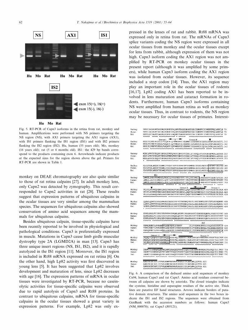
pressed in the lenses of rat and rabbit. Rt88 mRNA wasexpressed only in retina from rat. The mRNAs of Capn3splice variants coding the NS region were expressed in allocular tissues from monkey and the ocular tissues exceptfor lens from rabbit, although expression of them was nothigh. Capn3 isoform coding the AX1 region was not am-pli¢ed by RT-PCR on monkey ocular tissues in thepresent report (although it was ampli¢ed by some prim-ers), while human Capn3 isoform coding the AX1 regionwas isolated from ocular tissues. However, its sequenceincluded a stop codon [14]. Thus, the AX1 region mayplay an important role in the ocular tissues of rodents[16,17]. Lp82 coding AX1 has been reported to be in-volved in lens maturation and cataract formation in ro-dents. Furthermore, human Capn3 isoforms containingNS were ampli¢ed from human retina as well as monkeyocular tissues. Thus, in contrast to rodents, the NS regionmay be necessary for ocular tissues of primates. Interest-
Fig. 5. RT-PCR of Capn3 isoforms in the retina from rat, monkey andhuman. Ampli¢cations were performed with NS primers targeting theNS region (NS), with AX1 primers targeting the AX1 region (AX1),with IS1 primers £anking the IS1 region (IS1) and with IS2 primers£anking the IS2 region (IS2). Hu, human (55 years old); Mo, monkey(16 years old); rat (5 or 6 months old). IS1: the 429 bp bands corre-spond to the products containing exon 6. Arrowheads indicate productsat the expected sizes for the region shown above the gel. Primers forRT-PCR are shown in Table 1.
Fig. 6. A comparison of the deduced amino acid sequences of monkeyCn94, human Capn3 and rat Capn3. Amino acid residues conserved be-tween all calpains are shown by asterisks. The closed triangles indicatethe cysteine, histidine and asparagine residues of the active site. Thicklines are putative EF hand structures. Arrows indicate borders of puta-tive domain structures. The amino acid sequences in the two boxes in-dicate the IS1 and IS2 regions. The sequences were obtained fromGenBank with the accession numbers as follows: human Capn3(NM_000070); rat Capn3 (J05121).
BBAEXP 93526 29-5-01
T. Nakajima et al. / Biochimica et Biophysica Acta 1519 (2001) 55^6462
ingly, both the AX1 and NS regions were present in theocular tissues from rabbit.
Cn94 (Capn3 splice variant lacking exon 15) was pref-erentially expressed in monkey cornea, although quanti¢-cation of mRNAs for Capn3 splice variants by TaqManPCR showed that the expression level in corneal epitheli-um was 22-fold lower than in muscle (data not shown). AsCn94 includes the IS1 region, it may be labile. Overexpres-sion of Lp82 with an inserted IS1 region caused break-down of the Lp82 protein and loss of enzymatic activity[29]. Besides, Cn94 lacks the nuclear translocation signal-like sequence (PXKKKKXKP = exon 15) [30]. Capn3protein did not show nuclear localization when Capn3lacking a nuclear translocation signal was transfectedinto COS cells [30]. Interestingly, Lp85, which containedthe IS3 region, also translocated with the perinuclear re-gion of the nucleated lens ¢bers [29]. Moreover, it has alsobeen suggested that di¡erentiation in the skeletal musclecell culture system was accompanied by a change in ex-pression pattern of Capn3 isoforms [15]. Thus, Capn3splice variants containing Cn94 may have speci¢c func-tions in monkey eye as do Lp82 and Rt88 in rats.
The previous studies have shown the human-mouse dif-ferences in embryonic expression patterns of Capn3 cDNA[14]. The expression of human Capn3 was observed inmuscle and embryonic heart, and its mouse orthologuewas expressed in muscle and embryonic heart, lens andsmooth muscle. Thus, the di¡erent expression patternsfor Capn3 splice variants among the animals are likelydue to susceptibility of tissue-speci¢c genes for accumula-tion of mutations on promoter and intron regions. Thisappears to happen less frequently for ubiquitous calpaingenes. Homology of the amino acid sequences for Capn3splice variants among the animal was as highly conservedas for ubiquitous calpains. These mutations may cause thediversity of expression pattern represented by severalCapn3 splice variants (ex. Capn3/p94, Lp82, Rt88 andCn94). These genes may be related to mammalian evolu-tion.
Acknowledgements
The authors appreciate Y. Nakamura, M. Higashineand K. Mizutani for the excellent technical assistance forthis work. Support in part by Grant NIH grant EY05786to TRS, C5.
References
[1] T. Murachi, K. Tanaka, M. Hatanaka, T. Murakami, IntracellularCa2�-dependent protease (calpain) and its high-molecular-weight en-dogenous inhibitor (calpastatin), Adv. Enzyme Regul. 19 (1980) 407^424.
[2] H. Sorimachi, S. Imajoh-Ohmi, Y. Emori, H. Kawasaki, S. Ohno, Y.Minami, K. Suzuki, Molecular cloning of a novel mammalian calci-
um-dependent protease distinct from both m- and mu-types. Speci¢cexpression of the mRNA in skeletal muscle, J. Biol. Chem. 264 (1989)20106^20111.
[3] H. Sorimachi, S. Ishiura, K. Suzuki, A novel tissue-speci¢c calpainspecies expressed predominately in the stomach comprises two alter-native splicing products with and without Ca(2+)-binding domain,J. Biol. Chem. 268 (1993) 19476^19482.
[4] H.J. Lee, H. Sorimachi, S.Y. Jeong, S. Ishiura, K. Suzuki, Molecularcloning and characterization of a novel tissue-speci¢c calpain pre-dominantly expressed in the digestive tract, Biol. Chem. 379 (1998)175^183.
[5] H. Ma, C. Fukiage, M. Azuma, T.R. Shearer, Cloning and expres-sion of mRNA for calpain Lp82 from rat lens: splice variant of p94,Invest. Ophthalmol. Vis. Sci. 39 (1998) 454^461.
[6] M. Azuma, C. Fukiage, M. Higashine, T. Nakajima, H. Ma, T.R.Shearer, Identi¢cation and characterization of a retina-speci¢c cal-pain (Rt88) from rat, Curr. Eye Res. 21 (2000) 710^720.
[7] D. Branca, A. Gugliucci, D. Bano, M. Brini, E. Carafoli, Expression,partial puri¢cation and functional properties of the muscle-speci¢ccalpain isoform p94, Eur. J. Biochem. 265 (1999) 839^846.
[8] H. Sorimachi, S. Ishiura, K. Suzuki, Structure and physiologicalfunction of calpains, Biochem. J. 328 (1997) 721^732.
[9] T.C. Saido, H. Sorimachi, K. Suzuki, Calpain: new perspectivesin molecular diversity and physiological-pathological involvement,FASEB J. 8 (1994) 814^822.
[10] M.S. Lee, Y.T. Kwon, M. Li, J. Peng, R.M. Friedlander, L.H. Tsai,Neurotoxicity induces cleavage of p35 to p25 by calpain, Nature 405(2000) 360^364.
[11] K. Kinbara, H. Sorimachi, S. Ishiura, K. Suzuki, Skeletal muscle-speci¢c calpain, p94, Biochem. Pharmacol. 56 (1998) 415^420.
[12] I. Richard, J.S. Beakmann, Calpain: Calpain 3 (p94) in Limb-girdleMuscular Dystrophy Type 2A, Taylor and Francis, Philadelphia, PA,1999, pp. 369^390.
[13] I. Richard, O. Broux, V. Allamand, F. Fougerousse, N. Chiannilkul-chai, N. Bourg, L. Brenguier, C. Devaud, P. Pasturaud, C. Roudaut,D. Hillaire, M. Passos-Bueno, M. Zatz, J.A. Tisch¢eld, M. Fardeau,C.E. Jackson, D. Cohen, J.S. Beckmann, Mutations in the proteolyticenzyme calpain 3 cause limb-girdle muscular dystrophy type 2A, Cell81 (1995) 27^40.
[14] F. Fougerousse, P. Bullen, M. Herasse, S. Lindsay, I. Richard, D.Wilson, L. Suel, M. Durand, S. Robson, M. Abitbol, J.S. Beckmann,T. Strachan, Human-mouse di¡erences in the embryonic expressionpattern of developmental control genes and disease genes, Hum. Mol.Genet. 9 (2000) 165^173.
[15] M. Herasse, Y. Ono, F. Fougerousse, E. Kimura, D. Stockholm, C.Beley, D. Montarras, C. Pinset, K. Sorimachi, K. Suzuki, J.S. Beck-mann, I. Richard, Expression and functional characteristics of cal-pain 3 isoforms generated though tissue-speci¢c transcriptional andposttranscriptional events, Mol. Cell. Biol. 19 (1999) 4047^4055.
[16] T.R. Shearer, H. Ma, M. Shih, I. Hata, C. Fukiage, Y. Nakamura,M. Azuma, Lp82 calpain during rat lens maturation and cataractformation, Curr. Eye Res. 17 (1998) 1037^1043.
[17] Y. Nakamura, C. Fukiage, M. Shin, H. Ma, L.L. David, M. Azuma,T.R. Shearer, Contribution of calpain Lp82-induced proteolysis toexperimental cataractogenesis in mice, Invest. Ophthalmol. Vis. Sci.41 (2000) 1460^1466.
[18] H. Ma, I. Hata, M. Shin, C. Fukiage, Y. Nakamura, M. Azuma,T.R. Shearer, Lp82 is the dominant form of calpain in young mouselens, Exp. Eye Res. 68 (1999) 447^456.
[19] F. Fougerousse, M. Durand, L. Suel, O. Pourquie, A.L. Delezoide,N.B. Romero, M. Abitbol, J.S. Beckmann, Expression of genes(CAPN3, SGCA, SGCB, and TTN) involved in progressive musculardystrophies during early human development, Genomics 48 (1998)145^156.
[20] J.B. Jonas, S.S. Hayreb, In£uence of experimental chronic high pres-sure glaucoma on age-related macular degeneration in rhesus mon-keys, Invest. Ophthalmol. Vis. Sci. 41 (2000) 2972^2977.
BBAEXP 93526 29-5-01
T. Nakajima et al. / Biochimica et Biophysica Acta 1519 (2001) 55^64 63
[21] S. Kawashima, H. Akanuma, K. Asaoka, Comparison of calpainsfrom rabbit, monkey, human and rat, Biol. Chem. 379 (1998) 201^204.
[22] K. Hara, Y. Ichihara, K. Takahashi, Puri¢cation and characteriza-tion of a calcium-activated neutral protease from monkey cardiacmuscle, J. Biochem. 93 (1983) 1435^1445.
[23] T. Yamashita, T.C. Saido, M. Takita, A. Miyazawa, J. Yamano, A.Miyakawa, H. Nishijyo, J. Yamashita, S. Kawashima, T. Ono, T.Yoshioka, Transient brain ischemia provokes Ca2�, PIP2 and calpainresponse prior to delayed neuronal death in monkeys, Eur. J. Neuro-sci. 8 (1996) 1932^1944.
[24] K.J. Raser, A. Posner, K.K. Wang, Casein zymography: a method tostudy mu-calpain, m-calpain, and their inhibitory agents, Arch. Bio-chem. Biophys. 319 (1995) 211^216.
[26] T. Murachi, M. Hatanaka, Y. Yasumoto, N. Nakayama, K. Tanaka,
A quantitative distribution study on calpain and calpastatin in rattissues and cells, Biochem. Int. 2 (1981) 651^656.
[27] P. Tsung, J.B. Lombardini, Identi¢cation of low-Ca2�- and high-Ca2�-requiring neutral proteinase in rat retina, Exp. Eye Res. 41(1985) 97^103.
[28] L.L. David, T.R. Shearer, Puri¢cation of calpain II from rat lens anddetermination of endogenous substrate, Exp. Eye Res. 42 (1986) 227^238.
[29] H. Ma, M. Shih, C. Fukiage, M. Azuma, M.K. Duncan, N.A. Reed,I. Richard, J.S. Beckmann, T.R. Shearer, In£uence of speci¢c regionsin Lp82 calpain on protein stability, activity, and localization withinlens, Invest. Ophthalmol. Vis. Sci. 41 (2000) 4232^4239.
[30] H. Sorimachi, N. Toyama-sorimachi, T.C. Saido, H. Kawasaki, H.Sugita, M. Miyasaka, K. Arahata, S. Ishiura, K. Suzuki, Muscle-speci¢c calpain, p94, is degraded by autolysis immediately after trans-lation, resulting in disappearance from muscle, J. Biol. Chem. 268(1993) 10593^10605.
BBAEXP 93526 29-5-01
T. Nakajima et al. / Biochimica et Biophysica Acta 1519 (2001) 55^6464