Dominican Scholar Dominican Scholar Graduate Master's Theses, Capstones, and Culminating Projects Student Scholarship 5-2018 Different Methodologies to Characterize and Diagnose Sickle Cell Different Methodologies to Characterize and Diagnose Sickle Cell Disease in Both Developed and Developing Nations Disease in Both Developed and Developing Nations Mohammed AlHarbi Dominican University of California https://doi.org/10.33015/dominican.edu/2018.cls.05 Survey: Let us know how this paper benefits you. Recommended Citation AlHarbi, Mohammed, "Different Methodologies to Characterize and Diagnose Sickle Cell Disease in Both Developed and Developing Nations" (2018). Graduate Master's Theses, Capstones, and Culminating Projects. 305. https://doi.org/10.33015/dominican.edu/2018.cls.05 This Master's Thesis is brought to you for free and open access by the Student Scholarship at Dominican Scholar. It has been accepted for inclusion in Graduate Master's Theses, Capstones, and Culminating Projects by an authorized administrator of Dominican Scholar. For more information, please contact [email protected].
Transcript
Dominican Scholar Dominican Scholar
Graduate Master's Theses, Capstones, and Culminating Projects Student Scholarship
5-2018
Different Methodologies to Characterize and Diagnose Sickle Cell Different Methodologies to Characterize and Diagnose Sickle Cell
Disease in Both Developed and Developing Nations Disease in Both Developed and Developing Nations
Mohammed AlHarbi Dominican University of California
Recommended Citation AlHarbi, Mohammed, "Different Methodologies to Characterize and Diagnose Sickle Cell Disease in Both Developed and Developing Nations" (2018). Graduate Master's Theses, Capstones, and Culminating Projects. 305. https://doi.org/10.33015/dominican.edu/2018.cls.05
This Master's Thesis is brought to you for free and open access by the Student Scholarship at Dominican Scholar. It has been accepted for inclusion in Graduate Master's Theses, Capstones, and Culminating Projects by an authorized administrator of Dominican Scholar. For more information, please contact [email protected].
HbF = 50-80%. Hemoglobin electrophoresis is a method used to discriminate between
these different types of hemoglobin under alkaline conditions using variety of sieving
materials that includes gel and paper. Under alkaline condition various forms of
hemoglobin have a net negative charge and move towards the positive electrode.
Different hemoglobin form migrates to different lengths on the gel based on the charge
carried by these molecules, pore size of the medium and the ionic strength of the buffer
solution. A condition can be determined based on the various bands observed on the
gel. The advantage of this methodology is rapid screening of small number of samples
5
is possible. After electrophoretic separation, densitometry is used to quantify various
forms of hemoglobin observed on the gel. However, at low sample concentration
densitometry suffer from inaccuracy of result. The resolution between HbF and HbS is
very low and hard to distinguish in neonates having high concentration of HbF.
For high-throughput screening a capillary zone electrophoresis (CZE) is carried out in
a U-shaped narrow-bore fused-silica capillary tube. Like HPLC, CZE is an automated
method and has been shown in various studies to be an effective methodology to detect
monoclonal gammopathies and protein abnormalities present in the serum. Migration
in CZE is similar to conventional electrophoretic methods in which the protein migrates
based on its net charge when an electric field in applied. The buffers used in CZE differ
from gel electrophoresis where barbital buffer is used. Barbital buffer has a
disadvantage since it has a high absorbance at 200nm wavelength, which coincides with
peptide bonds and thus interferes with the detection system. To avoid this CZE uses
borate-based buffers that do not cause the above interference (Dolník et al. 1995).
The negative charge of the fused silica used to make the capillaries provides a negative
surface charge to the migrating protein molecules. Also, the narrow lumen (about 50μm
in diameter) inside the capillary tubes makes the surface area relatively large. This
combined effect of large surface area and charge on the capillaries causes a more
effective movement of proteins in the sample towards the cathode than could have been
achieved by only voltage difference between cathode and anode. This high charge and
surface area offered by the capillaries causes a higher resolution of the hemoglobin
variants present in the samples. A detector is usually present at the cathodal end of the
capillary, which records the optical density at a wavelength of 200nm (peptide bond
absorption) as the proteins migrate past it. The Hb variants pass by the detector in the
following order-γ, β, α2, and α1 globulins (Keren et al. 1998).
6
High resolution, ease of automation, shorter run time at a high voltage and no additional
requirement for densitometer are the advantages of the CZE methodology for diagnosis
of SCD. However, CZE is a new technology and the limitations haven’t been properly
investigated so far. One major drawback is this methodology does not have any gel that
can be examined. The bands represent those substances that have absorbency at around
200 nm, which are usually proteins. Non-protein substances having similar absorbency
parameters can therefore also produce bands that can be confused with hemoglobin
variants (Keren et al. 1998; Bain, B. J. 2008). CZE requires a detector, which records
optical density at a wavelength of 200nm and this is connected to a computer for data
acquisition and also needs high voltage power supply. All these arrangements are
expensive and are not readily available in remote areas hence CZE-based SCD
screening methodology is not easy to implement in developing or in underdeveloped
countries.
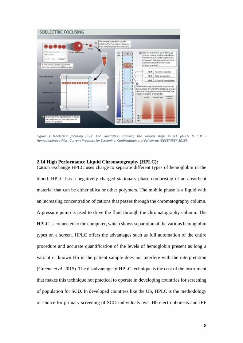
2.13 Isoelectric focusing (IEF):
Every protein carries a net charge that varies according to the pH of the surrounding
environment. An isoelectric point (pI) is defined as a point in pH gradient where a
protein carries a net zero charge. Hemoglobin samples are run in a gel medium across
which an electrical gradient is applied, and a fixed pH gradient is maintained throughout
the gel (Fig. 1). Different types of hemoglobins present in the blood sample travel and
stop at the location in the gel at their respective pI. Resolution of IEF is better than Hb
electrophoresis and thus this technique is useful in distinguishing between higher
populations of individuals with variant Hb (Bain, B. J. 2008). The disadvantage of this
technique is that the interpretation of result sometimes becomes difficult due to the
presence of a higher number of bands on the gel. Also, the technique is more expensive
compared to Hb electrophoresis method. Like Hb electrophoresis, the quantitation
depends on densitometric analysis, that sometimes gives inaccurate interpretation.
7
However, IEF is the method of choice for screening newborns in many clinical settings
since this methodology can operate at a very small volume and even gives reliable
results from dried blood spot samples (World Health Organization 2006; APLH & CDC
- Hemoglobinopathies -Current Practices for Screening, Confirmation and Follow-up -
DECEMBER 2015).
In a pilot study conducted in Tunisia, a low cost neonatal screening method using IEF
assay for the detection of SCD and other Hb variants has been reported (Hajer et al.
2012). In this study samples from 9148 newborns were collected on blotting paper
(Whatman grade BFC 180) at maternity centers and using a general office-use printer
these blotting papers were printed. IEF methodology was then carried out by a lab-
prepared agarose gel to test the dried blood samples from these newborns. This low cost
IEF on lab prepared agarose gel along with office printer printed blotting paper for
sample collection proved to be an effective method since this method successfully
detected the newborns who had abnormal Hbs (HbS, HbC, HbO and HbG). The
accuracy of the data collected in this screening method was also verified by comparing
with previously established national data. The families of these newborns were made
aware of the result and guidelines for treatment along with genetic counseling was also
provided.
8
Figure 1 Isoelectric focusing (IEF): The illustration showing the various steps in IEF (APLH & CDC - Hemoglobinopathies -Current Practices for Screening, Confirmation and Follow-up -DECEMBER 2015).
2.14 High Performance Liquid Chromatography (HPLC):
Cation exchange HPLC uses charge to separate different types of hemoglobin in the
blood. HPLC has a negatively charged stationary phase comprising of an absorbent
material that can be either silica or other polymers. The mobile phase is a liquid with
an increasing concentration of cations that passes through the chromatography column.
A pressure pump is used to drive the fluid through the chromatography column. The
HPLC is connected to the computer, which shows separation of the various hemoglobin
types on a screen. HPLC offers the advantages such as full automation of the entire
procedure and accurate quantification of the levels of hemoglobin present as long a
variant or known Hb in the patient sample does not interfere with the interpretation
(Greene et al. 2015). The disadvantage of HPLC technique is the cost of the instrument
that makes this technique not practical to operate in developing countries for screening
of population for SCD. In developed countries like the US, HPLC is the methodology
of choice for primary screening of SCD individuals over Hb electrophoresis and IEF
9
(Alapan et al .2016). HPLC is much less labor intensive, accurate and less time-
consuming method in comparison to the Hb electrophoresis. HPLC can also be used on
SCD individuals on hydroxyurea (HU) or transfusion therapies (Makani et al. 2013).
HU is the drug-of-choice to reduce the pain episode frequency in sickle cell patients.
HU raises the level of HbF that significantly decreases the rate of painful episodes by
about 50% (Agrawal et al. 2014). Blood transfusion lowers the amount of sickle
hemoglobin in the body. Since the normal red blood cells increase in the blood stream
separation, and (d) micro engineered electrophoresis. Each of these methodologies has
been explained in detail below.
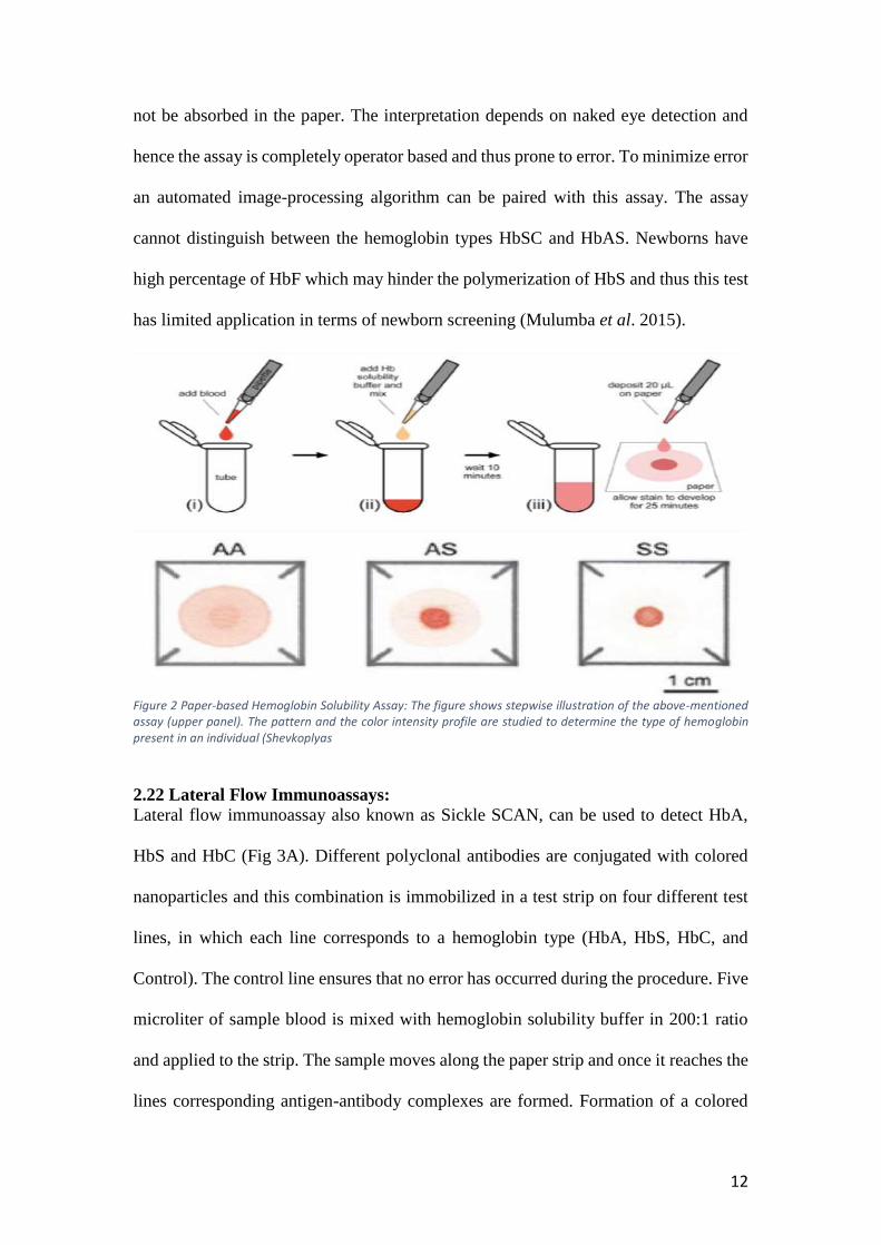
2.21 Paper-based Hemoglobin Solubility Assay:
The principle behind this technology is the insoluble property of hemoglobin and the
filtration ability of paper. This assay uses a microfluidic paper-based device known as
μPADs, to analyze the samples. In this way the HbS in the sample can be seen with our
eyes. A drop of blood (~20μL) is mixed with hemoglobin solubility buffer in a 1:10
ratio and then put onto a chromatography paper that is patterned (Fig. 2). Based on the
different capillary action pattern of different hemoglobin types, various bloodstain
patterns are observed for HbS that is polymerized and other hemoglobin types
(Shevkoplyas et al. 2017). Screening can be achieved within 20 minutes and the method
is 94.2% sensitive (see Table 1 below) (Yang et al. 2013). The advantage of this paper-
based is that it is cheaper compared to other methods, easy to interpret and sample
preparation is very easy. Individual tests can be performed without the need to batch
samples.
The technique however suffers from few disadvantages. First, if the blood clots before
adding to the μPAD, then the capillary action of the blood is lost, and the blood would
12
not be absorbed in the paper. The interpretation depends on naked eye detection and
hence the assay is completely operator based and thus prone to error. To minimize error
an automated image-processing algorithm can be paired with this assay. The assay
cannot distinguish between the hemoglobin types HbSC and HbAS. Newborns have
high percentage of HbF which may hinder the polymerization of HbS and thus this test
has limited application in terms of newborn screening (Mulumba et al. 2015).
Figure 2 Paper-based Hemoglobin Solubility Assay: The figure shows stepwise illustration of the above-mentioned assay (upper panel). The pattern and the color intensity profile are studied to determine the type of hemoglobin present in an individual (Shevkoplyas
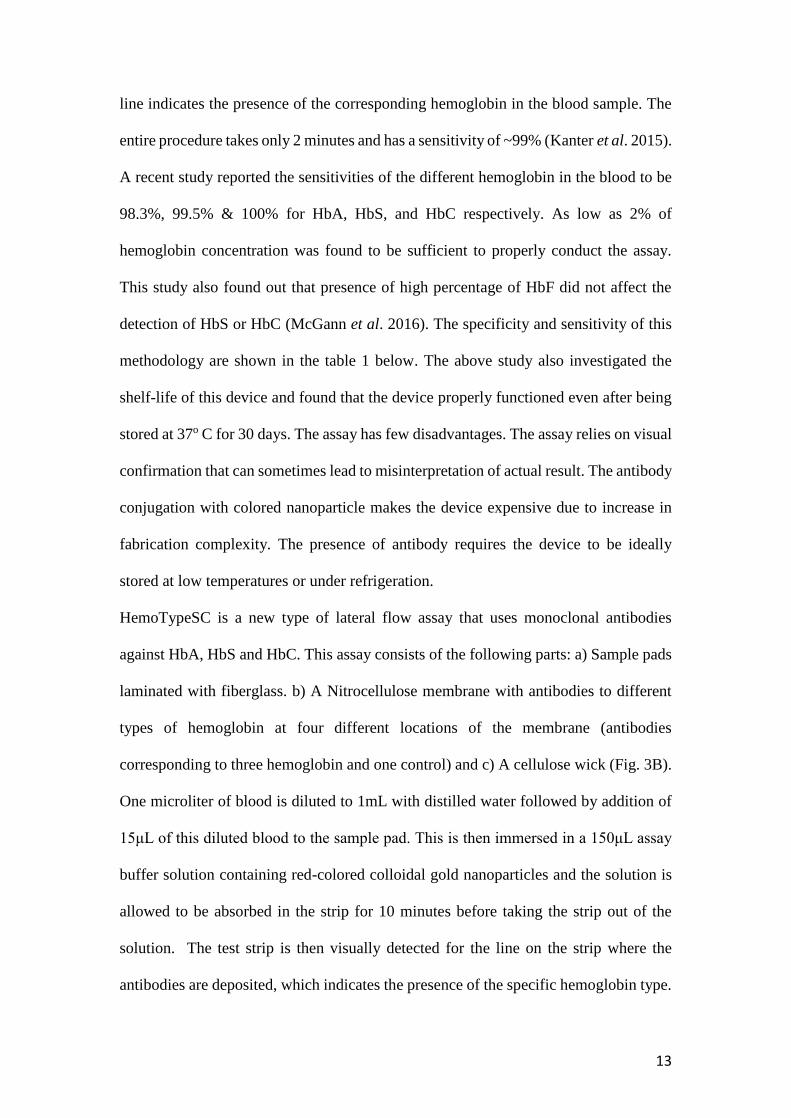
2.22 Lateral Flow Immunoassays:
Lateral flow immunoassay also known as Sickle SCAN, can be used to detect HbA,
HbS and HbC (Fig 3A). Different polyclonal antibodies are conjugated with colored
nanoparticles and this combination is immobilized in a test strip on four different test
lines, in which each line corresponds to a hemoglobin type (HbA, HbS, HbC, and
Control). The control line ensures that no error has occurred during the procedure. Five
microliter of sample blood is mixed with hemoglobin solubility buffer in 200:1 ratio
and applied to the strip. The sample moves along the paper strip and once it reaches the
lines corresponding antigen-antibody complexes are formed. Formation of a colored
13
line indicates the presence of the corresponding hemoglobin in the blood sample. The
entire procedure takes only 2 minutes and has a sensitivity of ~99% (Kanter et al. 2015).
A recent study reported the sensitivities of the different hemoglobin in the blood to be
98.3%, 99.5% & 100% for HbA, HbS, and HbC respectively. As low as 2% of
hemoglobin concentration was found to be sufficient to properly conduct the assay.
This study also found out that presence of high percentage of HbF did not affect the
detection of HbS or HbC (McGann et al. 2016). The specificity and sensitivity of this
methodology are shown in the table 1 below. The above study also investigated the
shelf-life of this device and found that the device properly functioned even after being
stored at 37o C for 30 days. The assay has few disadvantages. The assay relies on visual
confirmation that can sometimes lead to misinterpretation of actual result. The antibody
conjugation with colored nanoparticle makes the device expensive due to increase in
fabrication complexity. The presence of antibody requires the device to be ideally
stored at low temperatures or under refrigeration.
HemoTypeSC is a new type of lateral flow assay that uses monoclonal antibodies
against HbA, HbS and HbC. This assay consists of the following parts: a) Sample pads
laminated with fiberglass. b) A Nitrocellulose membrane with antibodies to different
types of hemoglobin at four different locations of the membrane (antibodies
corresponding to three hemoglobin and one control) and c) A cellulose wick (Fig. 3B).
One microliter of blood is diluted to 1mL with distilled water followed by addition of
15μL of this diluted blood to the sample pad. This is then immersed in a 150μL assay
buffer solution containing red-colored colloidal gold nanoparticles and the solution is
allowed to be absorbed in the strip for 10 minutes before taking the strip out of the
solution. The test strip is then visually detected for the line on the strip where the
antibodies are deposited, which indicates the presence of the specific hemoglobin type.
14
It takes around 20 minutes to obtain result by this assay (Quinn et al .2016). The
HemoTypeSC assay achieved 100% sensitivity and at the same time had a low
operating cost. The assay was able to distinguish between HbAA, HbAS, HbAC, HbSS,
HbSC, and HbCC but it could not detect HbF and HbA2 Hb types.
Figure 3 Lateral Flow Immunoassay: (A) The sample is added in the application pad, which then moves via capillary action to the test lines conjugated with different polyclonal antibodies as shown. The blue line that appears notifies the type of hemoglobin present.
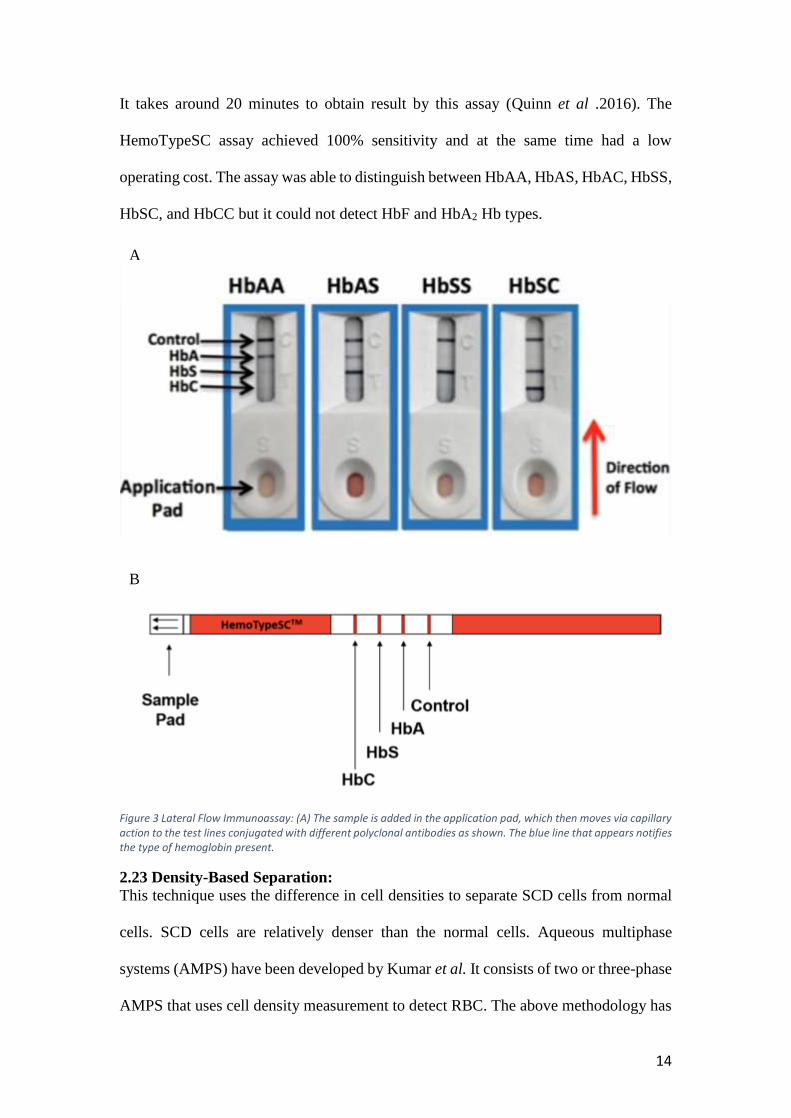
2.23 Density-Based Separation:
This technique uses the difference in cell densities to separate SCD cells from normal
cells. SCD cells are relatively denser than the normal cells. Aqueous multiphase
systems (AMPS) have been developed by Kumar et al. It consists of two or three-phase
AMPS that uses cell density measurement to detect RBC. The above methodology has
A
B
15
a sensitivity as high 90-91% and specificity ranging from 88-97% (Kumar et al. 2014).
Five microliters of blood are mixed with aqueous polymeric solution and then loaded
into capillary tubes (Fig. 4). The tubes are then centrifuged for 10 minutes that causes
dense RBC of SCD patients to settle. This method has several limitations that include
the higher cost due to inclusion of the centrifugation step. Centrifugation also makes it
difficult to conduct this test at POC settings. Besides samples need to be run in batches
increasing the turnaround time. Density based separation method cannot differentiate
between HbAA and HbAS. Also, newborn screening is not possible with this method
due to the presence of high % of HbF that restricts the occurrence of dense RBC. Other
factors like medication, health conditions, treatment processes and genetic factors affect
the density of RBC and hence the outcome of test result of this assay (see Table 1
below).
Figure 4 The figure shows the different outcome of the density-based assay. The sample blood moves through the different phases i.e. top , middle, and bottom and then accumulates at the interphases depending on the type of hemoglobin present (Kumar et al. 2014)
16
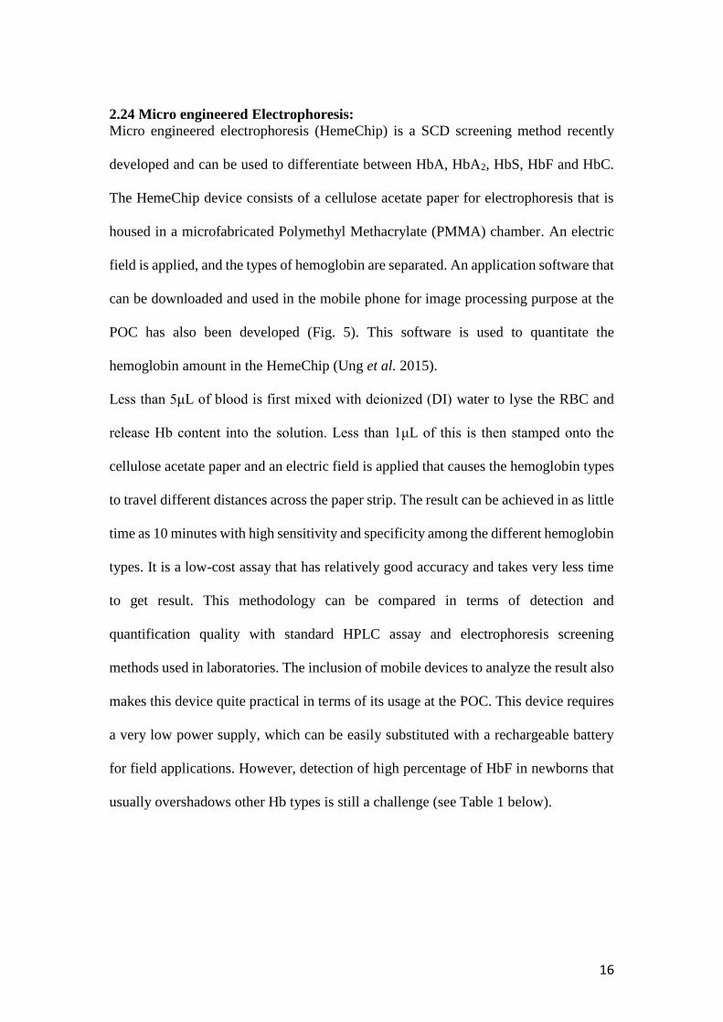
2.24 Micro engineered Electrophoresis:
Micro engineered electrophoresis (HemeChip) is a SCD screening method recently
developed and can be used to differentiate between HbA, HbA2, HbS, HbF and HbC.
The HemeChip device consists of a cellulose acetate paper for electrophoresis that is
housed in a microfabricated Polymethyl Methacrylate (PMMA) chamber. An electric
field is applied, and the types of hemoglobin are separated. An application software that
can be downloaded and used in the mobile phone for image processing purpose at the
POC has also been developed (Fig. 5). This software is used to quantitate the
hemoglobin amount in the HemeChip (Ung et al. 2015).
Less than 5μL of blood is first mixed with deionized (DI) water to lyse the RBC and
release Hb content into the solution. Less than 1μL of this is then stamped onto the
cellulose acetate paper and an electric field is applied that causes the hemoglobin types
to travel different distances across the paper strip. The result can be achieved in as little
time as 10 minutes with high sensitivity and specificity among the different hemoglobin
types. It is a low-cost assay that has relatively good accuracy and takes very less time
to get result. This methodology can be compared in terms of detection and
quantification quality with standard HPLC assay and electrophoresis screening
methods used in laboratories. The inclusion of mobile devices to analyze the result also
makes this device quite practical in terms of its usage at the POC. This device requires
a very low power supply, which can be easily substituted with a rechargeable battery
for field applications. However, detection of high percentage of HbF in newborns that
usually overshadows other Hb types is still a challenge (see Table 1 below).
17
Figure 5 A) The HemeChip with blood sample being separated into respective bands different hemoglobin type B) Graph showing Sensitivity and specificity C) Mobile Software for image processing and quantification of HemeChip results at the POC (Ung et al. 2015).
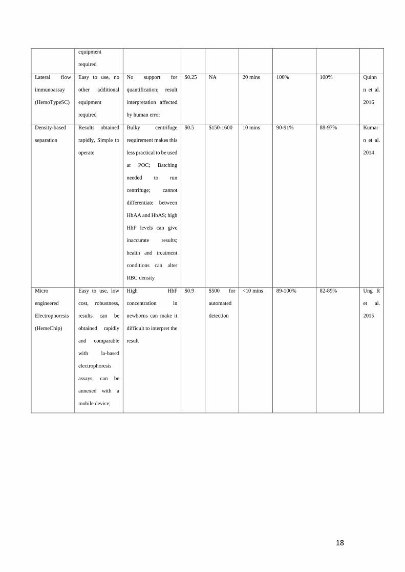
Table 1 Summary of methodology for screening SCD at Point of Care Testing.
Technique Advantages Disadvantages Cost
per
test
Equipment
cost
Turn
around
time
Sensitivity
Specificity Ref.
Paper-based
hemoglobin
solubility
Easy to use, no
batching required
Cannot distinguish
HbSC and HbAS,
Blood clotting issue;
result interpretation
affected by human
error
$0.7 $300-500
(for
automated
detection)
20 mins 94.2% 97.7% Yang et
al.
2013
Lateral flow
immunoassay
(Sickle SCAN)
Easy to use, result
can have obtained
rapidly, no other
additional
Cannot quantitate Hb;
result interpretation
affected by human
error
$5 NA 2 mins 99% 99% McGan
n et al.
2016
18
equipment
required
Lateral flow
immunoassay
(HemoTypeSC)
Easy to use, no
other additional
equipment
required
No support for
quantification; result
interpretation affected
by human error
$0.25 NA 20 mins 100% 100% Quinn
n et al.
2016
Density-based
separation
Results obtained
rapidly, Simple to
operate
Bulky centrifuge
requirement makes this
less practical to be used
at POC; Batching
needed to run
centrifuge; cannot
differentiate between
HbAA and HbAS; high
HbF levels can give
inaccurate results;
health and treatment
conditions can alter
RBC density
$0.5 $150-1600 10 mins 90-91% 88-97% Kumar
n et al.
2014
Micro
engineered
Electrophoresis
(HemeChip)
Easy to use, low
cost, robustness,
results can be
obtained rapidly
and comparable
with la-based
electrophoresis
assays, can be
annexed with a
mobile device;
High HbF
concentration in
newborns can make it
difficult to interpret the
result
$0.9 $500 for
automated
detection
<10 mins 89-100% 82-89% Ung R
et al.
2015
19
3. Discussion and Conclusions: Proper screening and early diagnosis of SCD individuals is vital to their management
and treatment. Improper screening or a delay results in higher mortality rates among
SCD patients. Several SCD screening techniques were developed in the early 1970s.
Unfortunately, many such methodologies proved to be unreliable since they gave false
positive or negative results that led to wrong treatment and confusion. In order to
standardize laboratory techniques and interpretation for SCD screening a
Hemoglobinopathy Reference Laboratory (HRL) was created at the Centers of Disease
Control (CDC) (Naik et al. 2015).
This review focused on the different methodologies that are currently used to screen
SCD individuals that include both newborns and adults. The most common screening
methodologies currently used comprise of sickle solubility testing, electrophoresis,
HPLC, CZE and IEF. Sickle solubility-based screening assays usually have lower cost
and they are based on the principle of insolubility of HbS in a reducing agent that causes
turbidity due to lysis of HbS- containing RBC in solution. The solubility test however
cannot differentiate between SCD and SCT individuals and gives false negative result
in newborns with higher HbF content. Hence additional tests that include hemoglobin
electrophoresis, HPLC and IEF are also used that serve the purpose of either being
primary identification of sickle hemoglobin types or as confirmatory tests. These
techniques can provide quantification of hemoglobin and clearly differentiate between
SCT and SCD individuals. Hemoglobin electrophoresis method is based on the
principles of gel electrophoresis and is an inexpensive method that separates types of
hemoglobin present in the blood based on their relative size and charge.
IEF is based on the principle that the net charge in the proteins varies based upon the
surrounding pH and at their respective isoelectric point (pI) all proteins carry zero
20
charge. Hemoglobin molecules present in the blood samples are thus run in a gel having
pH gradient under an electric field and they stop as they reach their respective pI points.
IEF methodology has relatively low cost and has high throughput abilities and thus is
the assay–of-choice in many clinical laboratories. In this context a pilot study conducted
in Tunisia, Africa has been mentioned here that uses a low cost SCD screening
technique by collecting blood samples on blotting paper printed in a common office
printer and then analyzing the samples in a laboratory prepared gel for IEF assay (Hajer
et al .2012).
HPLC although being an expensive method for hemoglobinopathy screening is used in
many clinical laboratories across developed nations for its accuracy in detection and
less time to get result.
An alternative methodology for screening SCD individuals is LC-MS/MS, which is
able to detect Hb peptides following digestion of blood spots with trypsin. This is also
an extremely accurate and fast method that can be applied for population screening for
identification of clinically important globin mutations.
SCD screening can also be achieved by DNA analysis, one of the methodology in this
category include RFLP assay, in which β-globin gene is first PCR amplified and then
cut with the restriction enzyme DdeI. Absence of this restriction site indicates the
presence of mutated β-globin gene since the point mutation eliminates the above
restriction site. Whole exome genome sequencing (WES) to look for SNVs in the β-
globin gene is also currently followed by several laboratories although sequencing is
an expensive technology and only developed nations can afford it.
The review also explains the emerging POC methodologies that are developed
considering the cost factor at a certain location, trained technicians available and
accessibility. Four such emerging methodologies have been discussed here that are as
21
follows, a) paper-based hemoglobin assays, b) lateral flow immunoassays, c) density-
based separation, and d) micro engineered electrophoresis (HemeChip).
The paper-based hemoglobin assay takes utilizes capillary action of blood sample when
applied to a paper-based device called μPADs that causes different hemoglobin types
of form unique patterns that can be easily distinguished from one another.
The lateral flow immunoassay comprises of a test strip that has polyclonal antibodies
against different hemoglobin types conjugated with colored nanoparticles. An antigen-
antibody complex is formed between respective hemoglobins once the blood sample
reaches the lines in the strip.
Density-based separation has an AMPS comprising of two and three phases that uses
cell density measurements to detect RBC. Blood samples are centrifuged in capillary
tubes and SCD cells that are of higher density settle at the lowest interphase.
The micro engineered electrophoresis (HemeChip) consists of a cellulose acetate paper
housed inside a PMMA chamber where sample blood electrophoresis is conducted to
separate different hemoglobin types. A mobile-based application software can be used
that is compatible with this assay to process image.
Most of these emerging methodologies are able to distinguish between HbA, HbA2,
HbS, HbF and HbF hemoglobin types. These emerging methodologies are based on
resources that are easily available at the POC of even economically challenged nations.
The advantages and disadvantages of each methodology for screening of SCD have
been discussed in this review. However, considering every factor that includes overall
cost, practicality of using an assay in both developed and developing nations, available
resources to perform the test, sensitivity, specificity, different Hb variants that can be
identified with certainty, the methodology-of-choice would be Lateral Flow
immunoassay (Sickle SCAN and HemoTypeSC). This methodology is categorized as a
22
POC assay because of its low cost and easy-of-use even in remote areas of developing
nations. As can be seen in Table 1, the lateral flow assay has both sensitivity and
specificity ranging between 99-100%. The cost of running the assay varies from $0.25-
$5, which is low compared to more expensive assays like HPLC and WES. A recent
study has been conducted in order to select the best methodology based on the low cost
of use, portability, easy-to-use diagnostic tests especially in resource-poor countries
(Nwegbu et al. 2017). In this study the performance characteristics of Lateral flow
assay (Sickle SCAN) was compared with cellulose acetate electrophoresis (CAE) and
high-performance liquid chromatography (HPLC) by evaluating several subjects for
HbSS, HbSC and HbAS. The SickleSCAN™ showed diagnostic sensitivity, specificity
and test efficiency of 100.0, 98.2 and 98.2%, respectively for sickle cell disease (Hb SS
and Hb SC) that was comparable to the other two methods but at a much lower cost and
easy-of-use at remote locations.
23
4. References:
1. Randolph, T. R., & Wheelhouse, J. (2012). Novel test method (sickle confirm)
to differentiate sickle cell anemia from sickle cell trait for potential use in