Page 1
ORIGINAL PAPER
Differential Activation of the ER Stress Factor XBP1by Oligomeric Assemblies
Diana L. Castillo-Carranza • Yan Zhang •
Marcos J. Guerrero-Munoz • Rakez Kayed •
Diego E. Rincon-Limas • Pedro Fernandez-Funez
Received: 8 December 2011 / Revised: 23 March 2012 / Accepted: 4 April 2012 / Published online: 21 April 2012
� The Author(s) 2012. This article is published with open access at Springerlink.com
Abstract Several neurodegenerative disorders are char-
acterized by protein misfolding, a phenomenon that results
in perturbation of cellular homeostasis. We recently iden-
tified the protective activity of the ER stress response factor
XBP1 (X-box binding protein 1) against Amyloid-ß1-42
(Aß42) neurotoxicity in cellular and Drosophila models of
Alzheimer’s disease. Additionally, subtoxic concentrations
of Aß42 soluble aggregates (oligomers) induced accumu-
lation of spliced (active) XBP1 transcripts, supporting the
involvement of the ER stress response in Aß42 neurotox-
icity. Here, we tested the ability of three additional disease-
related amyloidogenic proteins to induce ER stress by
analyzing XBP1 activation at the RNA level. Treatment of
human SY5Y neuroblastoma cells with homogeneous prep-
arations of a-Synuclein (a-Syn), Prion protein (PrP106–126),
and British dementia amyloid peptide (ABri1-34) confirmed
the high toxicity of oligomers compared to monomers and
fibers. Additionally, a-Syn oligomers, but not monomers or
fibers, demonstrated potent induction of XBP1 splicing. On
the other hand, PrP106–126 and ABri1-34 did not activate
XBP1. These results illustrate the biological complexity of
these structurally related assemblies and argue that oligo-
mer toxicity depends on the activation of amyloid-specific
cellular responses.
Keywords Oligomers � Neurodegeneration � ER stress �XBP1 � Amyloids
Abbreviations
Aß42 Amyloid-ß1-42
ABri1-34 British dementia amyloid peptide
AD Alzheimer’s disease
FBD Familial British dementia
PD Parkinson’s disease
PrD Prion disease
PrP Prion protein
a-Syn a-Synuclein
UPR Unfolded protein response
XBP1 X-box binding protein 1
Introduction
Neurodegenerative diseases encompass a complex group of
neurological disorders characterized by progressive and
widespread neuronal cell loss. The most common neuro-
degenerative disorders are caused by abnormal protein
deposition in the form of highly ordered intra- and/or
extracellular amyloid fibers. Alzheimer’s disease (AD) and
Parkinson’s disease (PD) are among the most prevalent
proteinopathies, affecting up to 40 % of the elderly popu-
lation [25, 33]. Upon autopsy, AD and PD are character-
ized by amyloid plaques rich in Amyloid-ß42 (Aß42) and
Lewy bodies containing a-Synuclein (a-Syn), respectively.
These large protein aggregates have a critical diagnostic
D. L. Castillo-Carranza � M. J. Guerrero-Munoz � R. Kayed
Department of Neurology, University of Texas Medical Branch,
Galveston, TX 77555, USA
Y. Zhang � D. E. Rincon-Limas (&)
Department of Neurology, McKnight Brain Institute, University
of Florida, 1149 South Newell Dr., Gainesville, FL 32611, USA
e-mail: [email protected]
P. Fernandez-Funez (&)
Departments of Neurology and Neurosciences, McKnight Brain
Institute, University of Florida, 1149 South Newell Dr.,
Gainesville, FL 32611, USA
e-mail: [email protected]
123
Neurochem Res (2012) 37:1707–1717
DOI 10.1007/s11064-012-0780-7
Page 2
value and were originally proposed to play a causative role
in disease. However, a modern understanding of the role of
amyloids in disease suggests that the large, fibrillar
aggregates may not be directly responsible for neurode-
generation [7, 32]. Large amyloid aggregates have also
been postulated to exert a defensive role by storing these
toxic proteins in cellular structures such as the aggresome
(Lewy bodies in PD and nuclear inclusions in polygluta-
mine diseases) or be inert byproducts of neuronal death
(amyloid plaques) [34].
In contrast, soluble aggregates (oligomers) seem to
correlate better with neurotoxicity in cellular and animal
models [5, 23, 27, 37]. Oligomeric assemblies are dynamic
structures that perturb many cellular processes, including
mitochondria and energy metabolism, cell signaling, cal-
cium homeostasis, oxidative stress and cell survival/apop-
tosis, and more by mechanisms not completely known
[13, 22]. Oligomers can bind to receptors in synaptic
membranes, including nicotinic and NMDA (N-Methyl-D-
aspartate) receptors and RAGE (Receptor for advance
glycation endproducts), causing synaptic dysfunction and
perturbation in signaling pathways [30]. They can also
form pores that integrate in the membrane and alter the
transport of essential ions, which may be responsible for
calcium dyshomeostasis [3, 11]. Finally, oligomers can be
actively transported by endocytic mechanisms, resulting in
intracellular accumulation and interaction with the protein
quality-control mechanism, including chaperones and the
proteasome [19]. So far, this new focus on oligomers has
identified common neurotoxic mechanisms among differ-
ent amyloidogenic proteins based on shared structures [13].
One problem with the idea that all amyloidogenic proteins
form structurally related oligomers is explaining the dif-
ferent cellular vulnerability and unique molecular pathol-
ogy of each disease. Thus, oligomeric assemblies from
different proteins must encompass unique biological
properties that explain the disease-specific phenotypes.
However, little is known at this time about the biological
differences among oligomers.
We have shown recently that overexpression of the ER
stress response factor XBP1 (X-box binding protein 1)
rescued the toxicity of human Aß42 expressed in trans-
genic flies [6]. Conversely, reduction of the endogenous
XBP1 function by RNAi increased the Aß42 phenotype,
supporting the physiological role of XBP1 in the response
to Aß42 neurotoxicity. XBP1 is a key component of the
unfolded protein response (UPR), a conserved protective
mechanism against misfolded proteins in the ER [15].
Three independent sensors regulate the UPR: PERK, ATF6
and IRE1. Upon ER stress, IRE1 autophosphorylates and
dimerizes, which activates its cytoplasmic RNase domain.
Active IRE1 then cleaves the XBP1 pre-mRNA in the
cytoplasm, which removes a 26-nt intron that changes the
reading frame in the second exon of XBP1. This uncon-
ventional splicing results in the production of the trans-
criptionally active (spliced) isoform XBP1s instead of the
inactive (unspliced) isoform XBP1u. XBP1s induces the
transcriptional upregulation of a large number of target
genes that contribute to reduce protein misfolding in the
ER [1]. In turn, activation of ATF6 results in a cleaved
fragment with potent transcriptional activity that induces
the expression of several key target genes, including XBP1.
Among our observations, we found that Aß42 activated ER
stress and induced the unconventional splicing of XBP1 in
both transgenic flies and rat pheochromocytoma cells
(PC12) treated with Aß42 oligomers [6]. Since subtoxic
concentrations of Aß42 oligomers induced robust XBP1
splicing in PC12 cells, ER stress and XBP1 splicing seemed
rapid and efficient cellular responses to Aß42 neurotox-
icity. Moreover, these observations suggested that XBP1
splicing could be used as a sensitive assay to detect Aß42
neurotoxicity and, possibly, the toxicity of other oligomers
linked to neurodegenerative diseases. The assay used so far
to detect XBP1 splicing consists on an RT-PCR with
primers straddling the small intron that amplifies both
isoforms, XBP1u and XBP1s. Then, the PCR products are
digested with PstI, which has a unique, evolutionarily
conserved restriction site in the intron of XBP1u, allowing
the diagnostic identification of XBP1s transcripts (resistant
to PstI). Using this approach Lee and col. identified
unconventional XBP1 splicing in the temporal cortex of
AD patients, but not in the cortex of Tg2576 mice, an AD
model with no cell loss [24]. Thus, activation of the IRE1-
XBP1 pathway seems to correlate better with neuronal
degeneration than with deposition of misfolded Aß42,
making XBP1 an attractive diagnostic tool for neurode-
generative conditions.
From these observations, we hypothesized that oligo-
mers from other amyloidogenic proteins, but not monomers
and fibers, should also induce XBP1 activation. In this
report, we asked three questions: (1) do other amyloido-
genic proteins induce XBP1 activation, (2) which quater-
nary conformations of the protein induces XBP1 activation,
and (3) what is the diagnostic potential of XBP1 splicing at
the RNA level? To answer these questions, we produced
monomeric, oligomeric, and fibrillar preparations for three
disease-related amyloids: full-length a-Syn; the amyloido-
genic fragment of the Prion protein (PrP106–126), and
ABri1-34, an Aß42-related peptide associated with familial
British dementia (FBD). Here we report that a-Syn oligo-
mers, similar to Aß42, induce strong XBP1 activation in
SH-SY5Y cells by a modified PCR procedure. Surpris-
ingly, PrP106–126 and ABri1-34 did not induce XBP1
splicing, although their oligomers were as toxic as a-Syn
oligomers. Overall, these results confirmed that oligomeric
assemblies of other amyloidogenic proteins can induce
1708 Neurochem Res (2012) 37:1707–1717
123
Page 3
unconventional splicing of XBP1, although this is not a
conserved activity of all amyloids.
Materials and Methods
Preparation of Monomers, Oligomers, and Fibers
Soluble oligomers were prepared as shown by Kayed [21]
by dissolving 0.3 mg of Aß42 (MW 4514), a-Syn (MW
14,460), PrP106–126 (MW 1912), and ABri1-34 (MW
3935) (previously re-solubilized in acetonitrile: water 1:1
and lyophilized) in 200 ll of hexafluoroisopropanol (HFIP)
for 20 min at room temperature. 200 ll of these solutions
were added to 1,000 ll DD H2O in a siliconized Eppendorf
tube for evaporation of the HFIP, resulting in pure mono-
mers at 0.3 mg/ml. The samples were then stirred at
500 rpm using a Teflon coated micro stir bar for 24–48 h at
22 �C for formation of oligomers. Before using oligomers
for cell treatment, the samples were sonicated to break
incipient fibers. Fibrils were prepared as described above
for oligomers, except they were stirred at room temperature
for 6–9 days. Fibril formation was monitored by thioflavin-
T fluorescence and UV light scattering. Once fibril for-
mation was complete, the solutions were centrifuged at
14,0009g for 20 min, the fibril pellet was washed three
times with double-distilled water and then resuspended in
the desired buffer. The morphology was verified by nega-
tive stain electron microscopy using standard procedures
[21]. For the treatment of cells, the molarity for each
peptide was calculated based on the amount of monomer.
Cell Culture, Oligomer Treatment, and Cell Toxicity
Human neuroblastoma SH-SY5Y cells (Sigma) were
grown in DMEM supplemented with 10 % FBS and dif-
ferentiated in 10 lM retinoic acid and 1 % FBS for 24 h.
To investigate toxic effects of different monomer, oligo-
mer, and fibrils preparations, SH-SY5Y cells were seeded
at 6,000 cells/well, in black 96-well clear bottom microtiter
plates (Corning). The next day, cells were treated with
2 lM of monomer, oligomers, or fibrils in triplicate. After
8 h of treatment, media containing oligomers was replaced
with 100 ll of fresh media and 10 ll of alamar blue
(Biosurce) per well. Fluorescence was measured at
530–560 nm excitation and at 590 nm emission, using a
fluorescence ELISA plate reader (Polar-star Omega BMG
Labtech). To collect enough RNA for XBP1 analysis, cells
were incubated in 6-well plates at 5 9 106 cells/well. After
differentiating for 24 h, we treated them with 10 lg/ml of
tunicamycin for 6 h to induce UPR as positive control for
XBP1s accumulation. For XBP1 analysis, the cells were
seeded in 6-well plates, treated with the peptides as
described above, and collected for RNA extraction after
8 h of treatment.
RT-PCR and Primers
10 lg of total RNA isolated from cultured cells (RNeasy
Mini kit, Qiagen) were subjected to RT-PCR using Super-
script III First Strand (Invitrogen). For amplification of both
isoforms of human XBP1, we used primers hxbp6F
50-GGAGTTAAGACAGCGCTTGG-30 and hxbp6R 50-AC
TGGGTCCAAGTTGTCCAG-30. In all RT-PCRs, a 323 bp
GAPDH fragment was amplified as internal control using
primers: hGAPDH-1F 50-CGAGATCCCTCCAAAATCA
A-30 and hGAPDH-1R 50-GTCTTCTGGGTGGCAGTGAT-30.
Electrophoretic Analysis of XBP1 Splicing
The procedure to detect XBP1s by digesting the XBP1u
isoform was described before [6]. Briefly, half of the RT-
PCR reaction was digested with PstI, which cleaves
XBP1u, but leaves XBP1s intact. For implementation of the
XBP1 electrophoresis without the hybrid band, we tried
different concentrations of formamide in the loading buffer
based on the TAE/hot formamide agarose electrophoresis
method [26]. Briefly, PCR products were mixed with
deionized formamide at different concentrations (up to
90 %), 1/10 sample volume of 10 9 loading dye (50 mM
Tris–HCl, pH 7.6, 0.25 % bromophenol blue, 60 % glyc-
erol) and ethidium bromide at a final concentration of
0.1 lg/ll. Samples were then denatured by heating at 95�for 5 min, immediately chilled on ice for 5 min and loaded
on 1.5 % agarose TAE gels. We also tried other denaturing
conditions including the addition of 8 M urea with and
without 0.1 % SDS to the loading dye, heating, and loading
on 1 M urea-agarose gels. The best results were obtained
when amplicons were resolved directly on 6 % Novex TBE
precast polyacrilamide gels (Invitrogen) at high voltage
15–20 V/cm, stained with ethidium bromide and visualized
with an Eagle Eye II Imaging system (Stratagene).
Purification and Sequencing of Hybrid
The hybrid band was cut out from an agarose gel, purified
by Qiaex II gel extraction kit (Qiagen) and the cDNA
fragment was sequenced using the forward primer: hxbp6F
50-GGAGTTAAGACAGCGCTTGG-30. The resulting sequence
was blasted against human sequences, which provided
the alignment of the first 57 nt. The rest of the alignment
with the XBP1u intron and XBP1s exon 2 was done
manually.
Neurochem Res (2012) 37:1707–1717 1709
123
Page 4
Quantitation, Statistical Analysis and Image Processing
Gels from three independent PCR experiments were
quantified by densitometry. Each band was quantified and
then the GAPDH band was used for normalization of each
lane. Then, the values for all XBP1 bands were normalized
against XBP1u in the untreated experiment (arbitrarily set
to 100). For the S/U ratio, the value of XBP1s was divided
by XBP1u in each treatment and normalized against the
untreated sample (arbitrarily set to 1). For statistical anal-
ysis, we used T test with one degree of freedom and con-
sidered p \ 0.05 as statistically significant.
Results
Detection of XBP1 Activation by RT-PCR
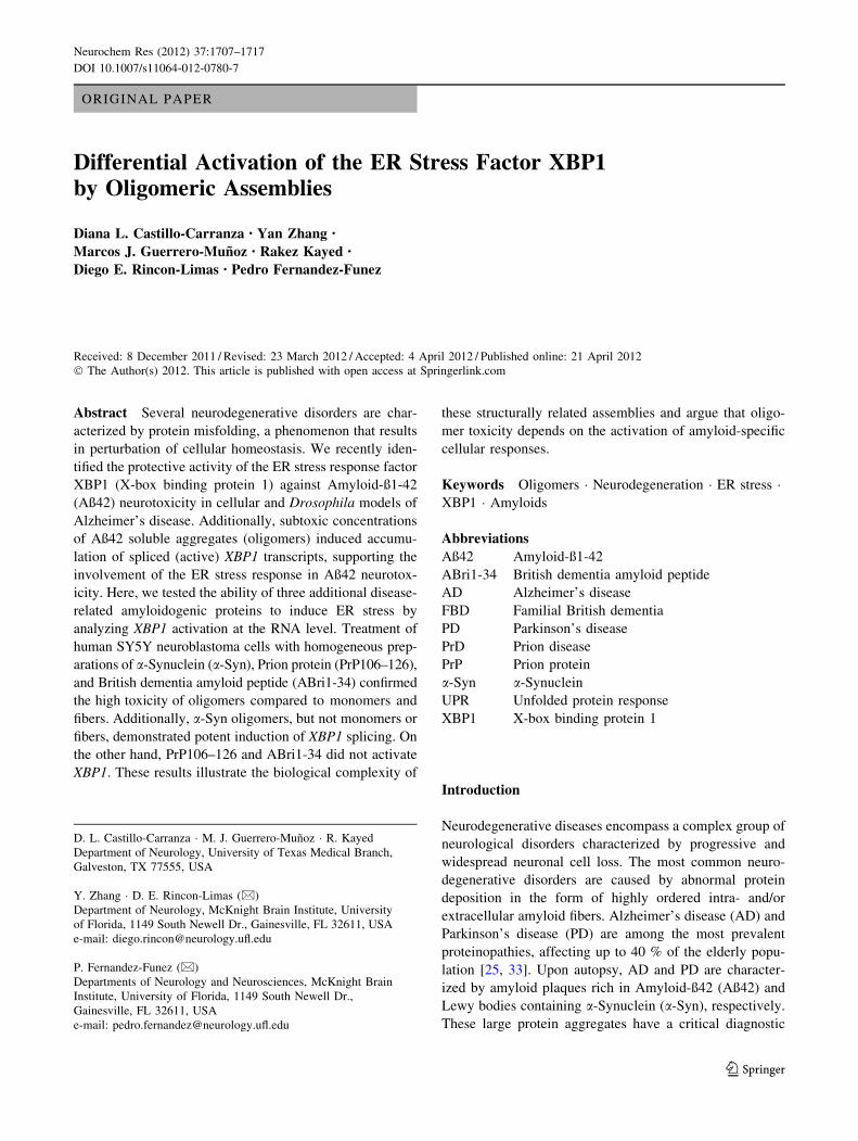
As described above, XBP1 activation can be easily detected
by RT-PCR through the elimination of the 26-nt intron
cleaved by IRE1 [6]. A detailed analysis of these gels
showed that the accumulation of XBP1s was accompanied
by the appearance of an extra band of slightly lower
molecular weight than XBP1s (Fig. 1a, red arrows). This
band was more prominent in conditions that favored XBP1s
accumulation (treatment with Aß42 oligomers), suggesting
that this was an artifact associated with the presence of two
almost identical PCR products, possibly a hybrid XBP1u/
XBP1s duplex. To verify that the extra band contained both
XBP1u and XBP1s, we purified it from an agarose gel and
sequenced it with the forward primer used for the RT-PCR.
The first 57 nt of the sequence matched both isoforms, as
expected, since the primer amplified a common region for
both isoforms (exon 1) (Fig. 1b). However, starting with nt
58 the electropherogram revealed two clear overlapping
peaks with half peak intensity in each position that indicated
the co-existence of two sequences (Fig. 1b, red arrow).
Reading the two peaks by eye, we identified the sequences
corresponding to the alternative intron (XBP1u) and to exon
2 (XBP1s) (Fig. 1b). This result demonstrated that the extra
band was a hybrid containing one copy of XBP1u and
another of XBP1s that forms due to the almost identical
sequence (except the 26-bp intron) of both PCR products.
Knowing the origin of the hybrid band still left the
technical problem of quantifying the XBP1u and XBP1s
isoforms. To eliminate the hybrid band during the elec-
trophoresis, we tried several procedures, including dena-
turing conditions that prevent the formation of secondary
structures in RNA and DNA gels. First, we tried a 60–90 %
formamide gradient combined with heating up the sample
to prevent the formation of the hybrids. Adding the form-
amide after heating the samples at 95 �C made the XBP1u
band more stable, but did not eliminate the hybrid band
(Fig. 1c, left). Heating the samples in the presence of
formamide promoted DNA denaturation and resulted in
weaker XBP1u and XBP1s bands (Fig. 1c, left). We also
tested the effect of running the samples in 1 M urea aga-
rose gels to prevent the formation of the hybrid. The urea
gels resolved the bands neatly, but did not affect the hybrid
(Fig. 1c, center). However, heating the samples at 95 �C
and adding up to 8 M urea alone or in combination with
0.1 % SDS to the loading buffer produced unexpected
effects on the samples that run in multiple weak bands
(Fig. 1c, center). Finally, we tried 6 % Novex TBE poly-
acrylamide gels at high voltage and found that the hybrid
band did not form in these conditions (Fig. 1c, right).
When the PCR products were digested with PstI, the
XBP1u band split in two smaller products, leaving a single,
neat band on top of the gel corresponding to XBP1s
(Fig. 1c, right). It is not entirely clear why the hybrid
disappeared in these conditions, although a number of
factors could contribute to its instability, including the
buffer and the heat generated by the high voltage.
Regardless of the mechanism mediating the elimination of
the hybrid band, these conditions resolving nicely the two
XBP1 isoforms allowed us to test the ability of other
amyloidogenic proteins to induce unconventional splicing
of XBP1.
Oligomeric Assemblies are Highly Toxic
Before we began testing the ability of a-Syn, PrP106-126,
and ABri1-34 to activate XBP1, we checked the cellular
toxicity of each amyloid in different states of aggregation.
For this, we generated monomeric, oligomeric, and fibrillar
forms of a-Syn, PrP106-126, and ABri1-34 and added each
into SY5Y cultures at 2 lM for 8 h in triplicate. The 2 lM
concentration is based on our previous experience and
extensive literature reporting a range of 500 nM to 2 lM
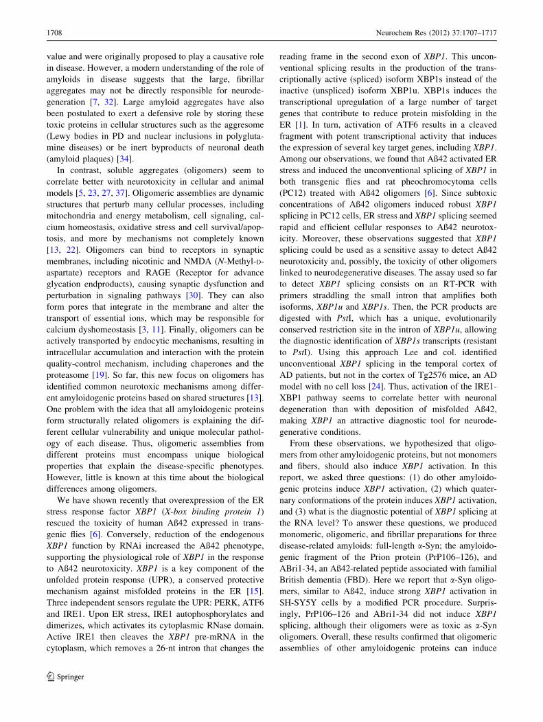
for synthetic assemblies [5, 22]. Then, we determined cell
viability using Alamar Blue, a compound that changes
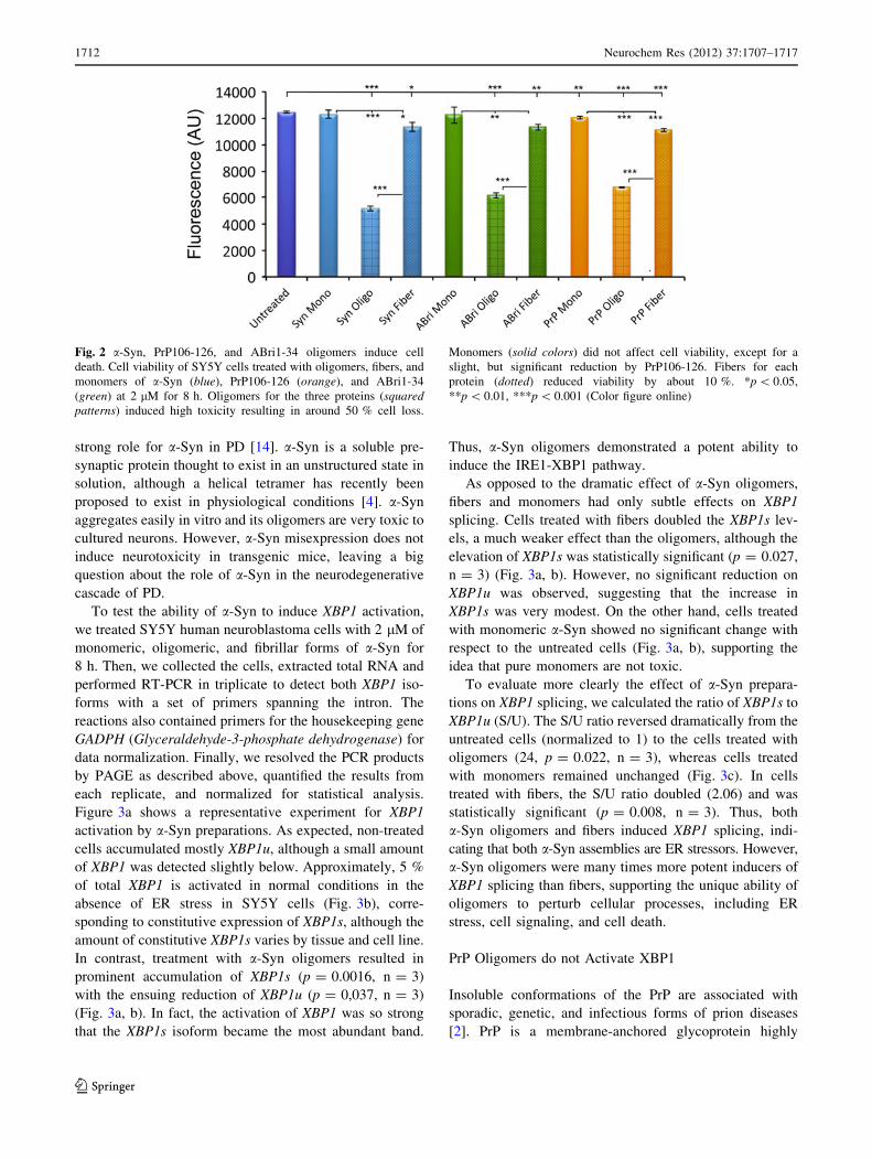
color when reduced inside living cells (Fig. 2). As expec-
ted, a-Syn monomers had no effect on viability, but olig-
omers reduced viability by almost 60 % (p \ 0.0001,
n = 3) (Fig. 2, blue). a-Syn fibers were slightly toxic,
reducing viability by 9 %, but significantly less toxic than
oligomers (p = 0.019, n = 3). Similarly, PrP106–126
oligomers were highly toxic to SY5Y cells, reducing via-
bility by 46 % (p \ 0.0001, n = 3) (Fig. 2, orange).
PrP106–126 fibers were also slightly toxic (89 % viability,
p \ 0.0001, n = 3), but significantly less than the oligo-
mers. Monomers reduced viability by only a few points
(96.5 % viability), although this difference was significant
(p \ 0.005, n = 3), indicating the robustness and sensi-
tivity of the assay. Finally, ABri1-34 showed the same
trend as the other proteins, with highly toxic oligomers
1710 Neurochem Res (2012) 37:1707–1717
123
Page 5
(49 % viability, p \ 0.0001, n = 3) and slightly toxic
fibers (91 % viability, p = 0.005, n = 3)) (Fig. 2, green).
These results demonstrated that oligomers were highly
toxic protein assemblies compared to the monomeric and
highly aggregated forms. These observations revealed that
all oligomers shared a common biological activity (cellular
toxicity) regardless of sequence. Thus, these structures are
ideal candidates to determine whether induction of ER
stress and activation of XBP1 unconventional splicing
are common pathogenic mechanisms mediated by toxic
oligomers.
a-Synuclein Oligomers Activate XBP1 at Low
Concentrations
PD is characterized pathologically by degeneration of
dopaminergic neurons in the substantia nigra and the
accumulation of protein aggregates in the cytosol known as
Lewy bodies and Lewy neurites [25]. a-Syn is highly
enriched in Lewy bodies and is thought to play a key role
in PD neuropathology. At least three mutations in the
coding region of SNCA, the a-Syn gene, as well as multi-
plications of SNCA lead to familial PD, supporting the
Fig. 1 Detection of XBP1s by RT-PCR. a SY5Y cells treated with
increasing concentrations of Aß42 oligomers induce XBP1 splicing
and accumulation of XBP1s, which is visualized by diagnostic
digestion of XBP1u by PstI. The non-digested samples that accumu-
late XBP1s contain a hybrid band that runs lower than XBP1u and
XBP1s (red arrows). The PstI-treated samples can help quantify
XBP1s by digestion of XBP1u, but incomplete digestions complicate
this effort. b Sequence of the hybrid band purified from an agarose
gel. The sequence in the electropherogram matches both XBP1
isoforms until it reaches the intron (red arrow). From this point, two
overlapping peaks were detected corresponding to the XBP1u intron
(red sequence) and XBP1s exon 2. c Elimination of the hybrid band in
gel electrophoresis. Denaturing conditions such as formamide (left)and urea (center) did not eliminate the hybrid band even when
combined with heat or SDS. The size for XBP1u (U) and XBP1s (S) is
shown in the left gel. Resolving the PCR in polyacrylamide gels
(PAGE) produced neat XBP1s and XBP1u bands. Upon digestion with
PstI, a single XBP1s band was left (Color figure online)
Neurochem Res (2012) 37:1707–1717 1711
123
Page 6
strong role for a-Syn in PD [14]. a-Syn is a soluble pre-
synaptic protein thought to exist in an unstructured state in
solution, although a helical tetramer has recently been
proposed to exist in physiological conditions [4]. a-Syn
aggregates easily in vitro and its oligomers are very toxic to
cultured neurons. However, a-Syn misexpression does not
induce neurotoxicity in transgenic mice, leaving a big
question about the role of a-Syn in the neurodegenerative
cascade of PD.
To test the ability of a-Syn to induce XBP1 activation,
we treated SY5Y human neuroblastoma cells with 2 lM of
monomeric, oligomeric, and fibrillar forms of a-Syn for
8 h. Then, we collected the cells, extracted total RNA and
performed RT-PCR in triplicate to detect both XBP1 iso-
forms with a set of primers spanning the intron. The
reactions also contained primers for the housekeeping gene
GADPH (Glyceraldehyde-3-phosphate dehydrogenase) for
data normalization. Finally, we resolved the PCR products
by PAGE as described above, quantified the results from
each replicate, and normalized for statistical analysis.
Figure 3a shows a representative experiment for XBP1
activation by a-Syn preparations. As expected, non-treated
cells accumulated mostly XBP1u, although a small amount
of XBP1 was detected slightly below. Approximately, 5 %
of total XBP1 is activated in normal conditions in the
absence of ER stress in SY5Y cells (Fig. 3b), corre-
sponding to constitutive expression of XBP1s, although the
amount of constitutive XBP1s varies by tissue and cell line.
In contrast, treatment with a-Syn oligomers resulted in
prominent accumulation of XBP1s (p = 0.0016, n = 3)
with the ensuing reduction of XBP1u (p = 0,037, n = 3)
(Fig. 3a, b). In fact, the activation of XBP1 was so strong
that the XBP1s isoform became the most abundant band.
Thus, a-Syn oligomers demonstrated a potent ability to
induce the IRE1-XBP1 pathway.
As opposed to the dramatic effect of a-Syn oligomers,
fibers and monomers had only subtle effects on XBP1
splicing. Cells treated with fibers doubled the XBP1s lev-
els, a much weaker effect than the oligomers, although the
elevation of XBP1s was statistically significant (p = 0.027,
n = 3) (Fig. 3a, b). However, no significant reduction on
XBP1u was observed, suggesting that the increase in
XBP1s was very modest. On the other hand, cells treated
with monomeric a-Syn showed no significant change with
respect to the untreated cells (Fig. 3a, b), supporting the
idea that pure monomers are not toxic.
To evaluate more clearly the effect of a-Syn prepara-
tions on XBP1 splicing, we calculated the ratio of XBP1s to
XBP1u (S/U). The S/U ratio reversed dramatically from the
untreated cells (normalized to 1) to the cells treated with
oligomers (24, p = 0.022, n = 3), whereas cells treated
with monomers remained unchanged (Fig. 3c). In cells
treated with fibers, the S/U ratio doubled (2.06) and was
statistically significant (p = 0.008, n = 3). Thus, both
a-Syn oligomers and fibers induced XBP1 splicing, indi-
cating that both a-Syn assemblies are ER stressors. However,
a-Syn oligomers were many times more potent inducers of
XBP1 splicing than fibers, supporting the unique ability of
oligomers to perturb cellular processes, including ER
stress, cell signaling, and cell death.
PrP Oligomers do not Activate XBP1
Insoluble conformations of the PrP are associated with
sporadic, genetic, and infectious forms of prion diseases
[2]. PrP is a membrane-anchored glycoprotein highly
Fig. 2 a-Syn, PrP106-126, and ABri1-34 oligomers induce cell
death. Cell viability of SY5Y cells treated with oligomers, fibers, and
monomers of a-Syn (blue), PrP106-126 (orange), and ABri1-34
(green) at 2 lM for 8 h. Oligomers for the three proteins (squaredpatterns) induced high toxicity resulting in around 50 % cell loss.
Monomers (solid colors) did not affect cell viability, except for a
slight, but significant reduction by PrP106-126. Fibers for each
protein (dotted) reduced viability by about 10 %. *p \ 0.05,
**p \ 0.01, ***p \ 0.001 (Color figure online)
1712 Neurochem Res (2012) 37:1707–1717
123
Page 7
expressed in the brain that is soluble in non-ionic detergents
and easily digested by proteases. In its disease-associated
‘scrapie’ conformation, PrP is detergent insoluble, resistant
to proteases and forms fibrillar aggregates [9]. PrP can
misfold due to mutations in different domains, but spo-
radic forms of the disease show similar conformational
changes as wild type PrP, indicating the intrinsic structural
instability of PrP. Although much is known about the
3-dimentional structure of PrP and its conformational
dynamics, it is still unclear how PrP causes neural loss and
disease.
For these studies, we used the PrP106–126 peptide to
take advantage of its reported ability to induce neurotox-
icity in cell culture [31]. This peptide contains the hydro-
phobic domain key for PrP fibrilization and forms amyloid
fibers in vitro, while full-length PrP requires a tissue
homogenate to do so, making it a less appropriate substrate
for cell culture studies. As described above for a-Syn, we
prepared homogenous PrP106–126 monomers, oligomers,
and fibrils, treated SY5Y cells, and determined the acti-
vation of XBP1. In Fig. 2 we showed that PrP106–126
oligomers are as toxic as a-Syn oligomers. To our surprise,
the effect of PrP106–126 assemblies on XBP1 was very
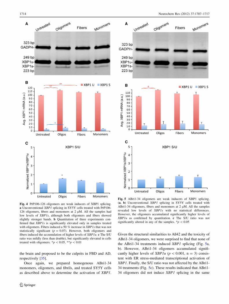
different from a-Syn. PrP106–126 oligomers at 2 lM
showed poor induction of XBP1 splicing (Fig. 4a). Upon
quantification, we detected small, albeit significant, chan-
ges in the cells treated with oligomers and fibers, but not
with monomers. PrP106–126 oligomers accumulated
slightly higher levels of XBP1s (p = 0.017, n = 3), indi-
cating weak induction of XBP1 splicing (Fig. 4b). In
addition, both PrP106–126 oligomers and fibers showed
significantly higher levels of XBP1u (p = 0.0025 and
p = 0.0021, respectively, n = 3) (Fig. 4a). This result
indicated ER stress-dependent transcriptional activation of
XBP1, which is typically mediated by the ATF6 sensor.
PrP106–126 monomers, on the other hand, showed no
significant changes in XBP1 compared with the untreated
controls (Fig. 4a, b), supporting the specific effects of
PrP106–126 assemblies on XBP1 levels. The S/U ratio was
low in all conditions, but the PrP106–126 oligomers
showed a slight increase that was statistically significant
(p = 0.027, n = 3). In summary, PrP106–126 oligomers
weakly activated XBP1 expression and splicing, demon-
strating critical differences with a-Syn oligomers.
ABri1-34 Oligomers Activate XBP1 at High
Concentrations
Familial British dementia (FBD) is an autosomal dominant
disorder characterized by progressive cognitive impairment
and cerebellar ataxia. These symptoms are associated with
amyloid deposition and neurofibrillary degeneration, shar-
ing some similarities with AD [36]. FBD is linked to a
mutation that eliminates the normal stop codon on BRI2,
resulting in a longer precursor protein that generates a
novel 34-residue amyloidogenic peptide named ABri1-34.
Although ABri1-34 and Aß42 do not have sequence
homology, they share many biological characteristics: both
are secreted peptides that accumulate amyloid plaques in
Fig. 3 a-Syn oligomers are strong inducers of XBP1 splicing.
a Unconventional XBP1 splicing in SY5Y cells treated with a-Syn
oligomers, fibers and monomers at 2 lM detected by RT-PCR.
Untreated samples along with samples treated with monomers show
high levels of XBP1u and very low levels of XBP1s. Samples treated
with oligomers show the opposite, with higher levels of XBP1s than
XBP1u. Samples treated with fibers show a subtle increase in XBP1s.
b Quantitation of three independent experiments confirmed that
monomers did not increase XBP1s, but fibers significantly increased
XBP1s. However, oligomers had the largest effect by far, significantly
reducing the levels of XBP1u. c The S/U ratio increases 24-fold in
cells treated with a-Syn oligomers. Fibers double the S/U ratio, a
significant difference with respect to untreated samples. a-Syn
monomers do not affect the S/U ratio. *p \ 0.05, **p \ 0.01
Neurochem Res (2012) 37:1707–1717 1713
123
Page 8
the brain and proposed to be the culprits in FBD and AD,
respectively [35].
Once again, we prepared homogenous ABri1-34
monomers, oligomers, and fibrils, and treated SY5Y cells
as described above to determine the activation of XBP1.
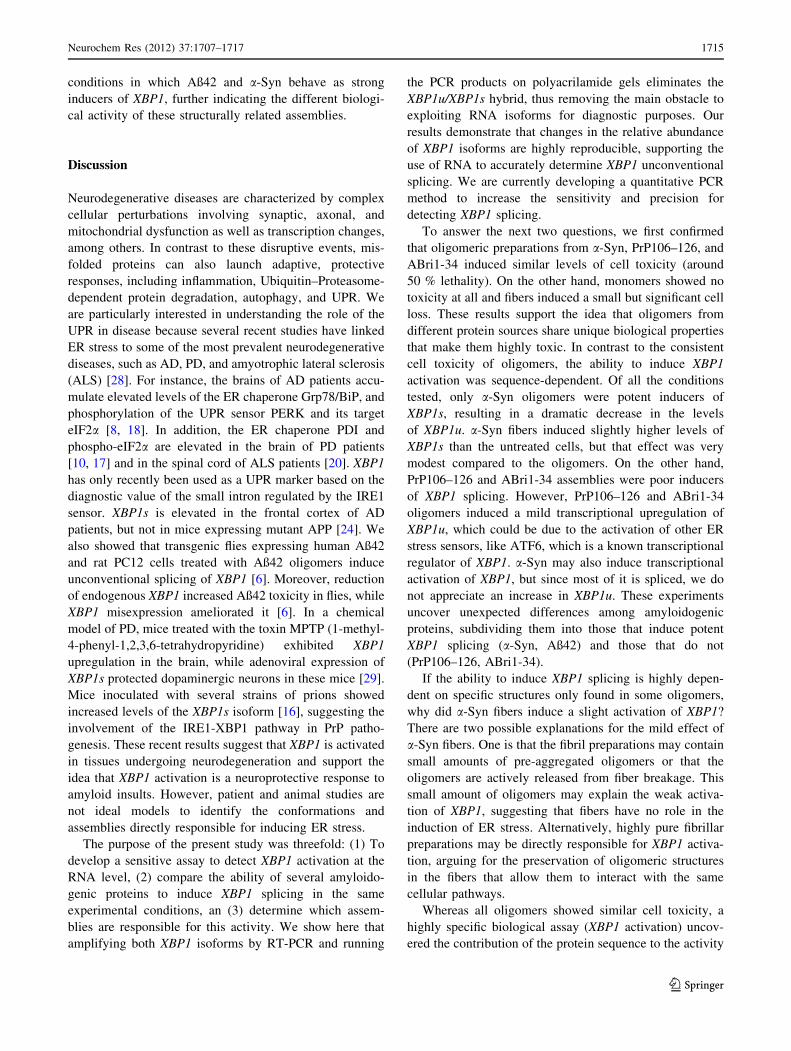
Given the structural similarities to Aß42 and the toxicity of
ABri1-34 oligomers, we were surprised to find that none of
the ABri1-34 treatments induced XBP1 splicing (Fig. 5a,
b). However, ABri1-34 oligomers accumulated signifi-
cantly higher levels of XBP1u (p \ 0.001, n = 3) consis-
tent with ER stress-mediated transcriptional activation of
XBP1. Finally, the S/U ratio was not affected by the ABri1-
34 treatments (Fig. 5c). These results indicated that ABri1-
34 oligomers did not induce XBP1 splicing in the same
Fig. 4 PrP106-126 oligomers are weak inducers of XBP1 splicing.
a Unconventional XBP1 splicing in SY5Y cells treated with PrP106-
126 oligomers, fibers and monomers at 2 lM. All the samples had
low levels of XBP1s, although both oligomers and fibers showed
slightly stronger bands. b Quantitation of three experiments con-
firmed that XBP1s is significantly elevated only in samples treated
with oligomers. Fibers induced a 50 % increase in XBP1s that was not
statistically significant (p = 0.07). However, both oligomers and
fibers induced the accumulation of higher levels of XBP1u. c The S/U
ratio was mildly (less than double), but significantly elevated in cells
treated with oligomers. *p \ 0.05, **p \ 0.01
Fig. 5 ABri1-34 oligomers are weak inducers of XBP1 splicing.
(a, b) Unconventional XBP1 splicing in SY5Y cells treated with
ABri1-34 oligomers, fibers and monomers at 2 lM. All the samples
revealed low levels of XBP1s with no statistical differences.
However, the oligomers accumulated significantly higher levels of
XBP1u as confirmed by quantitation. c The S/U ratio was not
significantly altered in any of the samples. *p \ 0.05
1714 Neurochem Res (2012) 37:1707–1717
123
Page 9
conditions in which Aß42 and a-Syn behave as strong
inducers of XBP1, further indicating the different biologi-
cal activity of these structurally related assemblies.
Discussion
Neurodegenerative diseases are characterized by complex
cellular perturbations involving synaptic, axonal, and
mitochondrial dysfunction as well as transcription changes,
among others. In contrast to these disruptive events, mis-
folded proteins can also launch adaptive, protective
responses, including inflammation, Ubiquitin–Proteasome-
dependent protein degradation, autophagy, and UPR. We
are particularly interested in understanding the role of the
UPR in disease because several recent studies have linked
ER stress to some of the most prevalent neurodegenerative
diseases, such as AD, PD, and amyotrophic lateral sclerosis
(ALS) [28]. For instance, the brains of AD patients accu-
mulate elevated levels of the ER chaperone Grp78/BiP, and
phosphorylation of the UPR sensor PERK and its target
eIF2a [8, 18]. In addition, the ER chaperone PDI and
phospho-eIF2a are elevated in the brain of PD patients
[10, 17] and in the spinal cord of ALS patients [20]. XBP1
has only recently been used as a UPR marker based on the
diagnostic value of the small intron regulated by the IRE1
sensor. XBP1s is elevated in the frontal cortex of AD
patients, but not in mice expressing mutant APP [24]. We
also showed that transgenic flies expressing human Aß42
and rat PC12 cells treated with Aß42 oligomers induce
unconventional splicing of XBP1 [6]. Moreover, reduction
of endogenous XBP1 increased Aß42 toxicity in flies, while
XBP1 misexpression ameliorated it [6]. In a chemical
model of PD, mice treated with the toxin MPTP (1-methyl-
4-phenyl-1,2,3,6-tetrahydropyridine) exhibited XBP1
upregulation in the brain, while adenoviral expression of
XBP1s protected dopaminergic neurons in these mice [29].
Mice inoculated with several strains of prions showed
increased levels of the XBP1s isoform [16], suggesting the
involvement of the IRE1-XBP1 pathway in PrP patho-
genesis. These recent results suggest that XBP1 is activated
in tissues undergoing neurodegeneration and support the
idea that XBP1 activation is a neuroprotective response to
amyloid insults. However, patient and animal studies are
not ideal models to identify the conformations and
assemblies directly responsible for inducing ER stress.
The purpose of the present study was threefold: (1) To
develop a sensitive assay to detect XBP1 activation at the
RNA level, (2) compare the ability of several amyloido-
genic proteins to induce XBP1 splicing in the same
experimental conditions, an (3) determine which assem-
blies are responsible for this activity. We show here that
amplifying both XBP1 isoforms by RT-PCR and running
the PCR products on polyacrilamide gels eliminates the
XBP1u/XBP1s hybrid, thus removing the main obstacle to
exploiting RNA isoforms for diagnostic purposes. Our
results demonstrate that changes in the relative abundance
of XBP1 isoforms are highly reproducible, supporting the
use of RNA to accurately determine XBP1 unconventional
splicing. We are currently developing a quantitative PCR
method to increase the sensitivity and precision for
detecting XBP1 splicing.
To answer the next two questions, we first confirmed
that oligomeric preparations from a-Syn, PrP106–126, and
ABri1-34 induced similar levels of cell toxicity (around
50 % lethality). On the other hand, monomers showed no
toxicity at all and fibers induced a small but significant cell
loss. These results support the idea that oligomers from
different protein sources share unique biological properties
that make them highly toxic. In contrast to the consistent
cell toxicity of oligomers, the ability to induce XBP1
activation was sequence-dependent. Of all the conditions
tested, only a-Syn oligomers were potent inducers of
XBP1s, resulting in a dramatic decrease in the levels
of XBP1u. a-Syn fibers induced slightly higher levels of
XBP1s than the untreated cells, but that effect was very
modest compared to the oligomers. On the other hand,
PrP106–126 and ABri1-34 assemblies were poor inducers
of XBP1 splicing. However, PrP106–126 and ABri1-34
oligomers induced a mild transcriptional upregulation of
XBP1u, which could be due to the activation of other ER
stress sensors, like ATF6, which is a known transcriptional
regulator of XBP1. a-Syn may also induce transcriptional
activation of XBP1, but since most of it is spliced, we do
not appreciate an increase in XBP1u. These experiments
uncover unexpected differences among amyloidogenic
proteins, subdividing them into those that induce potent
XBP1 splicing (a-Syn, Aß42) and those that do not
(PrP106–126, ABri1-34).
If the ability to induce XBP1 splicing is highly depen-
dent on specific structures only found in some oligomers,
why did a-Syn fibers induce a slight activation of XBP1?
There are two possible explanations for the mild effect of
a-Syn fibers. One is that the fibril preparations may contain
small amounts of pre-aggregated oligomers or that the
oligomers are actively released from fiber breakage. This
small amount of oligomers may explain the weak activa-
tion of XBP1, suggesting that fibers have no role in the
induction of ER stress. Alternatively, highly pure fibrillar
preparations may be directly responsible for XBP1 activa-
tion, arguing for the preservation of oligomeric structures
in the fibers that allow them to interact with the same
cellular pathways.
Whereas all oligomers showed similar cell toxicity, a
highly specific biological assay (XBP1 activation) uncov-
ered the contribution of the protein sequence to the activity
Neurochem Res (2012) 37:1707–1717 1715
123
Page 10
of oligomers from four protein sources. The different
ability of Aß42 and a-Syn oligomers to induce XBP1
splicing compared to ABri1-34 and PrP106–126 oligomers
support the existence of some degree of variation in the
conformation of these two groups of oligomers. Unfortu-
nately, it is unclear at this point what makes Aß42 and
a-Syn capable of activating IRE1-XBP1 and why ABri1-34
and PrP106–126 do not. The available experimental evi-
dence suggests that there may be little structural variation
among the oligomeric conformations. This is supported by
the ability of a few conformational antibodies to recognize
multiple oligomeric species obtained from synthetic or
biological sources and prepared by different methods [12].
These results argue for the existence of few stable con-
formations compatible with the formation of neurotoxic
oligomers. Also, most oligomers show the ability to perturb
membrane integrity and disrupt ion metabolism [11],
pointing to common biological activities [13]. Since acti-
vation of UPR requires the perturbation of an internal
organelle (the ER), exogenous Aß42 and a-Syn may be
more efficiently transported into the ER by endocytic
mechanisms. If this were the case, this would indicate the
differential recognition of some oligomeric conformations,
but not all, by specific receptors or transporters. Thus, we
report here that XBP1 and the ER stress play different roles
in neurodegenerative diseases, although the mechanisms
underlying these differences are not clear. Additional
structural approaches in the future may contribute to
resolve in more detail the similarities and differences
among these conformers critical in many chronic disorders.
Overall, we report here a strong connection of a-Syn to
induction of ER stress and the XBP1-IRE1 pathway.
Importantly, a-Syn misfolding and aggregation is an salient
pathological feature of other neurological disorders,
including dementia with Lewy bodies and multiple systems
atrophy, suggesting that ER stress may be a common
component of other synucleinopathies. Thus, identification
of the signals that result in UPR and amelioration of this
cellular response may contribute to the treatment of several
synucleinopathies. On the other hand, there seems to be
less consensus on the role of ER stress in prion diseases.
The inability of PrP106–126 to induce XBP1 splicing
agrees with the observation that elimination of XBP1 in
mice did not alter the course of prion disease [16], sug-
gesting that XBP1 plays no physiological role in prion
diseases. Finally, FBD is a rare dementia and little is
known about its specific pathobiology. Our results indicate
that despite the strong similarities between Aß42 and
ABri1-34 (two small, secreted, amyloidogenic peptides
that cause neurodegeneration), they may cause toxicity
through different cellular pathways. In conclusion, we
describe here the differential activation of XBP1 by four
amyloidogenic proteins, suggesting a complex involvement
of UPR in disease, a pathway that in the last few years has
been connected to a wide array of human maladies,
including cancer, ischemia, and several chronic disorders
[38].
Acknowledgments This work was supported by the NIH grant DP2
OD002721-01 to PF-F, start-up funds from the UF Department of
Neurology to DER-L and PF-F, and funding from the Mitchell Center
for Neurodegenerative Diseases at UTMB for RK.
Open Access This article is distributed under the terms of the
Creative Commons Attribution License which permits any use, dis-
tribution, and reproduction in any medium, provided the original
author(s) and the source are credited.
References
1. Acosta-Alvear D, Zhou Y, Blais A, Tsikitis M, Lents NH, Arias
C, Lennon CJ, Kluger Y, Dynlacht BD (2007) XBP1 controls
diverse cell type- and condition-specific transcriptional regulatory
networks. Mol Cell 27:53–66
2. Aguzzi A, Sigurdson C, Heikenwaelder M (2008) Molecular
mechanisms of prion pathogenesis. Annu Rev Pathol 3:11–40
3. Arispe N, Rojas E, Pollard HB (1993) Alzheimer disease amyloid
beta protein forms calcium channels in bilayer membranes:
blockade by tromethamine and aluminum. Proc Natl Acad Sci U
S A 90:567–571
4. Bartels T, Choi JG, Selkoe DJ (2011) alpha-Synuclein occurs
physiologically as a helically folded tetramer that resists aggre-
gation. Nature 477:107–110
5. Benilova I, Karran E, De Strooper B (2012) The toxic Abeta
oligomer and Alzheimer’s disease: an emperor in need of clothes.
Nat Neurosci 15:349–357
6. Casas-Tinto S, Zhang Y, Sanchez-Garcia J, Gomez-Velazquez
M, Rincon-Limas DE, Fernandez-Funez P (2011) The ER stress
factor XBP1 s prevents amyloid-beta neurotoxicity. Hum Mol
Genet 20:2144–2160
7. Caughey B, Lansbury PT (2003) Protofibrils, pores, fibrils, and
neurodegeneration: separating the responsible protein aggregates
from the innocent bystanders. Annu Rev Neurosci 26:267–298
8. Chang RC, Wong AK, Ng HK, Hugon J (2002) Phosphorylation
of eukaryotic initiation factor-2alpha (eIF2alpha) is associated
with neuronal degeneration in Alzheimer’s disease, NeuroReport
13:2429–2432
9. Colby DW, Prusiner SB (2011) Prions. Cold Spring Harb Per-
spect Biol 3:a006833
10. Conn KJ, Gao W, McKee A, Lan MS, Ullman MD, Eisenhauer
PB, Fine RE, Wells JM (1022) Identification of the protein
disulfide isomerase family member PDIp in experimental Par-
kinson’s disease and Lewy body pathology. Brain Res 2004:
164–172
11. Demuro A, Mina E, Kayed R, Milton SC, Parker I, Glabe CG
(2005) Calcium dysregulation and membrane disruption as a
ubiquitous neurotoxic mechanism of soluble amyloid oligomers.
J Biol Chem 280:17294–17300
12. Glabe CG (2008) Structural classification of toxic amyloid olig-
omers. J Biol Chem 283:29639–29643
13. Glabe CG, Kayed R (2006) Common structure and toxic function
of amyloid oligomers implies a common mechanism of patho-
genesis. Neurology 66:S74–S78
14. Hardy J, Lewis P, Revesz T, Lees A, Paisan-Ruiz C (2009) The
genetics of Parkinson’s syndromes: a critical review. Curr Opin
Genet Dev 19:254–265
1716 Neurochem Res (2012) 37:1707–1717
123
Page 11
15. Hetz C (2012) The unfolded protein response: controlling cell
fate decisions under ER stress and beyond. Nat Rev Mol Cell Biol
13:89–102
16. Hetz C, Lee AH, Gonzalez-Romero D, Thielen P, Castilla J, Soto
C, Glimcher LH (2008) Unfolded protein response transcription
factor XBP-1 does not influence prion replication or pathogene-
sis. Proc Natl Acad Sci U S A 105:757–762
17. Hoozemans JJ, van Haastert ES, Eikelenboom P, de Vos RA,
Rozemuller JM, Scheper W (2007) Activation of the unfolded
protein response in Parkinson’s disease. Biochem Biophys Res
Commun 354:707–711
18. Hoozemans JJ, Veerhuis R, Van Haastert ES, Rozemuller JM,
Baas F, Eikelenboom P, Scheper W (2005) The unfolded protein
response is activated in Alzheimer’s disease. Acta Neuropathol
(Berl) 110:165–172
19. Hu X, Crick SL, Bu G, Frieden C, Pappu RV, Lee JM (2009)
Amyloid seeds formed by cellular uptake, concentration, and
aggregation of the amyloid-beta peptide. Proc Natl Acad Sci U S
A 106:20324–20329
20. Ilieva EV, Ayala V, Jove M, Dalfo E, Cacabelos D, Povedano M,
Bellmunt MJ, Ferrer I, Pamplona R, Portero-Otin M (2007)
Oxidative and endoplasmic reticulum stress interplay in sporadic
amyotrophic lateral sclerosis. Brain 130:3111–3123
21. Kayed R, Glabe CG (2006) Conformation-dependent anti-
amyloid oligomer antibodies. Methods Enzymol 413:326–344
22. Kayed R, Head E, Thompson JL, McIntire TM, Milton SC,
Cotman CW, Glabe CG (2003) Common structure of soluble
amyloid oligomers implies common mechanism of pathogenesis.
Science 300:486–489
23. Lambert MP, Barlow AK, Chromy BA, Edwards C, Freed R,
Liosatos M, Morgan TE, Rozovsky I, Trommer B, Viola KL,
Wals P, Zhang C, Finch CE, Krafft GA, Klein WL (1998) Dif-
fusible, nonfibrillar ligands derived from Abeta1-42 are potent
central nervous system neurotoxins. Proc Natl Acad Sci U S A
95:6448–6453
24. Lee JH, Won SM, Suh J, Son SJ, Moon GJ, Park UJ, Gwag BJ
(2010) Induction of the unfolded protein response and cell death
pathway in Alzheimer’s disease, but not in aged Tg2576 mice.
Exp Mol Med 42:386–394
25. Lees AJ, Hardy J, Revesz T (2009) Parkinson’s disease. Lancet
373:2055–2066
26. Masek T, Vopalensky V, Suchomelova P, Pospisek M (2005)
Denaturing RNA electrophoresis in TAE agarose gels. Anal
Biochem 336:46–50
27. McLean CA, Cherny RA, Fraser FW, Fuller SJ, Smith MJ,
Beyreuther K, Bush AI, Masters CL (1999) Soluble pool of Abeta
amyloid as a determinant of severity of neurodegeneration in
Alzheimer’s disease. Ann Neurol 46:860–866
28. Paschen W, Mengesdorf T (2005) Endoplasmic reticulum stress
response and neurodegeneration. Cell Calcium 38:409–415
29. Sado M, Yamasaki Y, Iwanaga T, Onaka Y, Ibuki T, Nishihara S,
Mizuguchi H, Momota H, Kishibuchi R, Hashimoto T, Wada D,
Kitagawa H, Watanabe TK (2009) Protective effect against Par-
kinson’s disease-related insults through the activation of XBP1.
Brain Res 1257:16–24
30. Selkoe DJ (2002) Alzheimer’s disease is a synaptic failure.
Science 298:789–791
31. Singh N, Gu Y, Bose S, Kalepu S, Mishra RS, Verghese S (2002)
Prion peptide 106-126 as a model for prion replication and
neurotoxicity. Front Biosci 7:a60–a71
32. Terry RD (1996) The pathogenesis of Alzheimer disease: an
alternative to the amyloid hypothesis. J Neuropathol Exp Neurol
55:1023–1025
33. Thies W, Bleiler L (2011) Alzheimer’s disease facts and figures.
Alzheimers Dement 7(2011):208–244
34. Truant R, Atwal RS, Desmond C, Munsie L, Tran T (2008)
Huntington’s disease: revisiting the aggregation hypothesis in
polyglutamine neurodegenerative diseases. FEBS J 275:4252–4262
35. Tsachaki M, Ghiso J, Efthimiopoulos S (2008) BRI2 as a central
protein involved in neurodegeneration. Biotechnol J 3:1548–1554
36. Vidal R, Frangione B, Rostagno A, Mead S, Revesz T, Plant G,
Ghiso J (1999) A stop-codon mutation in the BRI gene associated
with familial British dementia. Nature 399:776–781
37. Walsh DM, Selkoe DJ (2004) Deciphering the molecular basis of
memory failure in Alzheimer’s disease. Neuron 44:181–193
38. Yoshida H (2007) ER stress and diseases. FEBS J 274:630–658
Neurochem Res (2012) 37:1707–1717 1717
123