jou rn al hom epage : w ww.elsev ier .com/ locate /an i r eprosc i
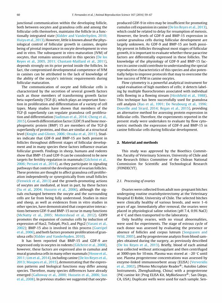
ifferential expression of GDF-9 and BMP- 15 duringollicular development in canine ovaries evaluated by flowytometry
omas Fernandeza, Jaime Palominoa, Victor H. Parraguezb, Oscar A. Peraltaa,onica De los Reyesa,∗
Laboratory of Animal Reproduction, Faculty of Veterinary Sciences, University of Chile, Casilla 2 Correo 15, Santiago, ChileLaboratory of Animal Physiology, Faculty of Veterinary Sciences, University of Chile, Casilla 2 Correo 15, Santiago, Chile
r t i c l e i n f o
rticle history:eceived 12 October 2015eceived in revised form 18 January 2016ccepted 1 February 2016vailable online 3 February 2016
eywords:ntral folliclesogrowth factorsstrus cycle
a b s t r a c t
Growth differentiation factor 9 (GDF-9) and bone morphogenetic protein 15 (BMP-15) playimportant functions in follicular and oocyte development in many species. This study eval-uated the dynamic expression of GDF-9 and BMP-15 in canine follicles cells using flowcytometry analysis. Follicular cells were removed from three sizes of antral follicles (small,medium and large) from ovaries of bitches throughout the estrus cycle. Cells were incu-bated with anti-human GDF-9 polyclonal and anti-mouse BMP-15 monoclonal antibodies.A size and complexity discriminatory gate was used for the cytometryc analysis in theinitial dot plot and, additionally, a CD45 marker for leukocyte and propidium iodide (PI)were used for erythrocyte and debris discrimination. The evidence corroborated the pres-ence of both proteins in canine follicle cells, but these proteins were not expressed equallyduring follicular development. The results analyzed by ANOVA showed that GDF-9 expres-sion decreased (P < 0.05) during follicular growth in anestrus and proestrous/estrous, butincreased in diestrus (P < 0.05). The expression levels of BMP-15 rose (P < 0.05) from small tomedium sizes in anestrous without changing at diestrus. Small antral follicles expressed thehighest values of GDF-9 at anestrus while only BMP-15 showed higher value in small antralfollicles at proestrous-estrus compared to diestrus and anestrus. Both proteins decreased
in proestrous/estrous (P < 0.05) with increasing follicle size, registering the lowest levels inlarge follicles. The flow cytometric assay was able to assess GDF-9 and BMP-15 expressionin canine follicular cells, showing that these proteins were differentially expressed duringfollicular development, possibly related to the special features of canine reproduction.
∗ Corresponding author at: University of Chile, Faculty of Veterinaryciences. Laboratory of Animal Reproduction, Santa Rosa 11735, Santiago,hile. Tel.: +56 2 29785534; fax: +56 2 29785611.
Oocyte development and follicle growth are closelyregulated by an ordered and complex series of signalingevents throughout folliculogenesis. The interplay between
oocytes and the somatic cells of ovarian follicles deter-mines the developmental ability of these cells. During thisprocess, meiotic competence of the oocyte is gained grad-ually (Eppig, 2001; Fair, 2003; Hussein et al., 2006) and gap
junctional communication within the developing follicle,both between oocytes and granulosa cells and among thefollicular cells themselves, maintains the follicle in a func-tionally integrated state (Kidder and Vanderhyden, 2010;Huiyu et al., 2013). However, little is known about the phys-iological control of follicular growth in canines, despitebeing of pivotal importance in oocyte development in vivoand in vitro. The subsequent in vitro maturation (IVM) ofoocytes, that remains unsuccessful in this species (De losReyes et al., 2005, 2011; Chastant-Maillard et al., 2011),depends strongly on its prior period inside the follicles. Infact, the compromised developmental competence of IVMin canines can be attributed to the lack of knowledge ofthe ability of the oocyte’s intrinsic requirements duringfollicular maturation.
The communication of oocyte and follicular cells ischaracterized by the secretion of several growth factorssuch as those members of transforming growth factorbeta superfamily (TGF-�), which plays an important func-tion in proliferation and differentiation of a variety of celltypes. Many studies have shown that members of theTGF-� superfamily can regulate granulosa cell prolifera-tion and differentiation (Sudiman et al., 2014; Cheng et al.,2015). Growth differentiation factor (GDF) 9 and bone mor-phogenetic protein (BMP) 15 are members of the TGF-�superfamily of proteins, and thus are similar at a structurallevel (Knight and Glister, 2006; Otsuka et al., 2011). Stud-ies indicate that GDF-9 and BMP-15 are both present infollicles throughout different stages of follicular develop-ment and in many species these factors influence ovarianfollicular growth. Findings in sheep, humans and rodentsshow that BMP-15 and GDF-9 can be considered to be newtargets for fertility regulation in mammals (Gilchrist et al.,2006; Persani et al., 2014), as they participate in signalingpathways that control the development of ovarian follicles.These proteins are thought to affect granulosa cell prolifer-ation independently or synergistically from small follicles(Fenwick et al., 2013) and the growth-promoting actionsof oocytes are mediated, at least in part, by these factors(Su et al., 2004; Hussein et al., 2006), although the sig-nals exchanged between the oocyte and the surroundingcells are far from being fully understood. Studies in miceand sheep, as well as evidences from in vitro studies inother species, have demonstrated that cooperative interac-tions between GDF-9 and BMP-15 occur in many functions(McNatty et al., 2005; Mottershead et al., 2012). GDF9promotes the expansion of cumulus cells by induction ofexpression of Has2, Tnfaip6, Ptx3, and Ptgs2 (Varani et al.,2002); BMP-15 also is involved in this process (Gueripelet al., 2006), and both factors promote proliferation of gran-ulosa cells (Kidder and Vanderhyden, 2010).
It has been reported that BMP-15 and GDF-9 areexpressed only in oocytes in rodents (Gilchrist et al., 2008),however, these factors are expressed also in cumulus andmural granulosa cells in many other mammals (Hosoe et al.,2011; Lim et al., 2014), including canine (De los Reyes et al.,2013; Maupeu et al., 2015), demonstrating that the expres-
sion patterns and biological functions may differ amongspecies. Therefore, many species differences have alreadyemerged (Galloway et al., 2000; Hussein et al., 2006; Sunet al., 2008). In previous studies we suggested that oocyte-
ion Science 167 (2016) 59–67
produced GDF-9 in vitro may be insufficient for promotingcumulus cell expansion in canine (De los Reyes et al., 2013),which could be related to delay for resumption of meiosis.However, the levels of GDF-9 and BMP-15 expression incanine granulosa cells during follicular development arelargely unknown. As GDF-9 and BMP-15 are both possi-bly present in follicles throughout most stages of folliculargrowth, it is important to evaluate whether these paracrinefactors are differentially expressed in these follicles. Theknowledge of the physiology of GDF-9 and BMP-15 fac-tors in canine could contribute to understanding the specialreproductive characteristics of this species and also poten-tially helps to improve protocols that may to overcome thelow success of IVM in canine oocytes.
Flow cytometry is a powerful analytical instrument forrapid evaluation of high numbers of cells; it detects label-ing by multiple fluorochromes associated with individualcells flowing in a flowing stream (Hirshfield et al., 1988).This technique has been successfully used for granulosacell analysis (Rao et al., 1991; De Neubourg et al., 1996;Douville and Sirard, 2014; Regan et al., 2015), providingan alternative method for assessing protein expression infollicular cells. Therefore, the experiments reported in thepresent study were undertaken to evaluate by flow cyto-metric methods the expression of GDF-9 and BMP-15 incanine follicular cells during follicular development.
2. Material and methods
This study was approved by the Bioethics Commit-tee, Faculty of Veterinary Sciences, University of Chile andthe Research Ethics Committee of the Chilean NationalCommission for Scientific and Technological Research(FONDECYT).
2.1. Processing of ovaries
Ovaries were collected from adult non-pregnant bitchesundergoing routine ovariohysterectomy at the VeterinaryHospital El Roble, University of Chile. The selected bitcheswere clinically healthy of various breeds, and were 1–6years of age. Immediately after removal, the ovaries wereplaced in physiological saline solution (pH 7.4, 0.9% NaCl)at 4 ◦C and then transported to the laboratory.
Only healthy ovaries, with no visual abnormalities,were used for experiments. The stage of estrous cycle ofeach donor was assessed by evaluating the presence orabsence of follicles and corpus luteum (Songsasen andWild, 2005), and by progesterone analysis from blood sam-ples obtained during the surgery, as previously described(De los Reyes et al., 2013). Briefly, blood of each animalwas collected without anticoagulant and then centrifugedat 3000 rpm for 10 min. Plasma was stored at −20 ◦C untiluse. Plasma progesterone concentrations was assessed byenzyme-linked immunosorbent assay (ELISA) (Ververidis
et al., 2002), (PHomo Microplate Reader®; Autobio LabtecInstruments, Zhenghaidong, China) with a progesterone(P4) canine kit (Prog ELISA Kit, MyBioSource®; San Diego,CA, USA). Duplicate wells were used for each sample. Sen-
producti
si
2
spio1dotMJcMcmifewicliwormt8
2
Ppf8aNtSswihatioBwtidsE
T. Fernandez et al. / Animal Re
itivity of the assay was 0.19 ng/mL. The mean intra-andnter assay precision was 5.1% and 6%, respectively.
.2. Follicles and granulosa cells isolation
All canine ovaries were washed in phosphate-bufferedaline (PBS) and the ovary cortex was dissected into smallieces under a stereomicroscope (Lieder MZ-730-J6 Amer-
can Scientific, Portland, OR, USA). The small pieces ofvaries were incubated with 1 mg/mL Collagenase (C0130-00MG Sigma, MO, USA) in PBS for 90 min, in order toisaggregate the ovarian tissue and facilitate the isolationf follicles. Antral follicles were retrieved free of stromalissue by using a 29 gauge needle (Nipro Corporation,
iami, FL, USA) under a stereomicroscope (Leider MZ-730-6 American Scientific, Portland, OR, USA) and separatelylassified into three sizes: (a) Small ∼0.25–0.39 mm; (b)edium ∼0.4–3.9 mm and (c) Large or preovulatory folli-
les 4–8 mm. The diameter of each individual follicle waseasured using a graticule in the stereomicroscope dur-
ng release of the intra-follicular contents. Fluid from eachollicle was collected by flushing followed by cutting openach follicle with a small scalped blade and gently scrapingith a 29-gauge needle (Nipro Corporation, Miami, FL, USA)
nto the same flushing fluid to recovery granulosa and thecaells. The oocyte of each follicle was discarded and cumu-us cells and cells of each selected follicle were transferrednto individual Eppendorf tubes and pooled in each trial
ithin estrus cycle and follicle size. Therefore, the contentsf several follicles pertaining to the same size group andeproductive phase were pooled in order to obtain enoughaterial for analysis. Follicular cells were washed by cen-
rifugation, 500× g for 10 min at 4 ◦C, using 1 mL of PBS, pH.2.
.3. Immunofluorescence
After centrifugation, the pellet was suspended in 1 mLBS. Cell suspensions were fixed with 100 �L of 4%araformaldehyde in PBS and incubated at 4 ◦C for 20 min,ollowed by permeabilization in 0.1% Triton X-100 -PBS (pH.2) (T878-50ML Sigma, St., Louis, MO, USA) for 10 min, tollow access of the antibodies to react with the proteins.on-specific binding sites were blocked by incubating
he cells for 1 h with PBS–BSA 0.2% (A2152–10 G Sigma,t., Louis, MO, USA) supplemented with 5% goat blooderum (G9023, Sigma, St., Louis, MO, USA) followed byashing two times in PBS. After that, the cells were
ncubated with 500 �L of first antibodies: polyclonal anti-uman GDF-9 (Ab93892 Abcam, Cambridge, MA, USA)nd monoclonal anti-mouse BMP-15 (AF2925 R&D Sys-em, Minneapolis, MN, USA) both at 1/100 dilution, andncubated over night at 4 ◦C. Both antibodies had previ-usly been proven to cross react with canine GDF-9 andMP-15 respectively (Maupeu et al., 2015). After threeashings of 10 min with 0.05% Tween-20 in PBS (pH 8,2)
o remove excess first antibodies, the second antibod-
es reactive with each first antibody were added: 1/500ilution of fluorescein-conjugated (FITC) goat anti-rabbitecond antibody (Ab97050 Abcam, Cambridge, MA, USA.mission range � 500–550 mn) for GDF-9 and 1/500 of
on Science 167 (2016) 59–67 61
Chlorophyl–Peridinine conjugated mouse anti-goat sec-ond antibody (PerCP) (Sc-45091 Santa Cruz BiotechnologyInc., Dallas, TX, USA. Emission range � 675–710 nm) forBMP-15, at room temperature (21 ◦C) in darkness for1 h. The cell samples incubated with second antibodiesalone were used as negative controls, as well as thoseincubated without antibodies to exclude emitted auto-fluorescence. After incubation, the samples were washedthree times in PBS/Tween-20 (0.05%) (P9416-50L Sigma,St., Louis, MO, USA) for 10 min. For DNA labeling the cellswere stained by propidium iodide (PI) solution (1 �g/mL,1351916 Molecular Probes, Eugene, OR, USA. Emissionrange � 562–588 nm), for 10 min and then washed againthree times with PBS. The cells were also stained with1 �g/mL anti-CD45-Phycoerithryn-texas Red (ECD) anti-body (117018, Beckman Coulter, Brea, CA, USA. Emissionrange � 606–635 nm) for 10 min, in order to discriminatethe CD45 leukocytes from the samples. Finally, each samplewas centrifuged in PBS at 800× g for 5 min, the supernatantfluid was discarded and the cells were suspended in 1 mLISO-Flow (Beckman Coulter, Brea, CA, USA) and analyzed ina flow cytometer.
2.4. Flow cytometry analysis
Flow Cytometric analysis was performed on a 6 colorGallius Cytometer (Beckman Coulter, Brea, CA, USA) withstandard setting, including compensation protocol forproper fluorophore discrimination.
To determine whether the different sizes of folliclescontained different levels of GDF-9 and BMP-15, 1 mLsuspensions of follicular cells in ISO-Flow medium wereanalyzed in each experimental replicate. The size gating ofthe follicular cell was used to eliminate debris and othercells. Thus, in each case events with very low forward scat-ter, representing debris, were excluded from analysis byforward and side-scatter gating.
In addition, samples stained with PI allowed separationof follicle cells expressing GDF-9 and erythrocytes in thedot plot FL1 vs FL2, respectively and GDF-9 to Leucocyteslabeled with anti CD45 in FL1 vs FL3 respectively. The samefor BMP-15, which were gated in the dot plot FL4 vs FL2 (PI)and FL4 vs FL3 (CD45).
Expression by follicular cells was quantified as the per-cent of cell expressing GDF-9 and BMP-15 on a logarithmicscale, according the proportion of labeled cells.
2.5. Statistical analysis
A total of 46 bitches was used, evaluating 522 antral fol-licles. Flow cytometry data were analyzed using softwareGallios Cytometer 1.2 List Mode Data Acquisition & Analy-sis Software, 2010 (Beckman Coulter, Brea, CA, USA). Morethan 10,000 cells were counted for each protein throughout3 experimental replicates.
The expression of GDF-9 and BMP-15 by follicles cellswere quantified by the percent of cells expressing these
proteins. Repeated measures analysis of variance (ANOVA)and Duncan’s test for significance were used to compareGDF-9 and BMP-15 expression among different folliclesizes and stages of estrus cycle (InfoStat, Professional
62 T. Fernandez et al. / Animal Reproduction Science 167 (2016) 59–67
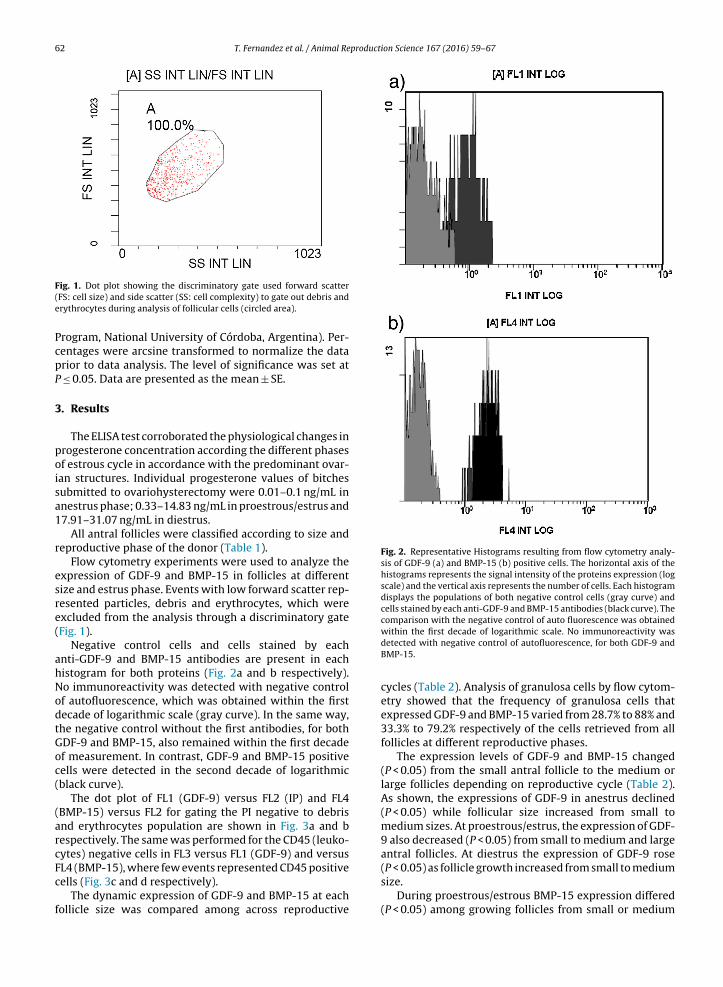
Fig. 2. Representative Histograms resulting from flow cytometry analy-sis of GDF-9 (a) and BMP-15 (b) positive cells. The horizontal axis of thehistograms represents the signal intensity of the proteins expression (logscale) and the vertical axis represents the number of cells. Each histogramdisplays the populations of both negative control cells (gray curve) andcells stained by each anti-GDF-9 and BMP-15 antibodies (black curve). Thecomparison with the negative control of auto fluorescence was obtained
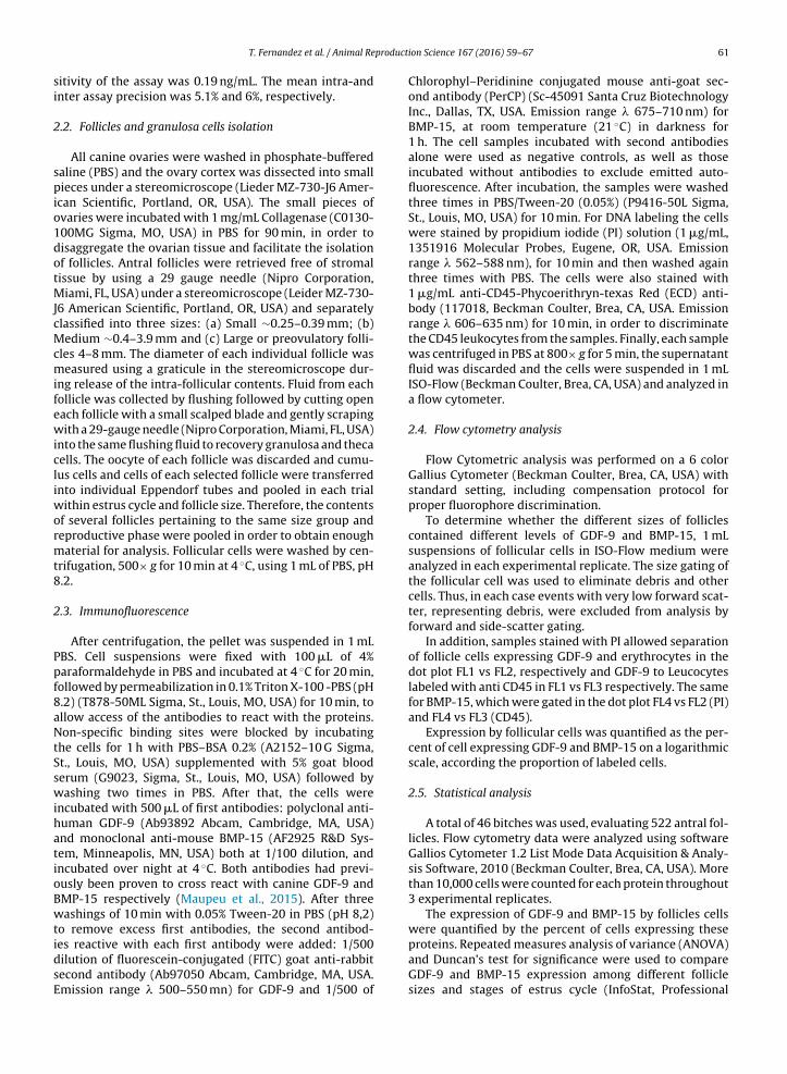
Fig. 1. Dot plot showing the discriminatory gate used forward scatter(FS: cell size) and side scatter (SS: cell complexity) to gate out debris anderythrocytes during analysis of follicular cells (circled area).
Program, National University of Córdoba, Argentina). Per-centages were arcsine transformed to normalize the dataprior to data analysis. The level of significance was set atP ≤ 0.05. Data are presented as the mean ± SE.
3. Results
The ELISA test corroborated the physiological changes inprogesterone concentration according the different phasesof estrous cycle in accordance with the predominant ovar-ian structures. Individual progesterone values of bitchessubmitted to ovariohysterectomy were 0.01–0.1 ng/mL inanestrus phase; 0.33–14.83 ng/mL in proestrous/estrus and17.91–31.07 ng/mL in diestrus.
All antral follicles were classified according to size andreproductive phase of the donor (Table 1).
Flow cytometry experiments were used to analyze theexpression of GDF-9 and BMP-15 in follicles at differentsize and estrus phase. Events with low forward scatter rep-resented particles, debris and erythrocytes, which wereexcluded from the analysis through a discriminatory gate(Fig. 1).
Negative control cells and cells stained by eachanti-GDF-9 and BMP-15 antibodies are present in eachhistogram for both proteins (Fig. 2a and b respectively).No immunoreactivity was detected with negative controlof autofluorescence, which was obtained within the firstdecade of logarithmic scale (gray curve). In the same way,the negative control without the first antibodies, for bothGDF-9 and BMP-15, also remained within the first decadeof measurement. In contrast, GDF-9 and BMP-15 positivecells were detected in the second decade of logarithmic(black curve).
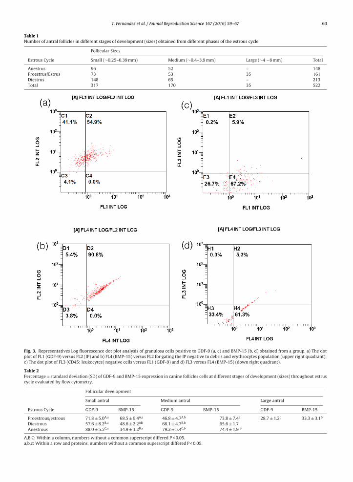
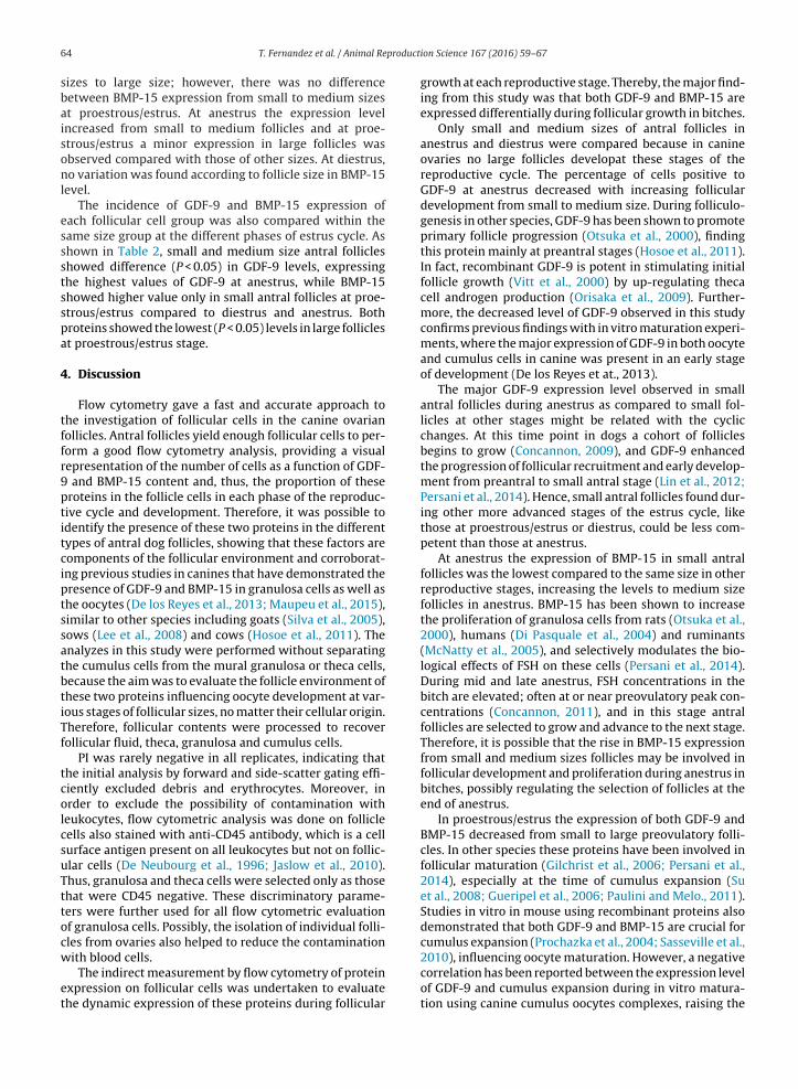
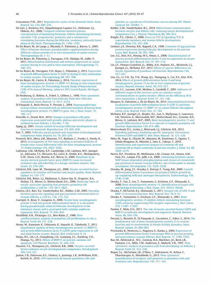
The dot plot of FL1 (GDF-9) versus FL2 (IP) and FL4(BMP-15) versus FL2 for gating the PI negative to debrisand erythrocytes population are shown in Fig. 3a and brespectively. The same was performed for the CD45 (leuko-cytes) negative cells in FL3 versus FL1 (GDF-9) and versus
FL4 (BMP-15), where few events represented CD45 positivecells (Fig. 3c and d respectively).
The dynamic expression of GDF-9 and BMP-15 at eachfollicle size was compared among across reproductive
within the first decade of logarithmic scale. No immunoreactivity wasdetected with negative control of autofluorescence, for both GDF-9 andBMP-15.
cycles (Table 2). Analysis of granulosa cells by flow cytom-etry showed that the frequency of granulosa cells thatexpressed GDF-9 and BMP-15 varied from 28.7% to 88% and33.3% to 79.2% respectively of the cells retrieved from allfollicles at different reproductive phases.
The expression levels of GDF-9 and BMP-15 changed(P < 0.05) from the small antral follicle to the medium orlarge follicles depending on reproductive cycle (Table 2).As shown, the expressions of GDF-9 in anestrus declined(P < 0.05) while follicular size increased from small tomedium sizes. At proestrous/estrus, the expression of GDF-9 also decreased (P < 0.05) from small to medium and largeantral follicles. At diestrus the expression of GDF-9 rose
(P < 0.05) as follicle growth increased from small to mediumsize.
During proestrous/estrous BMP-15 expression differed(P < 0.05) among growing follicles from small or medium
T. Fernandez et al. / Animal Reproduction Science 167 (2016) 59–67 63
Table 1Number of antral follicles in different stages of development (sizes) obtained from different phases of the estrous cycle.
Follicular Sizes
Estrous Cycle Small (∼0.25–0.39 mm) Medium (∼0.4–3.9 mm) Large (∼4 −8 mm) Total
Fig. 3. Representatives Log fluorescence dot plot analysis of granulosa cells positive to GDF-9 (a, c) and BMP-15 (b, d) obtained from a group. a) The dotplot of FL1 (GDF-9) versus FL2 (IP) and b) FL4 (BMP-15) versus FL2 for gating the IP negative to debris and erythrocytes population (upper right quadrant);c) The dot plot of FL3 (CD45; leukocytes) negative cells versus FL1 (GDF-9) and d) FL3 versus FL4 (BMP-15) (down right quadrant).
Table 2Percentage ± standard deviation (SD) of GDF-9 and BMP-15 expression in canine follicles cells at different stages of development (sizes) throughout estruscycle evaluated by flow cytometry.
A,B,C: Within a column, numbers without a common superscript differed P < 0.05.a,b,c: Within a row and proteins, numbers without a common superscript differed P < 0.05.
product
64 T. Fernandez et al. / Animal Re
sizes to large size; however, there was no differencebetween BMP-15 expression from small to medium sizesat proestrous/estrus. At anestrus the expression levelincreased from small to medium follicles and at proe-strous/estrus a minor expression in large follicles wasobserved compared with those of other sizes. At diestrus,no variation was found according to follicle size in BMP-15level.
The incidence of GDF-9 and BMP-15 expression ofeach follicular cell group was also compared within thesame size group at the different phases of estrus cycle. Asshown in Table 2, small and medium size antral folliclesshowed difference (P < 0.05) in GDF-9 levels, expressingthe highest values of GDF-9 at anestrus, while BMP-15showed higher value only in small antral follicles at proe-strous/estrus compared to diestrus and anestrus. Bothproteins showed the lowest (P < 0.05) levels in large folliclesat proestrous/estrus stage.
4. Discussion
Flow cytometry gave a fast and accurate approach tothe investigation of follicular cells in the canine ovarianfollicles. Antral follicles yield enough follicular cells to per-form a good flow cytometry analysis, providing a visualrepresentation of the number of cells as a function of GDF-9 and BMP-15 content and, thus, the proportion of theseproteins in the follicle cells in each phase of the reproduc-tive cycle and development. Therefore, it was possible toidentify the presence of these two proteins in the differenttypes of antral dog follicles, showing that these factors arecomponents of the follicular environment and corroborat-ing previous studies in canines that have demonstrated thepresence of GDF-9 and BMP-15 in granulosa cells as well asthe oocytes (De los Reyes et al., 2013; Maupeu et al., 2015),similar to other species including goats (Silva et al., 2005),sows (Lee et al., 2008) and cows (Hosoe et al., 2011). Theanalyzes in this study were performed without separatingthe cumulus cells from the mural granulosa or theca cells,because the aim was to evaluate the follicle environment ofthese two proteins influencing oocyte development at var-ious stages of follicular sizes, no matter their cellular origin.Therefore, follicular contents were processed to recoverfollicular fluid, theca, granulosa and cumulus cells.
PI was rarely negative in all replicates, indicating thatthe initial analysis by forward and side-scatter gating effi-ciently excluded debris and erythrocytes. Moreover, inorder to exclude the possibility of contamination withleukocytes, flow cytometric analysis was done on folliclecells also stained with anti-CD45 antibody, which is a cellsurface antigen present on all leukocytes but not on follic-ular cells (De Neubourg et al., 1996; Jaslow et al., 2010).Thus, granulosa and theca cells were selected only as thosethat were CD45 negative. These discriminatory parame-ters were further used for all flow cytometric evaluationof granulosa cells. Possibly, the isolation of individual folli-cles from ovaries also helped to reduce the contamination
with blood cells.
The indirect measurement by flow cytometry of proteinexpression on follicular cells was undertaken to evaluatethe dynamic expression of these proteins during follicular
ion Science 167 (2016) 59–67
growth at each reproductive stage. Thereby, the major find-ing from this study was that both GDF-9 and BMP-15 areexpressed differentially during follicular growth in bitches.
Only small and medium sizes of antral follicles inanestrus and diestrus were compared because in canineovaries no large follicles developat these stages of thereproductive cycle. The percentage of cells positive toGDF-9 at anestrus decreased with increasing folliculardevelopment from small to medium size. During folliculo-genesis in other species, GDF-9 has been shown to promoteprimary follicle progression (Otsuka et al., 2000), findingthis protein mainly at preantral stages (Hosoe et al., 2011).In fact, recombinant GDF-9 is potent in stimulating initialfollicle growth (Vitt et al., 2000) by up-regulating thecacell androgen production (Orisaka et al., 2009). Further-more, the decreased level of GDF-9 observed in this studyconfirms previous findings with in vitro maturation experi-ments, where the major expression of GDF-9 in both oocyteand cumulus cells in canine was present in an early stageof development (De los Reyes et at., 2013).
The major GDF-9 expression level observed in smallantral follicles during anestrus as compared to small fol-licles at other stages might be related with the cyclicchanges. At this time point in dogs a cohort of folliclesbegins to grow (Concannon, 2009), and GDF-9 enhancedthe progression of follicular recruitment and early develop-ment from preantral to small antral stage (Lin et al., 2012;Persani et al., 2014). Hence, small antral follicles found dur-ing other more advanced stages of the estrus cycle, likethose at proestrous/estrus or diestrus, could be less com-petent than those at anestrus.
At anestrus the expression of BMP-15 in small antralfollicles was the lowest compared to the same size in otherreproductive stages, increasing the levels to medium sizefollicles in anestrus. BMP-15 has been shown to increasethe proliferation of granulosa cells from rats (Otsuka et al.,2000), humans (Di Pasquale et al., 2004) and ruminants(McNatty et al., 2005), and selectively modulates the bio-logical effects of FSH on these cells (Persani et al., 2014).During mid and late anestrus, FSH concentrations in thebitch are elevated; often at or near preovulatory peak con-centrations (Concannon, 2011), and in this stage antralfollicles are selected to grow and advance to the next stage.Therefore, it is possible that the rise in BMP-15 expressionfrom small and medium sizes follicles may be involved infollicular development and proliferation during anestrus inbitches, possibly regulating the selection of follicles at theend of anestrus.
In proestrous/estrus the expression of both GDF-9 andBMP-15 decreased from small to large preovulatory folli-cles. In other species these proteins have been involved infollicular maturation (Gilchrist et al., 2006; Persani et al.,2014), especially at the time of cumulus expansion (Suet al., 2008; Gueripel et al., 2006; Paulini and Melo., 2011).Studies in vitro in mouse using recombinant proteins alsodemonstrated that both GDF-9 and BMP-15 are crucial forcumulus expansion (Prochazka et al., 2004; Sasseville et al.,
2010), influencing oocyte maturation. However, a negativecorrelation has been reported between the expression levelof GDF-9 and cumulus expansion during in vitro matura-tion using canine cumulus oocytes complexes, raising the
producti
ppadasr
cr2t2asvWadewis
clq(MfpoGhmLcbm
pplaltdtttic
ppa1gTBB
T. Fernandez et al. / Animal Re
ossibility that in dogs the major expression of GDF-9 isresent only in an early stage of development, possibly in
species specific manner which may be related with theelay in mucification (De los Reyes et al., 2013), because
low concentration of GDF-9 does not stimulate enzymesuch as Hyaluronan synthase 2 and cyclooxygegase-2, thategulate cumulus expansion (Varani et al., 2002).
Several connexins (Cx) are expressed in ovarian folli-les and Cx43 has been demonstrated to play an essentialole in folliculogenesis (Lenhart et al., 1998; Nitta et al.,010). Granulosa cells must be coupled via Cx43 gap junc-ions in order to respond optimally to GDF-9 (Gittens et al.,005). In dogs, the gene encoding Cx43, was found to differcross the reproductive cycle, showing a decreased expres-ion at estrus, rising levels during diestrus, and decliningalues during anestrus (Willingham-Rocky et al., 2007;illingham et al., 2004). These differences could be in
ccordance with variations of GDF-9 found in this studyuring the follicular development at different phases of thestrous cycle, because although expression levels of GDF-9ere high in small follicles at anestrus, these levels signif-
cantly decreased with increasing follicle size during thistage.
Studies in mice have shown that in growing folli-les Cx43 expression increases in response to the risingevel of follicle-stimulating hormone (FSH), but it subse-uently declines in response to the luteinizing hormoneLH) surge, since LH causes the closure of the channels via
AP kinase-dependent phosphorylation of Cx43 of ovarianollicle (Norris et al., 2008). In dogs during transition fromroestrous to estrous, FSH and LH are increasing to the pre-vulatory surge of LH (Concannon, 2011); the decreasedDF-9 levels in large follicles at proestrous/estrus founderein might be coincident with the increased LH surge ter-inating the proestrus in the bitch. Therefore, possibly the
H peak in dogs may restrict the flow of GDF-9 into the folli-le cells during estrus. This possibility would be reinforcedy the subsequent increase of GDF-9 found from small toedium sizes follicles at diestrus when LH levels decrease.The decreasing expression of BMP-15 during
roestrous–estrus appears somewhat confusing becausereviously it was found in canine ovaries that increasing
evels of this protein are expressed in both cumulus cellsnd oocytes during in vitro maturation experiments (Deos Reyes et al., 2014). To date, nothing is known abouthe mechanism by which BMP-15 modulates follicularevelopment in canines, but possibly the differences inhese results may be related to different expression duringhe estrous cycle, and thus the reproductive phase at whichhe follicles and oocytes for IVM were obtained, consider-ng the influence of the estrus cycle on cumulus–oocyteommunications (Luvoni et al., 2001).
Premature luteinization is associated with high serumrogesterone levels and BMP-15 suppresses progesteroneroduction in rodent granulosa cells (Otsuka et al., 2001)nd down-regulates Cx43 (Chang et al., 2014). Also, a BMP-5 inhibitory action on progesterone secretion in human
ranulosa cells lines has been described (Chang et al., 2013).he present data however, showed decreasing levels ofMP-15 at proestrous–estrous and lack of differences inMP-15 expression between small to medium sizes follicle
on Science 167 (2016) 59–67 65
at diestrus. In the bitch, serum progesterone rises slowlyduring end of proestrus, reflecting luteinization of large fol-licles visible histologically as early as six days before the LHpeak (Concannon, 2009). Therefore, estrus is concomitantwith subsequently declining phase of estradiol concentra-tion and increasing progesterone concentration (De Gieret al., 2006). This indicates that premature luteinizationthat is associated with high serum progesterone concentra-tion is a normal feature in the canine estrus cycle; therefore,species variation is also possible in the role of BMP-15 inantral follicle development, and thus, the level of this pro-tein for reproductive success may be not coincident acrossspecies. This suggest that the anti-luteinization function ofBMP-15 described in other mammals could be different indogs, or perhaps the decreasing expression of this factorobserved at proestrous/estrus in large follicles, is not suffi-cient to avoid progressive luteinization during this periodin canines.
In conclusion, the flow cytometric assay was able toassess protein expression in canine follicle cells. Althoughthe biological significance of GDF-9 and BMP-15 expres-sion in these cells has not been fully elucidated, thepresent study is the first evidence that canine follicu-lar GDF-9 and BMP-15 are differentially expressed duringdevelopment. The declining secretion of these factors inproestrous/estrus as compared to other species, might berelated, at least in part, to premature luteinization which isone of the main reproductive traits of dogs.
Conflict of interest
The authors declare that there is no conflict of interest.
Acknowledgments
This research was supported by Grant 1140658 (FONDE-CYT) and by Grant EQM120156 (FONDEQUIP), from TheNational Commission for Scientific and TechnologicalResearch, Chile (CONICYT).
We thank to veterinarians from El Roble Veterinary Hos-pital for procuring bitch ovaries for this study and also weare grateful to Prof. Donald L Palmquist for his helpful sug-gestions and comments on this manuscript.
References
Chang, H.M., Cheng, J.C., Klausen, C., Leung, P.C., 2013. BMP15 suppressesprogesterone production by down-regulating StAR via ALK3 inhuman granulosa cells. Mol. Endocrinol. 27, 2093–2104.
Chang, H.M., Cheng, J.C., Taylor, E., Leung, P.C., 2014. Oocyte-derivedBMP15 but not GDF9 down-regulates connexin43 expression anddecreases gap junction intercellular communication activity inimmortalized human granulosa cells. Mol. Hum. Reprod. 20,373–383.
Chastant-Maillard, S., Viaris de Lesegno, C., Chebrout, M., Thoumire, S.,Meylheue, T., Fontbonne, A., Chodkiewicz, M., Saint-Dizier, M.,Reynaud, K., 2011. The canine oocyte: uncommon features of in vivoand in vitro maturation. Reprod. Fertil. Dev. 23, 391–402.
Concannon, P.W., 2011. Reproductive cycles of the domestic bitch. Anim.Reprod. Sci. 124, 200–210.
De Gier, J., Kooistra, H.S., Djajadiningrat-Laanen, S.C., Dieleman, S.J.,Okkens, A.C., 2006. Temporal relations between plasmaconcentrations of luteinizing hormone, follicle-stimulating hormone,estradiol-17�, progesterone, prolactin, and a-melanocyte-stimulating hormone during the follicular, ovulatory, and early lutealphase in the bitch. Theriogenology 65, 1346–1359.
De los Reyes, M., de Lange, J., Miranda, P., Palomino, J., Barros, C., 2005.Effect of human chorionic gonadotrophin supplementation duringdifferent culture periods on in vitro maturation of canine oocytes.Theriogenology 64, 1–11.
De los Reyes, M., Palomino, J., Parraguez, V.H., Hidalgo, M., Saffie, P.,2011. Mitochondrial distribution and meiotic progression in canineoocytes during in vivo and in vitro maturation. Theriogenology 75,346–353.
De los Reyes, M., Parraguez, V.H., Rojas, C., Palomino, J., 2013. Expressionof growth differentiation factor 9 (GDF-9) during in vitro maturationin canine oocytes. Theriogenology 80, 587–596.
De los Reyes, M., Castro, R., Palomino, J., 2014. Dynamic expression ofbone morphogenetic protein 15 (BMP-15) during in vitro maturationof canine oocytes. In: Proceedings Society Study of Reproduction(SSR) 47th Annual Meeting, (abstract 295) Grand Rapids, MichiganUSA.
De Neubourg, D., Robins, A., Fishel, S., Gibbon, L., 1996. Flow cytometricanalysis of granulosa cells from follicular fluid after follicularstimulation. Hum. Reprod. 11, 2211–2214.
Di Pasquale, E., Beck-Peccoz, P., Persani, L., 2004. Hypergonadotropicovarian failure associated with an inherited mutation of human bonemorphogenetic protein-15 (BMP15) gene. Am. J. Hum. Genet. 75,106–111.
Douville, G., Sirard, M.A., 2014. Changes in granulosa cells geneexpression associated with growth, plateau and atretic phases inmedium bovine follicles. J. Ova Res. 7, 50.
Eppig, J.J., 2001. Oocyte control of ovarian follicular development andfunction in mammals. Reproduction 122, 829–838.
Fair, T., 2003. Follicular oocyte growth and acquisition of developmentalcompetence Anim. Reprod. Sci. 78, 203–216.
Fenwick, M.A., Mora, J.M., Mansour, Y.T., Baithun, C., Franks, S., Hardy, K.,2013. Investigations of TGF-beta signaling in preantral follicles offemale mice reveal differential roles for bone morphogenetic protein15. Endocrinology 154, 3423–3436.
Galloway, S.M., McNatty, K.P., Cambridge, L.M., Laitinen, M.P., Juengel,J.L., Jokiranta, T.S., McLaren, R.J., Luiro, K., Dodds, K.G., Montgomery,G.W., Davis, G.H., Beattie, A.E., Ritvos, O., 2000. Mutations in anoocyte-derived growth factor gene (BMP15) cause increasedovulation rate and infertility in a dosage-sensitive manner. Nat.Genet. 25, 279–283.
Gilchrist, R.B., Lane, M., Thompson, J.G., 2008. Oocyte-secreted factors:regulators of cumulus cell function and oocyte quality. Hum. Reprod.Update 14, 159–177.
Gittens, J.E.I., Barr, K.J., Vanderhyden, B.C., Kidder, G.M., 2005. Interplaybetween paracrine signaling and gap junctional communication inovarian follicles. J. Cell Sci. 118, 113–122.
Gueripel, X., Brun, V., Gougeon, A., 2006. Oocyte bone morphogeneticprotein 15 but not growth differentiation factor 9, is increasedduring gonadotropin-induced follicular development in theimmature mouse and is associated with cumulus oophorusexpansion. Biol. Reprod. 75, 836–843.
Hirshfield, A.N., Flickinger, G.L., Ben-Rafae, Z., 1988. Flowcytofluorometric analysis of granulosa cell proliferation in rats. J.Reprod. Fertil. 84, 231–238.
Hosoe, M., Kaneyam, K., Ushizawa, K., Hayashi, K., Takahashi, T., 2011.Quantitative analysis of bone morphogenetic protein 15 (BMP15)and growth differentiation factor 9 (GDF9) gene expression in calfand adult bovine ovaries. Reprod. Biol. Endocrinol. 9, 33.
Jaslow, C.R., Patterson, K.S., Cholera, S., Jennings, L.K., KeWilliam, R.W.,Kutteh, H., 2010. CD9 expression by human granulosa cells and
ion Science 167 (2016) 59–67
platelets as a predictor of fertilization success during IVF. Obstet.Gynecol. Int. 192461, 7.
Kidder, G.M., Vanderhyden, B.C., 2010. Bidirectional communicationbetween oocytes and follicle cells: ensuring oocyte developmentalcompetence Can. J. Physiol. Pharmacol. 88, 399–413.
Knight, P.G., Glister, C., 2006. Focus on TGF-� Signalling TGF-�superfamily members and ovarian follicle development.Reproduction 132, 191–206.
Lenhart, J.A., Downey, B.R., Bagnell, C.A., 1998. Connexin 43 gap junctionprotein expression during follicular development in the porcineovary. Biol. Reprod. 58, 583–590.
Lee, S.G., Kim, H.S., Hwang, W.S., Hwan, S., 2008. Characterization ofporcine growth differentiation factor-9 and its expression in oocytematuration. Mol. Reprod. Dev 75, 707–714.
Lin, J.Y., Pitman-Crawford, J.L., Bibby, A.H., Hudson, N.L., Mcintosh, C.J.,Juengel, J.L., McNatty, K.P., 2012. Effects of species differences onoocyte regulation of granulosa cell function. Reproduction 144,557–567.
Lim, Z.L., Li, Y.H., Xu, Y.N., Wang, Q.L., Namgong, S., Cui, X.S., Kim, N.H.,2014. Effects of growth differentiation factor 9 and bonemorphogenetic protein 15 on the in vitro maturation of porcineoocytes. Reprod. Domest. Anim. 49, 219–227.
Luvoni, G.C., Luciano, A.M., Modina, S., Gandolfi, F., 2001. Influence ofdifferent stages of the oestrous cycle on cumulus-oocytecommunications in canine oocytes: effects on the efficiency of invitro maturation. J. Reprod. Fertil. 57, 141–146.
Maupeu, D., Palomino, J., De los Reyes, M., 2015. Immunohistochemistrylocalization of growth differentiation factor 9 (GDF-9) and bonemorphogenetic protein 15 (BMP-15) in canine follicles throughoutestrus cycle. Reprod. Fertil. Dev. 27, 182.
McNatty, K.P., Juengel, J.L., Reader, K.L., Lun, S., Myllymaa, S., Lawrence,S.B., Western, A., Meerasahib, M.F., Mottershead, D.G., Groome, N.P.,Ritvos, O., Laitinen, M.P., 2005. Bone morphogenetic protein 15 andgrowth differentiation factor 9 co-operate to regulate granulosa cellfunction. Reproduction 129, 473–480.
Nitta, M., Yogo, K., Ohashi, M., Akiyama, M., Kunitomo, Y., Ogawa, T.,Ishida-Kitagawa, N., Miyoshi, J., Sato, E., Takeya, T., 2010.Identification and expression analysis of connexin-45 andconnexin-60 as major connexins in porcine oocytes. J. Anim. Sci. 88,3269–3279.
Norris, R.P., Freudzon, M., Mehlmann, L.M., Cowan, A.E., Simon, A.M.,Paul, D.L., Lampe, P.D., Jaffe, L.A., 2008. Luteinizing hormone causesMAP kinase-dependent phosphorylation and closure of connexin43gap junctions in mouse ovarian follicles: one of two paths to meioticresumption. Development 135, 3229–3238.
Paulini, F., Melo, E.O., 2011. The role of oocyte-secreted factors GDF9 andBMP15 in follicular development and oogenesis. Reprod. Domest.Anim. 46, 354–356.
Persani, L., Rossetti, R., Di Pasquale, E., Cacciatore, C., Fabre, S., 2014. Thefundamental role of bone morphogenetic protein 15 in ovarianfunction and its involvement in female fertility disorders. Hum.Reprod. Update 20, 869–883.
Prochazka, R., Nemcova, L., Nagyova, E., Kanka, J., 2004. Expression ofgrowth differentiation factor 9 messenger RNA in porcine growingand preovulatory ovarian follicles. Biol. Reprod. 71, 1290–1295.
Rao, M., Allsbrook Jr., W.C., Conway, B.A., Martinez, J.E., Beck, J.R.,Pantazis, C.G., Mills, T.M., Anderson, E., Mahesh, V.B., 1991. Flowcytometric analysis of granulosa cells from developing rat follicles. J.
Reprod. Fertil. 91, 521–530.
Regan, S.L., McFarlane, J.R., O’Shea, T., Andronicos, N., Arfuso, F.,Dharmarajan, A., Almahbobi, G., 2015. Flow cytometricquantification of receptors and apoptosis in granulosa cells andovulation rate. Reproduction 150, 151–163.
ilva, J.R., Van Den Hurk, R., Van Tol, H.T.A., Roelen, B.A., Figueiredo, J.R.,2005. Expression of growth differentiation factor 9 (GDF9), and bonemorphogenetic protein 15 (BMP15), and BMP receptors in theovaries of goats. Mol. Reprod. Dev. 70, 11–19.
ongsasen, N., Wild, D., 2005. Size of the donor follicle but not stage ofreproductive cycle or seasonality, influences meiotic competency ofselected domestic dog oocytes. Mol. Reprod. Dev. 72, 113–119.
udiman, J., Ritter, L.J., Feil, D.K., Wang, X., Chan, K., Mottershead, D.G.,Robertson, D.M., Thompson, J.G., Gilchrist, R.B., 2014. Effects ofdiffering oocyte-secreted factors during mouse in vitro maturationon subsequent embryo and fetal development. J. Assist. Reprod.Genet. 31, 295–306.
u, Y.Q., Wu, X., O’Brien, M.L., Pendola, F.L., Denegre, J.N., Matzuk, M.M.,Eppig, J.J., 2004. Synergistic roles of the oocyte-cumulus cell complexin mice: genetic evidence for an oocyte–granulosa cell regulatoryloop. Dev. Biol. 276, 64–73.
u, Y.Q., Sugiura, K., Wigglesworth, K., O’Brien, M.J., Affourtit, J.P., Pangas,S.A., Matzuk, M.M., Eppig, J.J., 2008. Oocyte regulation of metaboliccooperativity between mouse cumulus cells and oocytes: BMP15and GDF9 control cholesterol biosynthesis in cumulus cells.Development 135, 111–121.
on Science 167 (2016) 59–67 67
Sun, Y.Q., Liu, K., Kikuchi, K., 2008. Oocyte-specific knockout a novelin vivo approach for studying gene functions during folliculogenesis.oocyte maturation. fertilization, and embryogenesis. Biol. Reprod.79, 1014–1020.
Varani, S., Elvin, J.A., Yan, C., De Mayo, J., De Mayo, F.J., Horton, H.F.,Byrne, M.C., Matzuk, M.M., 2002. Knockout of pentraxin 3 adownstream target of growth differentiation factor-9, causes femalesubfertility. Mol. Endocrinol. 16, 1154–1167.
Ververidis, H.N., Boscos, C.M., Stefanakis, A., Krambovitis, E., 2002. Use ofenzyme-immunoassay for oestradiol-17� and progesteronequantification in canine serum. Anim. Reprod. Sci. 69, 53–64.
Vitt, U.A., Hayashi, M., Klein, C., Hsueh, A.J., 2000. Growth differentiationfactor-9 stimulates proliferation but suppresses thefollicle-stimulating hormone-induced differentiation of culturedgranulosa cells from small antral and preovulatory rat follicles. Biol.Reprod. 62, 370–377.