c Indian Academy of Sciences RESEARCH NOTE Differential expression of PARP1 mRNA in leucocytes of patients with Down’s syndrome MICHELE SALEMI 1† , CONCETTA BARONE 2† , CARMELO ROMANO 1 , FEDERICO RIDOLFO 1 , ELEONORA GULOTTA 1 , CATALDO SCAVUZZO 1 , MARIA GRAZIA SALLUZZO 1 , MARIACONCETTA GIAMBIRTONE 1 , FILIPPO CARACI 3 , CORRADO ROMANO 2 and PAOLO BOSCO 1 ∗ 1 Laboratory of Cytogenetics, 2 Unit of Pediatrics and Medical Genetics, Oasi Institute for Research on Mental Retardation and Brain Aging, 94018 Troina, Italy 3 Department of Pharmaceutical Sciences, University of Catania, 95100 Catania, Italy [Salemi M., Barone C., Romano C., Ridolfo F., Gulotta E., Scavuzzo C., Salluzzo M. G., Giambirtone M., Caraci F., Romano C. and Bosco P. 2011 Differential expression of PARP1 mRNA in leucocytes of patients with Down’s syndrome. J. Genet. 90, 469–472] Introduction Down’s syndrome (DS) is one of the most common numer- ical chromosomal aberrations, usually caused by trisomy of chromosome 21, and is the most frequent genetic cause of mental retardation. People with DS can develop some traits of Alzheimer disease at an earlier age than subjects with- out trisomy 21 (Wisniewski et al. 1985). It may be assumed that these individuals would experience a neurodegenerative process because of the presence of an extra copy of amy- loid precursor protein (APP) gene located on chromosome 21, even if this hypothesis has not been entirely confirmed by the literature (Arriagada et al. 2010). In addition, it has been widely demonstrated that genes related to apoptosis play a crucial role in neurodegenerative processes (Engidawork et al. 2001; Fromage and Anglade 2002). The role of the apoptotic pathways in neurodegenerative processes and in cancer proliferation is determinant. The latter is favoured when the apoptotic surveillance is, for any reason, decreased (Hu and Kavanagh 2003); conversely, when the apoptotic process is somewhat encouraged, neurodegenerative pro- cesses, such as those related to Alzheimer disease, will be prevailing (Elmore 2007). Poly (ADP-ribose) polymerase 1 (PARP1) gene is located at 1q42 and is 43-kb long and splits into 23 exons (OMIM 173870). Grube and Bürkle (1992) suggested that higher PARP1 action capacity may contribute to the efficient main- tenance of genome integrity. Yu et al. (2006) showed that PARP1 activation is required for translocation of apoptosis- inducing factor (AIF) from the mitochondria to the nucleus. PARP1 is proteolytically cleaved at the onset of apoptosis ∗ For correspondence. E-mail: [email protected]. † These authors contributed equally to this work. by caspase-3 (Nicholson et al. 1995); further, PARP1 activ- ity and poly (ADP-ribose) (PAR) polymer, mediate PARP1- induced cell death (Yu et al. 2006). Genetic and pharmacological studies found that overex- pression of PARP1 is a key mediator of programmed-necrotic cell death in vivo. PARP1 appears to be also involved in programmed cell death processes besides necrosis, such as apoptosis or macroautophagocytotic cell death (Hassa and Hottiger 2008). The aim of the present study was to evalu- ate the possible differential expression of PARP1 mRNA in leucocytes of peripheral blood of DS subjects compared with the normal population. Materials and methods A total of 36 subjects were enrolled for this study at the Unit of Pediatrics and Medical Genetics of the IRCCS Oasi Institute, Troina, Italy, a specialized centre for patients with mental disability mainly from Sicily. They included: 18 DS patients (7 males and 11 females) with a mean age of 38.44 ± 10.51 (range 20–55 years) and 18 normal subjects (7 males and 11 females) with a mean age of 38.33 ± 11.28 (range 19–55 years). The DS cases and controls were recruited after family and personal informed consent. RNA extraction from leucocytes of peripheral blood was performed using RNeasy Mini Handbook (Qiagen, Germantown, USA), following the manufacturer’s protocol. The RNA quality and quantity were checked by spectropho- tometry. To avoid any genomic DNA contamination during qRT- PCR, a brief incubation of the samples at 42 ◦ C with a specific Wipeout buffer (QuantiTect Reverse Transcription, Qiagen, Germantown, USA) was carried out. Retrotranscription of Keywords. Down’s syndrome; PARP1 gene; mRNA; qRT-PCR expression. Journal of Genetics, Vol. 90, No. 3, December 2011 469
MARIACONCETTA GIAMBIRTONE1, FILIPPO CARACI3, CORRADO ROMANO2 and PAOLO BOSCO1∗
1Laboratory of Cytogenetics, 2Unit of Pediatrics and Medical Genetics, Oasi Institute for Research on Mental Retardationand Brain Aging, 94018 Troina, Italy
3Department of Pharmaceutical Sciences, University of Catania, 95100 Catania, Italy
[Salemi M., Barone C., Romano C., Ridolfo F., Gulotta E., Scavuzzo C., Salluzzo M. G., Giambirtone M., Caraci F., Romano C. and BoscoP. 2011 Differential expression of PARP1 mRNA in leucocytes of patients with Down’s syndrome. J. Genet. 90, 469–472]
Introduction
Down’s syndrome (DS) is one of the most common numer-ical chromosomal aberrations, usually caused by trisomy ofchromosome 21, and is the most frequent genetic cause ofmental retardation. People with DS can develop some traitsof Alzheimer disease at an earlier age than subjects with-out trisomy 21 (Wisniewski et al. 1985). It may be assumedthat these individuals would experience a neurodegenerativeprocess because of the presence of an extra copy of amy-loid precursor protein (APP) gene located on chromosome21, even if this hypothesis has not been entirely confirmed bythe literature (Arriagada et al. 2010). In addition, it has beenwidely demonstrated that genes related to apoptosis playa crucial role in neurodegenerative processes (Engidaworket al. 2001; Fromage and Anglade 2002). The role of theapoptotic pathways in neurodegenerative processes and incancer proliferation is determinant. The latter is favouredwhen the apoptotic surveillance is, for any reason, decreased(Hu and Kavanagh 2003); conversely, when the apoptoticprocess is somewhat encouraged, neurodegenerative pro-cesses, such as those related to Alzheimer disease, will beprevailing (Elmore 2007).
Poly (ADP-ribose) polymerase 1 (PARP1) gene is locatedat 1q42 and is 43-kb long and splits into 23 exons (OMIM173870). Grube and Bürkle (1992) suggested that higherPARP1 action capacity may contribute to the efficient main-tenance of genome integrity. Yu et al. (2006) showed thatPARP1 activation is required for translocation of apoptosis-inducing factor (AIF) from the mitochondria to the nucleus.PARP1 is proteolytically cleaved at the onset of apoptosis
∗For correspondence. E-mail: [email protected].†These authors contributed equally to this work.
by caspase-3 (Nicholson et al. 1995); further, PARP1 activ-ity and poly (ADP-ribose) (PAR) polymer, mediate PARP1-induced cell death (Yu et al. 2006).
Genetic and pharmacological studies found that overex-pression of PARP1 is a key mediator of programmed-necroticcell death in vivo. PARP1 appears to be also involved inprogrammed cell death processes besides necrosis, such asapoptosis or macroautophagocytotic cell death (Hassa andHottiger 2008). The aim of the present study was to evalu-ate the possible differential expression of PARP1 mRNA inleucocytes of peripheral blood of DS subjects compared withthe normal population.
Materials and methods
A total of 36 subjects were enrolled for this study at theUnit of Pediatrics and Medical Genetics of the IRCCS OasiInstitute, Troina, Italy, a specialized centre for patients withmental disability mainly from Sicily. They included: 18 DSpatients (7 males and 11 females) with a mean age of 38.44 ±10.51 (range 20–55 years) and 18 normal subjects (7 malesand 11 females) with a mean age of 38.33 ± 11.28 (range19–55 years). The DS cases and controls were recruited afterfamily and personal informed consent.
RNA extraction from leucocytes of peripheral bloodwas performed using RNeasy Mini Handbook (Qiagen,Germantown, USA), following the manufacturer’s protocol.The RNA quality and quantity were checked by spectropho-tometry.
To avoid any genomic DNA contamination during qRT-PCR, a brief incubation of the samples at 42◦C with a specificWipeout buffer (QuantiTect Reverse Transcription, Qiagen,Germantown, USA) was carried out. Retrotranscription of
Journal of Genetics, Vol. 90, No. 3, December 2011 469
Michele Salemi et al.
600 ng of total RNA from each samples was then per-formed in a final volume of 20 μL and generated cDNA wasused as a template for real-time quantitative PCR analysisusing gene expression products. For each sample, real-timePCR reactions were carried out in duplicate using 2.5 μL ofcDNA and QuantiTect Probe PCR Master Mix kit (Qiagen,Germantown, USA) in a total volume of 50 μL. PARP1and GAPDH assays were obtained from Applied Biosys-tems (Carlsbad, USA). The thermal cycling conditions con-sisted of one cycle for 2 min at 50◦C, one cycle of 15 minat 95◦C and 40 cycles for 15 s at 94◦C followed by 1 minat 60◦C. Real-time analysis was performed on Light Cycler480 (Roche, Mannheim, Germany). The amplified transcriptswere quantified using the comparative CT method (Livak andSchmittgen 2001) and relative quantification analysis datawere played using the comparative ��Ct method includedin the software version 1.5 supplied with the LightCycler
480. PARP1 gene expression level was normalized toGAPDH level and target mean Cp definition was used toindicate the mean normalized cycle threshold.
Results and discussion
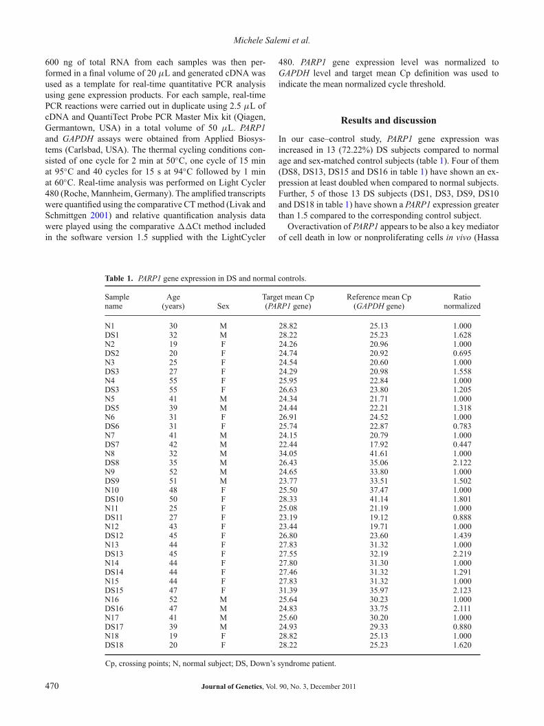
In our case–control study, PARP1 gene expression wasincreased in 13 (72.22%) DS subjects compared to normalage and sex-matched control subjects (table 1). Four of them(DS8, DS13, DS15 and DS16 in table 1) have shown an ex-pression at least doubled when compared to normal subjects.Further, 5 of those 13 DS subjects (DS1, DS3, DS9, DS10and DS18 in table 1) have shown a PARP1 expression greaterthan 1.5 compared to the corresponding control subject.
Overactivation of PARP1 appears to be also a key mediatorof cell death in low or nonproliferating cells in vivo (Hassa
Table 1. PARP1 gene expression in DS and normal controls.
Sample Age Target mean Cp Reference mean Cp Rationame (years) Sex (PARP1 gene) (GAPDH gene) normalized
N1 30 M 28.82 25.13 1.000DS1 32 M 28.22 25.23 1.628N2 19 F 24.26 20.96 1.000DS2 20 F 24.74 20.92 0.695N3 25 F 24.54 20.60 1.000DS3 27 F 24.29 20.98 1.558N4 55 F 25.95 22.84 1.000DS3 55 F 26.63 23.80 1.205N5 41 M 24.34 21.71 1.000DS5 39 M 24.44 22.21 1.318N6 31 F 26.91 24.52 1.000DS6 31 F 25.74 22.87 0.783N7 41 M 24.15 20.79 1.000DS7 42 M 22.44 17.92 0.447N8 32 M 34.05 41.61 1.000DS8 35 M 26.43 35.06 2.122N9 52 M 24.65 33.80 1.000DS9 51 M 23.77 33.51 1.502N10 48 F 25.50 37.47 1.000DS10 50 F 28.33 41.14 1.801N11 25 F 25.08 21.19 1.000DS11 27 F 23.19 19.12 0.888N12 43 F 23.44 19.71 1.000DS12 45 F 26.80 23.60 1.439N13 44 F 27.83 31.32 1.000DS13 45 F 27.55 32.19 2.219N14 44 F 27.80 31.30 1.000DS14 44 F 27.46 31.32 1.291N15 44 F 27.83 31.32 1.000DS15 47 F 31.39 35.97 2.123N16 52 M 25.64 30.23 1.000DS16 47 M 24.83 33.75 2.111N17 41 M 25.60 30.20 1.000DS17 39 M 24.93 29.33 0.880N18 19 F 28.82 25.13 1.000DS18 20 F 28.22 25.23 1.620
Cp, crossing points; N, normal subject; DS, Down’s syndrome patient.
470 Journal of Genetics, Vol. 90, No. 3, December 2011
PARP1 expression and Down’s syndrome
et al. 2006; Schreiber et al. 2006). Uncontrolled poly-ADP-ribosylation reactions can result in massive necrotic celldeath and tissue damage, which in turn often leads to severeinflammatory or neurodegenerative disorders (Hassa et al.2006; Yu et al. 2006). Further, overexpression of PARP1has been implicated in the pathogenesis of several diseases,including stroke, myocardial infarction, diabetes, shock andallergy (D’Amours et al. 1999).
The use of PARP1 inhibitors has been proposed as a pro-tective therapy in decreasing cell death and other tissue dam-age in inflammatory and neurodegenerative disorders (Hassaet al. 2006; Yu et al. 2006).
Indeed, recent studies using DNA damaging agents suchas N-methyl-N’-nitro-N-nitrosoguanidine (MNNG), hydro-gen peroxide (H2O2) or peroxynitrite, which are well knownto induce necrosis at high concentrations, showed that phar-macological inhibition of PARP1 activity or knockout of thePARP1 gene blocks programmed-necrotic cell death inducedby these agents (Jagtap and Szabo 2005; Hassa et al. 2006;Schreiber et al. 2006).
Eustermann et al. (2011) in their study concluded that itis clear that a full description of the activation mechanismof PARP1 will require significant further work to character-ize its interdomain interactions and their DNA-dependence,both at the structural and functional levels. Nonetheless, theresults presented in the above mentioned study, provide valu-able insights into recognition of chromosomal DNA single-strand breaks, the crucial first step of the activation pro-cess, by the PARP1 DNA binding domain. In fact, PARP1is a highly abundant chromatin-associated enzyme presentin all higher eukaryotic cell nuclei, where it plays key rolesin maintenance of genomic integrity, chromatin remodellingand transcriptional control. PARP1 gene product binds toDNA single-strand and double-strand breaks through anN-terminal region containing two zinc fingers, F1 and F2(Langelier et al. 2010). The C-terminal catalytic domainof PARP1 protein is activated via an unknown mechanism,causing formation and addition of the polyadenosine-ribose(PAR) complex to acceptor proteins including PARP1 itself(Kannan et al. 2011).
Several reports from various laboratories indicate thatinhibition or absence of PARP1 provides remarkable protec-tion in disease models such as septic shock, diabetes, stroke,myocardial infarction and ischaemia, which are characterizedpredominantly by programmed-necrotic cell death (Jagtapand Szabo 2005; Schreiber et al. 2006).
Kannan et al. (2011) showed the interaction of hairy/enhancer of split1 (HES1) and PARP1 in B cell acute lym-phoblastic leukaemia (B-ALL). In effect, they report thatHES1 regulates proapoptotic signals via the novel interactingprotein PARP1. This mechanism reveals a cell type-specificproapoptotic pathway which may lead to a new cancer ther-apy. Also, PARP1 activation has been causally connected tophotoreceptor cell death (Paquet-Durand et al. 2007) and thisobservation was confirmed by Sahaboglu et al. (2010) whoshowed a causal involvement of PARP1 in retinal degener-
ation, a neurodegenerative diseases affecting photoreceptorsand causing blindness in humans.
We tried to investigate a possible relationship betweenoverexpression and neurophenotype of the subjects exam-ined. The small number of subjects does not allow a sta-tistical evaluation. Nonetheless, the group of patients withoverexpression at least doubled when compared to normal,including people with profound degree of intellectual dis-ability (ID) and drug-resistant epilepsy. All remaining groupsinclude people with severe or modest ID.
In conclusion, the PARP1 overexpression at least dou-bled in comparison to normal, seems to be associated witha severe ID phenotype. The data obtained from our exper-iments need to be verified finding a link between overex-pression of the gene PARP1 and activation of the apoptoticpathways both in early ageing and neurodegenerative pro-cesses in DS. The confirmation of these data and further stud-ies on larger samples, could lead to the hypothesis of usinginhibitors of PARP1 gene as a protective therapy also to pre-vent neurodegeneration and premature ageing of individualswith DS. Currently, an evaluation of the expression of thePARP1 gene in other tissues and in cell cultures of fibroblastsobtained from subjects with DS is in progress, to increase ourknowledge and strengthen the evidences obtained.
Further, it would be very interesting to examine in subse-quent studies whether the repair function of PARP1 is impli-cated in Down’s syndrome or other diseases associated withmental retardation and/or cerebral involution.
Acknowledgements
This study was supported by funds obtained from the ItalianMinistry of Health (Ricerca Corrente).
References
Arriagada C., Bustamante M., Atwater I., Rojas E., Caviedes R.and Caviedes P. 2010 Apoptosis is directly related to intracellularamyloid accumulation in a cell line derived from the cerebral cor-tex of a trisomy 16 mouse, an animal model of Down syndrome.Neurosci. Lett. 470, 81–85.
D’Amours D., Desnoyers S., D’Silva I. and Poirier G. G. 1999Poly (ADP-ribosyl) ation reactions in the regulation of nuclearfunctions. Biochem J. 342, 249–268.
Elmore S. 2007 Apoptosis: a review of programmed cell death.Toxicol. Pathol. 35, 495–516.
Engidawork E., Balic N., Juranville J. F., Fountoulakis M., DierssenM. and Lubec G. 2001 Unaltered expression of Fas (CD95/APO-1), caspase-3, Bcl-2 and annexins in brains of fetal Down syn-drome: evidence against increased apoptosis. J. Neural. Transm.Suppl. 61, 149–162.
Eustermann S., Videler H., Yang J. C., Cole P. T., Gruszka D.,Veprintsev D. et al. 2011 The DNA-binding domain of humanPARP-1 interacts with DNA single-strand breaks as a monomerthrough its second zinc finger. J. Mol. Biol. 407, 149–170.
Fromage B. and Anglade P. 2002 The aging of Down’s Syndromesubjects. Encephale 28, 212–226.
Journal of Genetics, Vol. 90, No. 3, December 2011 471
Michele Salemi et al.
Grube K. and Bürkle A. 1992 A Poly(ADP-ribose) polymeraseactivity in mononuclear leukocytes of 13 mammalian species cor-relates with species-specific life span. Proc. Natl. Acad. Sci. USA89, 11759–11763.
Hassa P. O. and Hottiger M. O. 2008 The diverse biological roles ofmammalian PARPs, a small but powerful family of poly-ADP-ribose polymerases. Front Biosci. 13, 3046–3082.
Hassa P. O., Haenni S. S., Elser M. and Hottiger M. O. 2006 NuclearADP-ribosylation reactions in mammalian cells: where are wetoday and where are we going? Microbiol. Mol. Biol. Rev. 70,789–829.
Hu W. and Kavanagh J. J. 2003 Anticancer therapy targeting theapoptotic pathway. Lancet Oncol. 4, 721–729.
Jagtap P. and Szabo C. 2005 Poly (ADP-ribose) polymerase andthe therapeutic effects of its inhibitors. Nat. Rev. Drug Discov. 4,421–440.
Kannan S., Fang W., Song G., Mullighan C. G., Hammitt R.,McMurray J. et al. 2011 Notch/HES1-mediated PARP1 activa-tion: a cell-type specific mechanism for tumor suppression. Blood117, 2891–2900.
Langelier M. F., Ruhl D. D., Planck J. L., Kraus W. L. andPascal J. M. 2010 The Zn3 domain of human poly(ADP-ribose)polymerase-1 (PARP-1) functions in both DNA-dependentpoly(ADP-ribose) synthesis activity and chromatin compaction.J. Biol. Chem. 285, 18877–18887.
Livak K. J. and Schmittgen T. D. 2001 Analysis of relative geneexpression data using real-time quantitative PCR and the 2(-DeltaDelta C(T)). Method 4, 402–408.
Nicholson D. W., Ali A., Thornberry N. A., Vaillancourt J. P., DingC. K., Gallant M. et al. 1995 Identification and inhibition of theICE/CED-3 protease necessary for mammalian apoptosis. Nature376, 137–143.
Paquet-Durand F., Silva J., Talukdar T., Jonhnson L. E., AzadiS., van Veen T. et al. 2007 Excessive activation of poly(ADP-ribose) polymerase contributes to inherited photoreceptor degen-eration in the retinal degeneration 1 mouse. J. Neurosci. 27,10311–10319.
Sahaboglu A., Tanimoto N., Kaur J., Sancho-Pelluz J., Huber G.,Fahl E. et al. 2010 PARP1 gene knock-out increases resistanceto retinal degeneration without affecting retinal function. PLoSONE 11, e15495.
Schreiber V., Dantzer F., Ame J. C. and de Murcia G. 2006Poly(ADP-ribose): novel functions for an old molecule. Nat. Rev.Mol. Cell. Biol. 7, 517–528.
Wisniewski K. E., Dalton A. J., McLachlan C., Wen G. Y. andWisniewski H. M. 1985 Alzheimer’s disease in Down’s syn-drome: clinicopathologic studies. Neurology 35, 957–961.
Yu S. W., Andrabi S. A. and Wang H. 2006 Apoptosis-inducingfactor mediates poly (ADP-ribose) (PAR) polymer-induced celldeath. Proc. Natl. Acad. Sci. USA 103, 18314–18319.
Received 29 November 2010, in revised form 4 February 2011; accepted 15 February 2011Published on the Web: 17 October 2011
472 Journal of Genetics, Vol. 90, No. 3, December 2011