Differential Involvement of Histidine Kinase Receptors in PseudohyphalDevelopment, Stress Adaptation, and Drug Sensitivity
of the Opportunistic Yeast Candida lusitaniae�
Florence Chapeland-Leclerc,* Pamela Paccallet, Gwenael Ruprich-Robert, David Reboutier,Christiane Chastin, and Nicolas Papon
Programme Chimioresistance des Levures Pathogenes, EA209 Eucaryotes Pathogenes: Transports Membranaires et Chimioresistance,UFR des Sciences Pharmaceutiques et Biologiques, Universite Paris-Descartes, 75006 Paris, France
Received 2 May 2007/Accepted 19 July 2007
Fungal histidine kinase receptors (HKRs) sense and transduce many extracellular signals. We investigatedthe role of HKRs in morphogenetic transition, osmotolerance, oxidative stress response, and mating ability inthe opportunistic yeast Candida lusitaniae. We isolated three genes, SLN1, NIK1, and CHK1, potentiallyencoding HKRs of classes VI, III, and X, respectively. These genes were disrupted by a transformation systembased upon the “URA3 blaster” strategy. Functional analysis of disruptants was undertaken, except for the sln1nik1 double mutant and the sln1 nik1 chk1 triple mutant, which are not viable in C. lusitaniae. The sln1 mutantrevealed a high sensitivity to oxidative stress, whereas both the nik1 and chk1 mutants exhibited a moremoderate sensitivity to peroxide. We also showed that the NIK1 gene was implicated in phenylpyrrole anddicarboximide compound susceptibility while HKRs seem not to be involved in resistance toward antifungalsof clinical relevance. Concerning mating ability, all disruptants were still able to reproduce sexually in vitro inunilateral or bilateral crosses. The most important result of this study was that the sln1 mutant displayed aglobal defect of pseudohyphal differentiation, especially in high-osmolarity and oxidative-stress conditions.Thus, the SLN1 gene could be crucial for the C. lusitaniae yeast-to-pseudohypha morphogenetic transition. Thisimplication is strengthened by a high level of SLN1 mRNAs revealed by semiquantitative reverse transcription-PCR when the yeast develops pseudohyphae. Our findings highlight a differential contribution of the threeHKRs in osmotic and oxidant adaptation during the morphological transition in C. lusitaniae.
Like bacteria, fungi and plants sense and transduce many ex-tracellular signals through histidine kinase receptors (HKRs). Ineukaryotic cells, these phosphorelay proteins often carry botha histidine kinase component and a response regulator do-main. In response to an external signal, the histidine kinasecomponent autophosphorylates a conserved histidine residue.The phosphate on this histidine is then transferred to a con-served aspartic acid in the response regulator domain. Suchhistidine-to-aspartate phosphotransfers initiate intracellularpathways mediated in particular by mitogen-activated protein(MAP) kinases (6, 40). In fungi, it is now accepted that HKR-mediated transduction pathways are implicated in regulatingdiverse processes, including osmoregulation, morphogenesis,and virulence expression (62). Based on results from the fungalgenome sequencing project (http//www.broad.mit.edu), a re-cent study provided a classification of several HKR genes iden-tified in the Ascomycota and revealed that fungal HKRs fallinto 11 classes (15). Most of these classes are encountered ineach examined filamentous fungus, such as Neurospora crassaand Botryotinia fuckeliana, whereas Saccharomyces cerevisiae
encodes only one HKR and the yeast species Schizosaccharo-myces pombe and Candida albicans only three.
The best-documented HKR-mediated signaling pathway infungi is the HOG cascade, modulated by an Sln1p-Ypd1p-Ssk1p phosphorelay system, which is involved in adaptation tooxidative and osmotic stresses in S. cerevisiae (61). More pre-cisely, under high-osmolarity conditions, the autokinase activ-ity of the S. cerevisiae HKR Sln1p (class VI) is turned off, whichleads to the activation of a MAP kinase pathway via the phos-phorelay elements Ypd1p and Ssk1p. Finally, phosphorylationof the MAP kinase Hog1p modulates the activation of severalnuclear transcription factors of osmoresponse genes (39). In S.pombe the three HKRs (SpMak1p, class V; and SpMak2p andSpMak3p, class X) are also implicated in the response to oxi-dative stress and cell cycle control (5, 12). In the filamentousfungi N. crassa, B. fuckeliana, Alternaria alternata, Cochliobolusheterostrophus, Alternaria brassicicola, and Monilinia fructicola,the mutation of class III HKR (also named Nik1p-like HKR)genes is responsible for severe osmosensitivity and dicarbox-imide resistance (8, 21, 22, 44, 53, 70). Furthermore, the NcNIK1gene (also referred as OS-1) was shown to be essential forhyphal development of N. crassa (2, 64). Additionally, thecorresponding genes of B. fuckeliana (named BOS1) and M.fructicola (named MfOS1), have been clearly demonstratedto be virulence factors in these two plant-pathogenic species(44, 67).
Concerning human-pathogenic fungi, only a few HKR geneshave been fully described. In Aspergillus fumigatus, a putativeHKR gene, AfFOS1 (class IV), has been isolated, and the fos1
* Corresponding author. Mailing address: Programme Chimioresis-tance des Levures Pathogenes, EA209 Eucaryotes Pathogenes: Trans-ports Membranaires et Chimioresistance, UFR des Sciences Pharma-ceutiques et Biologiques, Universite Paris-Descartes, 4 avenue del’Observatoire, 75006 Paris, France. Phone: (33) 1 53 73 96 42. Fax:(33) 1 53 73 96 40. E-mail: [email protected].
deletion strain showed significantly reduced virulence com-pared with the parental wild-type strain. Thus, the AfFos1pHKR was proposed to be a key virulence factor of A. fumigatus(19). The genome of Cryptococcus neoformans encodes sevenHKRs, and it was recently demonstrated that two of these,CnTco1p (class III) and CnTco2p (unclassified HKR), areimplicated in the regulation of stress responses, drug suscep-tibility, sexual development and virulence (9, 18). A recentwork demonstrated that the HKR Drk1p (class III) senses hostsignals and triggers the transition from mold to yeast in thedimorphic species Histoplasma capsulatum and Blastomycesdermatitidis. This HKR was also implicated in the regulation ofcell wall integrity, sporulation, and expression of virulencegenes in vivo (50).
The pathogenic fungus in which the functions of HKRs havebeen the most extensively studied remains C. albicans, whichharbors three HKRs, i.e., CaSln1p (class VI), CaNik1p (classIII), and CaChk1p (class X) (17, 35). A few years ago, furtherstudies had shown the contribution of HKRs in the morpho-genesis of C. albicans (3, 13, 36, 49, 69). Moreover, deletions ofCaNIK1 or CaSLN1 attenuate virulence, while deletion ofCaCHK1 abolishes virulence of the strains (14, 69). More re-cently, it has been shown that Cachk1 null mutation couldaffect cell wall biosynthesis and flocculation of yeasts (13, 36)and that CaChk1p was involved in the quorum sensing of C.albicans (37). C. albicans yeast cells (round to ovoid cells) canalternatively form true hyphae (long continuous tubes withsepta) or pseudohyphae (chains of distinct cells remaining at-tached each other) (11, 65). This morphological switching isinfluenced by environmental factors, such as temperature, pH,nitrogen sources, carbon sources, and physical contact withsurfaces (23). It has been well demonstrated that the threeHKRs CaSln1p, CaNik1p, and CaChk1p play a crucial role intrue-hypha formation in C. albicans (3, 13, 49, 69), but the
occurrence of HKRs in C. albicans pseudohyphal growth re-mains unknown. Additionally, to our knowledge, the role ofHKRs in other Candida species that only form pseudohyphaehas never been studied.
As described for S. cerevisiae (29, 41), C. albicans (23), andCandida glabrata (20), the budding yeast Candida lusitaniae(teleomorph Clavispora lusitaniae) can efficiently switch topseudohyphal growth when cultured on solid medium depletedof a nitrogen source (30). It is also an emerging pathogenwhich is characterized by its propensity to develop resistance toantifungal agents during treatment (16, 24, 31, 32, 47, 52, 58,60). The recent achievement of C. lusitaniae whole genomesequencing clearly reflects a growing interest of the scientificcommunity in this opportunistic yeast. As a laboratory model,C. lusitaniae is an experimentally tractable haploid organismoffering formal genetic tools based upon a complete sexualcycle reproducible in vitro (27, 51, 72) and an integrative trans-formation system for gene disruption using the URA3 gene asa selection marker (26, 71, 72).
In this regard, we report here on the cloning and the char-acterization of three genes, SLN1, NIK1, and CHK1, poten-tially encoding HKRs in C. lusitaniae. These genes were dis-rupted by homologous recombination, and functional analysisof the transformants was undertaken. The roles of HKRs ingrowth, osmotolerance, oxidative stress response, drug suscep-tibility, and mating ability were studied. The most importantfinding of this study is the involvement of the SLN1 gene inearly steps of pseudohyphal development. The global occur-rence of HKRs in the morphogenesis of C. lusitaniae is alsodiscussed.
MATERIALS AND METHODS
Strains and standard growth conditions. C. lusitaniae strains (Table 1) wereroutinely cultivated in liquid YPD medium (1% yeast extract, 2% peptone, 2%
TABLE 1. Candida lusitaniae strains
Strain or abbreviatedgenotype Genotype Parent Mating type
glucose) at 35°C under agitation (250 rpm). Solid media were prepared with 2%agar (Sigma).
Sequence analysis. The complete open reading frames (ORFs) of S. cerevisiaeScSln1p (GenBank accession number NP_012119) and C. glabrata CgSln1p(GenBank accession number XP_447081) were retrieved from the NCBI data-base. Similarity searches in the yeast databases were performed with the BLASTalgorithm (4) using CaSln1p (GenBank accession number AB006362), CaNik1p(GenBank accession number AF029092), and CaChk1p (GenBank accessionnumber AF013273) of C. albicans.
The complete ORFs of the C. lusitaniae SLN1, NIK1, and CHK1 genes andcorresponding homologues from Candida guilliermondii (CguiSln1p, ORF lo-cated in supercontig 1.3: 1682656 to 1685910; CguiNik1p, supercontig 1.1,109500 to 112860; CguiChk1p, supercontig 5, 828047 to 835330) and Candidatropicalis (CtSln1p, supercontig 1, 2247091 to 2250819; CtNik1p, supercontig 7,629227 to 632733; CtChk1p, supercontig 2: 186020 to 193255) were retrievedfrom their respective genome databases, available on the Broad Institute FungalGenome website (http://www.broad.mit.edu).
Protein alignments were generated using the ClustalW software (66), and thededuced phylogenetic tree was built with TreeView PPC software (56). Trans-membrane helices and secondary structures were predicted using the TMHMM(34) and SOPMA (28) software, respectively.
DNA and RNA extraction. Genomic DNA was extracted by following theprotocol described by Scherer and Stevens (63) except that zymolyase was re-placed by lyticase (60 U/ml) for production of spheroplasts. Total RNAs wereextracted using the RNeasy minikit (QIAGEN) associated with the RNase-freeDNase set (QIAGEN) according to the manufacturer’s instructions.
PCR amplifications and plasmid constructions. PCRs were performed withBD Advantage 2 polymerase mix (BD Biosciences). PCR conditions for ampli-fication were those indicated by the supplier. All primers were synthesized byInvitrogen and are listed in Table 2. The PCR and endonuclease-digested prod-ucts were purified using QIAquick PCR purification (QIAGEN) according to themanufacturer’s instructions.
The plasmid pG-ura3[�360] was constructed as follow. The plasmid pGEM-U(26) was digested with EcoRV (a unique site located in the central region of theURA3 gene), and the two generated extremities were cut back with 5 U ofnuclease BAL31 (Fermentas) during 2 min and then ligated. We screened aplasmid, named pG-ura3[�360], which harbors a 360-bp deletion (nucleotide 213to 572) located in the core of the ura3 gene.
The backbone plasmids pG-SLN1, pG-NIK1, and pG-CHK1 were built bycloning in the pGEM-T easy vector (Promega) PCR amplification fragmentsoverlapping the SLN1 (4,523 bp), NIK1 (4,078 bp), and CHK1 (8,563 bp) genes,respectively.
For constructing the plasmid pGUN, a 327-bp cassette (nucleotides 19 to 345)of the NPTI gene (encoding prokaryotic neomycin phosphostransferase) fromthe pVAX1 plasmid (Invitrogen) was amplified by PCR with the primersNEOREP1 and NEOREP2 (Table 2), thus flanking the cassette with the NcoI/
SpeI and SacII/NsiI restriction sites, respectively. The resulting PCR amplifica-tion fragment was then successively subcloned into the SpeI-NsiI and NcoI-SacIIrestriction sites, located upstream and downstream, respectively, of the URA3gene from the pGEM-U plasmid (26), to yield the plasmid pGUN, containing theREP-URA3-REP fragment (abbreviated “GUN” sequence).
To obtain plasmids pG-�SLN1/GUN and pG-�CHK1/GUN, plasmids pG-SLN1 and pG-CHK1 were both digested with BclI (producing compatible endswith BglII) to release 2,528-bp and 1,992-bp central fragments from the SLN1and CHK1 genes, respectively. The resulting digested plasmids were indepen-dently ligated to the GUN fragment previously amplified from pGUN withprimers BGLUN1 and BGLUN2 (Table 2) and digested with BglII. In the sameway, plasmid pG-�NIK1/GUN was constructed from plasmid pG-NIK1, replacinga 335-bp MfeI-digested central fragment from the NIK1 gene with a GUNsequence amplified with the primers MFEUN1 and MFEUN2 (Table 2). Thus,the three resulting plasmids, pG-�SLN1/GUN, pG-�NIK1/GUN, and pG-�CHK1/GUN, harbor 5� end-SLN1-GUN-SLN1-3�end, 5� end-NIK1-GUN-NIK1-3� end, and 5� end-CHK1-GUN-CHK1-3� end disruption cassettes, respec-tively.
For construction of the complementation vectors, the plasmid pGEM-U wasdigested with SpeI and NotI to release a 1,536-bp fragment containing the C.lusitaniae URA3 gene. This fragment was subcloned into the SpeI/NotI restric-tion sites of the plasmid pVAX1, resulting in the plasmid pVAX-URA3. plasmidspG-SLN1, pG-NIK1, and pG-CHK1 were digested with NotI to release frag-ments overlapping the SLN1, NIK1, and CHK1 genes, respectively. Finally, thesefragments were subcloned into the unique NotI restriction site from plasmidpVAX-URA3, resulting in the complementation plasmids pVAX-URA3-SLN1,pVAX-URA3-NIK1, and pVAX-URA3-CHK1, respectively.
Semiquantitative reverse transcription (RT)-PCR. RNA (0.5 �g) was reversetranscribed by using the RevertAid H Minus First Strand cDNA synthesis kit(Fermentas) and oligo(dT)18 primers. PCR was performed with Hot Star TaqDNA polymerase (QIAGEN). One microliter of cDNA was used as a templatefor PCR. Oligonucleotides used in this experiment are listed in Table 2. Condi-tions for amplification were 15 min at 95°C, followed by 30 cycles of 30 s at 94°C,30 s at 58°C, 30 s at 72°C, and a final extension of 5 min at 72°C. The PCRproducts were visualized by electrophoresis in 2% agarose gels and quantified byusing a DC290 camera coupled with the Kodak 1D 3.5.3 software. Expression ofthe C. lusitaniae actin-encoding gene (ACT1) was used as an internal control.
Yeast transformation. Strains were transformed by the electroporation pro-cedure as previously described (26).
To obtain the auxotrophic strain 6936 ura3[�360] (MATa), the wild-type strain6936 was transformed with the plasmid pG-ura3[�360] and the ura3[�360] geno-type was selected by plating cells onto YNB solid medium supplemented with 1mg ml1 5-fluoroorotic acid (5-FOA) (Fermentas) and 25 �g ml1 uracil (Fer-mentas).
For HKR gene disruption experiments, strains were transformed with the 5�end-SLN1-GUN-SLN1-3� end, 5� end-NIK1-GUN-NIK1-3� end, and 5� end-
TABLE 2. Oligonucleotides
Primer Sequence (5� to 3�) Used for PCR amplification of:
CHK1-GUN-CHK1-3� end cassettes released by digestion with NotI from plas-mids pG-�SLN1/GUN, pG-�NIK1/GUN, and pG-�CHK1/GUN, respectively.For complementation experiments, 5-FOA-resistant strains were transformedwith plasmid pVAX-URA3-SLN1, pVAX-URA3-NIK1, or pVAX-URA3-CHK1,previously linearized with endonuclease BglII, BsrGI, or ClaI, respectively, whichwas located in an HKR ORF, in order to obtain a greater efficiency of plasmidintegration at the relevant homologous locus. Ura� transformants were selectedon YNB medium (0.67% yeast nitrogen base without amino acids [Difco Labo-ratoires], 2% glucose) supplemented with 1 M sorbitol. Transformants appearedafter 3 days of incubation at 35°C.
Southern hybridization. For Southern blot analysis, yeast genomic DNA wasdigested with appropriate restriction enzymes, separated by electrophoresis intoa 0.8% agarose gel, and transferred onto a Hybond N� nylon membrane (RocheMolecular Biochemicals). Membranes were hybridized with digoxigenin-labeledprobes synthesized with a PCR DIG probe synthesis kit (Roche MolecularBiochemicals), as indicated by the supplier. The SLN1, NIK1, and CHK1 DNAprobes were synthesized by PCR amplification with the PHK1-PHK2, PHK3-PHK4, and PHK7-PHK8 sets of specific primers (Table 2), respectively. TheREP DNA probe, which was homologous to the REP fragment contained indisruption cassettes, was synthesized by PCR amplification with the NEOREP1-NEOREP2 set of primers (Table 2). For each experiment, probe-target hybridswere visualized by a chemiluminescence assay with the DIG luminescent detec-tion kit (Roche Molecular Biochemicals) according to the manufacturer’s in-structions and exposure of the blot to X-ray film for 3 h.
Sensitivity test for stress responses, methylglyoxal (MG), and antifungal com-pounds. Each strain was incubated overnight at 35°C in liquid YPD medium,washed, serially diluted (102 to 105 dilutions) in distilled water, and spotted (4 �l)on solid YPD medium. This medium was supplemented with 1 or 1.5 M of NaCl,KCl, or sorbitol (Sigma-Aldrich) to test osmosensitivity of spotted cells or with10, 15, or 20 mM MG (Sigma-Aldrich) to test sensitivity toward this compound.To test sensitivity to UV, spotted cells were exposed to UV for 4 s (960 J/m2), 8 s(1,920 J/m2), or 12 s (2880 J/m2) using a UV table (Fischer Bioblock Scientific).To test temperature sensitivity, YPD plates were incubated at 30, 35, or 40°C.
An oxidative stress tolerance study was conducted using the method outlinedby Pedreno et al. (59). Exponentially growing cells (optical density at 600nm 0.8 to 1.3) were divided into several identical aliquots, which were treatedwith 2, 5, or 10 mM of H2O2 (or maintained without H2O2 as a control) andincubated at 35°C for 1 h. Viability was determined after appropriate dilution ofthe samples with sterile water by plating in triplicate on solid YPD. Between 100and 1,000 colonies were counted per plate. The percentage of survival wasnormalized to a H2O2-treated control sample of the wild-type strain (100%).
Stock solutions of fluconazole (FLC) (ICN Biomedicals Inc.) and flucytosine(5FC) (Sigma-Aldrich) were prepared by dissolving these antifungal agents inwater at concentrations of 3.2 mg ml1 and 12.8 mg ml1, respectively. A stocksolution of amphotericin B (AmB) (Bristol-Myers Squibb) (1.6 mg ml1) wasprepared in dimethyl sulfoxide, as iprodione (100 mg ml1) and fenpiclonil (100mg ml1), kindly provided by P. Leroux (INRA, Versailles, France). For FLCand 5FC antifungal susceptibility testing, we used RPMI 1640 AutoMod (mod-ified for autoclaving) agar plates. For AmB susceptibility testing, we used anti-biotic medium 3 (Difco Laboratories) supplemented with 2% agar, whereas YPDagar plates were employed to test iprodione and fenpiclonil susceptibility. A tinyyeast colony isolated on an agar plate was picked up with a toothpick andresuspended in sterile water to a final concentration of 106 cells ml1. A 5-�lcalibrated loop was then used to stake yeast cells on each drug-containing agarplate.
Mating test. Genetic crosses were performed under the same conditions de-scribed previously (27, 51). The reference mating type tester strains were the6936 MATa strain and the Cl38 MAT� strain. To obtain an auxotrophic strain,
MAT� ura3[�360], we crossed the strain Cl38 MAT� with the strain 6936 MATaura3[�360]. A progeny clone named PC1(�) with the genotype ura3[�360] MAT�was selected and transformed with plasmid pG-�SLN1/GUN, pG-�NIK1/GUN,or pG-�CHK1/GUN, as described in the “Yeast transformation” section, inorder to generate genotypes MAT�, ura3[�360], sln1�::REP-URA3-REP (abbre-viated sln1::GUN�), MAT�, ura3[�360], nik1�::REP-URA3-REP (abbreviatednik1::GUN�) and MAT�, ura3[�360], chk1�::REP-URA3-REP (abbreviatedchk1::GUN�), respectively (Table 1). The number of conjugation tubes andproduced tetrads (echinulate ascospores) was evaluated on three representativereplicates for each unilateral and bilateral genetic cross (27, 51).
Pseudohyphal growth study and morphological observations. Pseudohyphalgrowth was triggered by spotting a 5-�l drop (106 cells) on YCB solid medium(1.17% yeast carbon base [Difco laboratories]) supplemented with sorbitol (0.5to 1.5 M), NaCl (0.3 to 1 M), KCl (0.3 to 1 M), MG (10 to 20 mM), or H2O2 (0.5to 5 mM).
Pseudohypha length was measured from the edge of the spotted colony after48 h of growth with an inverted Leitz microscope fitted with a micrometereyepiece. All pictures were taken with an Olympus BX41 microscope using DPController software. Pictures of spotted cells producing pseudohyphae weretaken directly on agar plates at magnification �100.
For the kinetic study, cells were observed at 3, 6, 12, and 24 h after spotting atmagnification �400.
RESULTS
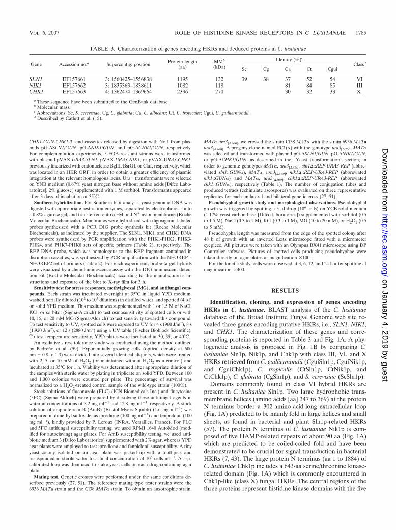
Identification, cloning, and expression of genes encodingHKRs in C. lusitaniae. BLAST analysis of the C. lusitaniaedatabase of the Broad Institute Fungal Genome web site re-vealed three genes encoding putative HKRs, i.e., SLN1, NIK1,and CHK1. The characterization of these genes and corre-sponding proteins is reported in Table 3 and Fig. 1A. A phy-logenetic analysis is proposed in Fig. 1B by comparing C.lusitaniae Sln1p, Nik1p, and Chk1p with class III, VI, and XHKRs retrieved from C. guilliermondii (CguiSln1p, CguiNik1p,and CguiChk1p), C. tropicalis (CtSln1p, CtNik1p, andCtChk1p), C. glabrata (CgSln1p), and S. cerevisiae (ScSln1p).
Domains commonly found in class VI hybrid HKRs arepresent in C. lusitaniae Sln1p. Two large hydrophobic trans-membrane helices (amino acids [aa] 347 to 369) at the proteinN terminus border a 302-amino-acid-long extracellular loop(Fig. 1A) predicted to be mainly fold in large helices and smallsheets, as found in bacterial and plant Sln1p-related HKRs(57). The protein N terminus of C. lusitaniae Nik1p is com-posed of five HAMP-related repeats of about 90 aa (Fig. 1A)which are predicted to be coiled-coiled fold and have beendemonstrated to be crucial for signal transduction in bacterialHKRs (7, 43). The large protein N terminus (aa 1 to 1884) ofC. lusitaniae Chk1p includes a 643-aa serine/threonine kinase-related domain (Fig. 1A) which is commonly encountered inChk1p-like (class X) fungal HKRs. The central regions of thethree proteins represent histidine kinase domains with the five
TABLE 3. Characterization of genes encoding HKRs and deduced proteins in C. lusitaniae
Gene Accession no.a Supercontig: position Protein length(aa)
a These sequence have been submitted to the GenBank database.b Molecular mass.c Abbreviations: Sc, S. cerevisiae; Cg, C. glabrata; Ca, C. albicans; Ct, C. tropicalis; Cgui, C. guilliermondii.d Described by Catlett et al. (15).
VOL. 6, 2007 ROLE OF HISTIDINE KINASE RECEPTORS IN C. LUSITANIAE 1785
typical boxes (H, N, G1, F, and G2) that define the catalyticcore of histidine kinases and include the presumptive auto-phosphorylated histidine residue. The C-terminal regions ofSln1p, Nik1p, and Chk1p are commonly composed of canoni-cal receiver domains which exhibit acidic pockets containingphosphorylable aspartate residues (68).
It was previously shown that pseudohyphal formation in C.lusitaniae was triggered on YCB solid medium but not in YCBliquid medium or in a complete YPD medium (30). We thencompared the mRNA levels of HKR genes by semiquantitativeRT-PCR using the wild-type strain 6936 in YCB or YPD,liquid or solid medium (Fig. 2A and B). The most striking
result was the high level of SLN1 transcripts detected onlywhen the 6936 strain was cultured in YCB solid medium. Theaccumulation of SLN1 mRNAs is concomitant with the forma-tion of pseudohyphae. This first preliminary experiment couldpoint out a correlation between SLN1 gene transcription andthe morphological state of C. lusitaniae cells.
Disruption of genes encoding HKRs in C. lusitaniae. LinearDNA cassettes 5� end-SLN1-GUN-SLN1-3� end, 5� end-NIK1-GUN-NIK1-3� end, and 5�end-CHK1-GUN-CHK1-3� end wereeach used to transform strain 6936 ura3[�360] to prototrophy.Correct insertion of the disrupting cassette was verified at eachlocus by Southern analysis of the genomic DNA of a subset of20 Ura� transformants, randomly selected from each transfor-mation experiment, along with the DNA of the control strain,6936 ura3[�360]. Genomic DNAs were digested with XhoI, PstI,or SacII and hybridized with SLN1, NIK1, and CHK1 probesaccording to the experiment. All fragments of the expected sizeare shown in Fig. 3. Southern blot analysis revealed thathomologous integration of the 5� end-SLN1-GUN-SLN1-3�end, 5� end-NIK1-GUN-NIK1-3� end, and 5� end-CHK1-GUN-CHK1-3� end cassettes at the corresponding target locus oc-curred in half of transformants analyzed and was derived fromgene replacement, resulting in disruption of the target geneand in the genotypes ura3[�360] sln1�::REP-URA3-REP (abbre-viated sln1::GUN), ura3[�360] nik1�::REP-URA3-REP (abbre-viated nik1::GUN), and ura3[�360] chk1�::REP-URA3-REP(abbreviated chk1::GUN), respectively. The molecular eventswere confirmed by hybridization with the REP DNA probe.For the remaining Ura� transformants, the hybridization pat-tern revealed that they were derived from gene replacement atthe ura3 locus (results not shown).
In order to obtain double-mutant genotypes, representativesln1::GUN, nik1::GUN, and chk1::GUN Ura� transformants wereplated onto YNB supplemented with 5-FOA and uracil. Thefrequency of 5-FOA-resistant colonies was about 1 � 105. Thegenetic organization of 10 5-FOA-resistant Ura clones fromeach sln1::GUN, nik1::GUN, and chk1::GUN transformants wasconfirmed by Southern blot analysis (results not shown). The5-FOA-resistant clones displayed the hybridization fragments ex-pected from deletion of the URA3 gene and one of the flankingREP fragments. The genotypes ura3[�360] sln1�::REP (abbrevi-ated sln1::REP), ura3[�360] nik1�::REP (abbreviated nik1::REP),and ura3[�360] chk1�::REP (abbreviated chk1::REP) were as-signed to the 5-FOA-resistant clones.
The linear 5� end-NIK1-GUN-NIK1-3� end cassette was usedto transform the chk1::REP strain. The double-mutant geno-type ura3[�360] chk1�::REP nik1�::REP-URA3-REP (abbrevi-ated chk1::REP nik1::GUN) was assigned to transformants har-boring the expected hybridization profile as screened in Fig. 3.In the same way, the linear 5� end-NIK1-GUN-NIK1-3� endand 5� end-CHK1-GUN-CHK1-3� end cassettes were each usedto transform the sln1::REP strain. Similarly, the double-mutantgenotype ura3[�360] sln1�::REP chk1�::REP-URA3-REP (ab-breviated chk1::GUN sln1::REP) was assigned to transformantsharboring the expected hybridization profile as screened in Fig. 3.
We failed to obtain homologous integration of the 5� end-NIK1-GUN-NIK1-3� end cassette in the sln1::REP strain and inthe chk1::REP sln1::REP strain (counterselected by cultivatingthe chk1::REP sln1::GUN strain on 5-FOA-containing YNBmedium). In the same way, the SLN1 disruption in the
FIG. 1. (A) Structure of the C. lusitaniae HKR proteins. The pu-tative phosphorylable residues are indicated with triangles. EL, extra-cellular loop; HKcd, histidine kinase catalytic domain; R, repeatedsequence; RD, receiver domain; S/TKrd, serine/threonine kinase-re-lated domain; TMH, transmembrane helix. (B) Dendrogram gener-ated after alignment of the predicted sequences of C. lusitaniae HKRs(ClSln1p, ClNik1p, and ClChk1p) with sequences retrieved from C.guilliermondii (CguiSln1p, CguiNik1p, and CguiChk1p), C. tropicalis(CtSln1p, CtNik1p, and CtChk1p), C. glabrata (CgSln1p), and S. cer-evisiae (ScSln1p). Alignment utilizes the neighborhood-joining methodfrom TreeView PPC software. Distances along the branches representthe divergence between two cognate sequences.
nik1::REP deletant could never be achieved. Indeed, Southernblot analysis of 20 Ura� transformants revealed that they de-rived only from ectopic integrations of disruption cassettes(40%) or from gene replacement at the ura3 locus (60%)
(results not shown). These two last results suggested that thesln1 nik1 double mutant and the sln1 chk1 nik1 triple mutantare lethal in C. lusitaniae.
The linear DNA disruption cassettes were also used to trans-
FIG. 2. Morphology of C. lusitaniae cells and expression analysis of genes encoding HKRs. (A) Morphology of pseudohyphae emerging fromthe edge of a colony when cells are plated on YCB solid medium. The morphology of budding yeast cultured in YCB liquid medium is shown inthe top left of the picture. (B) Semiquantitative RT-PCR of the three genes encoding HKRs (SLN1, NIK1, and CHK1). A representative RT-PCRanalysis is shown. For each target gene, the amount of transcription was compared to that of the ACT1 gene (relative level of transcription). Thehistogram presents the mean values of results from three independent experiments. Error bars show standard deviation. Wild-type cells werecultured during 48 h in YPD liquid medium (lane 1), in YPD solid medium (lane 2), in YCB liquid medium (lane 3), or in YCB solid medium(lane 4). ph, pseudohyphal formation.
FIG. 3. Southern blot hybridization and schematic representation of resident loci SLN1, NIK1, and CHK1 and of molecular events thatoccurred in transformants. Signals revealed by the labeled probes (each marked with an asterisk) correspond to those expected from the genomicrestriction map. (A) Hybridization pattern with SLN1 and REP probes of XhoI-digested genomic DNA from 6936 ura3[�360] (a) or a representativesln1::GUN transformant (b). (B) Hybridization pattern with NIK1 and REP probes of PstI-digested genomic DNA from 6936 ura3[�360] (a) or arepresentative nik1::GUN transformant (b). (C) Hybridization pattern with CHK1 and REP probes of SacII-digested genomic DNA from 6936ura3[�360] (a) or a representative chk1::GUN transformant (b). DNA fragment sizes are indicated in kilobases.
VOL. 6, 2007 ROLE OF HISTIDINE KINASE RECEPTORS IN C. LUSITANIAE 1787
form the strain PC1(�) to prototrophy in order to generatesln1::GUN�, nik1::GUN�, and chk1::GUN� MAT� disruptantstrains.
Complementation of the sln1, nik1, and chk1 null mutantalleles. To obtain reintegrant strains, linearized plasmidspVAX-URA3-SLN1, pVAX-URA3-NIK1, and pVAX-URA3-CHK1 were used to transform to prototrophy sln1::REP,nik1::REP, and chk1::REP mutants, respectively. We verifiedby Southern blotting (not shown) that homologous integrationof the whole plasmids pVAX-URA3-SLN1, pVAX-URA3-NIK1, and pVAX-URA3-CHK1 occurred at the sln1, nik1,and chk1 loci, respectively, resulting in the relevant geno-types ura3[�360], sln1�::[REP pVAX-URA3-SLN1] (abbreviatedsln1�SLN1), ura3[�360] nik1�::[REP pVAX-URA3-NIK1] (ab-breviated nik1�NIK1), and ura3[�360] chk1�::[REP pVAX-URA3-CHK1] (abbreviated chk1�CHK1). In the same way,linearized plasmids pVAX-URA3-SLN1 and pVAX-URA3-NIK1 were used to transform to prototrophy sln1::REPchk1::REP and chk1::REP nik1::REP (counterselected by cul-tivating the chk1::REP nik1::GUN strain on 5-FOA-containingYNB medium) strains, respectively. By this way, we obtainedchk1 sln1�SLN1 and chk1 nik1�NIK1 reintegrant strains.
Growth, osmotolerance, and oxidative stress response ofmutants. We investigated the putative involvement of thethree HKRs in C. lusitaniae in perception and transduction ofvarious environmental stresses. We started the phenotypiccharacterization of engineered mutants by comparing theirgrowth kinetics. All mutants and the wild-type strain 6936exhibited similar doubling times in liquid YPD medium (datanot shown). No differences were observed in the development(colony length and aspect) of all strains cultured in solid YPDmedium.
We next examined the osmotolerance of mutants. For that,drop plate assays were performed to determine the sensitivitiesof wild-type, sln1::GUN, nik1::GUN, chk1::GUN, chk1::GUNsln1::REP, and chk1::REP nik1::GUN strains to NaCl (1 to 1.5M), KCl (1 to 1.5 M), and sorbitol (1 to 1.5 M). The growth ofall simple and double mutants on these high-osmolarity mediawas similar to that of the wild-type strain (data not shown).These results imply that deletion of one or two HKR genes inC. lusitaniae has no effect on the growth and capacity of adap-tation of yeast cells to hyperosmotic conditions.
Furthermore, none of the HKR mutants exhibited hyper-sensitivity to UV irradiation or high temperature (40°C) (datanot shown), indicating that HKRs are not essential for theresponse to these physical stresses.
We also tested the effect of MG on HKR mutants, a meta-bolic by-product whose toxic action on cells can be counter-acted by triggering activation of the HOG signaling pathway(1, 45). We evaluated the sensitivities of each strain by usingdifferent inoculum concentrations (102 to 105) in the presenceof 10, 15, or 20 mM MG. We demonstrated that the sensitiv-ities of disruptant strains to this compound were completelyidentical to that of the wild-type strain. Indeed, concentrationsabove and below 15 to 20 mM MG either were too toxic or hadno effect on the strains (data not shown).
The effect of H2O2 on our engineered mutants was alsostudied (Fig. 4). For this purpose, YPD-grown exponential-phase cultures from all strains (wild type and simple and dou-ble disruptants) were incubated with 2, 5, or 10 mM H2O2. The
percentage of survival was expressed with respect to an H2O2-treated control sample of the wild-type strain. The 10 mMH2O2 concentration was too deleterious for C. lusitaniae toestimate cell survival. Interestingly, at 2 and 5 mM H2O2, thesln1::GUN mutant exhibited the greatest sensitivity to H2O2
(39% � 0.5% and 12% � 5%, respectively). The nik1::GUNmutant was more resistant to H2O2 than the sln1::GUN mutant(49% � 4% and 26% � 5%, respectively). Moreover, thechk1::GUN strain displayed moderate sensitivity at 2 and 5 mMof peroxide (85% � 5% and 51% � 6%, respectively). Thegenetically engineered sln1�SLN1, nik1�NIK1, andchk1�CHK1 revertants showed H2O2 susceptibilities similar tothat of wild-type strain 6936 (data not shown). Such a resultcould suggest the involvement of the three HKRs, especiallySln1p, in the regulation of oxidative stress.
Susceptibilities of mutants to antifungal compounds. Wefirst monitored the effects of the clinical antifungals AmB,5FC, and FLC. Neither hypersensitivity nor resistance towardthese antifungals was observed. Indeed, the cell-mediated im-munities (CMIs) of all the deletant strains were similar to thatof the wild-type strain (AmB CMI, �1 �g ml1; 5FC CMI, �4�g ml1; FLC CMI, �8 �g ml1) (data not shown).
Because in various filamentous fungi, such as N. crassa andC. heterostrophus, the mutation of class III HKR genes is re-sponsible for severe dicarboximide and phenylpyrrole resis-tance (53, 70), we studied the effects of these antifungals atdifferent concentrations (1 to 16 �g ml1) on our transfor-mants. Figure 5 shows that wild-type strain 6936 and thechk1::GUN mutant were resistant to iprodione (dicarboxim-ide) up to 4 �g ml1, whereas the nik1::GUN mutant wasresistant to iprodione in concentrations up to 16 �g ml1.Interestingly, the sln1::GUN mutant was hypersusceptible toiprodione (resistance only up to 2 �g ml1). The wild-type
FIG. 4. Measurement of cell survival after oxidative stress imposedby the addition of 2 mM or 5 mM H2O2 in simple mutants (sln1::GUN,nik1::GUN, and chk1::GUN) and double mutants (chk1::GUNsln1::REP and chk1::REP nik1::GUN). The percent survival is ex-pressed with respect to that of an H2O2-treated control sample of thewild-type strain (100%). Standard deviation bars are based on threeindividual replicates.
strain, 6936, and the chk1::GUN mutant were resistant to fen-piclonil up to 1 �g ml1, whereas the nik1::GUN mutant wasresistant to fenpiclonil to concentrations up to 16 �g ml1.Moreover, the sln1::GUN mutant was hypersusceptible to fen-piclonil (CMI, �1 �g ml1). The chk1::REP nik1::GUN andchk1::GUN sln1::REP double mutants presented the same an-tifungal susceptibility as the nik1::GUN and sln1::GUN simplemutants, respectively. The genetically engineered sln1�SLN1,nik1�NIK1, chk1 sln1�SLN1, and chk1 nik1�NIK1 revertantsshowed antifungal susceptibilities similar to those of wild-typestrain 6936. Therefore, we concluded that in C. lusitaniae cells,NIK1 gene disruption confers fenpiclonil and iprodione resis-tance and that sln1 deletion increases the susceptibility to theseantifungals.
Mating abilities of mutants. It was recently demonstratedthat an HKR (Tco1p) promotes sexual reproduction in C.neoformans (9). We next investigated if the deletion of theSLN1, NIK1, and CHK1 genes could have an effect upon the invitro mating ability of C. lusitaniae. We demonstrated that thesimple or double disruptants, like the parental 6936 MATawild-type strain, were still able to reproduce sexually in vitrowhen mated unilaterally with the appropriate opposite mating-type strain, Cl38 MAT�. Moreover, all mutants exhibited nor-mal wild-type mating in bilateral mutant crosses (sln1::GUNMATa � sln1::GUN MAT�; nik1::GUN MATa � nik1::GUNMAT�, and chk1::GUN MATa � chk1::GUN MAT�) (data notshown). Therefore, the HKR proteins seem not to participatein the mating process of C. lusitaniae.
Pseudohyphal growth abilities of mutants. We investigatedthe capacity of transformants to differentiate pseudohyphae.
Approximately 106 cells (contained in drops of 5 �l) of wild-type strain 6936 and of each mutant were spotted on YCB solidmedium supplemented or not with sorbitol, NaCl, KCl, MG, orH2O2 (Fig. 6). The lengths of pseudohyphae emerging fromthe edge of the colony are reported in Table 4.
Homogeneously distributed pseudohyphae were readily ob-tained for all strains on unsupplemented YCB medium. Engi-neered mutants present no significant length variation com-pared with the wild-type strain (560 � 5 �m), except thesln1::GUN mutant, which displayed a reproducible reduction(440 � 24 �m) of pseudohypha length. Interestingly, thechk1::GUN sln1::REP double mutant presented no significantreduction (547 � 22 �m) of pseudohypha length. Figure 6shows representative pictures of pseudohypha formation after48 h of growth on YCB medium supplemented with the mostdiscriminatory concentrations of sorbitol (1 M), NaCl (0.5 M),KCl (0.5 M), MG (15 mM), and H2O2 (1 mM). Indeed, con-centrations above and below either were too toxic or had noeffect on the pseudohyphal development of strains.
The addition of 1 M sorbitol produces a homogeneous re-duction of pseudohyphal growth of the wild-type strain and ofchk1::GUN, nik1::GUN, and chk1::REP nik1::GUN mutantscompared to growth of the strains plated on unsupplementedYCB solid medium. The most important result of this experi-ment is the complete inhibition of pseudohyphal developmentof sln1::GUN and chk1::GUN sln1::REP mutants. Similar re-sults were obtained with the presence of 0.5 M NaCl or KCl.The reintroduction of a functional SLN1 allele in sln1::REP(reintegrant sln1�SLN1 strain) and chk1::REP sln1::REPmutants (chk1 sln1�SLN1 reintegrant strain) was sufficient
FIG. 5. Susceptibilities of C. lusitaniae mutants to iprodione and fenpiclonil. The wild-type strain and representative simple mutants(sln1::GUN, nik1::GUN, and chk1::GUN), double mutants (chk1::GUN sln1::REP and chk1::REP nik1::GUN), and reintegrant strains (sln1�SLN1,nik1�NIK1, chk1 sln1�SLN1, and chk1 nik1�NIK1) were grown for 48 h on YPD plates containing fenpiclonil or iprodione at concentrationsindicated.
VOL. 6, 2007 ROLE OF HISTIDINE KINASE RECEPTORS IN C. LUSITANIAE 1789
to restore pseudohyphal development similar to that of wild-type strain 6936 on YCB solid medium supplemented or notwith sorbitol, NaCl, or KCl (Fig. 6). Thus, the SLN1 geneseems to play a key role in pseudohyphal development of C.lusitaniae during growth on high-osmolarity media.
We then tested the effect of MG. The addition of 15 mMMG produces a homogeneous reduction of pseudohyphalgrowth of the wild-type strain and of chk1::GUN, nik1::GUN,and chk1::REP nik1::GUN mutants compared to growth ofstrains plated on unsupplemented YCB solid medium (Fig. 6).Furthermore, sln1::GUN pseudohyphal formation was com-pletely abolished. The chk1::GUN sln1::REP mutants formedsome sporadic bunch-like pseudohyphae displaying a signifi-cant reduction of their lengths (133 � 41 �m). Furthermore,the reintroduction of a functional SLN1 allele (sln1�SLN1and chk1 sln1�SLN1 reintegrant strains) was sufficient to re-
store pseudohyphal development similar to that of wild-typestrain 6936 on YCB solid medium supplemented with 15mM MG.
Finally, we monitored the effect of oxidative stress. Theaddition of 1 mM H2O2 produces a reduction of pseudohy-phal growth of the wild-type strain (387 � 16 �m) (Fig. 6).Of all the strains, the sln1::GUN and chk1::GUN sln1::REPmutants exhibited the greatest sensitivity to peroxide, sincepseudohyphal growth was completely inhibited. The nik1::GUNmutant presented a significant reduction (167 � 50 �m) and thechk1::GUN strain a moderate reduction (280 � 28 �m) inpseudohypha length. The reintroduction of the SLN1 allele wassufficient to restore partially (chk1 sln1�SLN1 reintegrant strain)or completely (sln1�SLN1 reintegrant strain) a pseudohyphaldevelopment similar to that of wild-type strain 6936 cultured onYCB solid medium supplemented with 1 mM H2O2. We also
FIG. 6. Pseudohyphal growth abilities of mutants. Cells (106; contained in drops of 5 �l) of the wild-type strain and representative simplemutants (sln1::GUN, nik1::GUN, and chk1::GUN), double mutants (chk1::GUN sln1::REP and chk1::REP nik1::GUN), and reintegrant strains(sln1�SLN1 and chk1 sln1�SLN1) were spotted on YCB solid medium supplemented or not with discriminatory concentrations of sorbitol (1 M),NaCl (0.5 M), KCl (0.5 M), MG (15 mM), and H2O2 (1 mM). Observations were done 48 h after spotting. This experiment was done in triplicate,and pictures present representative structures observed for each sample.
TABLE 4. Pseudohyphal differentiation of C. lusitaniae strains on various supplemented YCB media
a Abbreviated genotypes are given. WT, wild-type strain 6936.b The length of pseudohyphae was measured from the edge of the spotted colony. The values (�m) are means � standard deviations based on three individual
replicates. “�” indicates YCB medium supplemented as indicated.c ND, not determined.
obtained wild-type pseudohyphal growth after the reintroductionof the NIK1 allele in the nik1::REP strain and the CHK1 allele inthe chk1::REP strain (Table 4). These results demonstrate thecrucial role of Sln1p and also the involvement of Nik1p andChk1p in the regulation of oxidative stress during pseudohyphaldevelopment in C. lusitaniae.
Because we observed that the sln1 deletion leads to a reduc-tion of pseudohyphal formation in C. lusitaniae, we comparedthe time course of pseudohyphae differentiation in wild-typestrain 6936, the sln1::GUN mutant, and the sln1�SLN1 rein-tegrant strain during 24 h (Fig. 7). The strains were simulta-neously plated on YCB agar medium in order to inducepseudohyphal development. The first cell elongations whichinitiate chain formations (around the colony) of the wild-typestrain and the sln1�SLN1 reintegrant strain were well detectedafter 6 h of incubation. However, a slight start of cell elonga-tions was observed for the sln1::GUN mutant. After 12 h ofgrowth, the wild-type strain and the sln1�SLN1 reintegrantstrain presented readily homogeneously distributed pseudohy-phae whereas the sln1::GUN mutant showed a heterogeneousdistribution of shorter pseudohyphae. However, the number ofbranching of lateral cells along the pseudohyphae did not sig-nificantly vary between the sln1::GUN mutant and the wild-type strain. We thus concluded that the SLN1 disruption in-duced a delay in this morphogenetic transition and a reductionof pseudohypha growth.
DISCUSSION
Fungal HKRs are involved in some essential cellular pro-cesses, such as osmosensing, virulence expression, oxidativestress response, and cell cycle control (62). Various investiga-tions also report the implication of HKRs in yeast morpholog-ical switching. Indeed, in C. albicans, it has been shown that thethree HKRs CaSln1p, CaNik1p, and Cahk1p play a predomi-
nant role in true-hypha formation (3, 13, 49, 69). Moreover, arecent study provides firm genetic evidence that the Nik1p-likeHKRs (class III) were essential in the mold-to-yeast transitionin the dimorphic pathogens H. capsulatum and B. dermatitidis(50). These findings point out a broad role of HKRs in mor-phological switching in nonrelated yeast species.
We undertook the characterization of HKRs in C. lusitaniaebecause, to our knowledge, the involvement of HKRs in yeastswhich are only able to form pseudohyphae has never been fullystudied. The only data available focused on the potential im-plication of the HOG pathway in pseudohyphal formation in S.cerevisiae (55). Moreover, C. lusitaniae represents an emerginghuman pathogen which is characterized by its propensity todevelop resistance to antifungal agents during treatment.HKR-mediated transduction pathways, which are not encoun-tered among the animal kingdom, could provide promisingapplications in antimicrobial compound research (10, 33, 46).BLAST analysis of the C. lusitaniae genome database allowedus to identify three genes, namely SLN1, NIK1, and CHK1,encoding HKRs of class VI, III, and X, respectively. An inter-esting fact is that each of their three homologues was found inthe recently fully sequenced genomes of C. guilliermondii andC. tropicalis, as in C. albicans. In contrast, Nik1p-like andChk1p-like HKRs were absent in C. glabrata and S. cerevisiae,each harboring a sole Sln1p-like HKR (class VI). A recentphylogeny study based on whole-genome analysis indicatesthat Saccharomycotina could be subdivided within two majorgroups: (i) species whose genomes have undergone a whole-genome duplication (referred to as the WGD clade), includingC. glabrata and other yeasts from the Saccharomyces genus,and (ii) species that translate CTG as serine instead of leucine(referred to as the CTG clade), including C. guilliermondii, C.tropicalis, C. albicans, and C. lusitaniae (25). Thus, our analysissuggests that fungal species belonging to the WGD clade prob-ably encode a unique class VI HKR, and interestingly, species
FIG. 7. Delay of pseudohyphal development of the sln1::GUN mutant. The wild-type strain, the sln1::GUN simple mutant, and the corre-sponding sln1�SLN1 reintegrant strain were simultaneously spotted on YCB solid medium. Observations were done at 3, 6, 12, and 24 h afterspotting. This experiment was done in triplicate, and pictures present representative structures observed in each sample.
VOL. 6, 2007 ROLE OF HISTIDINE KINASE RECEPTORS IN C. LUSITANIAE 1791
belonging to the CTG clade encodes three HKRs from classesVI, III, and X, respectively.
We started this work by comparing the transcriptional reg-ulation of the SLN1, NIK1, and CHK1 genes when C. lusitaniaegrows either as budding blastospores or as pseudohyphae. Wedetected a high level of SLN1 transcripts when the wild-typestrain was cultured in YCB solid medium, indicating that theaccumulation of SLN1 mRNAs is concomitant with the forma-tion of pseudohyphae. This experiment could point out a cor-relation between SLN1 gene transcription and the pseudohy-phal developing process in C. lusitaniae.
We then disrupted the SLN1, NIK1, and CHK1 genes by atransformation system based upon the “URA3 blaster” strategythat we developed for C. lusitaniae in a previous study (58). Inthis way, we then selected sln1, nik1, and chk1 simple mutantsas well as sln1 chk1 and nik1 chk1 double mutants. Neverthe-less, we failed to obtain an sln1 nik1 double mutant and an sln1chk1 nik1 triple mutant, suggesting that these deletants are notviable in C. lusitaniae, as demonstrated for C. albicans (69).Thus, since the combination of sln1 and nik1 mutations re-sulted in synthetic lethality, it could suggest that Sln1p andNik1p act in two complementary pathways and that the lack ofone of these proteins could be compensated by the other.Independent studies have demonstrated that (i) ScYpd1p in-teracts with ScSln1p HKR in S. cerevisiae (42, 48, 61) and (ii)ScYpd1p interacts with MgNik1p HKR (Hik1p, from the riceBLAST fungus Magnaporthe grisea) (48). Preliminary data inour lab showed that the C. lusitaniae YPD1 gene, encoding ahistidine-containing phosphotransfer protein, is essential (un-published result). This could suggest that Ypd1p is a cross talkkey component of the Sln1p and Nik1p pathways in C. lus-itaniae.
When growing as budding yeast, all mutants and the wild-type strain exhibited similar doubling times and no differencewas observed between the development of strains in normaland hyperosmotic conditions. However, since the sln1 mutantwas hypersensitive to oxidative stress, we hypothesized a con-tribution of Sln1p in oxidative stress adaptation. These resultsare slightly different of those described for C. albicans: (i) sln1transformants were viable under normal and high osmolarity,but growth retardation was observed under hyperosmotic con-ditions (49), and (ii) only chk1 mutants were moderately hy-persensitive to oxidative stress (38). C. lusitaniae also behavesdifferently from several filamentous fungi, such as N. crassaand M. fructicola, for which nik1 mutants are sensitive to os-motic stress (44, 53).
Important morphological changes occur during sexual re-production of C. lusitaniae, notably during conjugation (27).Moreover, in the human pathogen C. neoformans, it has beenshown that the Tco1p HKR and the cognate stress-activatedHog1p MAPK system governed several cellular events, includ-ing sexual reproduction (9). So, we investigated if the deletionof the C. lusitaniae SLN1, NIK1, and CHK1 genes modified thein vitro mating ability of the yeast. We demonstrated that all ofthe strains were still able to reproduce sexually when crossedwith the opposite-mating-type strain.
In several filamentous fungi, the mutation of genes encodingHKRs of class III has been found to confer resistance todicarboximides, such as iprodione, and to phenylpyrroles, suchas fenpiclonil or fludioxonil (8, 21, 22, 44, 53, 70). A similar
study was carried out with C. albicans sln1, chk1, and nik1disruptants. Because the wild-type strain CAI4, used to con-struct the corresponding HKR mutants, was intrinsically resis-tant to both antifungals, it was impossible to study the specificrole of CaNik1p in dicarboximide and phenylpyrrole resistance(54). We thus tested the susceptibilities of C. lusitaniae HKRdisruptants and the wild-type strain 6936 to iprodione andfenpiclonil. The present study clearly demonstrated that the6936 strain was susceptible to both antifungals whereas thenik1 genotype conferred resistance to these compounds. It hasbeen shown that the target of these compounds was probablythe osmotic stress signal transduction pathway, which involvedthe class III HKRs (21). Furthermore, a recent study showedthat expression of MgHIK1, a NIK1 orthologue from M. grisea,could confer fungicide susceptibility on S. cerevisiae, which wasintrinsically resistant (48). Moreover, two-hybrid experimentsrevealed an interaction between MgHik1p and the yeast phos-phorelay protein Ypd1p. It was thus suggested that Nik1p-likeHKRs were direct targets of the fungicides or were mediatorsof antifungal action which were transmitted to the Hog1p path-way via Ypd1p. To date, since disruption of the C. lusitaniaeNIK1 gene confers resistance to iprodione and fenpiclonil andsince sequences homologuous to those of the S. cerevisiaeYPD1, HOG1, and SSK1 genes are located in the C. lusitaniaegenome (not shown; personal data), similar targeting processesand signaling pathways could occur in Candida species, such asC. lusitaniae. Finally, we also found that sln1 deletion increasesthe susceptibilities of strains to fenpiclonil and iprodione, aswas previously described for C. albicans (54). This might indi-cate that in Candida species, the signal transduction of Nik1p-like HKRs interacts with that of Sln1p-like HKRs. Neverthe-less, Sln1p, Nik1p, and Chk1p seem not to be implicated inresistance to antifungals of clinical relevance, such as FLC,5FC, and AmB.
In order to determine if HKRs could be involved in theyeast-to-pseudohypha morphological transition in C. lusitaniae,we compared the development of the HKR disruptants withthat of the wild-type strain on YCB plates, an appropriatemedium efficiently triggering pseudohyphal differentiation(30). Homogeneously distributed pseudohyphae were readilyobtained for all mutants, but a reproducible reduction of about20% of pseudohypha length was observed for the sln1 geno-type. More precisely, we showed that the defect of pseudohy-phae forming by the sln1 mutant was due to a global delay ofthe development process. Interestingly, the disruption of theCHK1 gene in the sln1 genotype partially restored wild-type-like pseudohyphal development. These results underlined acouple of conclusions compared to C. albicans true hyphaldevelopment. First, although HKRs are all clearly implicatedin a dramatic defect in true hypha formation when deleted inC. albicans, only one gene, namely SLN1, appears to be crucialfor pseudohyphal differentiation in our model. This clearlyillustrates that the information leading the yeast switching to-ward true hyphae or pseudohyphae is controlled by a set ofspecific genes. HKRs seem thus to be more closely involved inthe true hyphal shaping of C. albicans than they are in thepseudohyphal shaping of C. lusitaniae. Another possibility isthat the HKRs in C. albicans behave similarly to those in C.lusitaniae when C. albicans is undergoing pseudohyphal forma-tion. Secondly, similar effects were observed when the CHK1
gene was deleted in the sln1 strain, since it partially restoredwild-type pseudohyphal development in C. lusitaniae andpartly restored true hyphal growth in C. albicans. This supportsthe hypothesis that Candida sp. Sln1p-like HKRs would beable to regulate negative effectors of Chk1p-like HKRs (69).Moreover, pseudohyphal development on the hyperosmoticsurface and oxidant environment of the sln1 disruptant is dra-matically affected comparing to that of the wild-type strain.Interestingly, these growth conditions have no effect (hyperos-motic condition) or have a lesser effect (oxidant stress) onother HKR-disrupted mutants. This implies a differential con-tribution of the three HKRs in osmotic and oxidant adaptationduring the morphological transition.
In summary, this study reveals that C. lusitaniae, like otherspecies belonging to the CTG clade of the subphylum Saccha-romycotina, harbors three HKR genes. The NIK1 gene, whichencodes a class III HKR, is clearly involved in dicarboximideand phenylpyrrole resistance when the corresponding gene ismutated. The SLN1 gene encoding a transmembrane class VIHKR appears to be crucial (i) for oxidative stress adaptationduring C. lusitaniae strain growth as budding yeast and (ii) inthe early steps of pseudohyphal development, especially inhyperosmotic and oxidant conditions. Future studies will aimto characterize downstream elements, such as histidine-con-taining phosphotransfer protein and response regulators, inorder to clarify fungal HKR-mediated pathways and cross talkevents that regulate the morphological transition of thisemerging pathogen.
ACKNOWLEDGMENTS
We acknowledge the Broad Institute Fungal Genome Initiative formaking the complete genome sequence of Candida lusitaniae available.We thank Susan Fox for critical reading of the manuscript.
REFERENCES
1. Aguilera, J., S. Rodriguez-Vargas, and J. A. Prieto. 2005. The HOG MAPkinase pathway is required for the induction of methylglyoxal-responsivegenes and determines methylglyoxal resistance in Saccharomyces cerevisiae.Mol. Microbiol. 56:228–239.
2. Alex, L. A., K. A. Borkovich, and M. I. Simon. 1996. Hyphal development inNeurospora crassa: involvement of a two-component histidine kinase. Proc.Natl. Acad. Sci. USA 93:3416–3421.
3. Alex, L. A., C. Korch, C. P. Selitrennikoff, and M. I. Simon. 1998. COS1, atwo-component histidine kinase that is involved in hyphal development inthe opportunistic pathogen Candida albicans. Proc. Natl. Acad. Sci. USA95:7069–7073.
4. Altschul, S. F., T. L. Madden, A. A. Schaffer, J. Zhang, Z. Zhang, W. Miller,and D. J. Lipman. 1997. Gapped BLAST and PSI-BLAST: a new generationof protein database search programs. Nucleic Acids Res. 25:3389–3402.
5. Aoyama, K., H. Aiba, and T. Mizuno. 2001. Genetic analysis of the His-to-Asp phosphorelay implicated in mitotic cell cycle control: involvement ofhistidine-kinase genes of Schizosaccharomyces pombe. Biosci. Biotechnol.Biochem. 65:2347–2352.
6. Appleby, J. L., J. S. Parkinson, and R. B. Bourret. 1996. Signal transductionvia the multi-step phosphorelay: not necessarily a road less traveled. Cell86:845–848.
7. Aravind, L., and C. P. Ponting. 1999. The cytoplasmic helical linker domainof receptor histidine kinase and methyl-accepting proteins is common tomany prokaryotic signalling proteins. FEMS Microbiol. Lett. 176:111–116.
8. Avenot, H., P. Simoneau, B. Iacomi-Vasilescu, and N. Bataille-Simoneau.2005. Characterization of mutations in the two-component histidine kinasegene AbNIK1 from Alternaria brassicicola that confer high dicarboximide andphenylpyrrole resistance. Curr. Genet. 47:234–243.
9. Bahn, Y. S., K. Kojima, G. M. Cox, and J. Heitman. 2006. A unique fungaltwo-component system regulates stress responses, drug sensitivity, sexualdevelopment, and virulence of Cryptococcus neoformans. Mol. Biol. Cell17:3122–3135.
10. Barrett, J. F., and J. A. Hoch. 1998. Two-component signal transduction asa target for microbial anti-infective therapy. Antimicrob. Agents Chemother.42:1529–1536.
11. Berman, J. 2006. Morphogenesis and cell cycle progression in Candidaalbicans. Curr. Opin. Microbiol. 9:595–601.
12. Buck, V., J. Quinn, T. Soto Pino, H. Martin, J. Saldanha, K. Makino, B. A.Morgan, and J. B. Millar. 2001. Peroxide sensors for the fission yeast stress-activated mitogen-activated protein kinase pathway. Mol. Biol. Cell 12:407–419.
13. Calera, J. A., and R. Calderone. 1999. Flocculation of hyphae is associatedwith a deletion in the putative CaHK1 two-component histidine kinase genefrom Candida albicans. Microbiology 145:1431–1442.
14. Calera, J. A., X. J. Zhao, F. De Bernardis, M. Sheridan, and R. Calderone.1999. Avirulence of Candida albicans CaHK1 mutants in a murine model ofhematogenously disseminated candidiasis. Infect. Immun. 67:4280–4284.
15. Catlett, N. L., O. C. Yoder, and B. G. Turgeon. 2003. Whole-genome analysisof two-component signal transduction genes in fungal pathogens. Eukaryot.Cell 2:1151–1161.
16. Chapeland-Leclerc, F., J. Bouchoux, A. Goumar, C. Chastin, J. Villard, andT. Noel. 2005. Inactivation of the FCY2 gene encoding purine-cytosine per-mease promotes cross-resistance to flucytosine and fluconazole in Candidalusitaniae. Antimicrob. Agents Chemother. 49:3101–3108.
17. Chauhan, N., J. P. Latge, and R. Calderone. 2006. Signalling and oxidantadaptation in Candida albicans and Aspergillus fumigatus. Nat. Rev. Micro-biol. 4:435–444.
18. Chun, C. D., O. W. Liu, and H. D. Madhani. 2007. A link between virulenceand homeostatic responses to hypoxia during infection by the human fungalpathogen Cryptococcus neoformans. PLoS Pathog. 3:e22.
19. Clemons, K. V., T. K. Miller, C. P. Selitrennikoff, and D. A. Stevens. 2002.fos-1, a putative histidine kinase as a virulence factor for systemic aspergil-losis. Med. Mycol. 40:259–262.
20. Csank, C., and K. Haynes. 2000. Candida glabrata displays pseudohyphalgrowth. FEMS Microbiol. Lett. 189:115–120.
21. Cui, W., R. E. Beever, S. L. Parkes, P. L. Weeds, and M. D. Templeton. 2002.An osmosensing histidine kinase mediates dicarboximide fungicide resis-tance in Botryotinia fuckeliana (Botrytis cinerea). Fungal Genet. Biol. 36:187–198.
22. Dry, I. B., K. H. Yuan, and D. G. Hutton. 2004. Dicarboximide resistance infield isolates of Alternaria alternata is mediated by a mutation in a two-component histidine kinase gene. Fungal Genet. Biol. 41:102–108.
23. Ernst, J. F. 2000. Transcription factors in Candida albicans—environmentalcontrol of morphogenesis. Microbiology 146:1763–1774.
24. Favel, A., A. Michel-Nguyen, F. Peyron, C. Martin, L. Thomachot, A. Datry,J. P. Bouchara, S. Challier, T. Noel, C. Chastin, and P. Regli. 2003. Colonymorphology switching of Candida lusitaniae and acquisition of multidrugresistance during treatment of a renal infection in a newborn: case reportand review of the literature. Diagn. Microbiol. Infect. Dis. 47:331–339.
25. Fitzpatrick, D. A., M. E. Logue, J. E. Stajich, and G. Butler. 2006. A fungalphylogeny based on 42 complete genomes derived from supertree and com-bined gene analysis. BMC Evol. Biol. 6:99.
26. Francois, F., F. Chapeland-Leclerc, J. Villard, and T. Noel. 2004. Develop-ment of an integrative transformation system for the opportunistic patho-genic yeast Candida lusitaniae using URA3 as a selection marker. Yeast21:95–106.
27. Francois, F., T. Noel, R. Pepin, A. Brulfert, C. Chastin, A. Favel, and J.Villard. 2001. Alternative identification test relying upon sexual reproductiveabilities of Candida lusitaniae strains isolated from hospitalized patients.J. Clin. Microbiol. 39:3906–3914.
28. Geourjon, C., and G. Deleage. 1995. SOPMA: significant improvements inprotein secondary structure prediction by consensus prediction from multi-ple alignments. Comput. Appl. Biosci. 11:681–684.
29. Gimeno, C. J., P. O. Ljungdahl, C. A. Styles, and G. R. Fink. 1992. Unipolarcell divisions in the yeast S. cerevisiae lead to filamentous growth: regulationby starvation and RAS. Cell 68:1077–1090.
30. Goumar, A., A. Brulfert, C. Martin, J. Villard, and T. Noel. 2004. Selectionand genetic analysis of pseudohyphae defective mutants with attenuatedvirulence in Candida lusitaniae. J. Med. Mycol. 14:3–11.
31. Guinet, R., J. Chanas, A. Goullier, G. Bonnefoy, and P. Ambroise-Thomas.1983. Fatal septicemia due to amphotericin B-resistant Candida lusitaniae.J. Clin. Microbiol. 18:443–444.
32. Hawkins, J. L., and L. M. Baddour. 2003. Candida lusitaniae infections in theera of fluconazole availability. Clin. Infect. Dis. 36:e14–e18.
33. Koretke, K. K., A. N. Lupas, P. V. Warren, M. Rosenberg, and J. R. Brown.2000. Evolution of two-component signal transduction. Mol. Biol. Evol.17:1956–1970.
34. Krogh, A., B. Larsson, G. von Heijne, and E. L. Sonnhammer. 2001. Pre-dicting transmembrane protein topology with a hidden Markov model: ap-plication to complete genomes. J. Mol. Biol. 305:567–580.
35. Kruppa, M., and R. Calderone. 2006. Two-component signal transduction inhuman fungal pathogens. FEMS Yeast Res. 6:149–159.
36. Kruppa, M., T. Goins, J. E. Cutler, D. Lowman, D. Williams, N. Chauhan,V. Menon, P. Singh, D. Li, and R. Calderone. 2003. The role of the Candidaalbicans histidine kinase CHK1 gene in the regulation of cell wall mannanand glucan biosynthesis. FEMS Yeast Res. 3:289–299.
37. Kruppa, M., B. P. Krom, N. Chauhan, A. V. Bambach, R. L. Cihlar, and
VOL. 6, 2007 ROLE OF HISTIDINE KINASE RECEPTORS IN C. LUSITANIAE 1793
R. A. Calderone. 2004. The two-component signal transduction proteinChk1p regulates quorum sensing in Candida albicans. Eukaryot. Cell 3:1062–1065.
38. Li, D., V. Gurkovska, M. Sheridan, R. Calderone, and N. Chauhan. 2004.Studies on the regulation of the two-component histidine kinase gene CHK1in Candida albicans using the heterologous lacZ reporter gene. Microbiology150:3305–3313.
39. Li, S., A. Ault, C. L. Malone, D. Raitt, S. Dean, L. H. Johnston, R. J.Deschenes, and J. S. Fassler. 1998. The yeast histidine protein kinase, Sln1p,mediates phosphotransfer to two response regulators, Ssk1p and Skn7p.EMBO J. 17:6952–6962.
40. Loomis, W. F., G. Shaulsky, and N. Wang. 1997. Histidine kinases in signaltransduction pathways of eukaryotes. J. Cell Sci. 110:1141–1145.
41. Lorenz, M. C., and J. Heitman. 1998. Regulators of pseudohyphal differen-tiation in Saccharomyces cerevisiae identified through multicopy suppressoranalysis in ammonium permease mutant strains. Genetics 150:1443–1457.
42. Lu, J. M., R. J. Deschenes, and J. S. Fassler. 2003. Saccharomyces cerevisiaehistidine phosphotransferase Ypd1p shuttles between the nucleus and cyto-plasm for SLN1-dependent phosphorylation of Ssk1p and Skn7p. Eukaryot.Cell 2:1304–1314.
43. Lupas, A., M. Van Dyke, and J. Stock. 1991. Predicting coiled coils fromprotein sequences. Science 252:1162–1164.
44. Ma, Z., Y. Luo, and T. Michailides. 2006. Molecular characterization of thetwo-component histidine kinase gene from Monilinia fructicola. Pest Manag.Sci. 62:991–998.
45. Maeta, K., S. Izawa, and Y. Inoue. 2005. Methylglyoxal, a metabolite derivedfrom glycolysis, functions as a signal initiator of the high osmolarity glycerol-mitogen-activated protein kinase cascade and calcineurin/Crz1-mediatedpathway in Saccharomyces cerevisiae. J. Biol. Chem. 280:253–260.
46. Matsushita, M., and K. D. Janda. 2002. Histidine kinases as targets for newantimicrobial agents. Bioorg. Med. Chem. 10:855–867.
47. Minari, A., R. Hachem, and I. Raad. 2001. Candida lusitaniae: a cause ofbreakthrough fungemia in cancer patients. Clin. Infect. Dis. 32:186–190.
48. Motoyama, T., T. Ohira, K. Kadokura, A. Ichiishi, M. Fujimura, I. Yamaguchi,and T. Kudo. 2005. An Os-1 family histidine kinase from a filamentous fungusconfers fungicide-sensitivity to yeast. Curr. Genet. 47:298–306.
49. Nagahashi, S., T. Mio, N. Ono, T. Yamada-Okabe, M. Arisawa, H. Bussey,and H. Yamada-Okabe. 1998. Isolation of CaSLN1 and CaNIK1, the genesfor osmosensing histidine kinase homologues, from the pathogenic fungusCandida albicans. Microbiology 144:425–432.
50. Nemecek, J. C., M. Wuthrich, and B. S. Klein. 2006. Global control ofdimorphism and virulence in fungi. Science 312:583–588.
51. Noel, T., A. Favel, A. Michel-Nguyen, A. Goumar, K. Fallague, C. Chastin, F.Leclerc, and J. Villard. 2005. Differentiation between atypical isolates ofCandida lusitaniae and Candida pulcherrima by determination of matingtype. J. Clin. Microbiol. 43:1430–1432.
52. Noel, T., F. Francois, P. Paumard, C. Chastin, D. Brethes, and J. Villard.2003. Flucytosine-fluconazole cross-resistance in purine-cytosine permease-deficient Candida lusitaniae clinical isolates: indirect evidence of a flucon-azole uptake transporter. Antimicrob. Agents Chemother. 47:1275–1284.
53. Ochiai, N., M. Fujimura, T. Motoyama, A. Ichiishi, R. Usami, K. Horikoshi,and I. Yamaguchi. 2001. Characterization of mutations in the two-compo-nent histidine kinase gene that confer fludioxonil resistance and osmoticsensitivity in the os-1 mutants of Neurospora crassa. Pest Manag. Sci. 57:437–442.
54. Ochiai, N., M. Fujimura, M. Oshima, T. Motoyama, A. Ichiishi, H. Yamada-Okabe, and I. Yamaguchi. 2002. Effects of iprodione and fludioxonil onglycerol synthesis and hyphal development in Candida albicans. Biosci. Bio-technol. Biochem. 66:2209–2215.
55. O’Rourke, S. M., and I. Herskowitz. 1998. The Hog1 MAPK prevents cross
talk between the HOG and pheromone response MAPK pathways in Sac-charomyces cerevisiae. Genes Dev. 12:2874–2886.
56. Page, R. D. 1996. TreeView: an application to display phylogenetic trees onpersonal computers. Comput. Appl. Biosci. 12:357–358.
57. Papon, N., J. Bremer, A. Vansiri, G. Glevarec, M. Rideau, and J. Creche.2006. Molecular cloning and expression of a cDNA encoding a hybrid his-tidine kinase receptor in tropical periwinkle Catharanthus roseus. Plant Biol.(Stuttgart) 8:731–736.
58. Papon, N., T. Noel, M. Florent, S. Gibot-Leclerc, D. Jean, C. Chastin, J.Villard, and F. Chapeland-Leclerc. 2007. Molecular mechanism of flucy-tosine resistance in Candida lusitaniae: contribution of the FCY2, FCY1, andFUR1 genes to 5-fluorouracil and fluconazole cross-resistance. Antimicrob.Agents Chemother. 51:369–371.
59. Pendreno, Y., P. Gonzalez-Parraga, S. Conesa, M. Martinez-Esparza, A.Aguinaga, J. A. Hernandez, and J. C. Arguelles. 2006. The cellular resistanceagainst oxidative stress (H2O2) is independent of neutral trehalase (Ntc1p)activity in Candida albicans. FEMS Yeast Res. 6:57–62.
60. Pfaller, M. A., S. A. Messer, and R. J. Hollis. 1994. Strain delineation andantifungal susceptibilities of epidemiologically related and unrelated isolatesof Candida lusitaniae. Diagn. Microbiol. Infect. Dis. 20:127–133.
61. Posas, F., S. M. Wurgler-Murphy, T. Maeda, E. A. Witten, T. C. Thai, andH. Saito. 1996. Yeast HOG1 MAP kinase cascade is regulated by a multistepphosphorelay mechanism in the SLN1-YPD1-SSK1 “two-component” osmo-sensor. Cell 86:865–875.
62. Santos, J. L., and K. Shiozaki. 2001. Fungal histidine kinases. Sci. STKE2001:RE1.
63. Scherer, S., and D. A. Stevens. 1987. Application of DNA typing methods toepidemiology and taxonomy of Candida species. J. Clin. Microbiol. 25:675–679.
64. Schumacher, M. M., C. S. Enderlin, and C. P. Selitrennikoff. 1997. Theosmotic-1 locus of Neurospora crassa encodes a putative histidine kinasesimilar to osmosensors of bacteria and yeast. Curr. Microbiol. 34:340–347.
65. Sudbery, P., N. Gow, and J. Berman. 2004. The distinct morphogenic statesof Candida albicans. Trends Microbiol. 12:317–324.
66. Thompson, J. D., D. G. Higgins, and T. J. Gibson. 1994. CLUSTAL W:improving the sensitivity of progressive multiple sequence alignment throughsequence weighting, position-specific gap penalties and weight matrix choice.Nucleic Acids Res. 22:4673–4680.
67. Viaud, M., S. Fillinger, W. Liu, J. S. Polepalli, P. Le Pecheur, A. R. Kunduru,P. Leroux, and L. Legendre. 2006. A class III histidine kinase acts as a novelvirulence factor in Botrytis cinerea. Mol. Plant-Microbe Interact. 19:1042–1050.
68. West, A. H., and A. M. Stock. 2001. Histidine kinases and response regulatorproteins in two-component signaling systems. Trends Biochem. Sci. 26:369–376.
69. Yamada-Okabe, T., T. Mio, N. Ono, Y. Kashima, M. Matsui, M. Arisawa,and H. Yamada-Okabe. 1999. Roles of three histidine kinase genes in hyphaldevelopment and virulence of the pathogenic fungus Candida albicans. J.Bacteriol. 181:7243–7247.
70. Yoshimi, A., M. Tsuda, and C. Tanaka. 2004. Cloning and characterizationof the histidine kinase gene Dic1 from Cochliobolus heterostrophus thatconfers dicarboximide resistance and osmotic adaptation. Mol. Genet.Genomics 271:228–236.
71. Young, L. Y., C. M. Hull, and J. Heitman. 2003. Disruption of ergosterolbiosynthesis confers resistance to amphotericin B in Candida lusitaniae.Antimicrob. Agents Chemother. 47:2717–2724.
72. Young, L. Y., M. C. Lorenz, and J. Heitman. 2000. A STE12 homolog isrequired for mating but dispensable for filamentation in Candida lusitaniae.Genetics 155:17–29.