55
Difficult Vascular Access and Ultrasound-guided Peripheral Intravenous Access Carey Rivinius, MSN, FNP-C
Difficult Vascular Access and Ultrasound-guided Peripheral Intravenous AccessCarey Rivinius, MSN, FNP-C

Introduction
Photos by Carey Rivinius

ObjectivesDiscuss difficult vascular access
Interventions for difficult vascular access
Ultrasound mechanics
Identifying vessels with ultrasound
Technique for Ultrasound-guided Peripheral Intravenous Access (USGPIV)
Training recommendations

I have no disclosures
Difficult Vascular Access
Difficult Vascular Access
Vascular access commonly required in emergency departments
Certain patient factors cause difficult access
Challenging even for experienced clinicians
Patient care delays, staff frustration
(ENA, 2015)
Difficult Vascular AccessChronic medical conditions causing
difficult vascular access
Vascular disease (Adhikari, Schmier, & Marx, 2015)
Sickle cell disease (Adhikari et al., 2015)
Kidney failure (Adhikari et al., 2015)
Organ transplant (Adhikari et al., 2015)
Chronic illness such as cancer (Stone et al., 2013)
Patients who have had repeated venipuncture: (Stone et al.,
2013)
IV drug users (ENA, 2011)
Conditions requiring repeated venipuncture (Stone et al., 2013)
Difficult Vascular AccessAcute medical conditions
causing difficult vascular access Trauma (Ismailoglu, Zaybak, Akarca, & Kiyan, 2015)
Burns (Ismailoglu et al., 2015)
Dehydration (Arbique et al., 2014)
Shock (Ismailoglu et al., 2015)
Hypovolemia (ENA, 2011)
Peripheral edema (Arbique et al., 2014)
Hypothermia (Ismailoglu, 2015)
Difficult Vascular AccessPatient characteristics causing
difficult vascular access Obesity (Ismailoglu et al., 2015)
Pediatric patients (Egan et al., 2013)
Elderly patients (Calderdale & Huddersfield Medical Simulation Team, 2015)
Interventions for Difficult Vascular Access
Evidence Emergency Nurses Association Clinical
Practice Guidelines: -Ultrasound-guided PIV (USGPIV) access Level A recommendation (ENA, 2015)
Success rate of USGPIV access-3 studies consistently showed success (Egan et al., 2013; Ismailoglu et al., 2015; Stolz et al., 2015)
Decreased PIV attempts-1 systematic review and meta-analysis (Heinrichs et al., 2013)
Nurses performing USGPIV access-USGPIV can be completed by nurses (Adhikari, Schmier & Marx, 2015; Weiner et al., 2013)
Considerations
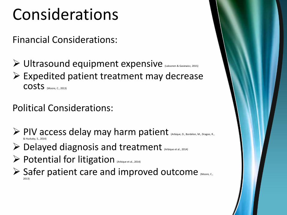
Financial Considerations:
Ultrasound equipment expensive (Laksonen & Gasiewicz, 2015)
Expedited patient treatment may decrease costs (Moore, C., 2013)
Political Considerations:
PIV access delay may harm patient (Arbique, D., Bordelon, M., Dragoo, R.,
& Huckaby, S., 2014)
Delayed diagnosis and treatment (Arbique et al., 2014)
Potential for litigation (Arbique et al., 2014)
Safer patient care and improved outcome (Moore, C.,
2013)
Cultural

Why Implement USGPIV
To bring evidence into practice!

Professional Guidelines Two sources of professional
guidelines for managing patients with difficult peripheral vascular access:
Emergency Nurses Association (ENA, 2011)
Infusion Nurses Society (INS, 2013)
Permission was obtained for use of this handout

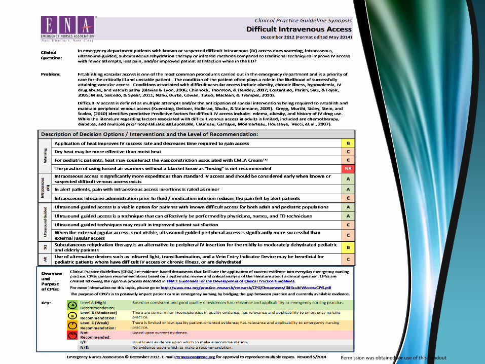
Interventions for Difficult Vascular Access
Emergency Nurses Association Clinical Guidelines
Ultrasound-guided peripheral intravenous access (Level A recommendation)
Intraosseous (Level A recommendation)
Subcutaneous rehydration therapy (Level B recommendation)
Warming (Level C recommendation)
(ENA, 2015)
Interventions for Difficult Vascular Access
Central line
PICC line
Risk of complications with central line, PICC not always available
(Meer, 2015)
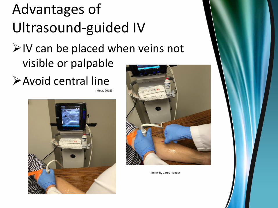
Advantages of Ultrasound-guided IV
IV can be placed when veins not visible or palpable
Avoid central line(Meer, 2015)
Photos by Carey Rivinius

Indications to Use USGPIV Known or suspected
difficult vascular access
(Adhikari et al., 2015; ENA, 2011)
Edema (Adhikari et al., 2015)
Multiple hospitalizations Adhikari
et al., 2015)
Obesity (Adhikari et al., 2015)
End-stage renal disease
(Adhikari et al., 2015)
Traditional technique fails
(Meer, 2015)
Severe dehydration(Meer, 2015)
Intravenous drug user or history (Meer, 2015)
Multiple IV catheters in past
(Meer, 2015)
Burns over IV site(Meer, 2015)
Pediatric or elderly patients(Calderdale & Huddersfield Medical
Simulation Team, 2015)
Ultrasound mechanics
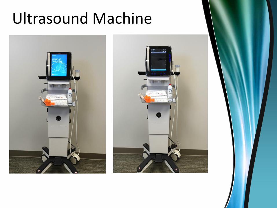
Ultrasound Machine

Ultrasound Machine Sonosite m-turbo was used in the training
courses attended
Mentioned in the literature as well (Emme, 2012)
Various options available
May vary depending on the facility needs
Image from www.ultrasoundportables.com
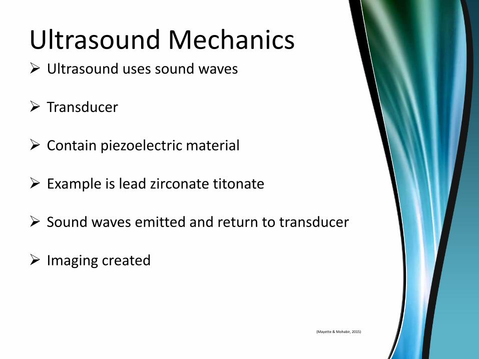
Ultrasound Mechanics Ultrasound uses sound waves
Transducer
Contain piezoelectric material
Example is lead zirconate titonate
Sound waves emitted and return to transducer
Imaging created
(Mayette & Mohabir, 2015)

Ultrasound Mechanics■Frequency ranges from 2 to 18 MHz
■Higher frequency= shorter wavelength
■Shorter wavelengths have higher resolution
Penetrate only to shallow depths
Linear transducer 5 to 10 MHz, superficial structures
Used for ultrasound-guided peripheral IV access
Up to 5 cm of depth
(Chiem, 2015)
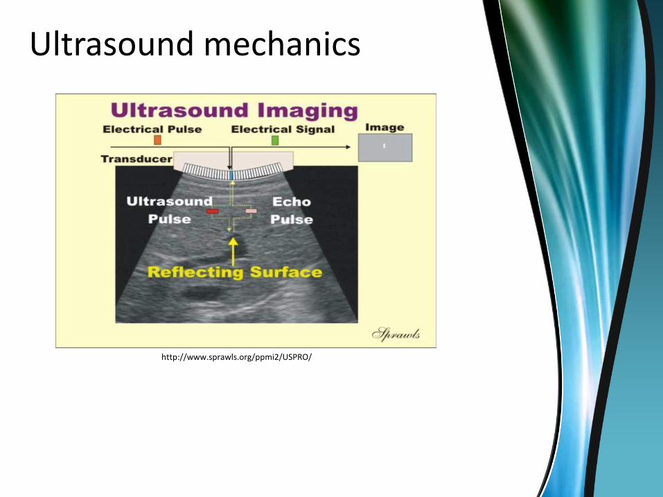
Ultrasound mechanics
http://www.sprawls.org/ppmi2/USPRO/

Transducers
http://www.usra.ca/transducer.php
Linear for ultrasound-guided IV
Vasculature
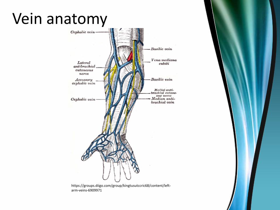
Vein anatomy
https://groups.diigo.com/group/kingtusutccric68/content/left-arm-veins-6909971

USGPIV Technique
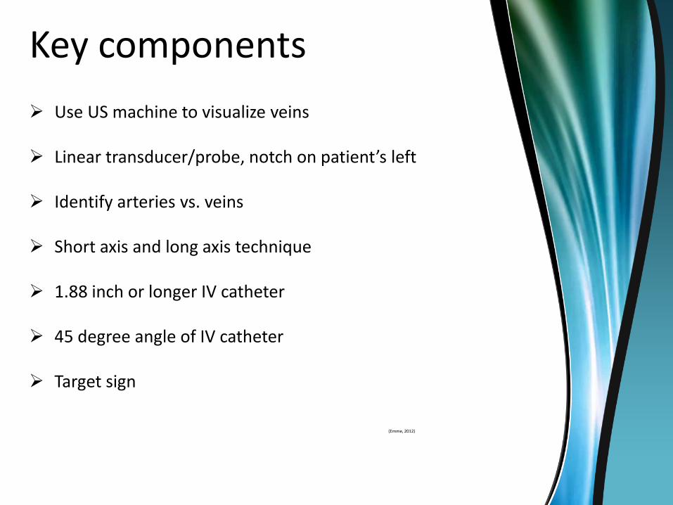
Key components
Use US machine to visualize veins
Linear transducer/probe, notch on patient’s left
Identify arteries vs. veins
Short axis and long axis technique
1.88 inch or longer IV catheter
45 degree angle of IV catheter
Target sign
(Emme, 2012)

Linear Probe
https://cdemcurriculum.files.wordpress.com/2016/04/venous-access-image-3.jpg
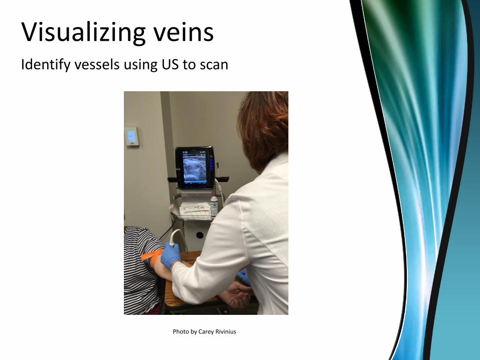
Visualizing veinsIdentify vessels using US to scan
Photo by Carey Rivinius

Visualizing veins
Image by Carey Rivinius
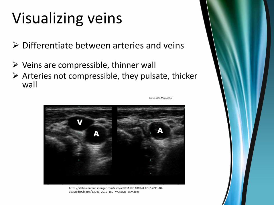
Visualizing veins
Differentiate between arteries and veins
Veins are compressible, thinner wall Arteries not compressible, they pulsate, thicker
wall Emme, 2012;Meer, 2015)
https://static-content.springer.com/esm/art%3A10.1186%2F1757-7241-18-39/MediaObjects/13049_2010_180_MOESM8_ESM.jpeg

Short Axis and Long Axis
https://pbs.twimg.com/media/B_f8Tz_VEAE93MB.png
https://i.ytimg.com/vi/lviC5wU-14U/hqdefault.jpg

Short Axis and Long Axis
https://www.researchgate.net/profile/Michael_Phelan2/publication/23293871/figure/fig6/AS:277353948303364@1443137742814/Figure-6-Photographs-of-ultrasound-probe-position-with-needle-placement-over-a.png

Short Axis and Long Axis, Target Sign
http://www.apicareonline.com/wordpress/wp-content/uploads/2015/12/Scanning-views-of-peripheral-vein.jpg

Long IV catheter Standard length 1.16 inch
Important to use longer IV catheter
1.88 inch or longer (Meer, 2015)
http://emedicine.medscape.com/article/1433943-overview?imageOrder=9
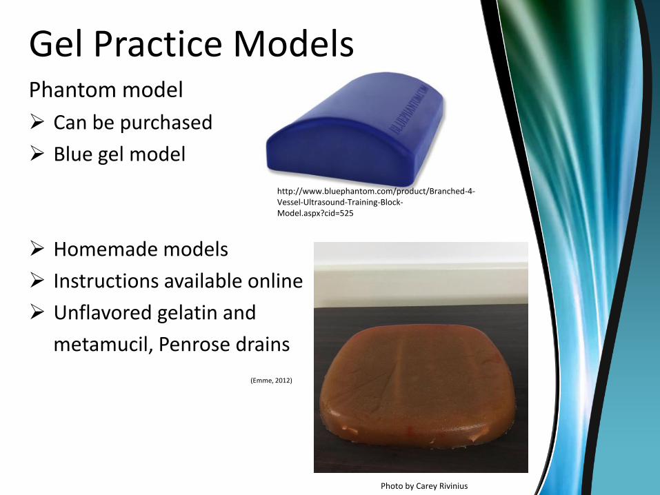
Gel Practice ModelsPhantom model
Can be purchased
Blue gel model
Homemade models
Instructions available online
Unflavored gelatin and
metamucil, Penrose drains
(Emme, 2012)
Photo by Carey Rivinius
http://www.bluephantom.com/product/Branched-4-Vessel-Ultrasound-Training-Block-Model.aspx?cid=525
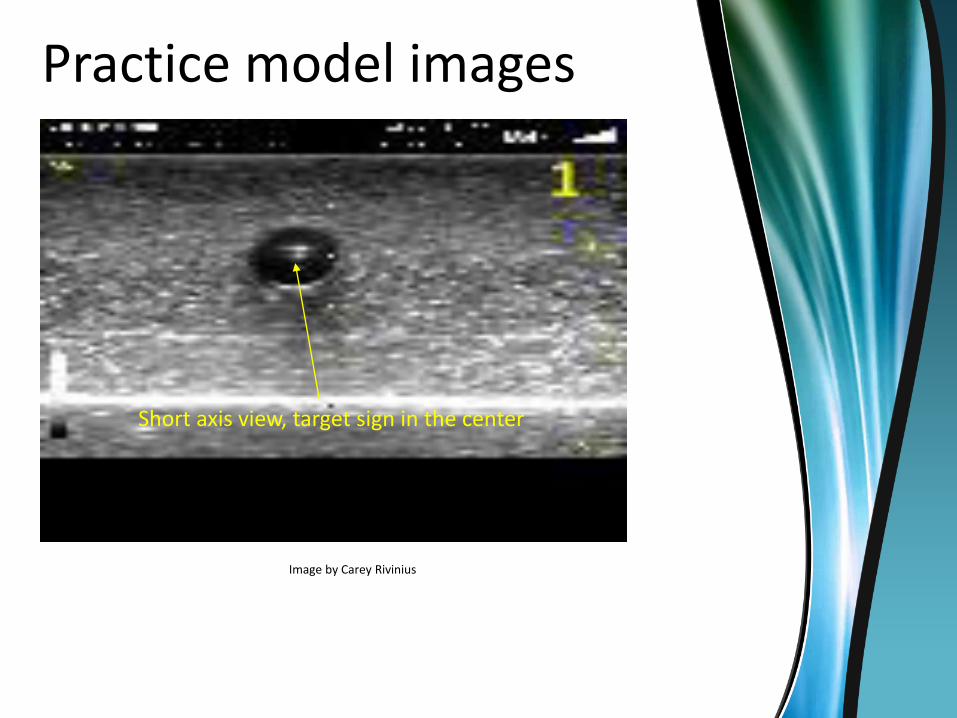
Practice model images
Short axis view, target sign in the center
Image by Carey Rivinius

Practice model images
Long axis view, angiocath is visible
Image by Carey Rivinius
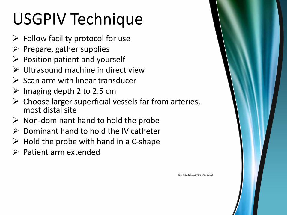
USGPIV Technique Follow facility protocol for use Prepare, gather supplies Position patient and yourself Ultrasound machine in direct view Scan arm with linear transducer Imaging depth 2 to 2.5 cm Choose larger superficial vessels far from arteries,
most distal site Non-dominant hand to hold the probe Dominant hand to hold the IV catheter Hold the probe with hand in a C-shape Patient arm extended
(Emme, 2012;Silverberg, 2015)
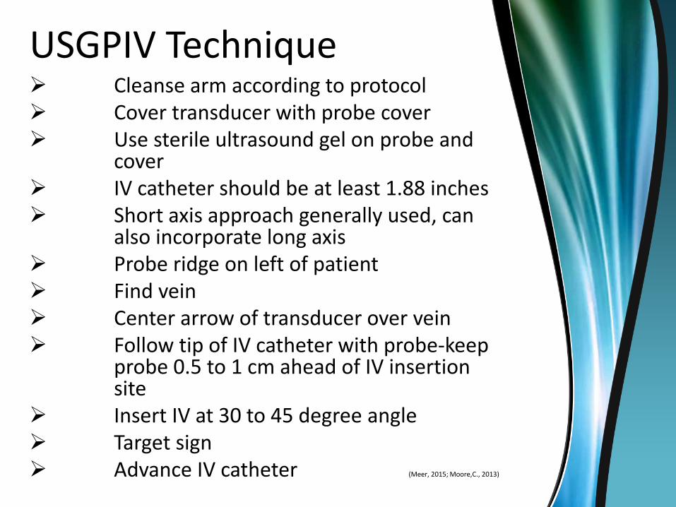
USGPIV Technique Cleanse arm according to protocol Cover transducer with probe cover Use sterile ultrasound gel on probe and
cover IV catheter should be at least 1.88 inches Short axis approach generally used, can
also incorporate long axis Probe ridge on left of patient Find vein Center arrow of transducer over vein Follow tip of IV catheter with probe-keep
probe 0.5 to 1 cm ahead of IV insertion site
Insert IV at 30 to 45 degree angle Target sign Advance IV catheter (Meer, 2015; Moore,C., 2013)
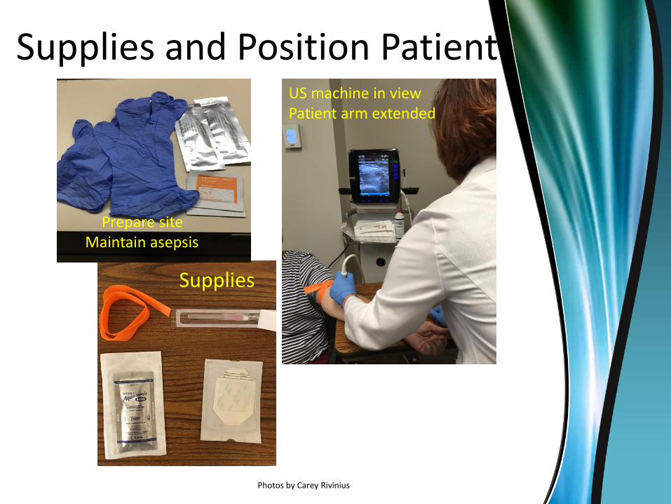
Supplies and Position Patient
Supplies
Photos by Carey Rivinius
US machine in viewPatient arm extended
Prepare siteMaintain asepsis

Transducer preparation
Photo by Carey Rivinius
Ultrasound gel and clear transparent cover over probe

Line it Up!Yellow notch over center of vessel on US screen correlates with line
on transducer over patient arm
Photos by Carey Rivinius
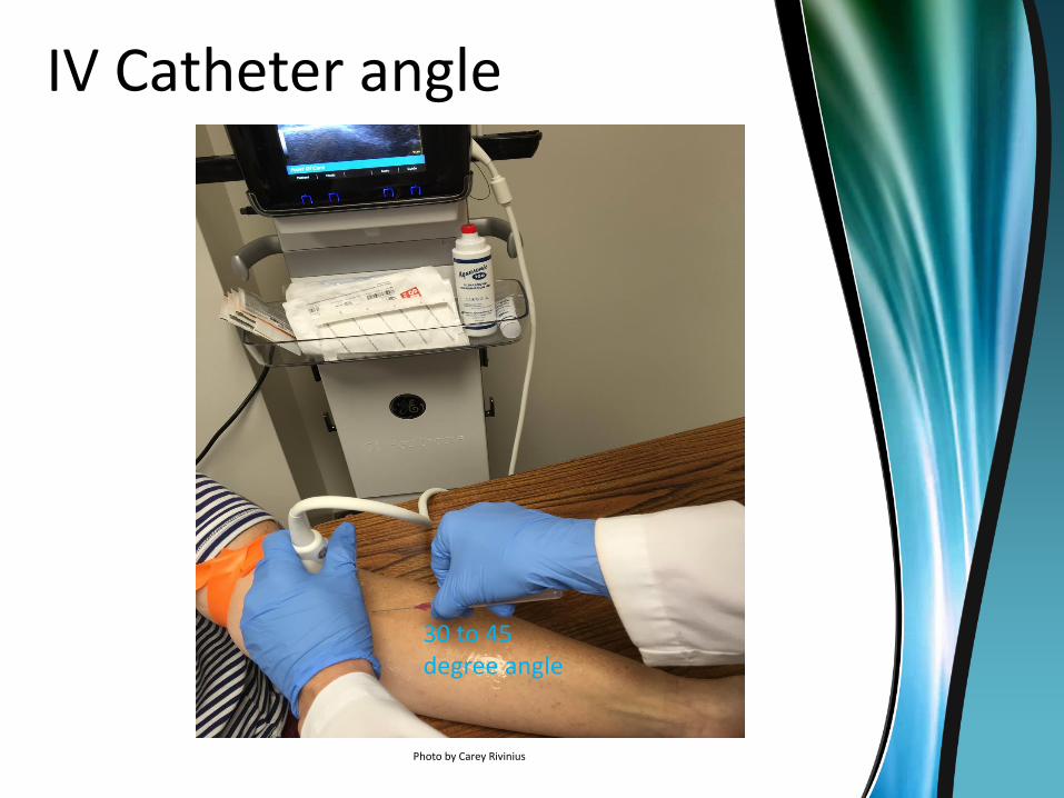
IV Catheter angle
Photo by Carey Rivinius
30 to 45 degree angle

Tips for Success
Superficial vessels, best at 1 cm depth or less, larger diameter better
Start distally on arm, scan for ideal vessel Use light pressure with probe Position US machine and patient properly Use US gel C-clamp probe with non-dominant hand 1.88 inch IV catheter or longer 45 degree angle IV catheter Short axis view generally easier but long axis can be helpful too Keep US probe ahead of IV catheter (0.5 to 1 cm) Probe marker on pt’s arm over center of vein, correlates with
arrow on US machine; keep it lined up! Watch for target sign in vessel May need 2 people at first until comfortable
(Emme, 2012)
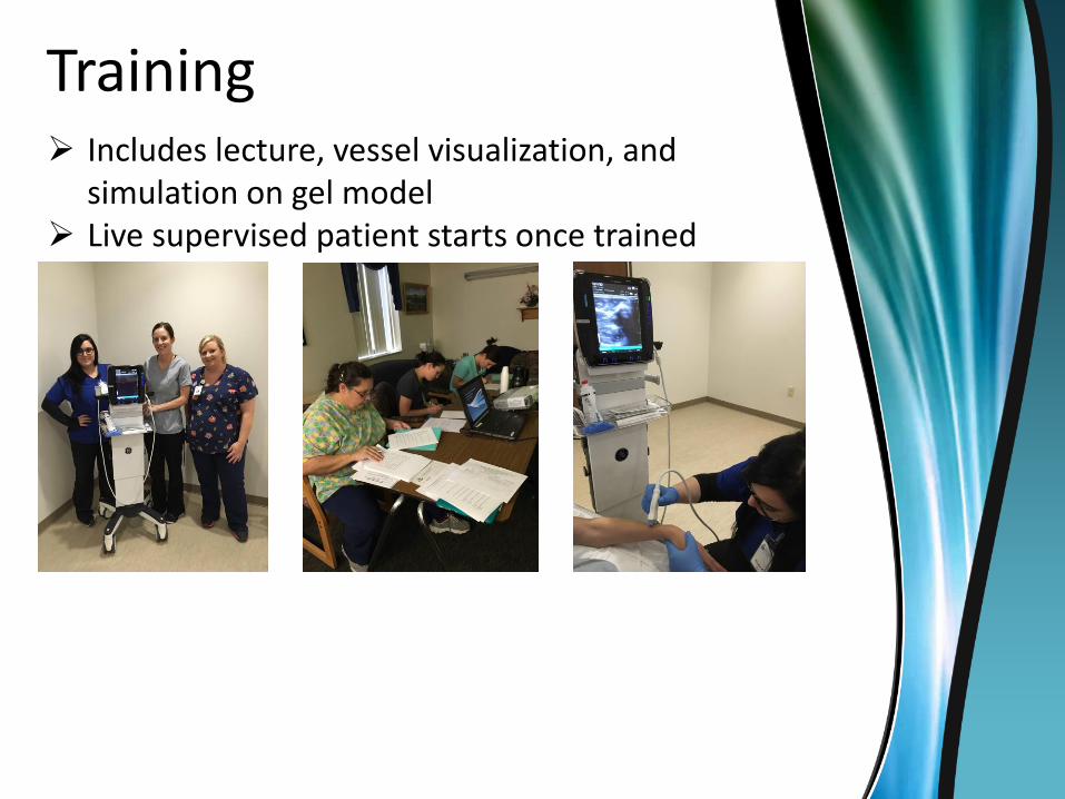
Training Includes lecture, vessel visualization, and
simulation on gel model Live supervised patient starts once trained

Implications for Practice
Difficult Vascular Access affects patient care, clinician challenges
USGPIV access brings evidence into practice
USGPIV can be used in adults and children
Nurses can perform USGPIV
USGPIV can be implemented in rural
hospital setting
Improve patient care,empowers clinicians
PodcastsThere are great podcasts available to demonstrate USGPIV:
Ultrasound guided peripheral IV course:Siegfried Emme, FNP:https://www.youtube.com/watch?v=d8VFgb9Edfw
(Emme, 2012)
Ultrasound-guided IV video https://www.youtube.com/watch?v=NgOF8f7408A
(Calderdale & Huddersfield Medical Simulation Team, 2015)
Ultrasound-guided IV video https://www.youtube.com/watch?v=Xofk-XBKZ6E
(Hsu, 2014)
Ultrasound-guided IV videohttp://www.ultrasoundpodcast.com/2013/10/ultrasound-guided-peripheral-iv-podcast-give-nurses-teach-providers-foamed/
(Dawson & Mallin (2013)

Questions?
Carey Rivinius, MSN, FNP-CJacobson Memorial Hospital601 E. St North Elgin, ND 58533701-584-3338Email: [email protected]
ReferencesAdhikari, S., Schmier, C., & Marx, J. (2015). Focused simulation training: Emergency
department nurses’ confidence and comfort level in performing ultrasound-guided vascular access. Journalof VascularAccess. doi:10.5301/jva.5000436
American Institute of Ultrasound in Medicine (AIUM) (2012). AIUM practice parameter for the use of ultrasound to guide vascular access procedures. Retrieved from http://www.aium.org/resources/guidelines/usgva.pdf
Arbique, D., Bordelon, M., Dragoo, R., & Huckaby, S. (2014). Ultrasound-guided access for peripheral intravenous therapy. Academy of Medical-Surgical Nurses, 23(3). Retrieved from https://www.amsn.org/sites/default/files/private/medsurg-matters-
newsletter- archives/mayjun14.pdfCalderdale & Huddersfield Medical Simulation Team (Producer). (2015, February 15).
Ultrasound guided peripheral venous access [Video podcast]. Retrieved fromhttps://www.youtube.com/watch?v=NgOF8f7408A
Chiem, A.T. (2015). Transducers. In N.J. Soni, R. Arntfield, & P. Kory (Eds.),Point-of-Care Ultrasound (19-24). Philadelphia, PA: Elsevier Saunders.
ReferencesEgan, G.; Healy, D.; O’Neill, H.; Clarke-Moloney, M.; Grace, P.A. & Walsh, S.R. (2013).
Ultrasound guidance for difficult peripheral venous access: Systematic review and meta-analysis. Emergency Medicine Journal, 30,
521-526. doi: 10.1136/emermed-2012-201652Emergency Nurses Association (ENA) (2011). Clinical Practice Guideline: Difficult
Intravenous Access Full Version. Retrieved from https://www.ena.org/practice-
research/research/CPG/Documents/DifficultIVAccessCPG.pdfEmergency Nurses Association (ENA) (2014). Clinical Practice Guideline Synopsis Difficult
Intravenous Access. Retrieved from https://www.ena.org/practice-
research/research/CPG/Documents/DifficultIVAccessSynopsis.pdfEmergency Med (Producer). (2014). Ultrasound-guided vascular access lecture [Video
podcast]. Retrieved fromhttps://www.youtube.com/watch?v=Xofk-XBKZ6EEmme, Siegfried (Producer). (2012, December 2). Ultrasound guided peripheral IV
course[Video podcast]. Retrieved from https://www.youtube.com/watch?v=d8VFgb9Edfw
Infusion Nurses Society (2013). Recommendations for Improving Safety Practices With Short Peripheral Catheters. INS Position Paper. Retrieved from:http://www.ins1.org/files/public/12_13_IV_Safety_Position%20Paper%20_
Board%20Final%20Draft.pdf
ReferencesIsmailoglu, E.G.; Zaybak, A.; Akarca, F.K.; & Kiyan, S. (2015). The effect of
the use of ultrasound in the success of peripheral venous catheterization. International Emergency Nursing, 23, 89-93.doi: 10.1016/j.ienj.2014.07.0101755-599X
Laksonen, R.P. & Gasiewicz, N.K. (2015). Implementing a program for ultrasound-guidedperipheral venous access: Training, policy and procedure development, protocol use,competency, and skill tracking. Nursing Clinics of North America, 50, 771-785.doi:10.1016/j.cnur.2015.07.010
Mayette, M. & Mohabir, P.K. (2015). Ultrasound physics. In N.J. Soni, R. Arntfield, & P. Kory (Eds.), Point-of-CareUltrasound (9-18). Philadelphia, PA: Elsevier Saunders.
Meer, J.M.; Euerle, B.; Hsu, S. (2015). Ultrasonography assisted peripheral line placement.Medscape Reference: Drugs, Diseases, & Procedures, 1-11. Retrieved fromhttp://emedicine.medscape.com/article/1433943-overview
Moore, C. (2013). An emergency department nurse-driven ultrasound-guided peripheralintravenous line program. Association for Vascular Access, 18, 45-51. doi:10.1016/j.java.2012.12.001
Moore, C.L. (2014). Ultrasound first, second, and last for vascular access.Journal of Ultrasound in Medicine, 33, 1135-1142.doi:10.7863/ultra.33.7.1135
Silverberg, M. (2015). Peripheral Venous Access. In N.J. Soni, R. Arntfield, & P. Kory (Eds.), Point-of-CareUltrasound (233-236). Philadelphia, PA: Elsevier Saunders.
Stone, P.; Meyer, B.; Aucoin, J.; Raynor, R.; Smith, N; Nelles, S.,…Grissom, J.(2013). Ultrasound-guided peripheral I.V. access:Guidelines for practice. American Nurse Today, 8(8), 1-5. Retrieved
from http://www.americannursetoday.com/ultrasound-guided-peripheral-i-v-access-guideline-for-practice/


