55 Diffuse large B cell lymphoma with chronic granulomatous inflammation Wint Wint Thu NYUNT MRCP, MMed.(IntMed), Yin Ping WONG* DrPath(UKM), MIAC, Wan Fariza WAN JAMALUDIN MBBS, MRCP and S Fadilah S ABDUL WAHID MD, PhD Cell therapy Centre and *Department of Pathology, University Kebangsaan Malaysia Medical Centre (UKMMC), Kuala Lumpur, Malaysia Abstract Non-necrotic epithelioid granulomas have been reported in association with neoplasms including Hodgkin and non-Hodgkin lymphoma. We report a case of diffuse large B cell lymphoma with chronic granulomatous inflammation to highlight awareness of obscure tumour cells within the granuloma, to avoid delay in diagnosis and management of lymphoma. A 39-year-old Malay lady with no past medical history, presented with a 2-month history of progressive worsening of difficulty in breathing, cough, low-grade fever, loss of weight and loss of appetite. Chest X-ray showed an anterior mediastinal mass and computed tomography (CT)-guided biopsy was reported as chronic granulomatous inflammation suggestive of tuberculosis. After 2 months of anti-TB treatment, her symptoms were not relieved. The patient underwent another CT-guided biopsy of the anterior mediastinal mass in another hospital and the histopathology revealed diffuse large B cell lymphoma. The patient was referred for treatment. On histopathological review, the first sample showed non- caseating granulomas engulfing tumour cells and large abnormal lymphoid cells which were CD20 positive and with high Ki-67 proliferative index. The patient was diagnosed with diffuse large B cell lymphoma stage IV B IPSS score 3. She underwent chemotherapy (R-EPOCH) and responded well to treatment. Keywords: granuloma, lymphoma, histopathology Address for correspondence: Dr. Wint Wint Thu Nyunt, Cell Therapy Centre, Level 2, University Kebangsaan Malaysia Medical Centre, Jalan Yaacob Latif, Bandar Tun Razak, Cheras, 56000, Kuala Lumpur, Malaysia. Email: [email protected]CASE REPORT INTRODUCTION Chronic granulomatous inflammation is histologically seen as a focal collection of inflammatory cells including epithelioid cells, multinucleated giant cells, macrophages, mononuclear cells and fibroblasts, with or without caseous necrosis in the centre. 1 Granulomas with caseation necrosis (soft tubercle) are associated with tuberculosis. Epithelioid granulomas can be associated with chronic persistent infections, sarcoidosis, vasculitis and neoplasms. It is postulated that the formation of granulomas is mediated through macrophage activity, Th1 cell response, B cell overactivity and a vast array of biological mediators. 1 It is usually as a result of the persistence of a non-degradable product and of active cell-mediated hypersensitivity. 1 In Hodgkin lymphoma and T-cell derived non- Hodgkin lymphoma, granulomatous reaction is probably evoked by aberrant cytokine production in the tumour cells or other cells composing the tumour background. 2 The purpose of chronic granulomatous inflammation is to eradicate foreign bodies, such as antigen, which are persistent and non-degradable. Over time, the granuloma may lead to fibrosis. Diffuse large B cell lymphoma (DLBCL) is the most common histological subtype of non-Hodgkin lymphoma (NHL), accounting for approximately 30 to 40 percent of NHL. 3 It is histologically seen as diffuse sheets of large cells (nuclei at least twice the size of a small lymphocyte) with vesicular nuclei, prominent nucleoli, basophilic cytoplasm and a moderate to high proliferation fraction with positive immunohistochemistry for B cell-associated antigens (CD19, CD20, CD22, CD79a). 3 There were case reports of chronic Malaysian J Pathol 2016; 38(1) : 55 – 59
Transcript
55
Diffuse large B cell lymphoma with chronic granulomatous inflammation
Wint Wint Thu NYUNT MRCP, MMed.(IntMed), Yin Ping WONG* DrPath(UKM), MIAC, Wan Fariza WAN JAMALUDIN MBBS, MRCP and S Fadilah S ABDUL WAHID MD, PhD
Cell therapy Centre and *Department of Pathology, University Kebangsaan Malaysia Medical Centre (UKMMC), Kuala Lumpur, Malaysia
Abstract
Non-necrotic epithelioid granulomas have been reported in association with neoplasms including Hodgkin and non-Hodgkin lymphoma. We report a case of diffuse large B cell lymphoma with chronic granulomatous inflammation to highlight awareness of obscure tumour cells within the granuloma, to avoid delay in diagnosis and management of lymphoma. A 39-year-old Malay lady with no past medical history, presented with a 2-month history of progressive worsening of difficulty in breathing, cough, low-grade fever, loss of weight and loss of appetite. Chest X-ray showed an anterior mediastinal mass and computed tomography (CT)-guided biopsy was reported as chronic granulomatous inflammation suggestive of tuberculosis. After 2 months of anti-TB treatment, her symptoms were not relieved. The patient underwent another CT-guided biopsy of the anterior mediastinal mass in another hospital and the histopathology revealed diffuse large B cell lymphoma. The patient was referred for treatment. On histopathological review, the first sample showed non-caseating granulomas engulfing tumour cells and large abnormal lymphoid cells which were CD20 positive and with high Ki-67 proliferative index. The patient was diagnosed with diffuse large B cell lymphoma stage IV B IPSS score 3. She underwent chemotherapy (R-EPOCH) and responded well to treatment.
Keywords: granuloma, lymphoma, histopathology
Address for correspondence: Dr. Wint Wint Thu Nyunt, Cell Therapy Centre, Level 2, University Kebangsaan Malaysia Medical Centre, Jalan Yaacob Latif, Bandar Tun Razak, Cheras, 56000, Kuala Lumpur, Malaysia. Email: [email protected]
CASE REPORT
INTRODUCTION
Chronic granulomatous inflammation is histologically seen as a focal collection of inflammatory cells including epithelioid cells, multinucleated giant cells, macrophages, mononuclear cells and fibroblasts, with or without caseous necrosis in the centre.1 Granulomas with caseation necrosis (soft tubercle) are associated with tuberculosis. Epithelioid granulomas can be associated with chronic persistent infections, sarcoidosis, vasculitis and neoplasms. It is postulated that the formation of granulomas is mediated through macrophage activity, Th1 cell response, B cell overactivity and a vast array of biological mediators.1 It is usually as a result of the persistence of a non-degradable product and of active cell-mediated hypersensitivity.1 In Hodgkin lymphoma and T-cell derived non-Hodgkin lymphoma, granulomatous reaction is
probably evoked by aberrant cytokine production in the tumour cells or other cells composing the tumour background.2 The purpose of chronic granulomatous inflammation is to eradicate foreign bodies, such as antigen, which are persistent and non-degradable. Over time, the granuloma may lead to fibrosis. Diffuse large B cell lymphoma (DLBCL) is the most common histological subtype of non-Hodgkin lymphoma (NHL), accounting for approximately 30 to 40 percent of NHL.3 It is histologically seen as diffuse sheets of large cells (nuclei at least twice the size of a small lymphocyte) with vesicular nuclei, prominent nucleoli, basophilic cytoplasm and a moderate to high proliferation fraction with positive immunohistochemistry for B cell-associated antigens (CD19, CD20, CD22, CD79a).3
There were case reports of chronic
Malaysian J Pathol 2016; 38(1) : 55 – 59
Malaysian J Pathol April 2016
56
granulomatous inflammation associated with neoplasms such as Hodgkin and non-Hodgkin lymphoma.4-6 Hodgkin lymphoma is more commonly associated with non-necrotic epithelioid granulomas.4 However, there have been many case reports on the finding of epithelioid granulomas in non-Hodgkin lymphoma such as small, non-cleaved cell lymphoma,5 splenic marginal B cell lymphoma7 and Burkitt’s lymphoma.2 The types of NHL associated with concurrent granulomas include B-cell NHL (low grade lymphoma, not otherwise specified [NOS]; follicular center cell lymphoma [FCCL]; small lymphocytic lymphoma [SLL]; large cell lymphoma, NOS [LCL, NOS]) and various types of T-cell NHL.4 The phenomenon of granulomas in Hodgkin lymphoma or small non-cleaved cell lymphoma confer a favorable prognosis.5,7 There was also a case report of lymphoma mimicking tuberculosis.8
To our knowledge, there is scarce data reporting adult diffuse large B cell lymphoma associated with chronic granulomatous inflammation. We reported a case of diffuse large B cell lymphoma associated with chronic granulomatous inflammation, which was previously misdiagnosed as tuberculosis and treated with anti-tuberculous drugs.
CASE REPORT
A 39-year-old Malay lady with no previous medical illness, presented with a 2-month-history of difficulty in breathing on and off, non-productive cough, low-grade fever on and off, loss of weight (about 10 kg within 2 months) and loss of appetite. She had no pruritus or night sweats. There was no history of close contact with persons with tuberculosis or chronic cough. Family history revealed no tuberculosis, cancer or blood disorders. She was a non-smoker. She was a housewife. The patient went to a private hospital in June 2014. Her chest X-ray showed an anterior mediastinal mass and left-sided pleural effusion. Computed tomography (CT) scan of thorax and CT-guided biopsy of the anterior mediastinal mass were performed. It was reported as chronic granulomatous inflammation suggestive of tuberculosis. Hence, the patient was treated with anti-tuberculous medication AKURIT-4. Unfortunately, the patient developed adverse reaction with AKURIT-4 and anti-tuberculous medication was changed into isoniazid, rifampicin, pyrazinamide and ethambutol. She
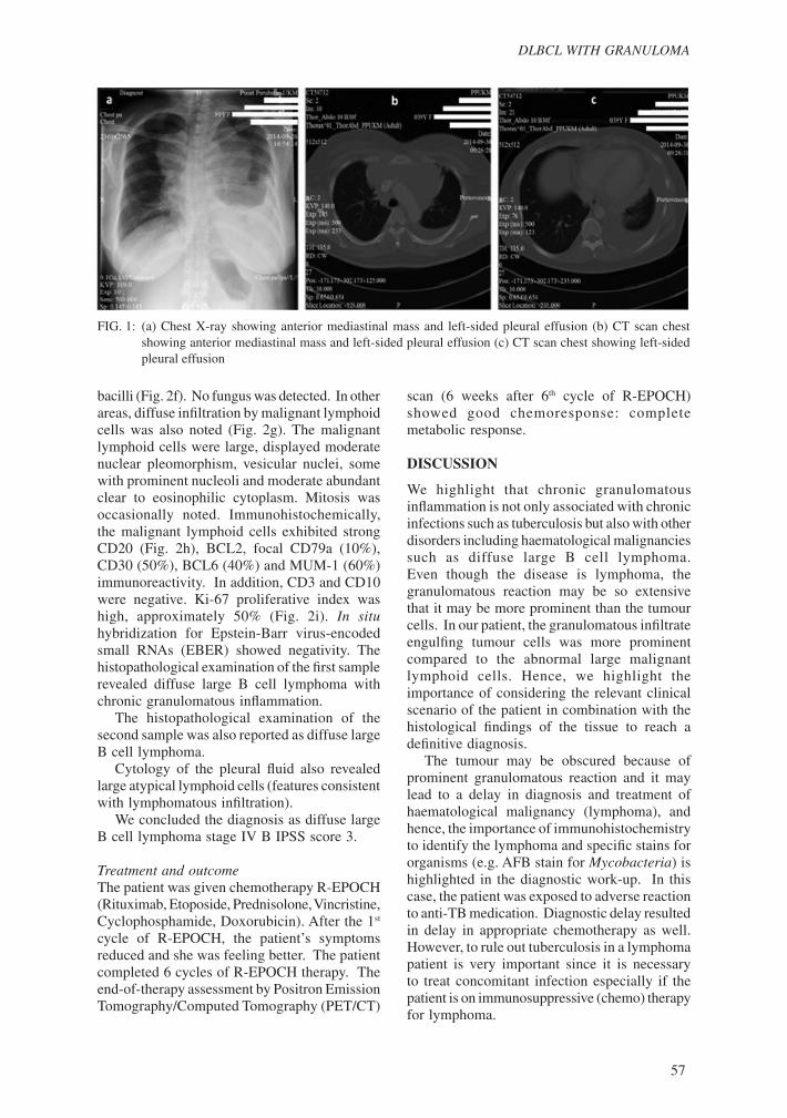
received anti-tuberculous medication for about 2 months. However, in view of no symptomatic relief and no response to treatment, the patient was referred to another hospital. In the second hospital, the patient underwent another CT-guided biopsy of the anterior mediastinal mass. Histopathological examination revealed diffuse large B-cell lymphoma. The patient was then referred to our hospital for further work-up and management of lymphoma. In our hospital, thorough history taking and physical examination were made. The patient had B symptoms (fever, loss of weight, loss of appetite). On physical examination, there were no signs of superior vena cava obstruction. Temperature, blood pressure, heart rate, respiratory rate and oxygen saturation were normal. There was no pallor, jaundice, lymph node enlargement palpable, hepatomegaly or splenomegaly. Initial blood investigations revealed: haemoglobin (Hb) 10.7 g/dL, white cell count 8.7x10⁹/L, platelets 174x109/L, lactate dehydrogenase (LDH) 511 U/L. Full blood picture revealed no leucoerythroblastic picture, abnormal lymphoid cells or blasts. A diagnostic work-up for tuberculosis was performed; all of the results were negative for Mycobacteria tuberculosis. [Erythrocyte sedimentation rate (ESR) 60 mm/1st hour, negative Mantoux test, negative acid fast bacilli (AFB) stain from bronchoalveolar lavage]. Chest X-ray showed anterior mediastinal mass and left-sided pleural effusion (Fig. 1a). CT scan of thorax, abdomen and pelvis revealed features consistent with lymphoma with bilateral subpleural nodules, left pleural effusion (Fig. 1b, 1c) and involvement of the mediastinum (Fig. 1b) and para-aortic lymph nodes. Bone marrow aspiration and trephine biopsy did not reveal lymphoid malignant cell infiltration. We arranged to obtain the tissue samples of the previous biopsies and both tissue samples were re-assessed again in our hospital.
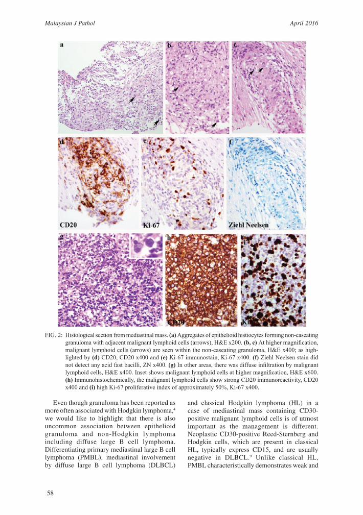
HistopathologyHistopathological findings of the first biopsy specimen revealed a few aggregates of epithelioid histiocytes forming non-caseating granuloma (Fig. 2a), within which scattered malignant lymphoid cells were seen (Fig. 2b, 2c). These were highlighted by CD20 positivity (Fig. 2d) and a high Ki-67 proliferative index (Fig. 2e). Ziehl-Neelsen stain did not reveal any acid fast
57
DLBCL WITH GRANULOMA
bacilli (Fig. 2f). No fungus was detected. In other areas, diffuse infiltration by malignant lymphoid cells was also noted (Fig. 2g). The malignant lymphoid cells were large, displayed moderate nuclear pleomorphism, vesicular nuclei, some with prominent nucleoli and moderate abundant clear to eosinophilic cytoplasm. Mitosis was occasionally noted. Immunohistochemically, the malignant lymphoid cells exhibited strong CD20 (Fig. 2h), BCL2, focal CD79a (10%), CD30 (50%), BCL6 (40%) and MUM-1 (60%) immunoreactivity. In addition, CD3 and CD10 were negative. Ki-67 proliferative index was high, approximately 50% (Fig. 2i). In situ hybridization for Epstein-Barr virus-encoded small RNAs (EBER) showed negativity. The histopathological examination of the first sample revealed diffuse large B cell lymphoma with chronic granulomatous inflammation. The histopathological examination of the second sample was also reported as diffuse large B cell lymphoma. Cytology of the pleural fluid also revealed large atypical lymphoid cells (features consistent with lymphomatous infiltration). We concluded the diagnosis as diffuse large B cell lymphoma stage IV B IPSS score 3.
Treatment and outcomeThe patient was given chemotherapy R-EPOCH (Rituximab, Etoposide, Prednisolone, Vincristine, Cyclophosphamide, Doxorubicin). After the 1st cycle of R-EPOCH, the patient’s symptoms reduced and she was feeling better. The patient completed 6 cycles of R-EPOCH therapy. The end-of-therapy assessment by Positron Emission Tomography/Computed Tomography (PET/CT)
scan (6 weeks after 6th cycle of R-EPOCH) showed good chemoresponse: complete metabolic response.
DISCUSSION
We highlight that chronic granulomatous inflammation is not only associated with chronic infections such as tuberculosis but also with other disorders including haematological malignancies such as diffuse large B cell lymphoma. Even though the disease is lymphoma, the granulomatous reaction may be so extensive that it may be more prominent than the tumour cells. In our patient, the granulomatous infiltrate engulfing tumour cells was more prominent compared to the abnormal large malignant lymphoid cells. Hence, we highlight the importance of considering the relevant clinical scenario of the patient in combination with the histological findings of the tissue to reach a definitive diagnosis. The tumour may be obscured because of prominent granulomatous reaction and it may lead to a delay in diagnosis and treatment of haematological malignancy (lymphoma), and hence, the importance of immunohistochemistry to identify the lymphoma and specific stains for organisms (e.g. AFB stain for Mycobacteria) is highlighted in the diagnostic work-up. In this case, the patient was exposed to adverse reaction to anti-TB medication. Diagnostic delay resulted in delay in appropriate chemotherapy as well. However, to rule out tuberculosis in a lymphoma patient is very important since it is necessary to treat concomitant infection especially if the patient is on immunosuppressive (chemo) therapy for lymphoma.
FIG. 1: (a) Chest X-ray showing anterior mediastinal mass and left-sided pleural effusion (b) CT scan chest showing anterior mediastinal mass and left-sided pleural effusion (c) CT scan chest showing left-sided pleural effusion
Malaysian J Pathol April 2016
58
Even though granuloma has been reported as more often associated with Hodgkin lymphoma,4 we would like to highlight that there is also uncommon association between epithelioid granuloma and non-Hodgkin lymphoma including diffuse large B cell lymphoma. Differentiating primary mediastinal large B cell lymphoma (PMBL), mediastinal involvement by diffuse large B cell lymphoma (DLBCL)
and classical Hodgkin lymphoma (HL) in a case of mediastinal mass containing CD30-positive malignant lymphoid cells is of utmost important as the management is different. Neoplastic CD30-positive Reed-Sternberg and Hodgkin cells, which are present in classical HL, typically express CD15, and are usually negative in DLBCL.9 Unlike classical HL, PMBL characteristically demonstrates weak and
FIG. 2: Histological section from mediastinal mass. (a) Aggregates of epithelioid histiocytes forming non-caseating granuloma with adjacent malignant lymphoid cells (arrows), H&E x200. (b, c) At higher magnification, malignant lymphoid cells (arrows) are seen within the non-caseating granuloma, H&E x400; as high-lighted by (d) CD20, CD20 x400 and (e) Ki-67 immunostain, Ki-67 x400. (f) Ziehl Neelsen stain did not detect any acid fast bacilli, ZN x400. (g) In other areas, there was diffuse infiltration by malignant lymphoid cells, H&E x400. Inset shows malignant lymphoid cells at higher magnification, H&E x600. (h) Immunohistochemically, the malignant lymphoid cells show strong CD20 immunoreactivity, CD20 x400 and (i) high Ki-67 proliferative index of approximately 50%, Ki-67 x400.
59
DLBCL WITH GRANULOMA
heterogeneous CD30 immunoexpression. Rarely, a diagnosis of grey zone (composite) lymphoma should be considered in cases exhibiting overlapping clinical, morphological and/or immunophenotypic features between classical HL and DLBCL.9 A patient with systemic DLBCL with secondary mediastinal involvement may be confused with PMBL. Radiological imaging and/or bone marrow biopsy can be helpful in confirming systemic involvement by the disease.9 Interestingly, mediastinal germ cell tumour particularly seminoma has been reported to be associated with chronic granulomatous inflammation. The coexistence of granulomatous inflammation and germ cell tumour, however, does not affect overall survival.10
The mechanism of granuloma formation in non-Hodgkin lymphoma, particularly B cell lymphoma, is poorly understood. This inspires further research to unravel its mechanism, clinical significance and implications. Some postulations published are: (1) T-cell mediated immune response to tumour antigen,7 (2) aberrant cytokine production in the tumour cells or other cells composing the tumour background,2 and (3) persistence of granuloma due to unremovable tumour antigen and then transformation to fibrous tissue.1
Our patient had previous hepatitis B infection but it was not clear whether the granuloma formation is influenced by this infection. Notably, Hepatitis B infection has been reported to be associated with non-Hodgkin lymphoma.11
The clinical significance of granulomatous reaction in diffuse large B cell lymphoma is not clear. It has been reported that patients with Hodgkin lymphoma associated with epithelioid granuloma have a better prognosis and favorable outcome (survival and relapse-free survival) compared to the patients without granuloma.7,12 Similarly, epithelioid granuloma formation conferred a favorable prognosis in patients with non-Hodgkin lymphoma (small non-cleaved cell lymphoma).5 Sporadic cases of EBV-positive Burkitt’s lymphoma also suggested the association between granulomatous response and favorable outcome.2 While EBV-positive DLBCL of the elderly is associated with inferior clinical outcomes when compared with the EBV-negative counterpart, the role of EBV in the younger age group (<50 years) remains controversial.9
ACKNOWLEDGEMENT
The patient gave informed written consent for publication of this case report. The authors have no conflict of interest to declare. This report is not supported by any funding agencies.
REFERENCES
1. James DG. A clinicopathological classification of granulomatous disorders. Postgrad Med J. 2000; 76: 457-65.
2. Haralambieva E, Rosati S, van Noesel C, et al. Florid granulomatous reaction in Epstein-Barr virus-positive nonendemic Burkitt lymphomas: report of four cases. Am J Surg Pathol. 2004; 28: 379-83.
3. Harris NL, Jaffe ES, Stein H, et al. A revised European-American classification of lymphoid neoplasms: a proposal from the International Lymphoma Study Group. Blood. 1994; 84: 1361-92.
4. Dunphy CH, Panella MJ, Grosso LE. Low-grade B-Cell lymphoma and concomitant extensive sarcoidlike granulomas: a case report and review of the literature. Arch Pathol Lab Med. 2000; 124: 152–6.
5. Hollingsworth HC, Longo DL, Jaffe ES. Small noncleaved cell lymphoma associated with florid epithelioid granulomatous response. A clinicopathologic study of seven patients. Am J Surg Pathol. 1993; 17: 51-9.
6. Braylan RC, Long JC, Jaffe ES, Greco FA, Orr SL, Berard CW. Malignant lymphoma obscured by concomitant extensive epithelioid granulomas: report of three cases with similar clinicopathologic features. Cancer. 1977; 39: 1146-55.
7. Laforga JB, Aranda FI, Gasent JM. Splenic marginal B-cell lymphoma with epithelioid granulomas. Report of a case with cytologic and immunohistochemical study: Case report. Cancer Therapy. 2009; 7: 21-6.
9. Swerdlow SH, Campo E, Harris NL, et al., editors. WHO classification of tumours of haematopoietic and lymphoid tissues. 4th ed. Lyon, France: IARC; 2008. P. 243-68.
10. Moran CA, Suster S, Przygodzki RM, Koss MN. Primary germ cell tumors of the mediastinum: II. Mediastinal seminomas – a clinicopathologic and immunohistochemical study of 120 cases. Cancer. 1997; 80: 691-8.
11. Marcucci F, Spada E, Mele A, Caserta CA, Pulsoni A. The association of hepatitis B virus infection with B-cell non-Hodgkin lymphoma – a review. Am J Blood Res. 2012; 2: 18-28.
12. Sacks EL, Donaldson SS, Gordon J, Dorfman RF. Epithelioid granulomas associated with Hodgkin’s disease: clinical correlations in 55 previously untreated patients. Cancer. 1978; 41: 562-7.