Diffusion Tensor Estimation by Maximizing Rician Likelihood Bennett Landman, Pierre-Louis Bazin, Jerry Prince Johns Hopkins University School of Medicine Baltimore, MD 21205 {blandma1,pbazin1}@jhmi.edu, [email protected]Abstract Diffusion tensor imaging (DTI) is widely used to char- acterize white matter in health and disease. Previous ap- proaches to the estimation of diffusion tensors have either been statistically suboptimal or have used Gaussian ap- proximations of the underlying noise structure, which is Rician in reality. This can cause quantities derived from these tensors — e.g., fractional anisotropy and apparent diffusion coefficient — to diverge from their true values, po- tentially leading to artifactual changes that confound clin- ically significant ones. This paper presents a novel maxi- mum likelihood approach to tensor estimation, denoted Dif- fusion Tensor Estimation by Maximizing Rician Likelihood (DTEMRL). In contrast to previous approaches, DTEMRL considers the joint distribution of all observed data in the context of an augmented tensor model to account for vari- able levels of Rician noise. To improve numeric stability and prevent non-physical solutions, DTEMRL incorporates a robust characterization of positive definite tensors and a new estimator of underlying noise variance. In simulated and clinical data, mean squared error metrics show con- sistent and significant improvements from low clinical SNR to high SNR. DTEMRL may be readily supplemented with spatial regularization or a priori tensor distributions for Bayesian tensor estimation. 1. Introduction Diffusion tensor imaging (DTI) provides unique insights into in vivo tissue structure through contrasts sensitive to the directional diffusion of water within restricted environ- ments [2]. Derived tensor contrasts, including fractional anisotropy (FA) and mean diffusivity (MD), have been widely applied to characterize cytoarchitectural changes re- lated to damage in cerebral white matter (e.g. see a review by Horsfield and Jones [11]). At low signal to noise ra- tios (SNRs), estimated contrasts tend to systematically di- verge from their true values (increased bias in addition to in- creased variability), which leads to artifactual changes that confound clinically significant ones [3]. This study presents a maximum likelihood (ML) approach of estimating ten- sors from DTI data that accounts for (1) non-Gaussian dis- tributed noise and (2) statistical dependence between ob- servations to minimize bias. This novel approach specifi- cally addresses the joint likelihood of all observations given a tensor model (as opposed to the marginal likelihood of each observation), denoted Diffusion Tensor Estimation by Maximizing Rician Likelihood (DTEMRL). In DTI studies, the degree and orientation of random thermal (Brownian) motion of water are inferred from the signal intensity obtained during sensitized, “diffusion weighted” (DW) acquisitions relative to a reference. A typ- ical DTI study consists of a reference image and series of 6 or more DW images, each of which are attenuated accord- ing to the diffusivity along a particular linear direction. The absolute intensity depends on the local relaxation properties (e.g., T1, T2, PD) of the tissue which are not directly linked to diffusivity. The ratio between the DW and reference sig- nals provides the relevant information. The 3D diffusivity is modeled as a tensor fit to these ratios. Current methods of computing tensors do not fully ac- count for the physical noise structure in magnetic resonance (MR) data. The noise on acquired images is well charac- terized by independent Rician distributions [10]. Indepen- dence between the noise on the intensities at the same lo- cation in different images is lost when the ratio between DW and reference images is computed because a common denominator is used. The most prevalent tensor estima- tion method, the log-linear minimum mean squared error (LLMMSE) approach [2], assumes the noise to be inde- pendently and identically Gaussian distributed on the log- arithms of the ratios. In support of the LLMMSE approach, Gudbjartsson et al.[10] demonstrated that the distribution of the logarithm of ratio of Rician random variables is “nearly Gaussian” for SNRs greater than 2:1. Methods for compensating for Rician bias in DTI have been proposed primarily in two categories, (1) characteriza- tion of the noise structure in single images and (2) spatial regularization of DTI data. Sijbers et al.[19] presented an 1
Transcript
Diffusion Tensor Estimation by Maximizing Rician Likelihood
Bennett Landman, Pierre-Louis Bazin, Jerry PrinceJohns Hopkins University School of Medicine
Diffusion tensor imaging (DTI) is widely used to char-acterize white matter in health and disease. Previous ap-proaches to the estimation of diffusion tensors have eitherbeen statistically suboptimal or have used Gaussian ap-proximations of the underlying noise structure, which isRician in reality. This can cause quantities derived fromthese tensors — e.g., fractional anisotropy and apparentdiffusion coefficient — to diverge from their true values, po-tentially leading to artifactual changes that confound clin-ically significant ones. This paper presents a novel maxi-mum likelihood approach to tensor estimation, denoted Dif-fusion Tensor Estimation by Maximizing Rician Likelihood(DTEMRL). In contrast to previous approaches, DTEMRLconsiders the joint distribution of all observed data in thecontext of an augmented tensor model to account for vari-able levels of Rician noise. To improve numeric stabilityand prevent non-physical solutions, DTEMRL incorporatesa robust characterization of positive definite tensors and anew estimator of underlying noise variance. In simulatedand clinical data, mean squared error metrics show con-sistent and significant improvements from low clinical SNRto high SNR. DTEMRL may be readily supplemented withspatial regularization or a priori tensor distributions forBayesian tensor estimation.
into in vivo tissue structure through contrasts sensitive tothe directional diffusion of water within restricted environ-ments [2]. Derived tensor contrasts, including fractionalanisotropy (FA) and mean diffusivity (MD), have beenwidely applied to characterize cytoarchitectural changes re-lated to damage in cerebral white matter (e.g. see a reviewby Horsfield and Jones [11]). At low signal to noise ra-tios (SNRs), estimated contrasts tend to systematically di-verge from their true values (increased bias in addition to in-creased variability), which leads to artifactual changes that
confound clinically significant ones [3]. This study presentsa maximum likelihood (ML) approach of estimating ten-sors from DTI data that accounts for (1) non-Gaussian dis-tributed noise and (2) statistical dependence between ob-servations to minimize bias. This novel approach specifi-cally addresses the joint likelihood of all observations givena tensor model (as opposed to the marginal likelihood ofeach observation), denoted Diffusion Tensor Estimation byMaximizing Rician Likelihood (DTEMRL).
In DTI studies, the degree and orientation of randomthermal (Brownian) motion of water are inferred fromthe signal intensity obtained during sensitized, “diffusionweighted” (DW) acquisitions relative to a reference. A typ-ical DTI study consists of a reference image and series of 6or more DW images, each of which are attenuated accord-ing to the diffusivity along a particular linear direction. Theabsolute intensity depends on the local relaxation properties(e.g., T1, T2, PD) of the tissue which are not directly linkedto diffusivity. The ratio between the DW and reference sig-nals provides the relevant information. The 3D diffusivityis modeled as a tensor fit to these ratios.
Current methods of computing tensors do not fully ac-count for the physical noise structure in magnetic resonance(MR) data. The noise on acquired images is well charac-terized by independent Rician distributions [10]. Indepen-dence between the noise on the intensities at the same lo-cation in different images is lost when the ratio betweenDW and reference images is computed because a commondenominator is used. The most prevalent tensor estima-tion method, the log-linear minimum mean squared error(LLMMSE) approach [2], assumes the noise to be inde-pendently and identically Gaussian distributed on the log-arithms of the ratios. In support of the LLMMSE approach,Gudbjartsson et al. [10] demonstrated that the distributionof the logarithm of ratio of Rician random variables is“nearly Gaussian” for SNRs greater than 2:1.
Methods for compensating for Rician bias in DTI havebeen proposed primarily in two categories, (1) characteriza-tion of the noise structure in single images and (2) spatialregularization of DTI data. Sijbers et al. [19] presented an
1
ML approach for Rician bias compensation of single MRimages. Koay et al. [13] demonstrated an exact solution andextended the method for images from multiple coils. Joneset al. [12] presented an estimation method that incorporatesnoise level estimation. Salvador et al. [18] reviews distri-bution assumptions and describes a weighted least squaresprocedure for addressing non-Gaussianity. Recent abstractsindicate that a Rician noise model is more accurate thanGaussian estimation (e.g., [1]). Spatial filtering of DTI datato compensate for Rician noise has been proposed in multi-ple contexts, including with wavelets [15], variational meth-ods [22], anisotropic smoothing [8], and edge preservingpartial differential equations [6].
In this work, spatial regularization is avoided to demon-strate improvements possible without compromise to spa-tial accuracy. The DTEMRL framework can readily in-corporate spatial consistency constraints as in the Frandsenet al. Bayesian tensor field regularization [9] or the Basuet al. maximum a posteriori smoothness integrating Ri-cian bias compensation [4]. In contrast to these approaches,DTEMRL assesses the likelihood as the joint probability ofobservations rather than as a spatial regularization problemgiven Rician marginal likelihoods.
In the tensor model of diffusion, the probabilistic mo-tion of water is independent along three orthogonal axes(i.e., tensor eigenvectors) with three independent diffusiv-ities (i.e., tensor eigenvalues). Since physical diffusivitiescannot be negative, tensors that arise from diffusion mustbe positive definite. However, common tensor estimationmethods, including LLMMSE, may result in tensors withnegative eigenvalues. Nonlinear approaches to limit solu-tions to the manifold of positive definite tensors have beenproposed by Tschumperle et al. [21], Niethammer [14], andCox et al. [7]. These methods have not specifically ad-dressed both the eigenvalue constraints and the Rician noisedistributions.
Despite the “near-Gaussian” distributions of DTI exper-imental data, DTEMRL offers substantial improvements athigh SNRs, up to and including 40:1. In simulations, meansquared error metrics demonstrate consistent and significantimprovements with low clinical to high SNR acquisitions.
2. Theory
2.1. Diffusion Tensor Imaging
The DW images that form the basis of DTI are typicallycreated by augmenting a conventional spin echo MR studywith sensitization magnetic field gradients. Relative to atraditional spin-echo reference, these “diffusion weightinggradients” produce intensity changes that are dependent onthe orientation of the magnetic field gradients relative to theunderlying tissue microstructure. The tensor model of dif-fusion models observed intensities by the Stejskal-Tanner
expression [2],
Si = S0e−bgTi Dgi (1)
where Si is the observed signal for the ith direction, S0 isthe reference signal, b is a signal attenuation constant (the“b-value”), gi is the unit direction vector of ith DW direc-tion, and D is the diffusion tensor, a symmetric 3x3 matrix,
D =∣∣∣∣Dxx Dxy DxzDxy Dyy DyzDxz Dyz Dzz
∣∣∣∣ . (2)
The DW directions and b-value are determined by experi-mental settings and are generally known before data acqui-sition. When a set of six or more non-collinear (and notmutually coplanar) DW directions are acquired, the tensormay be linearly estimated from the logarithm of ratios [2]:∣∣∣∣∣∣∣
ln(S1S0
)...
ln(SNS0
)∣∣∣∣∣∣∣ =
∣∣∣∣∣∣(g2x 2gxgy 2gxgz g
2y 2gygz g2z)1
......
......
......
(g2x 2gxgy 2gxgz g2y 2gygz g
2z)N
∣∣∣∣∣∣∣∣∣∣∣∣∣DxxDxyDxzDyyDyzDzz
∣∣∣∣∣∣∣ . (3)
Linear regression of the right hand side “G” matrix on theleft hand side “log-observations ratio” vector is an ML es-timate of the diffusion tensor when the noise on ln |Si/S0|is independently and identically Gaussian distributed. Theratio between DW and reference intensity is known as theapparent diffusion coefficient (ADC).
2.2. Noise in MR Imaging
The physical MR observations of complex-valuedFourier coefficient images are well described by Gaussiandistributions. In DTI however, the reference and DW im-ages are real-valued magnitude images. Thus, the noise onindividual MR observations follows a Rician distribution,
p(x; ν, σ) =x
σ2e−
x2+ν2
2σ2 I0
(xνσ2
), (4)
where x is the observed signal, ν is the true mean intensity,σ is the standard deviation of noise on the original com-plex valued image, and I0 is a zeroth order modified Besselfunction of the first kind. We define SNR as the magnitudeof the noise-free signal divided by the standard deviation ofthe noise on the original complex image (σ). The distribu-tion of the ratio of Ricians may be solved for special casesor evaluated numerically, but a convenient closed form ex-pression is not known. At low SNR, this distribution ex-hibits significant non-Gaussian skew. Furthermore, a singlecommon reference image is used for all DW images to com-pute ADCs, so the ADCs are not independent observations.Finally, the LLMMSE model makes no constraints on theeigenvalues of the resulting tensor, while physical diffusiv-ities (the quantity associated with the tensor eigenvalues)cannot be negative.
2.3. Maximum Likelihood Estimation
Consider a DTI study with a reference image andN DWimages. Since the noise on each acquired image is inde-pendent, the log-likelihood, L, of the observed data can becomputed by combining Eq. 1 and Eq. 4:
L(D, S0, σ0:N ;S0:N ) =N∑i=0
ln p(Si; S0e−bgTi Dgi , σi) (5)
where D is the tensor estimate, S0 is the reference signalestimate, and σi is the estimated noise level on image i. Fornotational convenience, g0 is defined as the zero vector.
Both the reference signal intensity and noise level areanatomically dependent, spatially varying, and, in general,unknown. The noise level is independent of DW, so a singleσ parameter per location is sufficient. If different numbersof averages are used for different images, then the σi param-eter may be computed from the single baseline parameterby a constant scalar. So, the ML approach requires estimat-ing eight parameters as opposed to a traditional LLMMSEmethod which only estimates the six tensor parameters.
2.4. Model Parameterization
The globally optimal ML solution does not depend onthe diffusion tensor parameterization, yet investigation re-veals differing susceptibilities to local maxima and sensi-tivity to numeric precision (not shown). Diffusion tensorsare commonly represented by the six unique matrix coef-ficients (Eq. 2). In DTEMRL, diffusion tensor are repre-sented by three degree of freedom rotation (R) and eigen-value (Λ) matrices:
D = RTΛR. (6)
This representation readily enables constraints on the eigen-values. In particular, the diffusion tensor is restricted tothe space of positive definite tensors by parameterizing eacheigenvalue by its logarithm:
Λ =∣∣∣∣ el1 el2
el3
∣∣∣∣ , (7)
where li is the ith eigenvalue parameter and eli is the itheigenvalue. The classical representation of rotation matricesuses Euler angles. Yet, Euler angles are numerically diffi-cult to jointly optimize because they are inhomogeneous,correlated, and have numerous singularities. DTEMRLexploits an alternative representation based on Rodrigues’analysis and related to the quaternion form [5]:
R =∣∣∣∣ 1−2b2−2c2 2ab−2cγ 2ac+2bγ
2ab+2cγ 1−2a2−2c2 2bc−2aγ‘
2ac−2bγ 2bc+2aγ 1−2a2−2b2
∣∣∣∣ , (8)
where a, b, and c are the angular parameters, γ =√1− a2 − b2 − c2, and a2 + b2 + c2 is restricted to [0, 1].
The derivatives of the parameters are equal for all parame-ters, and there is a single singularity at γ = 0. The remain-ing two parameters, S0 and σ, in DTEMRL are representedin their native forms for efficiency because non-negativityproblems were not encountered in practice.
3. Methods
3.1. Model Initialization
The numeric maximization of likelihood (Eq. 5) requiresan initial parameter estimate to seed the optimization. Theinitial tensor parameters (l10,l20,l30,a0,b0,c0) were derivedfrom an LLMMSE tensor estimate. Negative eigenvaluesand small magnitude eigenvalues (less than 1× 10⁻⁶) fromthe LLMMSE result were replaced with 1× 10⁻⁶. The es-timated reference signal S00 was initialized to the observedreference signal S0.
Initial estimation of the noise level σ0 in DTI is moreinvolved. It has often been proposed to estimate the noiselevel from background data [19]. Yet, clinical image re-construction programs employ background suppression andsignal equalization, especially with parallel imaging recon-struction (e.g. SENSE [17]). Therefore, the noise levelin the background is not representative of the noise levelwithin tissue. Furthermore, noise level varies with coil sen-sitivity, which may be readily visualized on clinical data[12]. In DTI, images are typically up-sampled by zero-padding complex-valued Fourier coefficient images, so thelocal noise structure is highly correlated. Hence, estimationof noise level from local, homogeneous regions is difficult.
The physical noise level (σ) at any given voxel is notdependent on diffusion weighting. However, the numberof averages used in the observed data must be taken intoaccount. An additional complicating factor is that signalintensity varies on the reference (S0) and each of the in-dividual DW images (Si). To overcome this difficulty, anestimate of noise level (σ) is formed based on a repeatedacquisition (i.e., a complete duplicate set of reference andDW images), which is commonly available in practice. Forvoxels with high signal intensity, the intensity distributionis approximately Gaussian but with variable mean. The dif-ferences between repeated observations with the same dif-fusion weighting are also approximately Gaussian, but withzero mean and
√2 increased standard deviation. Since the
distribution of the difference of Gaussian random variablesdoes not depend on the original mean, differences from thereference and DW images may be treated as repeated obser-vations from the same distribution. Accordingly, we formthe following estimator of σ based on the sample standarddeviation while correcting for the
√2 increase in standard
deviation,
σ =
√√√√ 12N
N∑i=0
(ξidi −1
N + 1
N∑i=0
ξidi)2, (9)
where di is the difference in signal intensities between theith repeated pair of images (with the same diffusion weight-ing) and ξi is the square root of the number of (k-space)averages acquired for the ith image.
Estimates of σ using Eq. 9 tend to exhibit low SNR andspatial variations that are inconsistent with coil sensitivityprofiles. Therefore, we regularize the initial noise field σ us-ing Chebyshev polynomial regression on non-backgroundvoxels with a third degree two-dimensional polynomial tocreate physically realistic noise level estimates (σ). Al-though other regularizers are possible, we note that Cheby-shev polynomials are numerically stable and have been pre-viously used to model coil sensitivity profiles [16].
3.2. Maximum Likelihood Estimation
ML estimates of tensor parameters were obtained by nu-meric optimization of Eq. 5 using the Nelder-Mead simplexalgorithm in Matlab (Mathworks, Natick, MA). To improvestability, optimization proceeded in three stages. First, a re-fined estimate of the tensor parameters was determined byfixing the noise and baseline intensity estimates,
Second, the estimates of reference intensity and noise levelwere refined holding the tensor definition constant,
{S0, σ} = argmaxS0,σ
L(•; l11, l21, l31, a1, b1, c1). (11)
Third, the tensor parameters were refined based on the up-dated estimated reference intensity and noise level,
{l1, l2, l3, a, b, c} = argmaxl1,l2,l3,a,b,c
L(•; S0, σ). (12)
In practice, iterative optimization is possible to avoid localminima. However, empirical simulations found that a singlepass was within 0.02 percent of the maximum likelihoodfound after 10 iterations under realistic clinical conditions.
3.3. Simulation Study
Simulation experiments were performed with prolatetensors (i.e., tensors with identical second and third eigen-values). The maximum (parallel) diffusivity was set to2 × 10−3 mm2/s and the radial diffusivities were adjustedto create tensors with fractional anisotropies of 0, 0.2, 0.5,and 0.8. Simulated DTI studies were conducted at b-valueof 1000 s/mm2 with the 30 DW directions described by
Jones and tabulated by Skare et al. [20]. One thousandMonte Carlo iterations were performed at each of 36 lin-early spaced noise levels between 5:1 and 40:1. First, thereliability of LLMMSE and DTEMRL tensor estimationmethods were assessed when noise estimates were correctlyinitialized (σ = σ). Second, the simulations were repeatedwith the initial noise level randomly set to either 80 percentor 120 percent of the correct value (σ = σ ± 0.2σ). Notethat DTEMRL adapts its estimate of noise level based onobserved data (Eq. 11) both when the noise level was cor-rectly and incorrectly specified. Both methods were eval-uated in terms of the mean squared errors (MSEs) on thetensor coefficients and derived scalar measures.
3.4. Empirical Study
Repeated acquisitions of a single subject were ac-quired from the Biomedical Informatics Research Net-work (BIRN) repository (http://www.nbirn.net). Briefly,the dataset consists of 15 DTI scans of a healthy 24 yearold male volunteer, acquired using a 1.5T MR unit (Intera,Philips Medical Systems, Best, The Netherlands) with bodycoil excitation and a six channel phased array SENSE head-coil for reception. Each DTI dataset was acquired with thefollowing imaging protocol. A multi-slice, single-shot EPI(SENSE factor = 2.0), spin echo sequence (90◦ flip angle,TR/TE = 3632/100 ms) was used to acquire 25 transverseslices parallel to the line connecting the anterior and pos-terior commissures, with no slice gap and 2.5 mm nomi-nal isotropic resolution (FOV = 240 x 240, data matrix =96 x 96, reconstructed to 256 x 256). A slice at the levelof the corpus callosum was selected for comparative pro-cessing. Diffusion weighting was applied along 30 DWdirections described by Jones et al. [20] with a b-value of1000 s/mm2. Five minimally weighted reference images(S0) were acquired and averaged on the scanner as part ofeach DTI dataset. The SNR on the resulting S0 images wasapproximately 23:1 within the corpus callosum (white mat-ter). The total scan time to acquire one DTI dataset was 2min 18s. DTI data were corrected for subject motion with apublic DTI software package.
Empirical noise level was estimated by computing thestandard deviation on each voxel measurement type (15 ob-servations per voxel per image), averaging across all images(1 reference and 30 DW images), and fitting a Chebyshevmodel. The pairwise noise estimation procedure was evalu-ated on all 105 combinations (15 choose 2) of two data setsto evaluate its reliability.
LLMMSE and DTEMRL methods were run on each ofthe 15 datasets. As the noise level initialization procedurerequires two datasets, an arbitrary second dataset was usedin the noise field initialization stage. To provide a “highSNR” comparison, both methods were also run on all ac-quired data (15 reference images and 450 DW images).
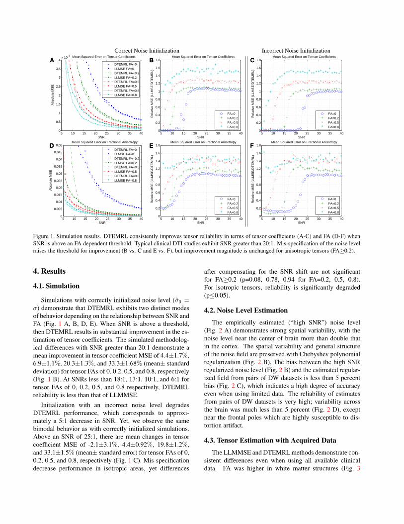
Figure 1. Simulation results. DTEMRL consistently improves tensor reliability in terms of tensor coefficients (A-C) and FA (D-F) whenSNR is above an FA dependent threshold. Typical clinical DTI studies exhibit SNR greater than 20:1. Mis-specification of the noise levelraises the threshold for improvement (B vs. C and E vs. F), but improvement magnitude is unchanged for anisotropic tensors (FA≥0.2).
4. Results
4.1. Simulation
Simulations with correctly initialized noise level (σ0 =σ) demonstrate that DTEMRL exhibits two distinct modesof behavior depending on the relationship between SNR andFA (Fig. 1 A, B, D, E). When SNR is above a threshold,then DTEMRL results in substantial improvement in the es-timation of tensor coefficients. The simulated methodolog-ical differences with SNR greater than 20:1 demonstrate amean improvement in tensor coefficient MSE of 4.4±1.7%,6.9±1.1%, 20.3±1.3%, and 33.3±1.68% (mean± standarddeviation) for tensor FAs of 0, 0.2, 0.5, and 0.8, respectively(Fig. 1 B). At SNRs less than 18:1, 13:1, 10:1, and 6:1 fortensor FAs of 0, 0.2, 0.5, and 0.8 respectively, DTEMRLreliability is less than that of LLMMSE.
Initialization with an incorrect noise level degradesDTEMRL performance, which corresponds to approxi-mately a 5:1 decrease in SNR. Yet, we observe the samebimodal behavior as with correctly initialized simulations.Above an SNR of 25:1, there are mean changes in tensorcoefficient MSE of -2.1±3.1%, 4.4±0.92%, 19.8±1.2%,and 33.1±1.5% (mean± standard error) for tensor FAs of 0,0.2, 0.5, and 0.8, respectively (Fig. 1 C). Mis-specificationdecrease performance in isotropic areas, yet differences
after compensating for the SNR shift are not significantfor FA≥0.2 (p=0.08, 0.78, 0.94 for FA=0.2, 0.5, 0.8).For isotropic tensors, reliability is significantly degraded(p≤0.05).
4.2. Noise Level Estimation
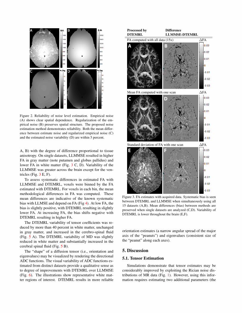
The empirically estimated (“high SNR”) noise level(Fig. 2 A) demonstrates strong spatial variability, with thenoise level near the center of brain more than double thatin the cortex. The spatial variability and general structureof the noise field are preserved with Chebyshev polynomialregularization (Fig. 2 B). The bias between the high SNRregularized noise level (Fig. 2 B) and the estimated regular-ized field from pairs of DW datasets is less than 5 percentbias (Fig. 2 C), which indicates a high degree of accuracyeven when using limited data. The reliability of estimatesfrom pairs of DW datasets is very high; variability acrossthe brain was much less than 5 percent (Fig. 2 D), exceptnear the frontal poles which are highly susceptible to dis-tortion artifact.
4.3. Tensor Estimation with Acquired Data
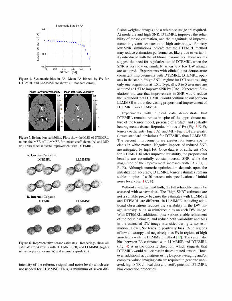
The LLMMSE and DTEMRL methods demonstrate con-sistent differences even when using all available clinicaldata. FA was higher in white matter structures (Fig. 3
a.u.A a.u.B
% Chg.C D % Chg.
Figure 2. Reliability of noise level estimation. Empirical noise(A) shows clear spatial dependence. Regularization of the em-pirical noise (B) preserves spatial structure. The proposed noiseestimation method demonstrates reliability. Both the mean differ-ence between estimate noise and regularized empirical noise (C)and the estimated noise variability (D) are within 5 percent.
A, B) with the degree of difference proportional to tissueanisotropy. On single datasets, LLMMSE resulted in higherFA in gray matter (note putamen and globus pallidus) andlower FA in white matter (Fig. 3 C, D). Variability of theLLMMSE was greater across the brain except for the ven-tricles (Fig. 3 E, F).
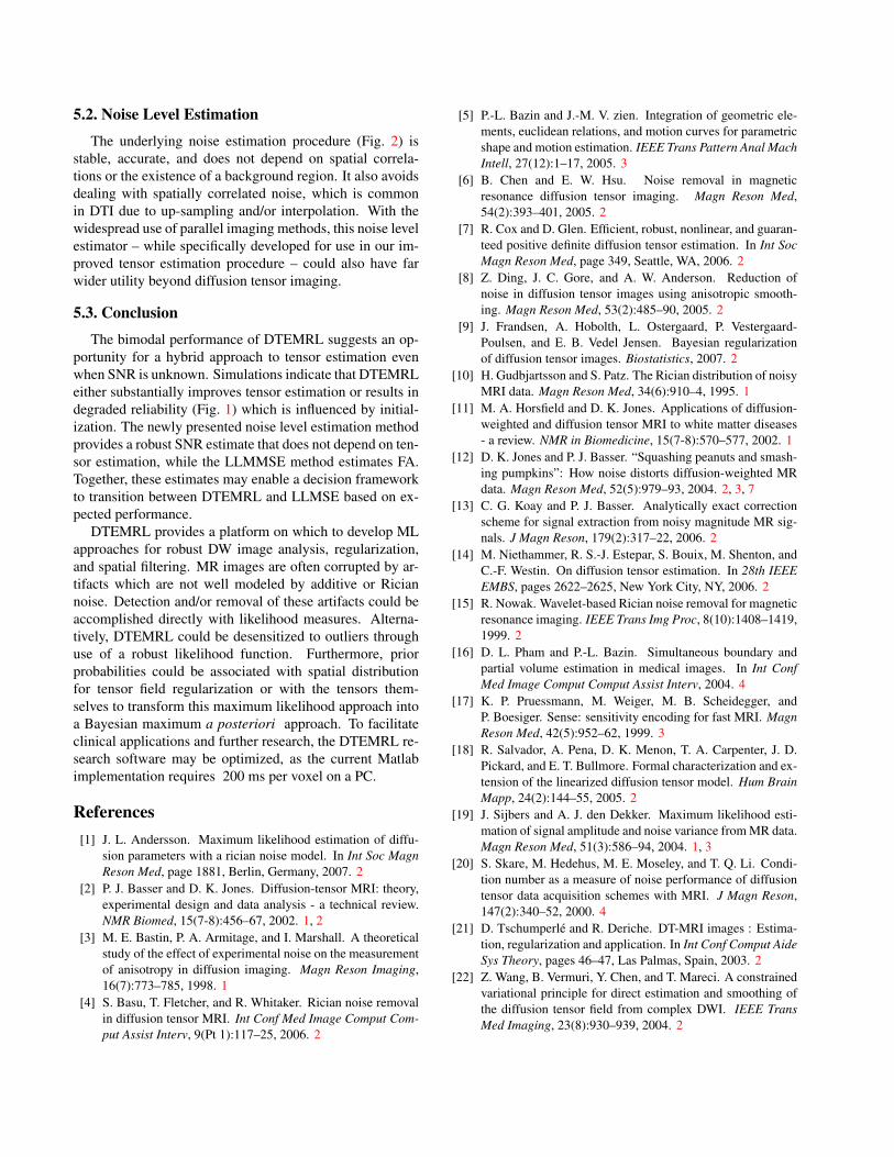
To assess systematic differences in estimated FA withLLMMSE and DTEMRL, voxels were binned by the FAestimated with DTEMRL. For voxels in each bin, the meanmethodological differences in FA was computed. Thesemean differences are indicative of the known systematicbias with LLMSE and depend on FA (Fig 4). At low FA, thebias is slightly positive, with DTEMRL resulting in slightlylower FA. At increasing FA, the bias shifts negative withDTEMRL resulting in higher FA.
The DTEMRL variability of tensor coefficients was re-duced by more than 40 percent in white matter, unchangedin gray matter, and increased in the cerebro-spinal fluid(Fig. 5 A). The DTEMRL variability of MD was slightlyreduced in white matter and substantially increased in thecerebral spinal fluid (Fig. 5 B).
The “shape” of a diffusion tensor (i.e., orientation andeigenvalues) may be visualized by rendering the directionalADC functions. The visual variability of ADC functions es-timated from distinct datasets provide a qualitative sense asto degree of improvements with DTEMRL over LLMMSE(Fig. 6). The illustrations show representative white mat-ter regions of interest. DTEMRL results in more reliable
Processed by DifferenceDTEMRL LLMMSE-DTEMRLFA computed with all data (15x) ∆FA
Mean FA computed with one scan ∆FA
Standard deviation of FA with one scan ∆FA
Figure 3. FA estimates with acquired data. Systematic bias is seenbetween DTEMRL and LLMMSE when simultaneously using all15 datasets (A,B). Mean differences (bias) between methods arepreserved when single datasets are analyzed (C,D). Variability ofDTEMRL is lower throughout the brain (E,F).
orientation estimates (a narrow angular spread of the majoraxis of the “peanuts”) and eigenvalues (consistent size ofthe “peanut” along each axes).
5. Discussion5.1. Tensor Estimation
Simulations demonstrate that tensor estimates may beconsiderably improved by exploiting the Rician noise dis-tributions of MR data (Fig. 1). However, using this infor-mation requires estimating two additional parameters (the
0 0.2 0.4 0.6 0.8 1−0.2
−0.1
0
0.1
DTEMRL [FA]
LLM
SE
−D
TE
MR
L [F
A]
Systematic Bias by FA
Figure 4. Systematic bias in FA. Mean FA binned by FA forDTEMRL and LLMMSE are shown (± standard error).
% DTensor Coefficient Variability
% DMean Diffusivity Variability
B
Figure 5. Estimation variability. Plots show the MSE of DTEMRLminus the MSE of LLMMSE for tensor coefficients (A) and MD(B). Dark tones indicate improvement with DTEMRL.
A. Corpus CallosumDTEMRL LLMMSE
B. Internal CapsuleDTEMRL LLMMSE
Figure 6. Representative tensor estimates. Renderings show allestimates for 4 voxels with DTEMRL (left) and LLMMSE (right)in the corpus callosum (A) and internal capsule (B).
intensity of the reference signal and noise level) which arenot needed for LLMMSE. Thus, a minimum of seven dif-
fusion weighted images and a reference image are required.At moderate and high SNR, DTEMRL improves the relia-bility of tensor estimation, and the magnitude of improve-ments is greater for tensors of high anisotropy. For verylow SNR, simulations indicate that the DTEMRL methodmay reduce estimation performance, likely due to variabil-ity introduced with the additional parameters. These resultssuggest the need for regularization of DTEMRL when theSNR is very low or, similarly, when very few DW imagesare acquired. Experiments with clinical data demonstrateconsistent improvements with DTEMRL. DTEMRL oper-ates in the stable, “high SNR” regime for DTI studies usingonly one acquisition at 1.5T. Typically, 3 to 5 averages areacquired at 1.5T to improve SNR by 70 to 120 percent. Sim-ulations indicate that improvement in SNR would reducethe likelihood that DTEMRL would continue to out performLLMMSE without decreasing proportional improvement ofDTEMRL over LLMMSE.
Experiments with clinical data demonstrate thatDTEMRL remains robust in spite of the approximate na-ture of the tensor model, presence of artifact, and spatiallyheterogeneous tissue. Reproducibilities of FA (Fig. 3 E, F),tensor coefficients (Fig. 5 A), and MD (Fig. 5 B) are greater(lower standard deviation) for DTEMRL than LLMMSE.The percent improvements are greatest for tensor coeffi-cients in white matter. Negative impacts of reduced SNRare mitigated by high FA. Once data is of sufficient SNRfor DTEMRL to offer improved reliability, the proportionalbenefits are essentially constant across SNR while themagnitude of the improvement increases with FA (Fig. 1B, E). Although numeric optimization depends upon theinitialization accuracy, DTEMRL tensor estimates remainstable in spite of a 20 percent mis-specification of initialnoise level (Fig. 1 C, F).
Without a valid ground truth, the full reliability cannot beassessed with in vivo data. The “high SNR” estimates arenot a suitable proxy because the estimates with LLMMSEand DTEMRL are different. In LLMMSE, including addi-tional observations reduces the variability in the DW im-age intensity, but also reinforces bias on each DW image.With DTEMRL, additional observations enable refinementof the noise estimate, and reduce both variability and biasin the estimated DW image intensities during tensor esti-mation. Low SNR tends to positively bias FA in regionsof low anisotropy and negatively bias FA in regions of highanisotropy with the LLMMSE method [12]. The systematicbias between FA estimated with LLMMSE and DTEMRL(Fig. 4) is in the opposite direction, which suggests thatDTEMRL would reduce bias in the estimated tensors. How-ever, additional acquisitions using k-space averaging and/orcomplex-valued imaging data are required to generate unbi-ased, high SNR clinical data and verify potential DTEMRLbias correction properties.
5.2. Noise Level Estimation
The underlying noise estimation procedure (Fig. 2) isstable, accurate, and does not depend on spatial correla-tions or the existence of a background region. It also avoidsdealing with spatially correlated noise, which is commonin DTI due to up-sampling and/or interpolation. With thewidespread use of parallel imaging methods, this noise levelestimator – while specifically developed for use in our im-proved tensor estimation procedure – could also have farwider utility beyond diffusion tensor imaging.
5.3. Conclusion
The bimodal performance of DTEMRL suggests an op-portunity for a hybrid approach to tensor estimation evenwhen SNR is unknown. Simulations indicate that DTEMRLeither substantially improves tensor estimation or results indegraded reliability (Fig. 1) which is influenced by initial-ization. The newly presented noise level estimation methodprovides a robust SNR estimate that does not depend on ten-sor estimation, while the LLMMSE method estimates FA.Together, these estimates may enable a decision frameworkto transition between DTEMRL and LLMSE based on ex-pected performance.
DTEMRL provides a platform on which to develop MLapproaches for robust DW image analysis, regularization,and spatial filtering. MR images are often corrupted by ar-tifacts which are not well modeled by additive or Riciannoise. Detection and/or removal of these artifacts could beaccomplished directly with likelihood measures. Alterna-tively, DTEMRL could be desensitized to outliers throughuse of a robust likelihood function. Furthermore, priorprobabilities could be associated with spatial distributionfor tensor field regularization or with the tensors them-selves to transform this maximum likelihood approach intoa Bayesian maximum a posteriori approach. To facilitateclinical applications and further research, the DTEMRL re-search software may be optimized, as the current Matlabimplementation requires 200 ms per voxel on a PC.
References[1] J. L. Andersson. Maximum likelihood estimation of diffu-
sion parameters with a rician noise model. In Int Soc MagnReson Med, page 1881, Berlin, Germany, 2007. 2
[2] P. J. Basser and D. K. Jones. Diffusion-tensor MRI: theory,experimental design and data analysis - a technical review.NMR Biomed, 15(7-8):456–67, 2002. 1, 2
[3] M. E. Bastin, P. A. Armitage, and I. Marshall. A theoreticalstudy of the effect of experimental noise on the measurementof anisotropy in diffusion imaging. Magn Reson Imaging,16(7):773–785, 1998. 1
[4] S. Basu, T. Fletcher, and R. Whitaker. Rician noise removalin diffusion tensor MRI. Int Conf Med Image Comput Com-put Assist Interv, 9(Pt 1):117–25, 2006. 2
[5] P.-L. Bazin and J.-M. V. zien. Integration of geometric ele-ments, euclidean relations, and motion curves for parametricshape and motion estimation. IEEE Trans Pattern Anal MachIntell, 27(12):1–17, 2005. 3
[6] B. Chen and E. W. Hsu. Noise removal in magneticresonance diffusion tensor imaging. Magn Reson Med,54(2):393–401, 2005. 2
[7] R. Cox and D. Glen. Efficient, robust, nonlinear, and guaran-teed positive definite diffusion tensor estimation. In Int SocMagn Reson Med, page 349, Seattle, WA, 2006. 2
[8] Z. Ding, J. C. Gore, and A. W. Anderson. Reduction ofnoise in diffusion tensor images using anisotropic smooth-ing. Magn Reson Med, 53(2):485–90, 2005. 2
[9] J. Frandsen, A. Hobolth, L. Ostergaard, P. Vestergaard-Poulsen, and E. B. Vedel Jensen. Bayesian regularizationof diffusion tensor images. Biostatistics, 2007. 2
[10] H. Gudbjartsson and S. Patz. The Rician distribution of noisyMRI data. Magn Reson Med, 34(6):910–4, 1995. 1
[11] M. A. Horsfield and D. K. Jones. Applications of diffusion-weighted and diffusion tensor MRI to white matter diseases- a review. NMR in Biomedicine, 15(7-8):570–577, 2002. 1
[12] D. K. Jones and P. J. Basser. “Squashing peanuts and smash-ing pumpkins”: How noise distorts diffusion-weighted MRdata. Magn Reson Med, 52(5):979–93, 2004. 2, 3, 7
[13] C. G. Koay and P. J. Basser. Analytically exact correctionscheme for signal extraction from noisy magnitude MR sig-nals. J Magn Reson, 179(2):317–22, 2006. 2
[14] M. Niethammer, R. S.-J. Estepar, S. Bouix, M. Shenton, andC.-F. Westin. On diffusion tensor estimation. In 28th IEEEEMBS, pages 2622–2625, New York City, NY, 2006. 2
[15] R. Nowak. Wavelet-based Rician noise removal for magneticresonance imaging. IEEE Trans Img Proc, 8(10):1408–1419,1999. 2
[16] D. L. Pham and P.-L. Bazin. Simultaneous boundary andpartial volume estimation in medical images. In Int ConfMed Image Comput Comput Assist Interv, 2004. 4
[17] K. P. Pruessmann, M. Weiger, M. B. Scheidegger, andP. Boesiger. Sense: sensitivity encoding for fast MRI. MagnReson Med, 42(5):952–62, 1999. 3
[18] R. Salvador, A. Pena, D. K. Menon, T. A. Carpenter, J. D.Pickard, and E. T. Bullmore. Formal characterization and ex-tension of the linearized diffusion tensor model. Hum BrainMapp, 24(2):144–55, 2005. 2
[19] J. Sijbers and A. J. den Dekker. Maximum likelihood esti-mation of signal amplitude and noise variance from MR data.Magn Reson Med, 51(3):586–94, 2004. 1, 3
[20] S. Skare, M. Hedehus, M. E. Moseley, and T. Q. Li. Condi-tion number as a measure of noise performance of diffusiontensor data acquisition schemes with MRI. J Magn Reson,147(2):340–52, 2000. 4
[21] D. Tschumperle and R. Deriche. DT-MRI images : Estima-tion, regularization and application. In Int Conf Comput AideSys Theory, pages 46–47, Las Palmas, Spain, 2003. 2
[22] Z. Wang, B. Vermuri, Y. Chen, and T. Mareci. A constrainedvariational principle for direct estimation and smoothing ofthe diffusion tensor field from complex DWI. IEEE TransMed Imaging, 23(8):930–939, 2004. 2