31
Diffusion Tensor Imaging (DTI) for the study of disorders of consciousness Stephen Larroque Coma Science Group, GIGA research University of Liège 24/03/2017
| Date post: | 22-Jan-2018 |
| Category: |
Science |
| Upload: | stephen-larroque |
| View: | 152 times |
| Download: | 2 times |
Diffusion Tensor Imaging (DTI) for the study of disorders of consciousness
Stephen Larroque
Coma Science Group, GIGA research
University of Liège
24/03/2017

Motivation
Connectivity is of paramount importance for consciousness
Study connectivity structure (micro and macro) from white matter
2

DTI preproc in 3 easy steps! (sort of…)
1. Using diffusion magnetic resonance imagery, acquire water
molecules (brownian) motion.
2. Estimate tensors ≈ mean motion of water molecules for each
brain’s voxel. We get isotropic (round, grey matter) and
anisotropic (ellipsoidic, white matter) shapes.
3
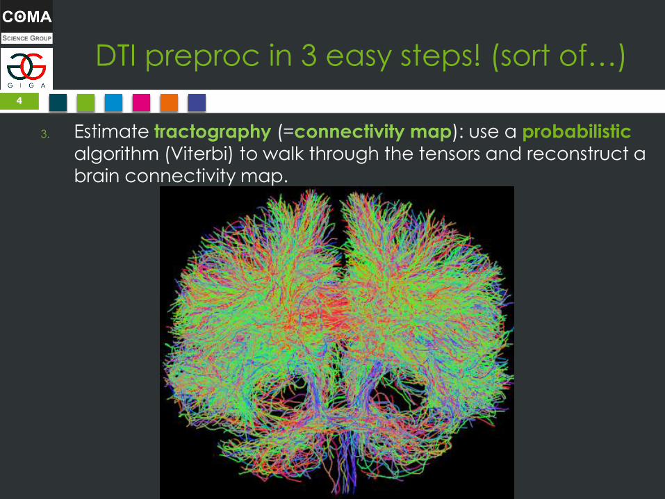
DTI preproc in 3 easy steps! (sort of…)
3. Estimate tractography (=connectivity map): use a probabilistic
algorithm (Viterbi) to walk through the tensors and reconstruct a
brain connectivity map.
4
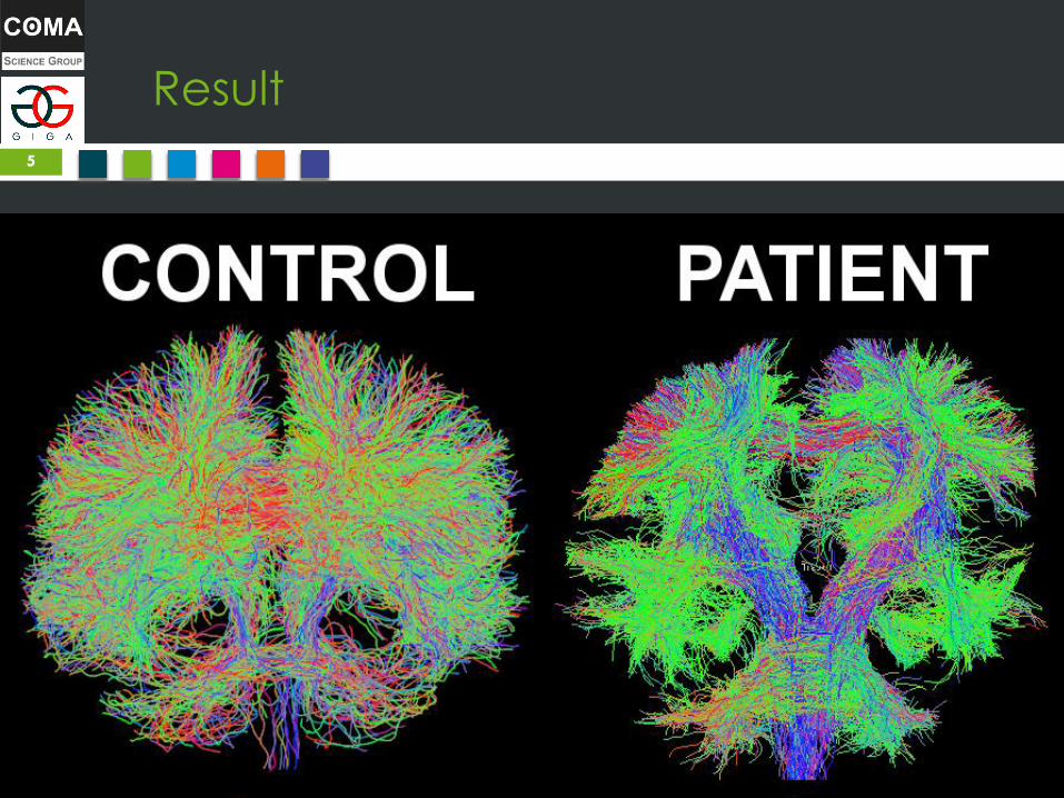
Result
5
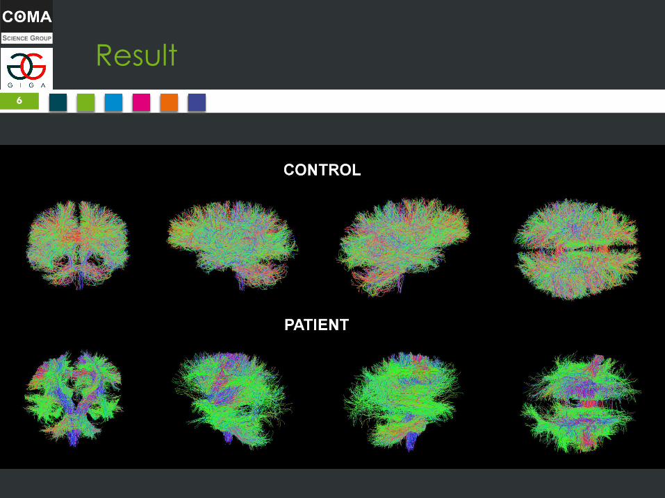
Result
6

DTI preprocessing theory vs reality
DTI preprocessing summary in theory:
1. Acquire DTI images (= hydrogen particles motion)
2. Estimate tensors (= mean particles motion)
3. Tractography (= reconstruct tracts and disambiguate
cross-sections)
7

DTI preprocessing theory vs reality
DTI preprocessing summary in practice: 1. Acquire DTI images + T1
2. Reorient both
3. Extract gradients (bvecs and bvals)
4. Brain Extraction (BET) mask on DWI and T1
5. Correct eddy currents
6. Estimate tensors & FA metrics
7. Segment T1
8. Coregister DWI on T1
9. Downsample T1
10. Estimate DWI response function
11. Tractography
12. And more steps depending on your objectives…
8
DTI is still in the process of
standardization… but not there yet!
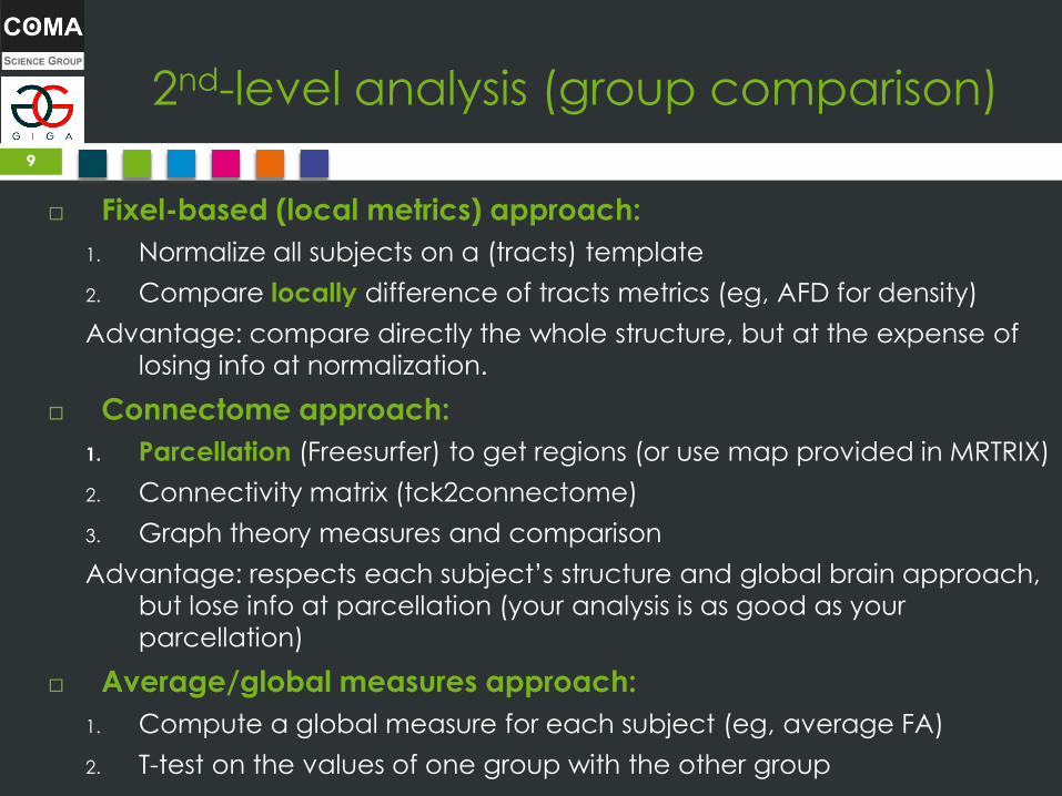
2nd-level analysis (group comparison)
Fixel-based (local metrics) approach:
1. Normalize all subjects on a (tracts) template
2. Compare locally difference of tracts metrics (eg, AFD for density)
Advantage: compare directly the whole structure, but at the expense of
losing info at normalization.
Connectome approach:
1. Parcellation (Freesurfer) to get regions (or use map provided in MRTRIX)
2. Connectivity matrix (tck2connectome)
3. Graph theory measures and comparison
Advantage: respects each subject’s structure and global brain approach,
but lose info at parcellation (your analysis is as good as your
parcellation)
Average/global measures approach:
1. Compute a global measure for each subject (eg, average FA)
2. T-test on the values of one group with the other group
9
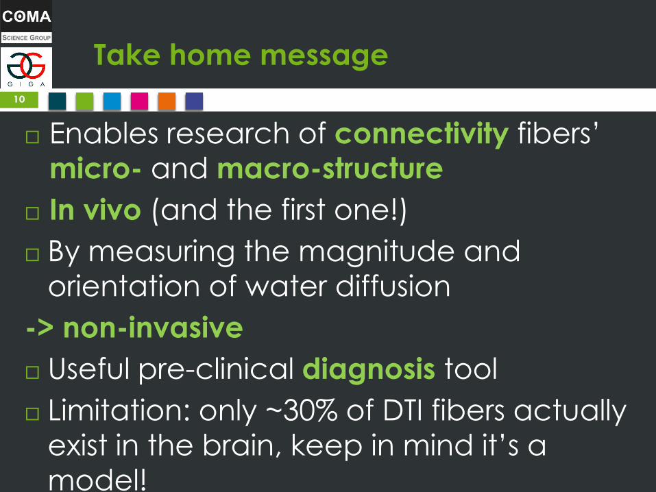
Take home message
Enables research of connectivity fibers’
micro- and macro-structure
In vivo (and the first one!)
By measuring the magnitude and
orientation of water diffusion
-> non-invasive
Useful pre-clinical diagnosis tool
Limitation: only ~30% of DTI fibers actually
exist in the brain, keep in mind it’s a
model!
10

To go further
MRTRIX3 whole documentation
Beginner’s DTI preprocessing pipeline (up to connectome analysis): http://community.mrtrix.org/t/beginner-connectome-pipeline-updated/373/2
Fixel-based analysis using MRTRIX3: http://mrtrix.readthedocs.io/en/latest/workflows/fixel_based_analysis.html
Connectome analysis using MRTRIX3 (tck2connectome): http://mrtrix.readthedocs.io/en/latest/workflows/structural_connectome.html
http://community.mrtrix.org/t/the-output-of-tck2connectome/345
Global measure analysis: see afdconnectivity and http://mrtrix.readthedocs.io/en/latest/workflows/DWI_preprocessing_for_quantitative
_analysis.html
FSL eddy (eddy currents + motion/realignment correction)
Subparcellation
Do Tromp’s DTI tutorials, diffusion-imaging.com, 2016
MRTRIX3 community forum! community.mrtrix.com
11
Thank you for your
attention References:
•Posterior cingulate cortex-related co-activation patterns: a resting state FMRI study in propofol-induced loss of
consciousness, Amico, Enrico, et al, PLoS One 9.6 (2014): e100012.
•Multimodal neuroimaging in patients with disorders of consciousness showing “functional hemispherectomy”, Van
Someren, E. J. W. (2011), Slow Brain Oscillations of Sleep, Resting State and Vigilance: Proceedings of the 26th International
Summer School of Brain Research, Held at the Royal Netherlands Academy of Arts and Sciences, Amsterdam, The
Netherlands, 29 June-2 July, 2010, 193, 323.
• Neural correlates of consciousness in patients who have emerged from a minimally conscious state: a cross-
sectional multimodal imaging study, Carol Di Perri & Mohamed Ali Bahri & Enrico Amico & Aurore Thibaut & Lizette Heine
et al., The Lancet Neurology, 2016
•Do Tromp, http://www.diffusion-imaging.com/, 2016
•Amico et al., Conf Proc IEEE Eng Med Biol Soc. 2015
12

BONUS SLIDES
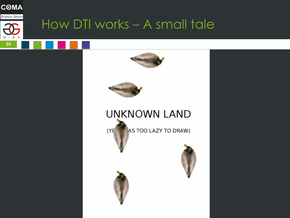
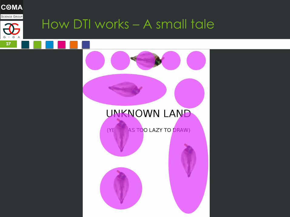
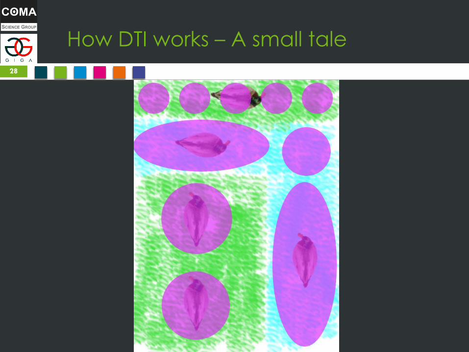
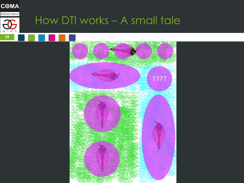
How DTI works – A small tale
14
A Ducks
Tale about Imagery
(DTI)

How DTI works – A small tale
15

How DTI works – A small tale
16

How DTI works – A small tale
17
How DTI works – A small tale
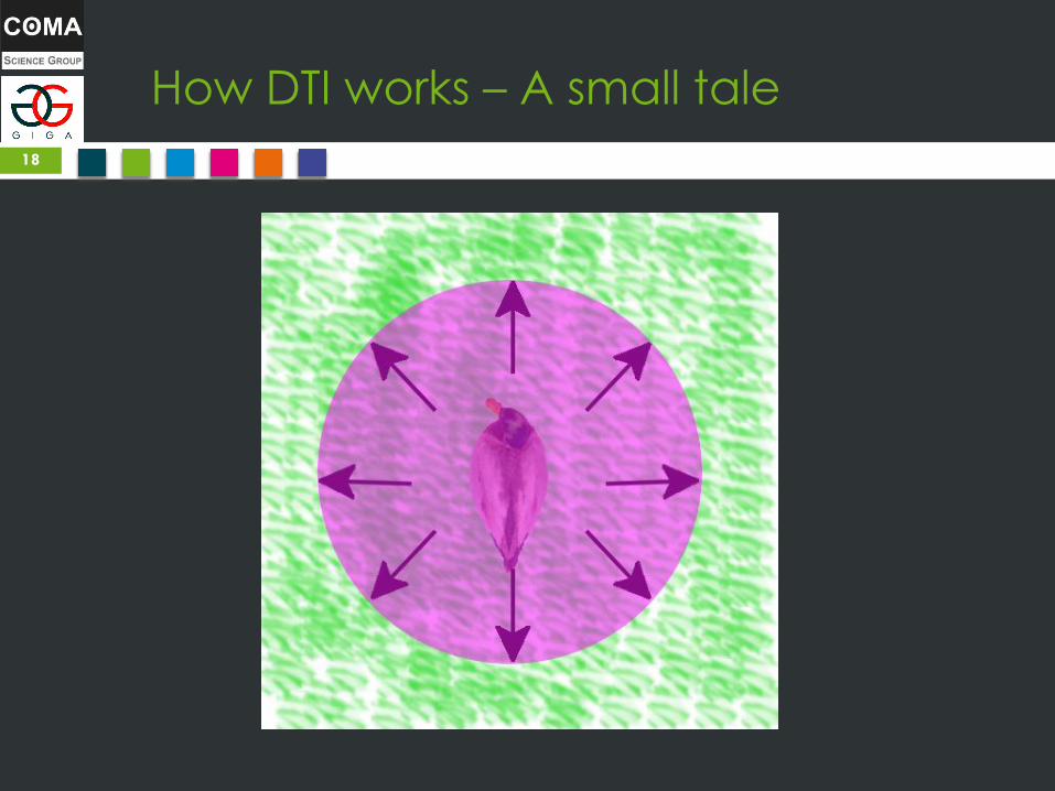
18
How DTI works – A small tale
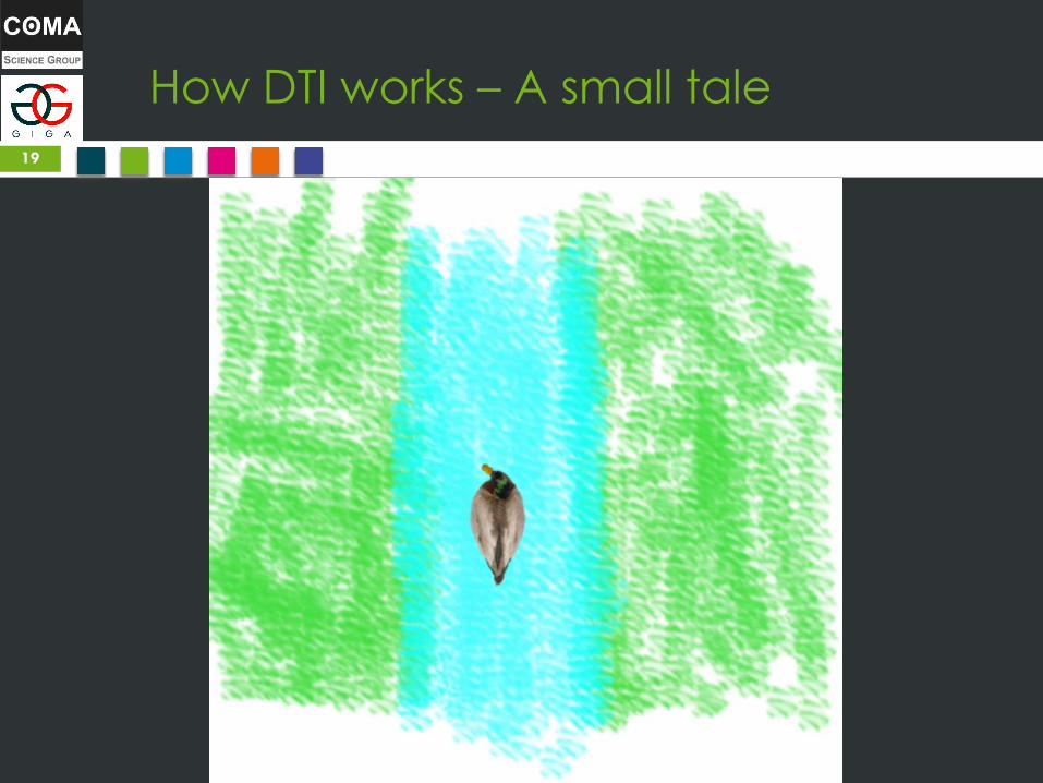
19

How DTI works – A small tale
20

How DTI works – A small tale
21
How DTI works – A small tale
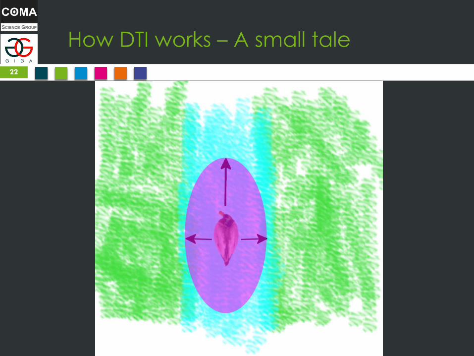
22
How DTI works – A small tale
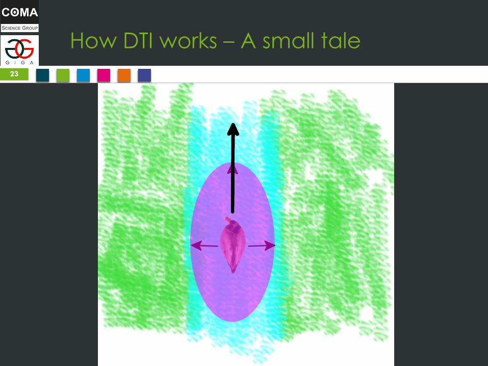
23

How DTI works – A small tale
24

How DTI works – A small tale
25
How DTI works – A small tale
26
How DTI works – A small tale
27
How DTI works – A small tale
28
How DTI works – A small tale
29
How DTI works – A small tale
30
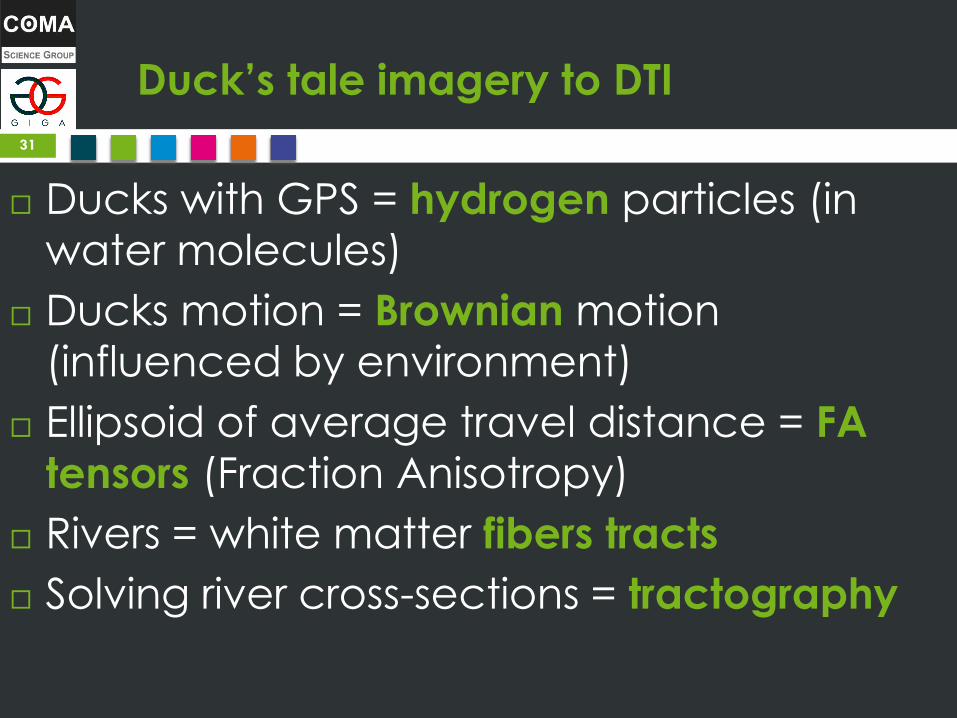
Duck’s tale imagery to DTI
Ducks with GPS = hydrogen particles (in
water molecules)
Ducks motion = Brownian motion
(influenced by environment)
Ellipsoid of average travel distance = FA
tensors (Fraction Anisotropy)
Rivers = white matter fibers tracts
Solving river cross-sections = tractography
31


![Diffusion Tensor Imaging (DTI): An overview of key concepts · Diffusion Tensor Imaging: Concepts and Applications. Journal of Magnetic Resonance Imaging 13, 534-546. [2] Johansen-Berg,](https://static.documents.pub/doc/80x56/5ed5b01e0a1a7f290d5f7199/diffusion-tensor-imaging-dti-an-overview-of-key-diffusion-tensor-imaging-concepts.jpg)


