Department of Human Anatomy and Cell Biology, The University of Liverpool,PO Box 147, Liverpool L69 3BX, UK
AND SUSAN W. HERRING
Department of Oral Anatomy, College of Dentistry, University of Illinois atChicago, 801 S Paulina Street, Chicago, Illinois 60612, USA
Accepted 30 November 1988
Summary
At incremental whole muscle lengths, active isometric and passive elastic forceswere recorded from the masseter and digastric muscles of anaesthetized miniaturepigs (Hanford) weighing 2-0-20-0 kg. Wet muscle mass and maximum tetanictension values for masseter exceed those for digastric and increase more rapidlywith body mass (age). At any body mass, masseter exceeds digastric in the ratio ofoptimum length (that length at which maximum tetanic tension is produced) to insitu muscle length (that length which corresponds to the jaw in a closed position)and the proportion of passive tension comprising total (passive plus active)tension. Passive elastic tension begins to rise in masseter at lengths as short as87 % of optimum (in younger pigs). In digastric, passive tension is absent until themuscle is stretched to a length slightly longer than optimum in younger pigs butoccurs at shorter lengths in older pigs.
Contractile properties explain functional differences between masseter anddigastric more clearly than they explain ontogenetic changes in either muscle. Thebehavioural transition from infant suckling to adult mastication of solid food isbest characterized by a disproportionate increase in mass (and force) of themasseter, relative to digastric, and increased reliance upon active (rather thanpassive) tension.
Introduction
It is a relatively well documented, empirically observed physiological phenom-enon that tetanic tension increases to its maximum (Po) at an optimum length (LQ)as a whole muscle or fibre is stretched and then declines with further lengthening(e.g. Ramsey & Street, 1940; Gordon et al. 1966; Close, 1972, citing others).Although readily perceptible for muscles having their fasciculi arranged in parallelwith the direction of whole muscle contraction, the length-tension relationship
becomes somewhat obscured with increasing complexity of muscle architecture.Despite recent advances using comprehensive examination of the contractileproperties of unipinnate muscles (e.g. Goslow et al. 1977; Muhl et al. 1978;Walmsley & Proske, 1981; Muhl, 1982), the effect of multipinnate or complex fibrearchitecture on the relationship between tension and whole muscle excursion,especially within the range in which a muscle can be expected to perform work,remains enigmatic.
Contractile properties of whole pinnate muscles are best known for thosestudied in the hindlimb, especially as applied to understanding the functionalconsequences of muscle length changes during locomotion (e.g. Goslow & VanDeGraaff, 1982; Stephens et al. 1975; De Koning et al. 1987). However, with rareexceptions (e.g. Nemeth et al. 1986), the few studies of the masticatory musclesthat are available indirectly measured tension and usually correlated forcesmeasured on the jaw with gape position following direct stimulation of individualwhole muscles (e.g. Nordstrom & Yemm, 1974; Yemm & Nordstrom, 1974;Thexton & Hiiemae, 1975; MacKenna & Turker, 1978). This approach wasprobably used because of the experimentally cumbersome geometry of the headand oral apparatus and the nonfusiform shape of most jaw muscles, which tend tomake length manipulation for isometric tension measurements difficult at best.One attempt to circumvent this problem has been to measure contractileproperties of small muscle bundles, but this necessitates severe extrapolation ofthe results (Faulkner et al. 1982; Maxwell et al. 1982). However, contraction of themuscle by direct stimulation less closely resembles the normal physiologicalcondition than does contraction by stimulation of the muscle nerve.
The masseter and digastric muscles of miniature domestic pigs (Sus scrofa)present an interesting comparison of changes in muscle function during postnatalontogeny. The transition from infant suckling behaviour to juvenile and adultmastication is characterized by increasing complexity of electromyographicactivity in the masseter (Herring & Wineski, 1986), whereas the adult EMGpattern in digastric resembles that observable in the newborn. In this study,isometric and passive tension and whole muscle lengths were recorded frommasseter and digastric following stepwise stretches and direct stimulation of themuscle nerve. The characteristic isometric contractile properties of these twomuscles account for their functional differences and behavioural changes duringontogeny.
Materials and methodsPreparation and apparatus
Active and passive tetanic tension were measured at increasing muscle lengthsin the masseter and digastric muscles of miniature swine (minipigs) of either sexand various strains (Table 1). Each animal was anaesthetized by gas inhalation( 1 % halothane, nitrous oxide/oxygen), initially administered with a snout cone
Length-tension of pig masticatory muscles 3
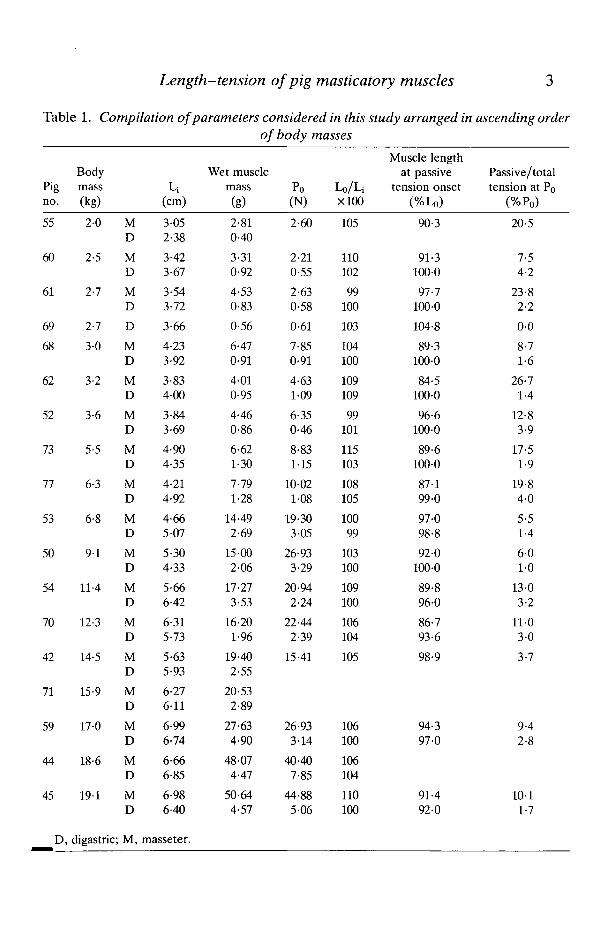
Table 1. Compilation of parameters considered in this study arranged in ascending orderof body masses
Pigno.
55
60
61
69
68
62
52
73
77
53
50
54
70
42
71
59
44
45
D,
Bodymass(kg)
2-0
2-5
2-7
2-7
3-0
3-2
3-6
5-5
6-3
6-8
9 1
11-4
12-3
14-5
15-9
17-0
18-6
19-1
digastric;
MD
MD
MD
D
MD
MD
MD
MD
MD
MD
MD
MD
MD
MD
MD
MD
MD
MD
M,
Li(cm)
3-052-38
3-423-67
3-543-72
3-66
4-233-92
3-834-00
3-843-69
4-904-35
4-214-92
4-665-07
5-304-33
5-666-42
6-315-73
5-635-93
6-27611
6-996-74
6-666-85
6-986-40
masseter.
Wet musclemass(g)
2-810-40
3-310-92
4-530-83
0-56
6-470-91
4-010-95
4-460-86
6-621-30
7-791-28
14-492-69
15-002-06
17-273-53
16-201-96
19-402-55
20-532-89
27-634-90
48-074-47
50-644-57
Po(N)
2-60
2-210-55
2-630-58
0-61
7-850-91
4-631-09
6-350-46
8-831-15
10-021-08
19-303-05
26-933-29
20-942-24
22-442-39
15-41
26-933-14
40-407-85
44-885-06
Lo/LixlOO
105
110102
99100
103
104100
109109
99101
115103
108105
10099
103100
109100
106104
105
106100
106104
110100
Muscle lengthat passive
tension onset(%Lo)
90-3
91-3100-0
97-71000
104-8
89-3100-0
84-5100-0
96-6100-0
89-6100-0
87-199-0
97-098-8
92-0100-0
89-8960
86-793-6
98-9
94-397-0
91-492-0
Passive/totaltension at Po
(%Po)
20-5
7-54-2
23-82-2
0-0
8-71-6
26-71-4
12-83-9
17-51-9
19-84-0
5-51-4
6-01-0
13-03-2
11-03-0
3-7
9-42-8
1011-7
4 F . ANAPOL AND S. W. HERRING
led from a Drager veterinary anaesthesia machine (North American Drager,Telford, PA).
The forehead, cheeks and ventral neck were shaved and cleansed with 70 %ethanol; an overhead heat lamp helped to maintain body temperature asmonitored by a rectal thermometer. The ventral neck skin was incised from themandibular symphysis to the jugular notch. The trachea was exposed by bluntdissection and cannulated 0-5 cm below the larynx; maintenance of surgical-planeanaesthesia was immediately relegated to the tracheostomy. A sagittal incision ofthe forehead skin and underlying periosteum allowed a headholder (25 mm x20 mm o.d. stainless-steel tubing) to be applied to the frontal bone between theorbits. This was accomplished by obliquely inserting 6-8 jeweller's screws (0,0-48-0-95 cm) in a circular formation slightly larger than the outer circumferenceof the tube. A flange was created by cementing the tube within the circle of screwsusing standard dental acrylic (Lang Dental Manufacturing Co., Chicago).
With the animal lying on its side, the masseter was exposed and a fine black silksuture (000, Ethicon, Somerville, New Jersey, USA) was carefully tied into thedeep fascia at the mandibular attachment of the muscle at gonion. A second suturewas placed into the tendinous attachment to the zygomatic arch at the oppositeend of a stress line (perceptible in the superficial aponeurotic tendon of masseter)from gonion. The distance between the sutures was defined as the in situ length ofthe muscle belly (Li) measured with the jaws in occlusion. Without injury to themasseter, the zygomaticomaxillary and temporal roots of the arch were cleanedand detached from the skull and the zygomaticomandibularis muscle was scrapedfrom the internal surface of the arch. The masseteric nerve was exposed by bluntdissection and stimulated with silver hook electrodes led from a Grass SIU-5Astimulus isolation unit (Grass Instrument Co., Quincy, Massachusetts, USA)driven by a Grass S-48 stimulator. Positive identification of the nerve was by directobservation of muscle contraction and palpation of the detached arch.
The arch was clamped to an adjustable rigid bar led from a 114 kg capacity forcetransducer (±0-03 linearity) (Omega Engineering, Inc., Stamford, CT, USA).The transducer was mounted on an adjustable collar which surrounded a35 mm x 400 mm vertical stainless-steel rod. The base of this upright was arectangular slide that could be secured anywhere along the edge of a stainless-steelplate (450 mm x 780 mm x 20 mm). Thus, the attitude of the transducer wasadjustable by translation in the vertical and horizontal planes and by horizontalrotation. A calibrated adjusting screw connecting the muscle and the forcetransducer allowed changes in length of up to 20 mm.
The cranium was stabilized by fastening (with a set screw) a 1 cm diameter rodinto the headholder. The rod extended from a fully adjustable ball joint mountedon the base plate. The lower jaw was fixed by an adjustable clamp mounted on asecond 25 mm x 245 mm upright rod whose attachment to the base plate could alsobe manipulated. The compliance of the apparatus (bar, force transducer, jawholder) was 0.092 mm kg"1.
Force and length measurements of the digastric either preceded or followed
Length-tension of pig masticatory muscles 5
completion of the masseter portion of the protocol. With the pig lying on its back,the digastric was exposed with blunt dissection. Sutures were tied into the mostrostral attachment of the muscle belly to the mandible and into the extrinsictendon at the attachment of the most caudal muscle fibres; L; was measured andrecorded. The branch of the mylohyoid nerve that innervates the digastric wasdissected and verified by stimulation at low voltage. The tendon was detachedfrom the skull, leaving a small fragment of the paracondyloid process in thesevered end, and tied to a wire hook with a silk ligature. The hook was attached tothe rigid bar that led from the force transducer.
Core temperature was maintained with a heating pad and monitored with arectal thermometer. The muscle was kept damp with a saline-soaked gauze spongeand the nerve preparation was bathed in mineral oil.
Physiological measurements
The remainder of the procedure was the same for either muscle. Resting lengthwas re-established and twitch contractions were induced by direct stimulation(6 ms pulses) of the nerve at increasing voltage (10 V increments). The signal fromthe force transducer was amplified through a Grass 7P1G low-level d.c. preampli-fier and 7DAG d.c. driver amplifier and displayed on a Tektronix 5116 digitaloscilloscope (Tektronix, Inc., Beaverton, Oregon, USA). The oscilloscope wasequipped with a Tektronix 5D10 dual-cursor waveform digitizer that enableddirect measurement of amplitude and time course of the signal. The voltage atwhich the amplitude of the twitch contraction no longer increased was selected fortetanic stimulation.
The muscle length was shortened 3-5 mm below L; and the length of the muscleand passive tension, if present, were recorded. The nerve was stimulated with a400-600 ms train at 40 Hz using the same pulse settings as before; active tetanictension (P) was recorded from the muscle. The muscle was lengthened 1 mm onthe scale and the procedure was repeated after a lmin interval. This continueduntil tetanic tension fell below maximum twitch tension. At the end of theexperiment, both masseter and digastric were removed, trimmed of extrinsictendon (digastric), blotted dry, and weighed. The pig was then administered alethal dose of sodium pentobarbital intraventricularly.
Data analysis
An equation to transform electrical data from the oscilloscope to force wasderived by least-squares linear regression. For each experiment, passive and activetension values were normalized by expressing each as a percentage of maximum oroptimum tetanic tension (Po) observed during that experiment. Muscle lengthswere normalized by expressing each as a percentage of optimum length LQ (themeasured length corresponding to Po at the maximum discrepancy between activeand passive tension).
Length-tension curves and related plots were generated with an IBM system370 computer at the University of Illinois at Chicago using the Statistical Analysis
6 F. ANAPOL AND S. W. HERRING
System (SAS Institute, Cary, North Carolina, USA). SAS was also used for thecomputation of statistics for the data analysis.
ResultsRepresentative oscilloscope traces of fused tetani recorded from masseter and
digastric muscles of a 3-2 kg minipig are presented in Fig. 1. Traces are includedfor muscle lengths approaching Lo (Fig. 1A,D), approximately at Lo (Fig. 1B,E)and following Lo (Fig. 1C,F). The curves typify those observed for most subjectsregardless of body mass.
The slight declinations of the plateaux at Lo are similar for both muscles andexhibit a slight 'sag', thought to be correlated to the presence of alkaline-stablemyosin adenosine triphosphatase, i.e. fast-twitch fibres (Burke etal. 1971;Reinking et al. 1975; Kernall et al. 1983). At lengths below Lo, the 'sag' is muchmore pronounced in digastric. However, at lengths greater than Lo, the plateau ismore horizontal, i.e. the sag is absent.
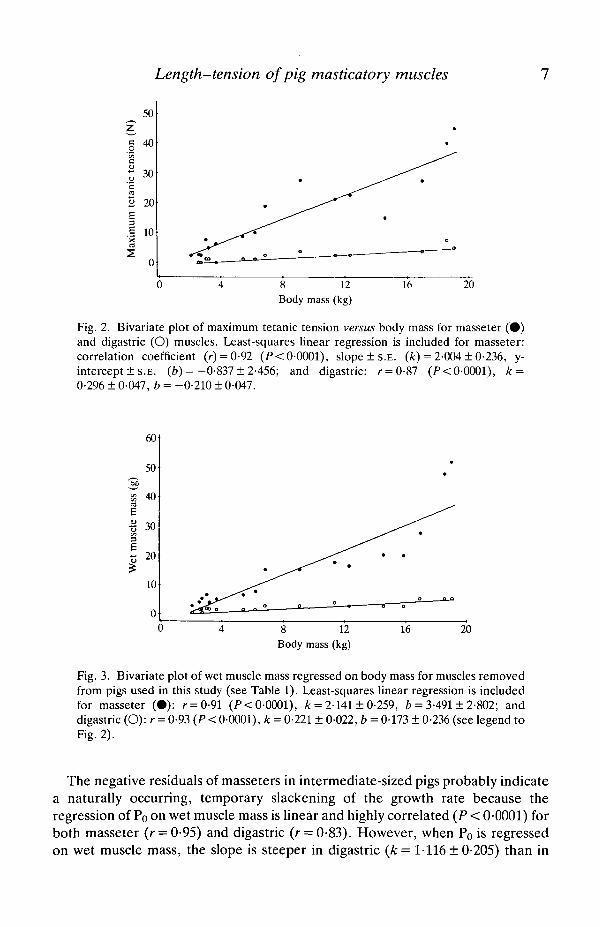
The results of least-squares regressions of Po and wet muscle mass on body massfor masseter and digastric muscles are shown in Figs 2 and 3. Although Po and wetmuscle mass are highly correlated with body mass for both masseter and digastric,only for digastric do the regressions appear to be linear. For both parameters formasseter, the portion of the curve corresponding to pigs =£16 kg has the mostgradual slope with the steepest portion corresponding to pigs >16kg. The slopesof the overall ontogenetic increases in both Po and wet muscle mass areconsiderably steeper in masseter than in digastric.
D
Fig. 1. Typical oscilloscope trace of maximum tetanic tension in a 3-2 kg miniature pigfor the masseter muscle at (A) approx. 89%Lo, (B) approx. 99%LQ and (C) approx.104%LQ; and the digastric muscle at (D) approx. 94%LQ, (E) approx. Lo and (F)approx. 103 %LQ.
Length-tension of pig masticatory muscles
50
g
~ 30c
2 20£1 10X
8 12Body mass (kg)
16 20
Fig. 2. Bivariate plot of maximum tetanic tension versus body mass for masseter ( • )and digastric (O) muscles. Least-squares linear regression is included for masseter:correlation coefficient (r) = 0-92 (P< 0-0001), slope ±S .E . (k) = 2-004 ± 0-236, y-intercept ± S.E. (b) = -0-837 ± 2-456; and digastric: r = 0-87 (P< 0-0001), k =0-296 ± 0-047, b = -0-210 ± 0-047.
60
50
¥ 40E
I 30
3a 20
10
0
8 12Body mass (kg)
16 20
Fig. 3. Bivariate plot of wet muscle mass regressed on body mass for muscles removedfrom pigs used in this study (see Table 1). Least-squares linear regression is includedfor masseter (•) : r = 0-91 (F<0-0001), k = 2-141 ± 0-259, b = 3-491 ± 2-802; anddigastric (O):r = 0-93 (P < 0-0001), k = 0-221 ± 0-022, b = 0-173 ± 0-236 (see legend toFig. 2).
The negative residuals of masseters in intermediate-sized pigs probably indicatea naturally occurring, temporary slackening of the growth rate because theregression of Po on wet muscle mass is linear and highly correlated (P < 0-0001) forboth masseter (r = 0-95) and digastric (r = 0-83). However, when Po is regressedon wet muscle mass, the slope is steeper in digastric {k = 1-116 ± 0-205) than in
8 F . ANAPOL AND S. W. HERRING
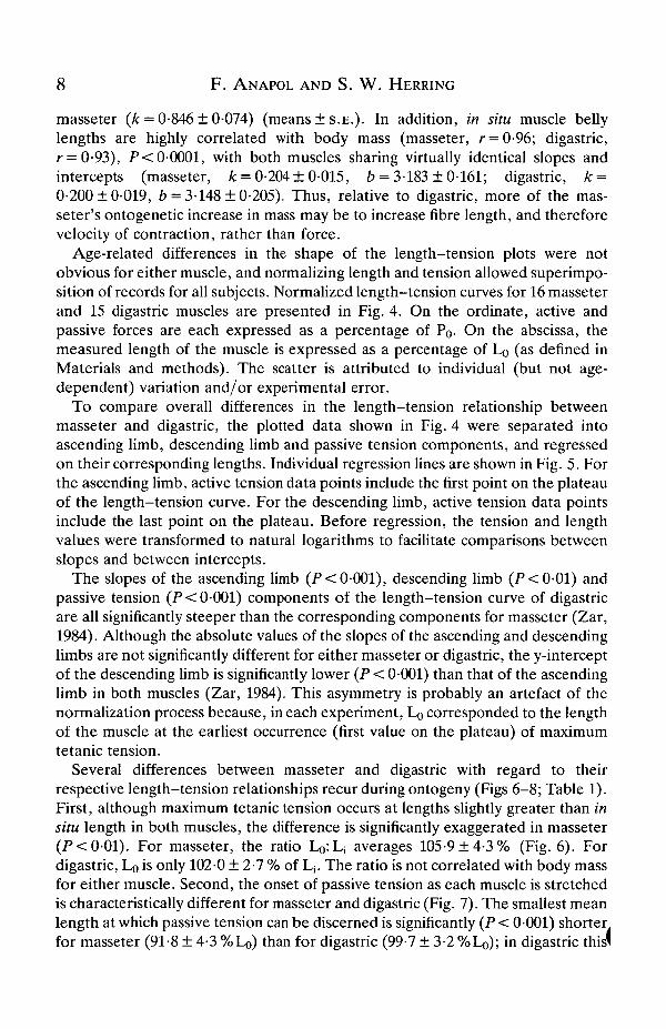
masseter (k = 0-846 ± 0-074) (means ± S . E . ) . In addition, in situ muscle bellylengths are highly correlated with body mass (masseter, r = 0-96; digastric,r = 0-93), P< 0-0001, with both muscles sharing virtually identical slopes andintercepts (masseter, k = 0-204 ±0-015, b = 3-183 ±0-161; digastric, k =0-200 ±0-019, b = 3-148 ±0-205). Thus, relative to digastric, more of the mas-seter's ontogenetic increase in mass may be to increase fibre length, and thereforevelocity of contraction, rather than force.
Age-related differences in the shape of the length-tension plots were notobvious for either muscle, and normalizing length and tension allowed superimpo-sition of records for all subjects. Normalized length-tension curves for 16 masseterand 15 digastric muscles are presented in Fig. 4. On the ordinate, active andpassive forces are each expressed as a percentage of Po- On the abscissa, themeasured length of the muscle is expressed as a percentage of Lo (as defined inMaterials and methods). The scatter is attributed to individual (but not age-dependent) variation and/or experimental error.
To compare overall differences in the length-tension relationship betweenmasseter and digastric, the plotted data shown in Fig. 4 were separated intoascending limb, descending limb and passive tension components, and regressedon their corresponding lengths. Individual regression lines are shown in Fig. 5. Forthe ascending limb, active tension data points include the first point on the plateauof the length-tension curve. For the descending limb, active tension data pointsinclude the last point on the plateau. Before regression, the tension and lengthvalues were transformed to natural logarithms to facilitate comparisons betweenslopes and between intercepts.
The slopes of the ascending limb (P< 0-001), descending limb (P<0-01) andpassive tension (P < 0-001) components of the length-tension curve of digastricare all significantly steeper than the corresponding components for masseter (Zar,1984). Although the absolute values of the slopes of the ascending and descendinglimbs are not significantly different for either masseter or digastric, the y-interceptof the descending limb is significantly lower (P < 0-001) than that of the ascendinglimb in both muscles (Zar, 1984). This asymmetry is probably an artefact of thenormalization process because, in each experiment, Lo corresponded to the lengthof the muscle at the earliest occurrence (first value on the plateau) of maximumtetanic tension.
Several differences between masseter and digastric with regard to theirrespective length-tension relationships recur during ontogeny (Figs 6-8; Table 1).First, although maximum tetanic tension occurs at lengths slightly greater than insitu length in both muscles, the difference is significantly exaggerated in masseter(P<0-01). For masseter, the ratio L^L; averages 105-9 ±4 -3% (Fig. 6). Fordigastric, LQ is only 102-0 ± 2-7 % of Lj. The ratio is not correlated with body massfor either muscle. Second, the onset of passive tension as each muscle is stretchedis characteristically different for masseter and digastric (Fig. 7). The smallest meanlength at which passive tension can be discerned is significantly (P < 0001) shorterfor masseter (91-8 ± 4-3 %Lo) than for digastric (99-7 ± 3-2 %L0); in digastric this(
100
80
60
40
20
100
80
60-
40
20-
o-
Length-tension of pig masticatory musclesMasseter i
.". .."•:: r: : : . • •• •
85 90 95 If
Digastric
• • • • • • • •
! . . . fl
! . • « . . .
• fl.• . • • • . .
• . , • • •• • • •
> • • •
» • • •• • • • • • • • •
10 105 110 115
• • : .
"• • •
•• •• t
. . . * • .
• • • • • •
• • *
• • •
85 90 95 100 105Percentage of optimum length
110 115
Fig. 4. Composite length-tension curves for 16 masseter and 15 digastric muscles inminiature pigs. A vertical dashed line transects optimum length (100%). Mean musclein situ length (with teeth in occlusion) is indicated by the short vertical line to the left ofoptimum. Dots are active tetanic force produced at each muscle length; asterisks arepassive force that occurred at each consecutive stretch of the muscle.
length is also inversely correlated to body mass. Third, at Lo, the portion of total(active plus passive) tension attributable to passive tension is significantly(P<0-001) higher in masseter (13-1 ±7-0%) than in digastric (2-3 ±1-2%)•(Fig. 8).
10 F. ANAPOL AND S. W. HERRING
100
50
Ascending limb —-^C^^• — • ^ —
• \ _ ^ - " ^
^^ Passive tension
*^ , ' Descending limb^^
— Masseter--- Digastric
80 90 100
Percentage of optimum length
110
Fig. 5. Least-squares linear regression of percentage of maximum tetanic tension onpercentage of optimum length for ascending and descending limbs of the activelength-tension curve and the passive tension curve for all pigs in this study. Data havebeen log-transformed (base e) to enable comparisons of slopes and intercepts.Ascending limb: masseter, coefficient of determination (r2) = 0-636, k = 9-039 ± 0-588,b= -36-867 ±2-676; digastric, r2 = 0-717, k = 13-850 ±0-928, b = -58-996 ± 4-234.Descending limb: masseter, r2 =0-474, k = -8-776 ±0-898, b = 45-049 ± 4-17; di-gastric, r2 = 0-637, k = -13-236 ± 1-111, b = 65-719 ± 5-164. Passive tension: masseter,r
2 = 0-794, k = 21-674±0-761, b= -97-255 ± 3-505; digastric, r2 = 0-677, k =32-515 ± 2-203, b = -148-281 ± 10-214.
J= 1 2 0
00
^ 110
• 5 100
90
1 80H.° 70
8 12
Body mass (kg)
16 20
Fig. 6. Bivariate plot of percentage optimum length/in situ length versus body mass.Masseter, ( • ) r = 0-19 (/>>0-48); digastric, (O) r= - 016 (P>0-5) (see legend toFig. 2).
Discussion
Accurate measurement of the contractile properties of the masseter muscle iscomplicated by both its gross anatomy and fibre architecture (figs 1 and 2 inHerring, 1980). Because the broad aponeurotic tendon of origin attaches along the
Length-tension of pig masticatory muscles 11
120
•£ noE r.
o. <uo >100
90
70
8 12Body mass (kg)
16 20
Fig. 7. Bivariate plot of percentage of optimum length at onset of passive tensionversus body mass. Masseter, (•) r = 0-15 (P>0-6); digastric, (O) r=-0-83(P< 0-0002), k = -0-475 ±0-093, b = 102-226 ±0-852 (see legend to Fig. 2).
D .
DO
50
40
30
20
10
8 12Body mass (kg)
16 20
Fig. 8. Bivariate plot of passive tension (as a percentage of the total of active pluspassive tension) at Po versus body mass. Masseter, (#) r = —0-48 (P > 0-06). Digastric,(O) r = 0-05 (P>0-85) (see legend to Fig. 2).
entire ventral margin of the zygomatic arch, the exact direction of contraction ofthe whole muscle is difficult to ascertain. Correct alignment of the whole musclefor tension measurements was achieved by adjustment of the apparatus untildeflection during tetani of the rigid bar linking the arch and the force transducerwas eliminated.
The inherent heterogeneity of force production within the pig masseter presentsa second problem. Architectural, histochemical and electrophysiological datasuggest that intramuscular compartments of this muscle may function indepen-dently during feeding (Herring et al. 1979; Herring, 1980; Wineski & Herring,1983; Anapol, 1985; Herring & Wineski, 1986). From several tendinous sheets
12 F. ANAPOL AND S. W. HERRING
invaginating from the superficial aponeurosis, short fasciculi arise and attachobliquely into portions of other tendinous sheets that insert into the massetericfossa of the mandible. When the jaw is in the closed position, sarcomeres in theanterior portion of the masseter are shorter than those in the posterior portion butbecome longer than the posterior sarcomeres as maximum gape is attained(Herring et al. 1979, 1984). Thus, it is doubtful whether maximum tension can begenerated in all parts of the muscle concurrently. Instead, different regions of themuscle may reach their maxima at different times and according to functionalrequirements at any moment. This regional variation may explain why the slope ofthe ascending limb (Figs 4, 5) of the masseter length-tension curve is moregradual than that of the digastric. Not all sarcomeres are at the same length whenthe mouth is closed and those which initially rest at lengths shorter than maximumoverlap are gradually stretched towards their plateau. Experimental control ofintramuscular variability would require individual length-tension measurementsfor subdivided portions of the muscle and was not attempted in this study.
In contrast, digastric (fig. 8 in Herring & Scapino, 1973) has a fusiform musclebelly with a circumscribed proximal tendinous attachment, a configurationgenerally more conducive to in situ physiological studies (Muhl et al. 1978; Muhl,1982; Anapol et al. 1987). This unipinnate jaw depressor is much less complex thanthe masseter. The thick, round and long extrinsic tendon of origin arises from theparacondyloid process of the occipital bone and broadens rostrally to ensheath themedioproximal surface of the muscle belly. Short fasciculi extend obliquely fromthe tendon to insert on the medial surface of the ventral border of the mandible.Most of the fasciculi arise from the lateral surface of the tendon with the remainderoriginating from the medial surface. The shape of the curves for the digastric of thepig are similar to those of comparable muscles, e.g. medial gastrocnemius of cat(Stephens et al. 1975; Walmsley & Proske, 1981) and rabbit digastric (Muhl et al.1978).
Functional implications
In isolated muscle fibres, 80% of maximum tetanic tension is maintainedbetween 0-8 and 1-2 times optimum length (Gans & Bock, 1965; after Ramsey &Street, 1940). In the current study, the range within which tetanic tension attainedor exceeded 80% of maximum was limited to 0-92-l-06L0 in masseter and0-93-1 -07L0 in digastric. In fact, tension was negligible or absent outside the range0-8-1-2Lo for either muscle. Other authors have argued that this shorter rangemay be a function either of pinnation (Goslow et al. 1977) or relatively longermuscle fibres (Muhl, 1982).
The striking physiological contrast between masseter and digastric parallelstheir architectural disparity. At maximum gape, the fibres of masseter stretch up toabout 150 % of their resting length (Herring et al. 1984), i.e. the working range ofmasseter begins at lengths greater than Lo- Because the excursion of the wholemuscle terminates at Lj, it includes a portion of both descending and ascendinglimbs of the length-tension curve. In contrast, the working range of digastrid
Length-tension of pig masticatory muscles 13
begins at Lj (which is shorter than Lo) and is limited to the ascending limb of thecurve.
Statistically significant differences between masseter and digastric occur both insize-related variables, e.g. wet mass, and maximum tetanic tension, and invariables that are independent of body mass but address the relationships betweenresting and optimum length and between active and passive tension, such as theratio of Lo to L,, the relative whole muscle length at which passive tension beginsto rise, and the proportion of passive tension comprising total tension at Lo. Inessence, Lj is further below Lo on the length-tension curve in masseter than indigastric. Passive tension begins to increase at shorter muscle lengths and at amore rapid rate in masseter, and contributes more significantly to total maximumtension; in digastric, passive tension is inconsequential at lengths shorter than Lj.
The resting posture of masseter, at which the muscle is slightly longer than in thejaws-closed position (Herring etal. 1984), can be maintained against gravity atrelatively little metabolic cost to the animal. Ample passive tension is present atlengths less than L;, and may result from the series elasticity within the contractilemachinery per se (Cavagna et al. 1980).
During jaw elevation, isotonic contraction of the masseter from Lj is, of course,impeded at occlusion. Its action more closely resembles the extensor muscles ofthe limbs than it does either the limb flexors or digastric in that it must be stretchedconsiderably beyond L, to generate maximum tetanic tension. This is analogous tothe contractile properties of cardiac muscle. At end-systole, the sarcomeres ofresting heart muscle are well below maximum overlap. As the heart becomesengorged with blood, the myocardium is stretched and the sarcomeres approachmaximum overlap where submaximal tension is generated for systole; thus, workis performed only on the ascending limb of the length-tension curve (Grimm &Whitehorn, 1966). Similarly, during feeding the parted jaws are 'engorged' withfood. The masseter is stretched towards or, perhaps, beyond the plateau of thelength-tension curve (and probably beyond maximum sarcomere overlap) so thatmaximum tension to crush the bolus is produced during closing.
When the masseter is stretched to a length on the descending limb of thelength-tension curve, the passive tension rises dramatically. This is probablyattributable to the extensive tendons of attachment, which provide stiff, parallelresistance to maintain the integrity of the sarcomeres and return the muscletowards Lo. Thus, the large passive forces occurring over a broad range of musclelengths both maintain resting posture and produce closing forces at wide gape.
The less complex architectural configuration of digastric more closely resemblesa flexor muscle of the limbs. Physiologically, its resting state approximatelycoincides with the jaws-closed position and represents the beginning of its in vivoworking range. Stretching beyond Lj to Lo could only occur at the conclusion of thepower stroke when jaw protrusion and contralateral deviation presage thebeginning of opening (Herring & Scapino, 1973). In this position, the maximumamount of active force would be required for jaw depression.
In the digastric, passive tension at lengths less than or equal to Lj would be
14 F. ANAPOL AND S. W. HERRING
counterproductive because the resting state of the closed jaw would be opposed.Whether digastric is ever stretched enough during mastication to generatefunctionally significant passive tension is doubtful. However, passive tension couldresist minimal anterior disarticulation of the mandible during other, non-masticatory behaviours.
Ontogenetic changes
Several, but not all, of the parameters examined in this study change duringpostnatal ontogeny. As one would expect, wet muscle mass, maximum tetanictension and in situ whole muscle length are highly correlated with body mass, andthus the age of the animal, for both masseter and digastric. Some of the increase inwet muscle mass in masseter may also reflect an increase in fibre length, and thusan increase in the velocity of contraction.
For digastric, the percentage of optimum length at which passive tensionbecomes perceptible decreases with body mass. The correlation coefficient is high,but the slope is slight and the onset of passive tension at decreasing muscle lengthsmay be attributable to the slight increase in muscle mass with age. The functionalsignificance of this phenomenon may be to increase the effect of digastric on'braking' jaw-closing as masticatory function increases in the maturing pig.
The proportion of passive tension comprising total (passive plus active) tensiondecreases with age in masseter. This must be a direct result of a larger increase inthe contractile muscle tissue relative to the non-contractile elastic component(tendon, mysia) of the whole muscle and is functionally related to the behaviouraltransition from suckling to adult mastication.
The ratio of optimum length to in situ length was not correlated with body massfor either muscle and was higher for masseter in all but two subjects. Thefunctional implication of this fundamental difference between masseter anddigastric is discussed above.
Behavioural adaptations
Alteration of individual length-tension relationships through architecturalelaboration of one or more muscles within a functional complex is a useful way inwhich a gross anatomical musculoskeletal bauplan can become adapted toaccommodate a wide variety of behavioural demands. Variations among indi-vidual muscles and within entire muscle groups have been relatively welldocumented for the locomotor anatomy (e.g. Goslow, 1972; Goslow & VanDeGraaff, 1982).
The present findings demonstrate a strong physiological distinction betweenmasseter and digastric in miniature pigs which is reconcilable with their in vivofunctional requirements. As in other mammals, e.g. opossum (Thexton &Hiiemae, 1975), cat (MacKenna & Turker, 1978) and rat (Nordstrom & Yemm,1974), the maximum forces in masseter are generated, not when the jaw is in theclosed or resting position, but when maximum gape is approached. Whethermaximum forces during feeding are actually produced at maximum gape, or
Length-tension of pig masticatory muscles 15
whether the instantaneous moment arm of the masseter is inversely proportionalto the forces at any gape so that the moment about the craniomandibular joint isheld constant, is still unclear.
The position at which maximum tension is produced in the digastric is similar tothat reported in rabbits (Anapol et al. 1987) and in domestic cats (MacKenna &Turker, 1978), in that L 0 >L; , i.e. optimum gape, is slightly less than 0°. Incontrast, optimum gape for digastric is 23° in opossum (Thexton & Hiiemae,1975). Although greater optimum gape for digastric in more carnivorous species isprobably adaptive for stabilizing the craniomandibular joint against strugglingprey (Scapino, 1975), the adaptive significance of differences in optimum gapeamong species of different dietary habits remains enigmatic despite the availablecomparative data.
As in other mammals, masticatory development in pigs entails a behaviouraltransition from suckling to adult mastication of solid food (Herring, 1985). Theresults presented here indicate that this transition is best characterized as a shift inemphasis from the digastric (during suckling) to the masseter (during mastication)by a disproportionate increase in mass (and force) and increased reliance onactive, rather than passive, tension during postnatal ontogeny.
This research was conducted in the Department of Oral Anatomy, College ofDentistry, University of Illinois at Chicago. Technical expertise provided by DrZane F. Muhl is gratefully acknowledged, as are comments and criticisms on themanuscript by Dr George E. Goslow, Jr and an anonymous reviewer. Support forthis work was generously provided by the National Institute of Dental Research(NIH) through the funding of NRSA DE-5427 to FA and PHS DE-5905 to SWH.
ReferencesANAPOL, F. C. (1985). Electromyographic and histochemical diversity within pig masseter
muscle. Am. Zool. 25, 121A.ANAPOL, F. C , MUHL, Z. F. & FULLER, J. H. (1987). The force-velocity relation of the rabbit
digastric muscle. Archs oral Biol. 32, 93-99.BURKE, R. E., LEVINE, D. N., ZAJACK, F. E., Ill, TSAIRIS, P. & ENGEL, W. K. (1971).
Mammalian motor units: Physiological-histochemical correlation in three types in catgastrocnemius. Science 174, 709-712.
CAVAGNA, G. A., CITTERIO, G. & JACINI, P. (1980). Elastic storage: Role of tendons andmuscles. In Comparative Physiology: Primitive Mammals (ed. K. Schmidt-Nielsen, L. Bolis,& C. R. Taylor), pp. 231-242. Cambridge: Cambridge University Press.
CLOSE, R. I. (1972). Dynamic properties of mammalian skeletal muscles. Physiol. Rev. 52,129-197.
DE KONING, J. J., VAN DER MOLEN, H. F., WonnEZ, R. D. & HUIJING, P. A. (1987). Functionalcharacteristics of rat gastrocnemius and tibialis anterior muscles during growth. J. Morph.194, 75-84.
FAULKNER, J. A., MCCULLY, K. K., CARLSON, D. S. & MCNAMARA, J. A. (1982). Contractileproperties of the muscles of mastication of rhesus monkeys (Macaca mulatto) followingincrease in muscle length. Archs oral Biol. 27, 841-845.
GANS, C. & BOCK, W. (1965). The functional significance of muscle architecture - a theoreticalanalysis. Ergebn. Anat. EntwGesch. 38, 115-142.
16 F. ANAPOL AND S. W. HERRING
GORDON, A. M., HUXLEY, A. F. & JULIAN, F. J. (1966). The variation in isometric tension withsarcomere length in vertebrate muscle fibres. J. Physiol., Lond. 184, 170-192.
GOSLOW, G. E., JR (1972). Adaptive significance of the raptor pelvic limb. Auk 89, 47-64.GOSLOW, G. E., JR, CAMERON, W. E. & STUART, D. G. (1977). Ankle flexor muscles in the cat:
Length-active tension and muscle unit properties as related to locomotion. J. Morph. 153,23-38.
GOSLOW, G. E., JR & VAN DEGRAAFF, K. M. (1982). Hindlimb joint angle changes and action ofthe primary ankle extensor muscles during posture and locomotion in the striped skunk{Mephitis mephitis). J. Zool., Lond. 197, 405-419.
GRIMM, A. F. & WHITEHORN, W. V. (1966). Characteristics of resting tension of myocardiumand localization of its elements. Am. J. Physiol. 210, 1362-1368.
HERRING, S. W. (1980). Functional design of cranial muscles: Comparative and functionalstudies in pigs. Am. Zool. 20, 283-293.
HERRING, S. W. (1985). The ontogeny of mammalian mastication. Am. Zool. 25, 339-349.HERRING, S. W., GRIMM, A. F. & GRIMM, B. R. (1979). Functional heterogeneity in a
multipinnate muscle. Am. J. Anat. 154, 563-575.HERRING, S. W., GRIMM, A. F. & GRIMM, B. R. (1984). Regulation of sarcomere number in
skeletal muscle: A comparison of hypotheses. Muscle Nerve 7,161-173.HERRING, S. W. &SCAPINO, R. P. (1973). Physiology of feeding in miniature pigs. 7. Morph. 141,
427-460.HERRING, S. W. & WINESKI, L. E. (1986). Development of the masseter muscle and oral
behavior in the pig. J. exp. Zool. 237, 191-207.KERNALL, D., EERBEEK, O. & VERHEY, B. A. (1983). Motor unit categorization on basis of
contractile properties: An experimental analysis of the composition of the cat's m. peroneuslongus. Expl Brain Res. 50, 211-219.
MACKENNA, B. R. & TURKER, K. S. (1978). Twitch tension in the jaw muscles of the cat atvarious degrees of mouth opening. Archs oral Biol. 23, 917-920.
MAXWELL, L. C , FAULKNER, J. A. & MURPHY, R. A. (1982). Relationship among fibre type,myosin ATPase activity and contractile properties. Histochem. J. 14, 981-997.
MUHL, Z. F. (1982). Active length-tension relation and the effect of muscle pinnation on fiberlengthening. J. Morph. 173, 285-292.
MUHL, Z. F., GRIMM, A. F. & GLICK, P. L. (1978). Physiologic and histologic measurements ofthe rabbit digastric muscle. Archs oral Biol. 23, 1051-1059.
NEMETH, P. A., DECHOW, P. C. & CARLSON, D. S. (1986). Contractile properties of rat jawelevators. J. dent. Res. 65, 216.
NORDSTROM, S. H. & YEMM, R. (1974). The relationship between jaw position and isometricactive tension produced by direct stimulation of the rat masseter muscle. Archs oral Biol. 19,353-359.
RAMSEY, R. W. & STREET, S. F. (1940). The isometric length-tension diagram of isolatedskeletal muscle fibers of the frog. J. cell. comp. Physiol. 15, 11-34.
REINKING, R. M., STEPHENS, J. A. & STUART, D. G. (1975). The motor units of cat medialgastrocnemius: Problem of their categorisation on the basis of mechanical properties. ExplBrain Res. 23, 301-313.
SCAPINO, R. P. (1975). Function of the digastric muscle in carnivores. J. Morph. 150, 843-860.STEPHENS, J. A., REINKING, R. M. & STUART, D. G. (1975). The motor units of cat medial
gastrocnemius: Electrical and mechanical properties as a function of muscle length. J. Morph.146, 495-512.
THEXTON, A. J. & HIIEMAE, K. M. (1975). The twitch-contraction characteristics of opossum jawmusculature. Archs oral Biol. 20, 743-748.
WALMSLEY, B. & PROSKE, U. (1981). Comparison of stiff ness of soleus and medial gastrocnemiusmuscles in cats. J. Neurophysiol. 46, 250-259.
WINESKI, L. E. & HERRING, S. W. (1983). Ontogeny of complex activity patterns in masticatorymuscles. Am. Zool. 23, 1009A.'
YEMM, R. & NORDSTROM, S. H. (1974). Forces developed by tissue elasticity as a determinant ofmandibular resting posture in the rat. Archs oral Biol. 19, 347-351.
ZAR, J. H. (1984). Biostatistical Analysis, 2nd edn. Englewood Cliffs, NJ: Prentice-Hall, Inc.