Digital image processing in veterinary medicine 510(k) application has been cleared by the FDA No. K070618 0482 FDA dicom vet PACS Digital Image Processing in Veterinary Medicine R Digital image processing with dicom vet PACS ®
Transcript
Digital image processingin veterinary medicine
510(k) application has been clearedby the FDA No. K0706180482 FDA
dicom vetPACSDigital Image Processing
in Veterinary Medicine
R
Dig
ital im
ag
e p
roce
ssin
g w
ith
dic
om
vet
PAC
S®
dicom vetPACSDigital Image Processing
in Veterinary Medicine
R
High qualityimage processingin veterinarymedicine
dicom vet
dicom vet
dicom vet
dicom vet
dicom vet
PACS
PACS
PACS
PACS
PACS
®
®
®
®
®
will make your dream of a paperless veterinary practice
come true. All images as well as any type of document (e.g. diagnostic
reports, records of healing processes, faxes) are stored by
in a digital patient file and can be accessed immediately with a simple
mouse click.
Well designed archiving and backup solutions guarantee fast access to
all data while observing the highest security standards in accordance with
the internationally recognised guidelines for human medicine. In addition,
can be integrated easily with all the popular practice
management systems.
The software includes acquisition, diagnosis, transfer
and archiving of image material. Since it has been designed and developed
in close cooperation with practising vets, you will find it easy to operate
and well suited to daily diagnosis.
Boasting more than 6,000 installed workstations locally and abroad (as of
January 2011), the system has proven itself many times over.
handles simple image processing requirements as brilliantly as complex
radiological networks.
Dig
ital im
ag
e p
roce
ssin
g w
ith
dic
om
vet
PAC
S®
of at one glancedicomPACS®
Benefits
2
Full diagnostic software for all workstations in your practice
(no 'light' versions)
User friendly and clearly arranged structure, minimal training
requirements and short familiarisation period
Individual adjustment of the user interface to your field of specialisation
and individual requirements
Flexible allocation of shortcut keys for many functions to allow fast
work without a mouse
Parallel processing (e.g. option to continue working during a CD
burning process)
Permanent online availability of all images and data in the network – no
need to store old images on CDs
“Perfect memory” – re-opening of images with all previous markings
and settings incl. zoom and orientation
Parallel diagnostic evaluation of several patients made possible by
opening any number of programme windows without loss of speed -
depending on the size of the working memory
Import of any external documents such as doctors' letters, faxes or X-ray
images – no additional module required
Installation with Windows, UNIX, LINUX or Apple Macintosh
Optimal data security, speed and compatibility by using standardised
SQL database technology
All images and documents are filed in the international DICOM
standard at all times
3
Boasting more than 6,000 installed workstations locally and abroad
(as of January 2011), the system has proven itself many times over.
4
Perfect integration of all imaging devices into your existing computer network
is an important condition for a smooth and reliable workflow. Apart from X-ray
systems, a wide range of devices including ultrasound, endoscopy, fluoroscopy,
CT and MRI systems as well as digital cameras can be connected.
In addition to imaging devices, you can also store documents such as faxes
and letters digitally in the digital patient file of your practice management system.
With , all data is immediately available and can even be easily
forwarded on request.
Continuous documentation and access to data over a period of many years
is only possible as a result of optimal integration of all information on your
animal patient.
dicom vetPACS®
Services offeredIntegrated modules and tools
ConnectivityThe diversity of dicom vetPACS
®
Image sources
Image output
Image viewing
Image processing
Image archiving
Multimonitorworkstation
Homeworkstation
Telemedicine/Web server
Interfaces to practicemanagement system
Archive server
DVD backupsystem
MRI/ CT/ NUKUltrasound
Diagnostic station
X-ray scanner
Laser printer
Laser imager
Viewing station
X-ray units
PatientCD burner
DR system
CR system
Video projector
Operationdocumentation
Documentscanner
Amadeo completeDR system
MediciDR retrofits
Divario CR solutionwith cassettes
Solutionsof OR Technologyincl. acquisition anddiagnostic softwareNetwork
5
dicom vetPACSDigital Image Processing
in Veterinary Medicine
R
Leonardo DRsuitcase solution
6
Prosthesis documentation
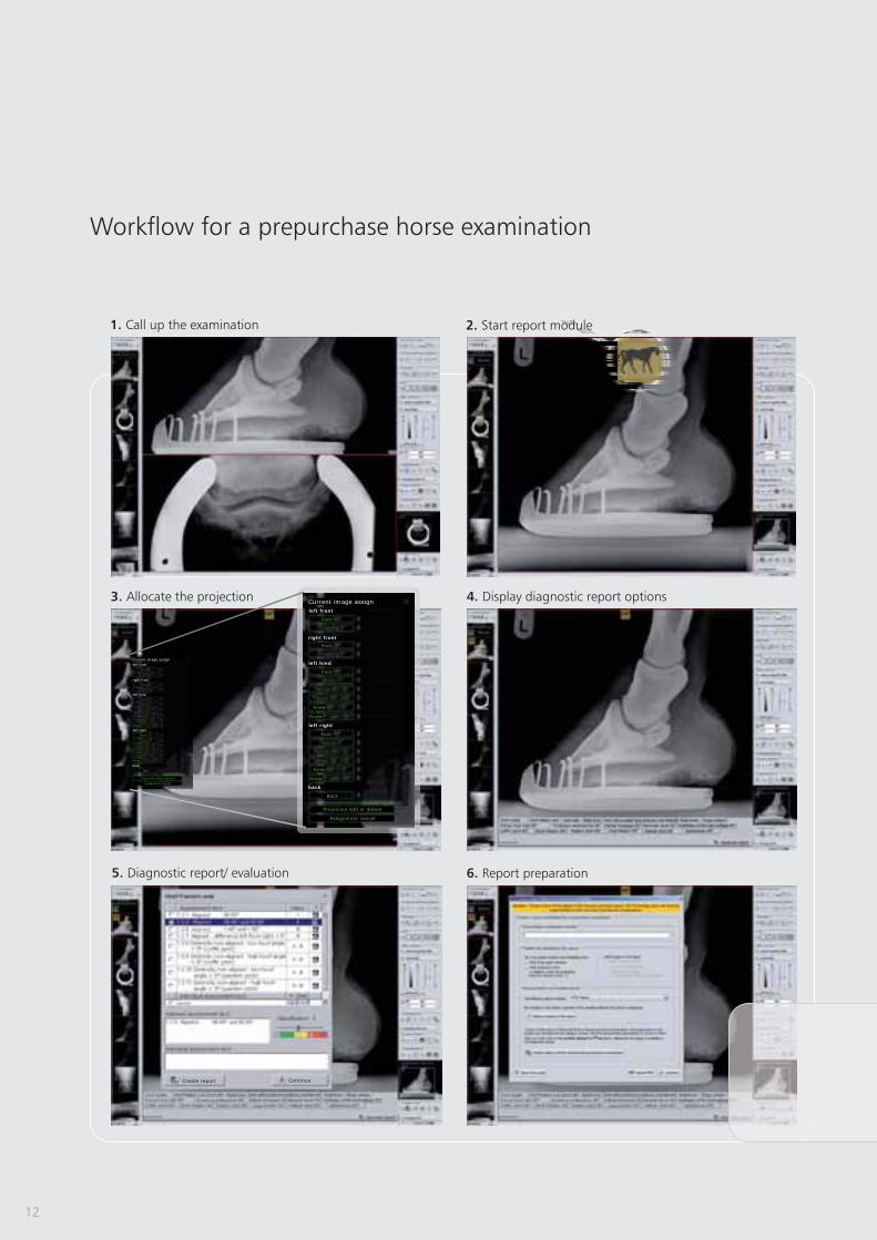
Report module for X-ray services relating to equine prepurchase
examinations
Special filter for the optimization of bones and soft parts
TPLO measuring function
TTA measuring tool
HD measuring technique for dogs
Measuring the distraction index
Buchanan‘s Vertebral Heart Score
- enables the user to plan operations
with digital prosthesis templates by one or more manufacturers
see page 18/ 19
[currently only available for Germany] - enables the quick
compilation of reports by automatically assembling X-ray images. It follows the
“X-ray guideline” by the German organisations “Gesellschaft für Pferdemedizin
e.V.” (non-profit organization for equine medicine) and “Bundestierärzte-
kammer e.V.” (Federal association of veterinarians).
see page 10/ 13
-
details of interest may be made visible by means of special filter magnifiers
(Tibial Plateau Leveling Osteotomy) - it serves to
theoretically optimize the existing slope of the tibial plateau in domestic dogs
see page 18/ 19
(Tibial Tuberosity Advancement) - the TTA measuring
technique is used to apply the translated length measurements at the
tuberositas tibiae in dogs
see page 18/ 19
- provides a
special tool to guarantee very fast and reliable determination of the Norberg
angle, including documentation
see page 18/ 19
- This measuring tool serves to
determine the displacement of the femoral head from the joint socket
of the hip joint in dogs
see page 18/ 19
- This annotation is a simple and
reliable method to determine the size of the heart - it has been desiged
specifically for cats and dogs
see page 18/ 19
dicom vetPACS®
dicom vetPACS®
features
Value
7
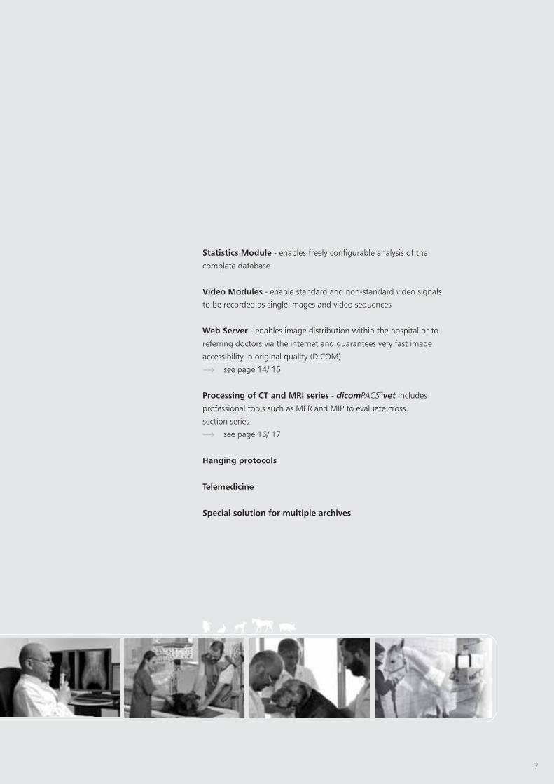
Statistics Module
Video Modules
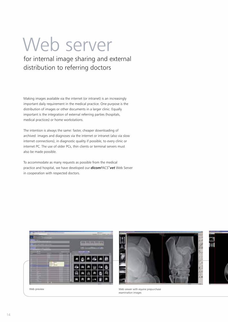
Web Server
Processing of CT and MRI series
Hanging protocols
Telemedicine
Special solution for multiple archives
- enables freely configurable analysis of the
complete database
- enable standard and non-standard video signals
to be recorded as single images and video sequences
- enables image distribution within the hospital or to
referring doctors via the internet and guarantees very fast image
accessibility in original quality (DICOM)
see page 14/ 15
- includes
professional tools such as MPR and MIP to evaluate cross
section series
see page 16/ 17
dicom vetPACS®
8
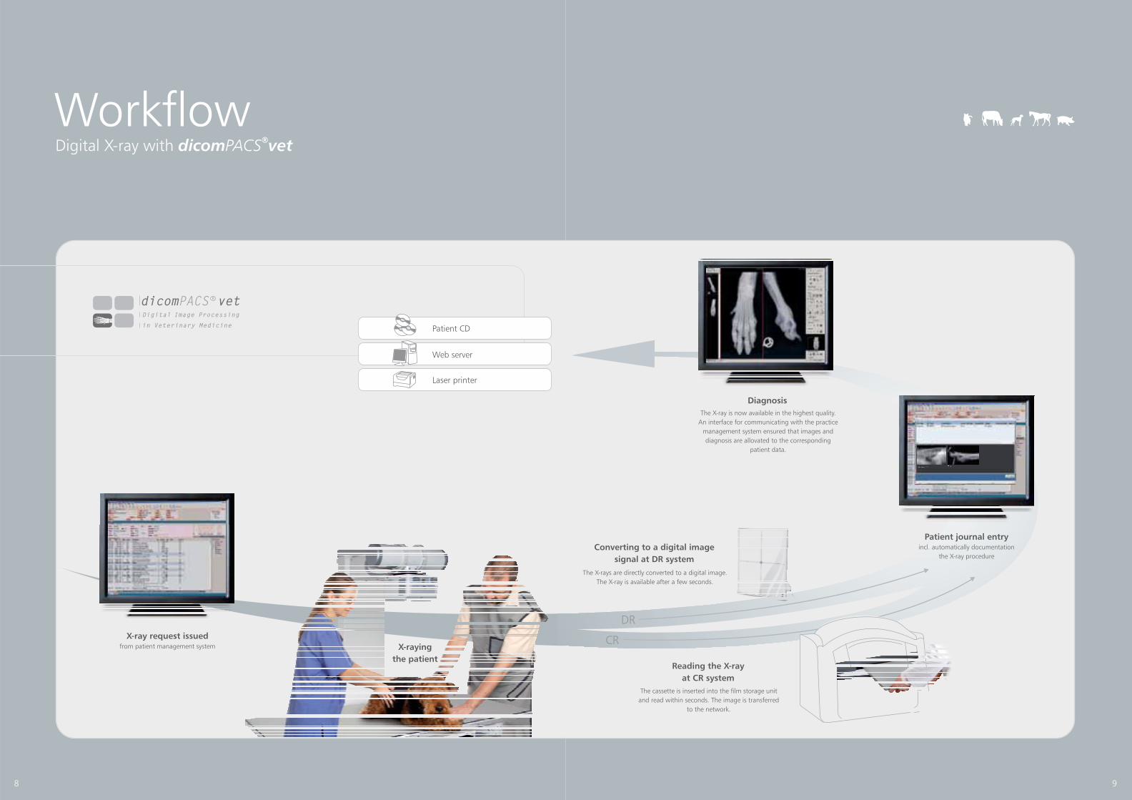
Digital X-ray with dicom vetPACS®
Workflow
X-ray request issuedfrom patient management system
Patient CD
Laser printer
Web server
X-raying
the patient
dicom vetPACSDigital Image Processing
in Veterinary Medicine
R
Reading the X-ray
at CR system
Converting to a digital image
signal at DR system
The X-rays are directly converted to a digital image.
The X-ray is available after a few seconds.
The cassette is inserted into the film storage unit
and read within seconds. The image is transferred
to the network.
DR
Diagnosis
The X-ray is now available in the highest quality.


![Title REV: TABLE OF CONTENTS Date...WCN_SW _CTRL [77] S DCARD ET_N [45] USB_P HY_PS [19] WMSS_RES ET [58] SWR _X DA A0 [20] SWR _TX _DATA1 [20] SW R _X DA A0 [20] SWR_RX_ DATA1 [20]](https://static.documents.pub/doc/80x56/61031e78cf23ee660e7b5d1f/title-rev-table-of-contents-date-wcnsw-ctrl-77-s-dcard-etn-45-usbp.jpg)


