Page 1
Accepted Manuscript
Diketoacid Chelating Ligands as Dual Inhibitors of HIV-1 Integration Process
Dominga Rogolino, Mauro Carcelli, Carlotta Compari, Laura De Luca, Stefania Ferro,Emilia Fisicaro, Gabriele Rispoli, Nouri Neamati, Zeger Debyser, Frauke Christ, AlbaChimirri
PII: S0223-5234(14)00284-0
DOI: 10.1016/j.ejmech.2014.03.070
Reference: EJMECH 6854
To appear in: European Journal of Medicinal Chemistry
Received Date: 19 December 2013
Revised Date: 7 March 2014
Accepted Date: 24 March 2014
Please cite this article as: D. Rogolino, M. Carcelli, C. Compari, L. De Luca, S. Ferro, E. Fisicaro,G. Rispoli, N. Neamati, Z. Debyser, F. Christ, A. Chimirri, Diketoacid Chelating Ligands as DualInhibitors of HIV-1 Integration Process, European Journal of Medicinal Chemistry (2014), doi: 10.1016/j.ejmech.2014.03.070.
This is a PDF file of an unedited manuscript that has been accepted for publication. As a service toour customers we are providing this early version of the manuscript. The manuscript will undergocopyediting, typesetting, and review of the resulting proof before it is published in its final form. Pleasenote that during the production process errors may be discovered which could affect the content, and alllegal disclaimers that apply to the journal pertain.
Page 2
MANUSCRIP
T
ACCEPTED
ACCEPTED MANUSCRIPT
Graphical Abstract
Page 3
MANUSCRIP
T
ACCEPTED
ACCEPTED MANUSCRIPT
1
Diketoacid Chelating Ligands as Dual Inhibitors of
HIV-1 Integration Process
Dominga Rogolino*a, Mauro Carcellia, Carlotta Comparib, Laura De Lucac, Stefania Ferroc,
Emilia Fisicarob, Gabriele Rispolia, Nouri Neamatid, Zeger Debysere, Frauke Christe and Alba
Chimirri** ,c
aDipartimento di Chimica and bDipartimento di Farmacia, Università di Parma, Parco Area delle
Scienze 17/A, I-43124 Parma, Italy, cDipartimento di Scienze del Farmaco e Prodotti per la Salute, Università di Messina, Viale
Annunziata, I-98168 Messina, Italy, dDepartment of Medicinal Chemistry, College of Pharmacy, University of Michigan, North Campus
Research Complex, 2800 Plymouth Road, Ann Arbor, MI 48109-2800 eLaboratory for Molecular virology and Gene Therapy, Division of Molecular Medicine, KU Leuven,
Kapucijnenvoer 33, 3000 Leuven, Belgium
*Corresponding author. Phone: +39 0521 905419. Email: [email protected] ** Corresponding author. Phone: +39 090 6766412. Email: [email protected] Abbreviations: 3-P, 3’-processing; AIDS, Acquired Immunodeficiency Syndrome; DKAs , α,β-
diketoacids; HAART, Highly Active Antiretroviral Therapy; HIV, Human Immunodeficiency Virus;
IN, Integrase; Lens Epithelium Derived Growth Factor, LEDGF/p75;ST, strand-transfer.
Page 4
MANUSCRIP
T
ACCEPTED
ACCEPTED MANUSCRIPT
2
Keywords: antiviral agents, HIV-1 integrase inhibitors, LEDGF/p75 protein-protein inhibitors,
magnesium complexes, dual inhibitors, diketoacid.
Abstract
HIV-1 Integrase (IN) represents a very attractive pharmacological target for the development of new
and more efficient drugs. Recently, an allosteric inhibitory approach also emerged, that targets the
interaction between IN and cellular cofactors, such as LEDGF/p75. Small molecules based on the
diketoacid pharmachophore were studied for their ability to inhibit at the same time integration and IN-
LEDGF/p75 interaction (dual inhibitors): in this study, we evaluated three indole diketoacid derivatives
and their magnesium(II) complexes for their ability to act as dual inhibitors.
Effectively, the metal complexes exhibited IN inhibition potency in low nanomolar/micromolar
concentration range; both the complexes and the free ligands are also able to inhibit the IN-LEDGF/p75
interaction at low µM values. Moreover, these magnesium compounds showed good antiviral activity,
suggesting the possibility to exploit metal coordination for the design of new antivirals.
Page 5
MANUSCRIP
T
ACCEPTED
ACCEPTED MANUSCRIPT
3
1. Introduction
Human Immunodeficiency Virus (HIV) is the etiological agent of the acquired immunodeficiency
syndrome (AIDS), which has become a major epidemic [1]. Highly Active Antiretroviral Therapy
(HAART) currently in use provides good results [2,3] but, as usual, problems related to drug toxicity
and to the emergence of drug resistant phenotypes urge for the identification of novel pharmacological
targets. In recent years, big efforts have been made to develop efficient inhibitors of HIV Integrase (IN),
the enzyme that catalyzes the integration of proviral cDNA into the host cell genome through two
different steps, 3’-processing (3-P) and strand-transfer (ST) [4-7]. By 3-P, the enzyme recesses the 3’-
terminal ends of the viral DNA to generate two CA-3’-hydroxyl ends, which are the reactive
intermediates required for the next step. IN, still bound to the 3’-processed viral DNA, translocates into
the nucleus of the infected cell, wherein the terminal 3’-OH of the viral DNA attacks the host DNA in
the ST step. IN contains a catalytic core domain that presents an amino acidic triad (the so-called
“D,D(35)E” motif), that coordinates two divalent Mg2+ cofactors [8,9]. These two ions are essential to
the catalytic process, according to the ‘two-metal-ion’ mechanism [10]. Chelation of the metal cofactors
within the active site has emerged as a successful strategy in the drug design of IN inhibitors and, in
general, in the development of inhibitors of viral enzymes containing magnesium, as HIV reverse
transcriptase, Hepatitis C polymerase and Influenza virus endonuclease [11]. A milestone in this sense
was the approval, in late 2007, of the chelating inhibitor Raltegravir (Isentress®) as the first drug
against HIV-1 IN [12-15]; other chelating IN inhibitors were recently approved or are under clinical
trials [16-20]. In recent years a great number of compounds have been studied as HIV-1 IN inhibitors
[5,21-28]. Among them, one of the most important classes is represented by the α,β-diketoacids
(DKAs) [29], that selectively inhibit the ST reaction and exhibit antiviral activity against HIV-1 infected
cells. They comprise a β-diketo moiety, an aromatic or heteroaromatic portion, and a carboxylic
functionality able to chelate divalent metal ions and therefore to block the interaction of the enzyme
with the DNA substrate. Some of us have synthesized a series of indole derivatives belonging to the
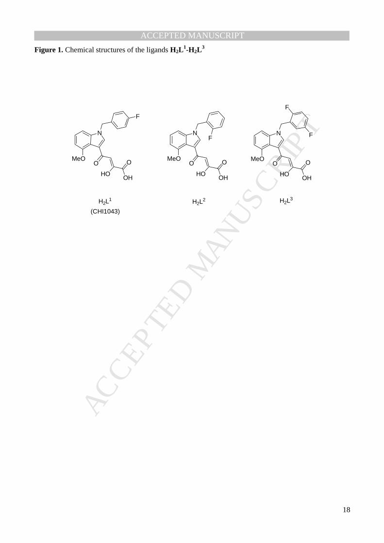
DKAs that are active IN inhibitors [30-34]; in particular H2L1 (CHI-1043, Fig. 1) presents very good
Page 6
MANUSCRIP
T
ACCEPTED
ACCEPTED MANUSCRIPT
4
activity both in enzymatic and in cellular assays with low toxicity (IC50 = 0.04 µM; EC50 = 0.59 µM; SI
= 70) [31]. Docking studies [35] highlighted that H2L1 has a binding mode similar to that observed for
other IN ST inhibitors crystallized within the active site of the enzyme [9], with the diketo acid moiety
coordinating the two metal cofactors. The diketo acid functionality chelates divalent metal ions in
solution, forming metal complexes with different stoichiometric ratios [36,37-39]. We also isolated
metal complexes with DKAs ligands, and we tested them for their ability to inhibit IN in enzymatic
assays, finding, quite surprisingly, that also some preformed complexes are active at a high nanomolar
to low micromolar range [37-39]. It is generally recognized that the study of the coordinating ability of
the DKA pharmachophore is of paramount importance, since it can lead to the design of more efficient
IN inhibitors [40].
Indole DKAs are also inhibitors of the interaction between HIV-1 IN and the cellular cofactor
LEDGF/p75 (Lens Epithelium Derived Growth Factor) [35-42]. Problems related to drug-resistant
strains observed with the use of Raltegravir has highlighted the necessity to identify molecules able to
target different steps in the integration process [43]. LEDGF/p75 is a cellular protein that has been
identified as a cellular cofactor of HIV integration and replication [44,45]. It binds HIV-1 IN via a small
IN-binding domain (LEDGFIBD) within its C-terminal region and several studies pointed out the
essential role of LEDGF/p75 in viral replication and fitness [46-51]. Therefore, the development of
protein-protein disrupting therapeutics is currently a very important pharmacological target to improve
available HAART. In particular, “dual inhibitors” able to interfere with distinct steps of the integration
process are very attractive, since a multimode mechanism of action could result in cooperative
inhibition of DNA integration and HIV-1 replication in infected cells.
It is also worth noting that data regarding the biological activity of isolated metal complexes towards
HIV IN, in particular in cellular assays, are surprisingly scarce [52-56].
With this in mind, we focused our attention on the potent IN ST inhibitor H2L1 and its analogues H2L
2
and H2L3 (Fig.1) [57] in order to isolate and characterized the corresponding magnesium complexes and
to test their ability to inhibit HIV-1 IN in enzymatic assays and to evaluate their activity against HIV-1
Page 7
MANUSCRIP
T
ACCEPTED
ACCEPTED MANUSCRIPT
5
infected cells. Finally, both the ligands and the magnesium complexes have been tested as IN-
LEDGF/p75 inhibitors.
2. Experimental
2.1 Material and methods.
All reagents of commercial quality were used without further purification. Purity of compounds was
determined by elemental analysis and verified to be ≥95% for all synthesised molecules. NMR spectra
were recorded at 27 °C on a Bruker Avance 400 FT spectrophotometer; IR spectra were obtained with a
Nicolet 5PCFT-IR spectrophotometer in the 4000-400 cm-1 range, in reflectance mode on the powder.
Elemental analyses were performed by using a Carlo Erba Model EA 1108 apparatus. Electrospray mass
spectral analyses (ESI-MS) were performed with an electrospray ionization (ESI) time-of-flight
Micromass 4LCZ spectrometer.
2.2 Synthesis.
Ligands H2L1-H2L
3 were synthesized as previously reported [31,33].
Synthesis of the complexes 1, 2, 3, general procedure: A suspension of magnesium hydroxide (0.27
mmol) in water (2 ml) was added to a methanolic solution (13 ml) of the ligand (0.27 mmol). The
reaction mixture was stirred at room temperature overnight. On concentrating the solution, a powder
was obtained, which was filtered, washed with cool water and dried on vacuum.
Mg2(L1)2·7H2O, 1. Yellow powder (45%). m.p. >350 °C. 1H NMR (CDCl3): δ=4.06 (s, 3H, OCH3), 5.32
(s, 2H, CH2), 6.77 (br,1H, Ar-H), 6.95 (br, 1H, Ar-H), 7.05 (br, 2H, Ar-H), 7.16 (br, 2H, Ar-H), 7.24
(br, 1H, Ar-H), 7.94 (s, br, 1H, =CH), 8.02 (s, br, 1H, =CH) ppm. 19F{1H}-NMR (CDCl3): δ = -113.18
ppm. IR (ATR): ν =1592, 1508 cm-1. ESI/MS (+) m/z 821.3 [M + K]+; 805.4 [M + Na]+. Anal. Calcd.
for C40H28F2Mg2N2O10⋅7H2O: C 52.83, H 4.66, N 3.08. Found: C 52.76, H 4.25 N 2.96.
Mg2(L2)2·5H2O, 2. Yellow powder (48%). m.p. >350 °C. 1H NMR (CDCl3): δ=4.05 (br, 3H, OCH3),
5.39 (br, 2H, CH2), 6.76-7.28 (m, br,7H, Ar-H), 7.93 (s, br, 1H, =CH), 8.05 (s, br, 1H, =CH) ppm.
19F{1H}-NMR (CDCl3): δ= -117.6 ppm. IR (ATR): ν=1592, 1501 cm-1. ESI/MS (+) m/z 805.4 [M +
Page 8
MANUSCRIP
T
ACCEPTED
ACCEPTED MANUSCRIPT
6
Na]+. Anal. Calcd. for C40H28F2Mg2N2O10⋅5H2O: C 55.01, H 4.39, N 3.21. Found: C 54.94, H 4.28, N
3.07.
Mg2(L3)2·7H2O, 3. Yellow powder (45%). m.p. >350 °C. 1H NMR (CDCl3): δ=4.01 (br, 3H, OCH3),
5.33 (br, 2H, CH2), 6.52-7.21 (m, br,7H, Ar-H), 7.92 (s, br, 1H, =CH), 8.01 (s, br, 1H, =CH) ppm.
19F{1H}-NMR (CDCl3): δ = -123.6, -117.0 ppm. IR (ATR): ν=1594, 1493 cm-1. ESI/MS (+) m/z 841.3
[M + Na]+. Anal. Calcd. for C40H26F4Mg2N2O10⋅7H2O: C 50.82, H 4.26, N 2.96. Found: C 51.07, H
3.71, N 2.84.
2.3 Potentiometric measurements
Equilibrium constants for protonation and complexation reactions were determined by means of
potentiometric measurements (pH = -log[H+]), carried out in methanol/water=9:1 v/v solution at ionic
strength 0.1 M KCl and 25 ± 0.1 °C, in the pH range 2.5–11 under N2. Temperature was controlled to ±
0.1 °C by using a thermostatic circulating water bath (ISCO GTR 2000 IIx). Appropriate aliquots of
ligand solution, prepared by weight, were titrated with standard KOH (solvent: methanol/water=9:1 v/v,
I= 0.1M KCl) with and without metal ions. Constant-speed magnetic stirring was applied throughout.
Freshly boiled methanol and bidistilled water, kept under N2, were used throughout. The experimental
procedure in order to reach very high accuracy in the determination of the equilibrium constants in this
mixed solvent has been described in detail elsewhere [58]. The protonation constants of H2L1 were
obtained by titrating 20-50 ml samples of the ligand (2·10-3 -7·10-3 M). For the complex formation
constants, the metal ion stock solution was prepared from MgCl2⋅6H2O (Carlo Erba) and the
concentration was determined by using EDTA as titrant and Eriochrome black T as indicator. The
titrations were performed with different ligand/metal ratios (1 up to 4). At least two measurements
(about 60 experimental points in each) were performed for each system. Potentiometric titrations were
carried out by a fully automated apparatus equipped with a CRISON GLP 21-22 digital voltmeter
(resolution 0.1 mV) and a 5 ml Metrohm Dosimat 655 autoburette, both controlled by a home-made
software, written in BASIC, working on an IBM computer. The electrodic chain (Crison 5250 glass
Page 9
MANUSCRIP
T
ACCEPTED
ACCEPTED MANUSCRIPT
7
electrode and KCl 0.1M in methanol/water=9:1 v/v calomel electrode, Radiometer 401) was calibrated
in terms of [H+] by means of a strong acid-strong base titration, by the Gran’s method [59], allowing the
determination of the standard potential, Eo (366.7± 0.1 mV) and of the ionic product of water, Kw (pKw =
14.38 ± 0.01) in the experimental conditions used. The software HYPERQUAD [60] was used to
evaluate the protonation and complexation constants from e.m.f. data.
2.4 Biological Materials, Chemicals, and Enzymes.
All compounds were dissolved in DMSO and the stock solutions were stored at –20 °C. The γ[32P]-ATP
was purchased from PerkinElmer. The expression system for wild-type IN was a generous gift of Dr.
Robert Craigie, Laboratory of Molecular Biology, NIDDK, NIH, Bethesda, MD.
2.5 Preparation of Oligonucleotide Substrates.
The oligonucleotides 21top, 5'-GTGTGGAAAATCTCTAGCAGT-3' and 21bot, 5'-
ACTGCTAGAGATTTTCCACAC-3' were purchased from Norris Cancer Center Core Facility
(University of Southern California) and purified by UV shadowing on polyacrylamide gel. To analyze
the extent of 3'-P and ST using 5'-end labeled substrates, 21top was 5'-end labeled using T4
polynucleotide kinase (Epicentre, Madison, WI) and γ [32P]-ATP (Amersham Biosciences or ICN). The
kinase was heat-inactivated and 21bot was added in 1.5-molar excess. The mixture was heated at 95 °C,
allowed to cool slowly to room temperature, and run through a spin 25 mini-column (USA Scientific) to
separate annealed double-stranded oligonucleotide from unincorporated material.
2.6 Integrase Assays.
To determine the extent of 3’-P and ST, wild-type IN was preincubated at a final concentration of 200
nM with the inhibitor in reaction buffer (50 mM NaCl, 1 mM 4-(2-hydroxyethyl)-1-piperazine
ethanesulfonic acid, pH 7.5, 50 µM EDTA, 50 µM dithiothreitol, 10% glycerol (w/v), 7.5 mM MnCl2,
0.1 mg/ml bovine serum albumin, 10 mM 2-mercaptoethanol, 10% DMSO, and 25 mM 3-(N-
morpholino)propanesulfonic acid, pH 7.2) at 30 °C for 30 min. Then, 20 nM of the 5'-end 32P-labeled
linear oligonucleotide substrate was added, and incubation was continued for an additional 1h.
Page 10
MANUSCRIP
T
ACCEPTED
ACCEPTED MANUSCRIPT
8
Reactions were quenched by the addition of an equal volume (16 µL) of loading dye (98% deionized
formamide, 10 mM EDTA, 0.025% xylene cyanol and 0.025% bromophenol blue). An aliquot (5 µL)
was electrophoresed on a denaturing 20% polyacrylamide gel (0.09 M tris-borate pH 8.3, 2 mM EDTA,
20% acrylamide, 8M urea).
Gels were dried, exposed in a PhosphorImager cassette, analyzed using a Typhoon 8610 Variable Mode
Imager (Amersham Biosciences) and quantitated using ImageQuant 5.2. Percent inhibition (% I) was
calculated using the following equation:
% I = 100 X [1 - (D - C)/(N - C)]
where C, N, and D are the fractions of 21-mer substrate converted to 19-mer (3’-proc product) or ST
products for DNA alone, DNA plus IN, and DNA plus IN plus drug, respectively. The IC50 values were
determined by plotting the logarithm of drug concentration versus percent inhibition to obtain
concentration that produced 50% inhibition.
2.7 LEDGF/p75-IN AlphaScreen proximity luminescent assay.
The AlphaScreen assay was performed according to the manufacturer’s protocol (Perkin Elmer,
Waltham, MA). Reactions were performed in 25 µl final volume in 384-well Optiwell™ microtiter
plates (Perkin Elmer). The reaction buffer contained 25 mM Tris–HCl (pH 7.4), 150 mM NaCl, 1 mM
MgCl2, 0.01% (v/v) Tween-20 and 0.1% (w/v) bovine serum albumin. Wild type IN with a His6-tag
(300 nM final concentration) was pre-incubated with each inhibitor for 30 min at 4 °C. Next, 100 nM
Flag peptide tagged-LEDGF/p75 was added to the reaction and incubated for an additional hour at 4 °C.
Subsequently 5 µl of Ni-chelate-coated donor beads and 5 µl anti-Flag acceptor beads were added to a
final concentration of 20 µg/ml for each beads. Proteins and beads were incubated for 1 h at 30 °C in
order to allow association to occur. Exposure of the reaction to direct light was omitted as much as
possible and the emission of light from the acceptor beads was measured in the EnVision plate reader
(Perkin Elmer).
2.8 In vitro anti-HIV and drug susceptibility assays.
Page 11
MANUSCRIP
T
ACCEPTED
ACCEPTED MANUSCRIPT
9
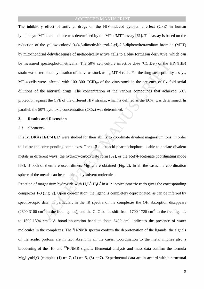
The inhibitory effect of antiviral drugs on the HIV-induced cytopathic effect (CPE) in human
lymphocyte MT-4 cell culture was determined by the MT-4/MTT-assay [61]. This assay is based on the
reduction of the yellow colored 3-(4,5-dimethylthiazol-2-yl)-2,5-diphenyltetrazolium bromide (MTT)
by mitochondrial dehydrogenase of metabolically active cells to a blue formazan derivative, which can
be measured spectrophotometrically. The 50% cell culture infective dose (CCID50) of the HIV(IIIB)
strain was determined by titration of the virus stock using MT-4 cells. For the drug-susceptibility assays,
MT-4 cells were infected with 100–300 CCID50 of the virus stock in the presence of fivefold serial
dilutions of the antiviral drugs. The concentration of the various compounds that achieved 50%
protection against the CPE of the different HIV strains, which is defined as the EC50, was determined. In
parallel, the 50% cytotoxic concentration (CC50) was determined.
3. Results and Discussion
3.1 Chemistry.
Firstly, DKAs H2L1-H2L
3 were studied for their ability to coordinate divalent magnesium ions, in order
to isolate the corresponding complexes. The α,β-diketoacid pharmachophore is able to chelate divalent
metals in different ways: the hydroxy-carboxylate form [62], or the acetyl-acetonate coordinating mode
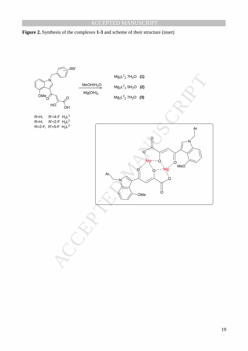
[63]. If both of them are used, dimers Mg2L2 are obtained (Fig. 2). In all the cases the coordination
sphere of the metals can be completed by solvent molecules.
Reaction of magnesium hydroxide with H2L1-H2L
3 in a 1:1 stoichiometric ratio gives the corresponding
complexes 1-3 (Fig. 2). Upon coordination, the ligand is completely deprotonated, as can be inferred by
spectroscopic data. In particular, in the IR spectra of the complexes the OH absorption disappears
(2800-3100 cm-1 in the free ligands), and the C=O bands shift from 1700-1720 cm-1 in the free ligands
to 1592-1594 cm-1. A broad absorption band at about 3400 cm-1 indicates the presence of water
molecules in the complexes. The 1H-NMR spectra confirm the deprotonation of the ligands: the signals
of the acidic protons are in fact absent in all the cases. Coordination to the metal implies also a
broadening of the 1H- and 19F-NMR signals. Elemental analysis and mass data confirm the formula
Mg2L2·nH2O (complex (1) n= 7, (2) n= 5, (3) n=7). Experimental data are in accord with a structural
Page 12
MANUSCRIP
T
ACCEPTED
ACCEPTED MANUSCRIPT
10
hypothesis that considers the acetyl, the hydroxy and the carboxylate groups coordinated to the metal
(Figure 2, inset), as we already observed with analogous DKA complexes [37-39]. In order to gain
insight on the complexing ability of these ligands towards Mg(II) ions in solution and to confirm the
proposed stoichiometry, potentiometric measurements have been carried out with ligand H2L1.
To avoid solubility problems, all the titrations were carried out in methanol/water 9/1 (v/v) and ionic
strength 0.1M KCl, where both the reactants and the complex species are soluble. The software
Hyperquad has been employed for the refinement of the trial equilibrium constants. The best fit of the
experimental titration curves was obtained by the set of species reported in Table 1.
H2L1 is a diprotic acid with protonation constants log β1= 11.75(0.01) and log β2= 16.34 (0.02). The
values are in good agreement with the protonation constant of the DKA analogue (2Z)-4-[1-(4-
fluorobenzyl)-1H-pyrrol-2-yl]-2-hydroxy-4-oxobut-2-enoic acid (log β1= 11.66(0.03) and log β2= 15.91
(0.06)), we have studied before [37]. The stoichiometry of the magnesium(II) complexes with H2L1 and
their formation constants have been determined. The best statistical parameters for the fit are obtained
by using the set of species MgL22-, Mg2L2, and, in alkaline environment, Mg2L2(OH) (Table 1). In
particular, at physiological pH the species Mg2L2 is predominant. The species M2L2+ [37] was always
rejected by the software. This species is very sensitive to the steric hindrance of the ligand [37],
therefore probably the presence of a bulky 4-methoxy indol group disfavours the simultaneous
interaction of two metal ions with the same ligand molecule. The distribution diagram for the M/L=1/4
is shown in Figure 3.
The formation constants of the monomeric species, ML and of the dimeric one, M2L2 cannot be refined
together, probably because of their high mathematical correlation. By means of potentiometry only, we
cannot surely reject the monomeric species, because the model with ML instead of M2L2 is only a little
worse. However, the model here reported shows better statistical values and it is in accord with the
chemical data (in particular, mass spectra) and with molecular modeling calculations with analogous
diketoacid ligands [36].
3.2 Biological activity.
Page 13
MANUSCRIP
T
ACCEPTED
ACCEPTED MANUSCRIPT
11
Indole DKA derivatives have been identified as good prototypes of small molecules able to inhibit two
different stages of the integration process: the IN ST and the IN-LEDGF/75 interaction [35, 64]. H2L1-
H2L3 were tested for their ability to inhibit 3’-P and ST catalytic activities by employing purified
enzyme (Table 2). They are effectively selective IN inhibitors, with good Selectivity Index for ST (SI,
Table 2).
H2L1-H2L
3 differ for the position or the numbers of the fluorine atoms in the aromatic ring. This
structural modification could be important, since it is known that the fluorophenyl ring is involved in the
displacement of the 3’-viral adenosine from the active site, leading to deactivation of the intasome
[9,65]; however, in this case, the different benzyl substituents do not influence significantly the activity.
Since some metal complexes have shown a good activity against IN in enzymatic assays [37-39,66,67],
we tried to verify if also the magnesium complexes of H2L1 - H2L
3 are active. 1-3 were tested for their
ability to inhibit 3’-P and ST catalytic activities by employing purified enzyme (Table 2). All the
compounds showed inhibition potency in low nanomolar/micromolar concentration range, but their
specificity for ST diminished; in fact, their SI is similar (compound 2) or worse than that of the
corresponding ligand.
Both the complexes and the free ligands are also able to inhibit the IN-LEDGF/p75 interaction at low
µM values (Table 2). Modification of the position of the fluorine substituent in the aromatic ring
resulted, in this case, in a variation of the activity, with H2L2 and H2L
3 five-fold more potent than H2L1.
The interactions that the metal complexes engage at the IN-LEDGF interface are quite different from
that of the free ligands: upon complexation, for instance, substantial modification of the hydrogen
bonding donor-acceptor capability occurs. In this sense, it is quite normal to observe IC50 values that are
different in H2L1 - H2L
3 and in their complexes. Anyway, these are the first data about metal complexes
able to inhibit the IN-LEDGF/p75 interaction and any generalization risks to be only speculative.
Finally, the ligands and the metal complexes were tested for their antiviral activity in HIV-infected MT-
4 cells, revealing good capability of inhibiting viral infection (Table 2). The EC50 values, in fact, ranges
from 0.14 to 0.53 µM; the magnesium complexes (1) and (2) substantially retain the activity of the
Page 14
MANUSCRIP
T
ACCEPTED
ACCEPTED MANUSCRIPT
12
corresponding free ligands, while (3) has a better EC50 than H2L3 (0.188 vs 0.53). Unfortunately, the
metal complexes are more cytotoxic of the corresponding free ligands, thus resulting in a worse
therapeutic index.
4. Conclusions
Coordination of metal cofactors represents a key aspect in the development of new and efficient
antivirals and it is at the basis of on-going research efforts [11,68]. Here we present the synthesis and
characterisation of magnesium complexes of HIV-1 IN inhibitors based on the DKA pharmachophore,
confirming the capability of these ligands to effectively coordinate the metals. In a scenario of few
available data on the activity of isolated metal complexes [52-56], we showed that the preformed
magnesium complexes of indole DKA inhibitors have both enzymatic and antiviral activity on infected
cells. Moreover, 1-3 revealed significant IN-LEDGF/p75 interaction inhibition, behaving as dual
inhibitors. These are, to the best of our knowledge, the first data regarding the activity of metal
complexes as allosteric inhibitors. The capability of magnesium coordination compounds to block the
IN-LEDGF/p75 protein-protein interactions, behaving as dual inhibitors with encouraging antiviral
activity, offers a promising approach to prevent viral replication and underlines the possibility to use
coordination chemistry to obtain unconventional scaffold to target enzymes.
Acknowledgements
The authors thank the “Centro Interdipartimentale Misure Giuseppe Casnati” of the University of Parma
for facilities. D.R, M.C. and G.R. thank Italian Ministero dell’Istruzione, dell’Università e della Ricerca
for financial support (PRIN 2010, 2010W2KM5L_003).
Page 15
MANUSCRIP
T
ACCEPTED
ACCEPTED MANUSCRIPT
13
References
[1] Joint United Nations Programme on HIV/AIDS and World Health Organization (UNAIDS).
AIDS Epidemic Update: December 2007.
[2] E. De Clercq, J. Med. Chem., 48 (2005) 1297-1313.
[3] S. Abel, D. J. Back, M. Vourvahis, Antivir. Ther., 14 (2009) 607-618.
[4] Y. Pommier, A. A. Johnson, C. Marchand, Nat. Rev. Drug Discov., 4 (2005) 236-248.
[5] N. Neamati in: HIV-1 Integrase: Mechanism of Action and Inhibitor Design, John Wiley & Sons
(Eds.), 2011.
[6] R. Dayam, L. Q. Al-Mawsawi, N. Neamati, Drugs R&D, 8 (2007) 155-168.
[7] B.A. Johns, A.C. Svolto, Expert. Opin. Ther. Patents, 18 (2008) 1225-1237.
[8] J. Kulkosky, K. S. Jones, R. A. Katz, J. P. Mack, A. M. Skalka, Mol. Cell Biol., 12 (1992) 2331-
2338.
[9] S. Hare, S. S. Gupta, E. Valkov, A. Engelman, P. Cherepanov, Nature (2010) 1-6.
[10] L. S Beese, T. A. Steitz, EMBO J., 10 (1991) 25-33.
[11] D. Rogolino, M. Carcelli, M. Sechi, N. Neamati, Coord. Chem. Rev., 256 (2012) 3063-3086.
[12] MK-0518 meeting transcript. Department of Health and Human Services; Food and Drug
Administration; Center for Drug Evaluation and Research; Antiviral Drugs Advisory Committee; (2007)
September 5. Available from: www.fda.gov/ohrms/dockets/ac/cder07.htm#AntiviralDrugs.
[13] C. Garrido, V. Soriano, C. de Mendoza, J. Antimicr. Chemother., 65 (2010) 218-223.
[14] M. Rowley, Progress Med. Chem., 46 (2008) 1-28.
[15] J. Cocohoba, B. Dong, J. Clinical Ther., 30 (2008) 1747-1765.
[16] FDA approves new drug to treat HIV infection; 2013 August 12. Available from:
http://www.fda.gov/NewsEvents/Newsroom/PressAnnouncements/ucm364744.htm.
[17] K. Shimura, E.N. Kodama, Antiv. Chem. Chemother., 20 (2009) 79-85.
Page 16
MANUSCRIP
T
ACCEPTED
ACCEPTED MANUSCRIPT
14
[18] M. Kobayashi, T. Yoshinaga, T. Seki, C. Wakasa-Morimoto, K. W. Brown, R. Ferris, S. A.
Foster, R. J. Hazen, S. Miki, A. Suyama-Kagitani, S. Kawauchi-Miki, T. Taishi, T. Kawasuji, B. A.
Johns, M. R. Underwood, E. P. Garvey, A. Sato, T. Fujiwara, Antimicrob. Agents Chemother., 55
(2011) 813-821.
[19] L. Vandekerckhove, Curr. Opin. Investig. Drugs, 11 (2010) 203-212.
[20] P. E. Sax, E. Dejesus, A. Mills, A. Zolopa, C. Cohen, D. Wohl, J. E. Gallant, H. C. Liu, L.
Zhong, K. Yale, K. White, B. P. Kearney, J. Szwarcberg, E. Quirk, A. K. Cheng, The Lancet, 379
(9835) (2012) 2439–2448
[21] P. Cotelle, Recent Patents Anti-Infect. Drug Disc., 1 (2006) 1-15.
[22] R. Dayam, J. Deng, N. Neamati, Med. Res. Rev., 26 (2006) 271-309.
[23] R. Dayam, R. Gundla, L. Q. Al-Mawsawi, N. Neamati, HIV Med. Res. Rev., 28 (2008) 118-154.
[24] E.E. Boros, C. E. Edwards, S. A. Foster, M. Fuji, T. Fujiwara, E. P. Garvey, P. L. Golden, R. J.
Hazen, J. L. Jeffrey, B. A. Johns, T. Kawasuji, R. Kiyama, C. S. Koble, N. Kurose, W. H. Miller, A. L.
Mote, H. Murai, A. Sato, J. B. Thompson, M. C. Woodward, T. Yoshinaga, J. Med. Chem., 52 (2009)
2754-2761.
[25] M.B. Plewe, S. L. Butler, K. R. Dress, Q. Hu, T. W. Johnson, J. E. Kuehler, A. Kuki, H. Lam,
W. Liu, D. Nowlin, Q. Peng, S. V. Rahavendran, S. P. Tanis, K. T. Tran, H. Wang, A. Yang, J. Zhang,
J. Med. Chem., 52 (2009) 7211-7219.
[26] G. Le, N. Vandegraaff, D.I. Rhodes, E.D. Jones, J.A.V. Coates, N. Thienthong, L.J. Winfield, L.
Lu, X. Li, C. Yu, X. Feng, J.J. Deadman, Bioorg. Med. Chem. Lett., 20 (2010) 5909-5912.
[27] J. Tang, K. Maddali, M. Metifiot, Y.Y. Sham, R. Vince, Y.Z. Wang, J. Med. Chem., 54 (2011)
2282-2292.
[28] R. Kiyama, T. Kawasuji, PCT Int. Appl., (2001) WO-01/95905.
[29] G. C. G. Pais, T. R. Burke, Drugs Fut., 27 (2002) 1101-1111.
[30] M.L. Barreca, S. Ferro, A. Rao, L. De Luca, M. Zappala, A.M. Monforte, Z. Debyser, M.
Witvrouw, A. Chimirri, J. Med. Chem., 48 (2005) 7084–7088.
[31] L. De Luca, M.L. Barreca, S. Ferro, N. Iraci, M. Michiels, F. Christ, Z. Debyser, M. Witvrouw,
A. Chimirri, Bioorg. Med. Chem. Lett., 18 (2008) 2891–2895.
Page 17
MANUSCRIP
T
ACCEPTED
ACCEPTED MANUSCRIPT
15
[32] L. De Luca, S. De Grazia, S. Ferro, R. Gitto, F. Christ, Z. Debyser, A. Chimirri, Eur. J. Med.
Chem., 46 (2011) 756–764.
[33] S. Ferro, L. De Luca, M.L. Barreca, N. Iraci, S. De Grazia, F. Christ, M. Witvrouw, Z. Debyser,
A. Chimirri, J. Med. Chem., 52 (2009) 569–573.
[34] S. Ferro, L. De Luca, M.L. Barreca, S. De Grazia, F. Christ, Z. Debyser, A. Chimirri, Bioorg.
Med. Chem., 18 (2010) 5510–5518.
[35] L. De Luca, R. Gitto, F. Christ, S. Ferro, S. De Grazia, F. Morreale, Z. Debyser, A. Chimirri,
Antiviral Research, 92 (2011) 201-107.
[36] C. Maurin, F. Bailly, E. Buisine, H. Vezin, G. Mbemba, J. F. Mouscadet, P. Cotelle, J. Med.
Chem., 47 (2004) 5583-5596.
[37] M. Sechi, A. Bacchi, M. Carcelli, C. Compari, E. Duce, E. Fisicaro, D. Rogolino, P. Gates, M.
Derudas, L. Q. Al-Mawsawi, N. Neamati, J. Med. Chem., 49 (2006) 4248-4260.
[38] A. Bacchi, M. Biemmi, M. Carcelli, F. Carta, C. Compari, E. Fisicaro, D. Rogolino, M. Sechi,
M. Sippel, C. Sotriffer, T.W. Sanchez, N. Neamati, J. Med. Chem., 51 (2008) 7253-7264.
[39] A. Bacchi, M. Carcelli, C. Compari, E. Fisicaro, N. Pala, G. Rispoli, D. Rogolino, T. W.
Sanchez, M. Sechi, N. Neamati, Mol. Pharmaceutics, 8 (2011) 507-519.
[40] A. Agrawa, J. DeSoto, J.L. Fullagar, K. Maddal, S. Rostami, D.D. Richman, Y. Pommier, S.M.
Cohen, Proc. Natl. Acad. Sci.U.S.A., (2012) 2251-2256.
[41] L. De Luca, M.L. Barreca, S. Ferro, F. Christ, N. Iraci, R. Gitto, A.M. Monforte, Z. Debyser, A.
Chimirri, Chem. Med. Chem., 4 (2009) 1311–1316.
[42] L. De Luca, S. Ferro, R. Gitto, M.L. Barreca, S. Agnello, F. Christ, Z. Debyser, A. Chimirri
Bioorg. Med. Chem., 18 (2010) 7515-21.
[43] F. Christ, Z. Debyser, Virology, 435 (2013) 102-109.
[44] K. Busschots, A. Voet, M. De Maeyer, J.C. Rain, S. Emiliani, R. Benarous, L. Desender, Z.
Debyser, F. Christ, J. Mol. Biol., 365 (2007) 1480–1492.
[45] W.C. Greene, Z. Debyser, Y. Ikeda, E.O. Freed, E. Stephens, W. Yonemoto, R.W. Buckheit,
J.A. Esté, T. Cihlar, Antiviral Res., 80 (2008) 251–265.
Page 18
MANUSCRIP
T
ACCEPTED
ACCEPTED MANUSCRIPT
16
[46] P. Cherepanov, A. L. Ambrosio, S. Rahman, T. Ellenberger, A. Engelman, Proc. Natl. Acad. Sci.
U.S.A., (2005), 102, 17308.
[47] S. Hare, M. C. Shun, S. S. Gupta, E. Valkov, A. Engelman, P. Cherepanov, PLoS Pathog., 5
(2009) e1000259.
[48] L. Vandekerckhove, F. Christ, B. Van Maele, J. De Rijck, R. Gijsbers, C. Van den Haute, M.
Witvrouw, Z. Debyser, J. Virol., 80 (2006) 1886-1896.
[49] A. Engelman, P. Cherepanov, PLoS Pathog., 4 (2008) e1000046.
[50] A. Ciuffi, M. Llano, E. Poeschla, C. Hoffmann, J. Leipzig, P. Shinn, J. R. Ecker, F. Bushman,
Nat. Med., 11 (2005) 1287.
[51] K. M. Tsutsui, K. Sano, O. Hosoya, T. Miyamoto, K. Tsutsui, Nucl. Acids Res., 39 (2011) 5067.
[52] G. Pelosi, F. Bisceglie, F. Bignami, P. Ronzi, P. Schiavone, M.C. Re, C. Casoli, E. Pilotti, J.
Med. Chem., 53 (2010) 8765-8769.
[53] S.A. Galal, A.S.A. El-All, K.H. Hegab, A.A. Magd-El-Din, N.S. Youssef, H. I. El Diwani, Eur.
J. Med. Chem., 45 (2010) 3035-3046.
[54] S.G. Gallego, J.S. Rodríguez, J.L. Jiménez, M. Cangiotti, M.F. Ottaviani, M.A. Muñoz-
Fernández, R. Gómez, F. J. De la Mata, Dalton Trans., 41 (2012) 6488-6499.
[55] S.G. Gallego, M.J. Serramía, E. Arnaiz, L. Díaz, M.A. Muñoz-Fernández, P. Gómez-Sal, M.F.
Ottaviani, R. Gómez, F. J. de la Mata, Eur. J. Inorg. Chem. (2011) 1657-1665.
[56] N.V. Loginova, T.V. Koval’chuk, G. I. Polozov, N. P. Osipovich, P. G. Rytik, I. I. Kucherov, A.
A. Chernyavskaya, V. L. Sorokin, O. I. Shadyro, Eur. J. Med. Chem., 43 (2008) 1536-1542.
[57] S. Ferro, L. De Luca, M.L. Barreca, N. Iraci, S. De Grazia, F. Christ, M. Witvrouw, Z. Debyser,
A. Chimirri, J. Med. Chem., 522 (2009) 569-573.
[58] E. Fisicaro, A. Braibanti, Talanta, 10 (1988) 769.
[59] G. Gran, Analyst, 77 (1952) 661.
[60] P. Gans, A. Sabatini, A. Vacca, Talanta, 43 (1996) 1739.
[61] R. Pauwels, J. Balzarini, M. Baba, R. Snoeck, D. Schols, P. Herdewijn, J. Desmyter, E. De
Clercq, J. Virol. Methods, 20 (1988) 309–321.
Page 19
MANUSCRIP
T
ACCEPTED
ACCEPTED MANUSCRIPT
17
[62] B. M. Gatehouse, M.J. O’Connor, Acta Cryst., B32 (1976) 3145-3148.
[63] H. Kawai, Y. Kitano, M. Mutoh, G. Hata, Chem. Pharm Bull., 41 (1993) 357-361.
[64] L. De Luca, F. Morreale, A. Chimirri. Chem. Inform. Mod., 52 (2012) 3245-3254.
[65] S. Hare, S.J. Smith, M. Metifiot, A. Jaxa-Chamiec, Y. Pommier, S.H. Hughes, P. Cherepanov,
Mol. Pharmac., 80 (2011) 565-572.
[66] A. Bacchi, M. Carcelli, C. Compari, E. Fisicaro, N. Pala, G. Rispoli, D. Rogolino, T. W.
Sanchez, M. Sechi, V. Sinisi, N. Neamati, J. Med Chem., 54 (2011) 8407-8420.
[67] M. Carcelli, A. Bacchi, P. Pelagatti, G. Rispoli, D. Rogolino, T. W. Sanchez, M. Sechi, N.
Neamati, J. Inorg. Biochem., 118 (2013) 74-82.
[68] E. Meggers, Chem. Commun. (2009) 1001-1010.
Page 20
MANUSCRIP
T
ACCEPTED
ACCEPTED MANUSCRIPT
18
Figure 1. Chemical structures of the ligands H2L1-H2L
3
N
O
HOOH
OMeO
F
F
(CHI1043)
N
O
HOOH
OMeO
N
O
HOOH
OMeO
F
F
H2L1 H2L2 H2L3
Page 21
MANUSCRIP
T
ACCEPTED
ACCEPTED MANUSCRIPT
19
Figure 2. Synthesis of the complexes 1-3 and scheme of their structure (inset)
N
O
HOOH
OOMe
MeOH/H2O
Mg2L12 7H2O (1)
RR'
R=H, R'=4-F H2L1
R=H, R'=2-F H2L2
R=2-F, R'=5-F H2L3
O O
O
O
OO
O
Mg
Mg
O
N
Ar
OMe
N
Ar
MeO
Mg(OH)2
Mg2L22 5H2O (2)
Mg2L32 7H2O (3)
Page 22
MANUSCRIP
T
ACCEPTED
ACCEPTED MANUSCRIPT
20
Figure 3. Distribution diagram for the system under investigation at L:M=4:1 (the concentration of the
ligand H2L1 is 4 mM).
4 6 8 10 12pH
0
20
40
60
80
100%
form
atio
n re
lativ
e to
M
Mg
M2L2 ML2
M2L2H-1
Page 23
MANUSCRIP
T
ACCEPTED
ACCEPTED MANUSCRIPT
21
Table 1. Logarithms of formation constants (βpqr = [MpLqHr]/ [M] p [L] q [H] r) in methanol/water=9:1
v/v, I = 0.1 M KCl at 25 °C for the ligand under study with Mg(II). Standard deviations are given in
parentheses. Charges are omitted for simplicity.
p
q
r
Log βpqr
M + 2L ⇌ ML2 1 2 0 11.56(0.15) 2M + 2 L ⇌ M2L2 2 2 0 17.09(0.20) 2M + 2L + OH ⇌ M2L2 (OH) 2 2 -1 6.81(0.63) L + H ⇌ LH 0 1 1 11.75 (0.01) L + 2H ⇌ LH2 0 1 2 16.34 (0.02)
Page 24
MANUSCRIP
T
ACCEPTED
ACCEPTED MANUSCRIPT
22
Table 2. Biological evaluation of the ligands H2L1-H2L
3 and of the corresponding magnesium
complexes 1-3
Compd. IN enzymatic activity IC50
(µM)
Selectivity
Index
LEDGF/p75-IN
IC50 (µM)
Activity in MT-4 cells
3’-P ST SIa HIV-1
EC50 (µM)b
Cytotoxicity
CC50 (µM)c]
TId
H2L1 2.10 0.04 52.5 14 0.238 47.5±0.5 200
H2L2 1.80 0.06 30.0 0.14 0.141 33.5±5.5 238
H2L3 2.00 0.03 66.7 0.53 0.534 24±5 45
1 1.00 0.08 12.5 4.80 0.310 24.5±2.5 79
2 0.94 0.02 47.0 8.50 0.165 15.5±0.5 94
3 0.85 0.05 17.0 17.00 0.188 15.00±3.0 80 aSelectivity Index. bEffective concentration required to reduce HIV-1-induced cytopathic effect by 50%
in MT-4 cells. cCytotoxic concentration to reduce MT-4 cell viability by 50%. dTherapeutic index:
CC50/EC50.
Page 25
MANUSCRIP
T
ACCEPTED
ACCEPTED MANUSCRIPTHighlights
• HIV-1 Integrase is an attractive target for the development of new antivirals
• Dual inhibitors can inhibit integration and interaction with cellular cofactors
• Diketoacid derivatives and their Mg(II) complexes were evaluated as dual inhibitors
• Both the complexes and the free ligands inhibit the IN-LEDGF/p75 interaction
• Magnesium compounds showed good antiviral activity