isoform, suggesting a potential role of this receptor in the development of the lesion. Although KSHV infection is a necessary element in KS pathogenesis, other co-factors are certainly involved. Deregulated expressions of inflammatory cytokines, growth factors and related receptors have been considered to play a role in inducing either the initial localization of the lesion and/or its progression [3]. Further studies are needed to analyze whether FGFR2-IIIc up-regulation varies in the history of the disease, and whether these variations might correlate with the clinical status of the disorder. In any case, our survey provides new elements to better understand the complex interplay between viral infection and host response in the development of KS. Acknowledgements This work was partially supported by grants from the MIUR, Cofin 2005, and from Regione Lazio grant issued for Progetto di Ricerca Finalizzata 2005. References [1] Gessain A, Duprez R. Spindle cells and their role in Kaposi’s sarcoma. Int J Biochem Cell Biol 2005;37:2457–65. [2] Moore PS, Chang Y. Detection of herpesvirus-like DNA sequences in Kaposi’s sarcoma in patients with and without HIV infection. N Engl J Med 1995;332:1181–5. [3] Gasperini P, Sakakibara S, Tosato G. Contribution of viral and cellular cytokines to Kaposi’s sarcoma-associated herpesvirus pathogenesis. J Leukoc Biol 2008 [Epub ahead of print]. [4] Eswarakumar VP, Lax I, Schlessinger J. Cellular signaling by fibroblast growth factor receptors. Cytokine Growth Factor Rev 2005;16:139–49. [5] Rubin JS, Bottaro DP, Chedid M, Miki T, Ron D, Cheon G, et al. Keratinocyte growth factor. Cell Biol Int 1995;19:399–411. [6] Miki T, Bottaro DP, Fleming TP, Smith CL, Burgess WH, Chan AM, et al. Deter- mination of ligand-binding specificity by alternative splicing: two distinct growth factor receptors encoded by a single gene. Proc Natl Acad Sci USA 1992;89:246–50. [7] Chaffer CL, Brennan JP, Slavin JL, Blick T, Thompson EW, Williams ED. Mesenchymal-to-epithelial transition facilitates bladder cancer metastasis: role of fibroblast growth factor receptor-2. Cancer Res 2006;66:11271–8. [8] Skroza N, Rotolo S, Ceccarelli S, Romano F, Innocenzi D, Frati L, et al. Modulation of the expression of the FGFR2-IIIb and FGFR2-IIIc molecules in dermatofi- broma. J Dermatol Sci 2008;51:53–7. [9] Givol D, Yayon A. Complexity of FGF receptors: genetic basis for structural diversity and functional specificity. FASEB J 1992;6:3362–9. [10] Tanimoto Y, Yokozeki M, Hiura K, Matsumoto K, Nakanishi H, Matsumoto T, et al. A soluble form of fibroblast growth factor receptor 2 (FGFR2) with S252W mutation acts as an efficient inhibitor for the enhanced osteoblastic differ- entiation caused by FGFR2 activation in Apert syndrome. J Biol Chem 2004;279:45926–34. Francesca Cottoni Institute of Dermatology, University of Sassari, Italy Simona Ceccarelli Department of Experimental Medicine, Sapienza University, Rome, Italy Maria Vittoria Masala Maria Antonietta Montesu Rosanna Satta Caterina Pirodda Institute of Dermatology, University of Sassari, Italy Sabrina Rotolo Luigi Frati Cinzia Marchese Antonio Angeloni* Department of Experimental Medicine, Sapienza University, Rome, Italy *Corresponding author at: Department of Experimental Medicine, Sapienza University, Viale Regina Elena 324, 00161 Rome, Italy. Tel.: +39 0649973012 E-mail address: [email protected](A. Angeloni) 14 April 2008 doi:10.1016/j.jdermsci.2008.07.012 Letter to the Editor Dipeptidyl peptidase II is not a marker for progression in melanoma Dipeptidyl peptidase II (EC 3.4.14.2, DPP II, identical to DPP7) a lysosomal serine protease can protect lymphocytes against apoptotic cell death [1]. The protease is involved in the terminal degradation of collagen [2] and an increase in DPP II activity may facilitate tumor progression. DPP II activity has been investigated only in few neoplastic lesions [3–6]: increased levels were detected but no correlation between stage and DPP II activity was obvious. In melanoma activity of proteases like cathepsin B, urokinase plasminogen activator and several metalloproteases are increased and serve as malignancy markers. On the other hand, the activity of dipeptidylpeptidase IV, neutral endopeptidase 24.11 and amino- peptidase N is decreased during tumor progression and/or metastasis in several tumor types [7]. Furthermore, high levels of neutral endopeptidase 24.11 in cancer tissue are regarded as positive predictor for survival in non-small cell lung cancer [8]. To reveal a potential role of DPP II in the transformation of melanocytic lesions, DPP II activity, immunoreactivity and mRNA were studied. DPP II immunoreactive cells in melanomas were characterized by double labeling with established markers for invasion, tumor progression, neo-angiogenesis, and prolifera- tion. Paraffin-embedded samples (5 each) of thin (<2 mm) and thick superficial spreading melanoma, metastases, compound and ARTICLE INFO Keywords: Melanoma Nevus Dipeptidylpeptidase II Dipeptidylpeptidase 7 Cathepsin B Letters to the Editor / Journal of Dermatological Science 53 (2009) 65–84 68
Transcript
Letters to the Editor / Journal of Dermatological Science 53 (2009) 65–8468
isoform, suggesting a potential role of this receptor in thedevelopment of the lesion. Although KSHV infection is a necessaryelement in KS pathogenesis, other co-factors are certainlyinvolved. Deregulated expressions of inflammatory cytokines,growth factors and related receptors have been considered to playa role in inducing either the initial localization of the lesion and/orits progression [3].
Further studies are needed to analyze whether FGFR2-IIIcup-regulation varies in the history of the disease, and whetherthese variations might correlate with the clinical status of thedisorder. In any case, our survey provides new elements to betterunderstand the complex interplay between viral infection and hostresponse in the development of KS.
Acknowledgements
This work was partially supported by grants from the MIUR,Cofin 2005, and from Regione Lazio grant issued for Progetto diRicerca Finalizzata 2005.
References
[1] Gessain A, Duprez R. Spindle cells and their role in Kaposi’s sarcoma. Int JBiochem Cell Biol 2005;37:2457–65.
[2] Moore PS, Chang Y. Detection of herpesvirus-like DNA sequences in Kaposi’ssarcoma in patients with and without HIV infection. N Engl J Med1995;332:1181–5.
[3] Gasperini P, Sakakibara S, Tosato G. Contribution of viral and cellular cytokinesto Kaposi’s sarcoma-associated herpesvirus pathogenesis. J Leukoc Biol 2008[Epub ahead of print].
[4] Eswarakumar VP, Lax I, Schlessinger J. Cellular signaling by fibroblast growthfactor receptors. Cytokine Growth Factor Rev 2005;16:139–49.
[5] Rubin JS, Bottaro DP, Chedid M, Miki T, Ron D, Cheon G, et al. Keratinocytegrowth factor. Cell Biol Int 1995;19:399–411.
[6] Miki T, Bottaro DP, Fleming TP, Smith CL, Burgess WH, Chan AM, et al. Deter-mination of ligand-binding specificity by alternative splicing: two distinctgrowth factor receptors encoded by a single gene. Proc Natl Acad Sci USA1992;89:246–50.
[7] Chaffer CL, Brennan JP, Slavin JL, Blick T, Thompson EW, Williams ED.Mesenchymal-to-epithelial transition facilitates bladder cancer metastasis:role of fibroblast growth factor receptor-2. Cancer Res 2006;66:11271–8.
[8] Skroza N, Rotolo S, Ceccarelli S, Romano F, Innocenzi D, Frati L, et al. Modulationof the expression of the FGFR2-IIIb and FGFR2-IIIc molecules in dermatofi-broma. J Dermatol Sci 2008;51:53–7.
[9] Givol D, Yayon A. Complexity of FGF receptors: genetic basis for structuraldiversity and functional specificity. FASEB J 1992;6:3362–9.
A R T I C L E I N F O
Keywords:
Melanoma
Nevus
Dipeptidylpeptidase II
Dipeptidylpeptidase 7
Cathepsin B
[10] Tanimoto Y, Yokozeki M, Hiura K, Matsumoto K, Nakanishi H, Matsumoto T,et al. A soluble form of fibroblast growth factor receptor 2 (FGFR2) with S252Wmutation acts as an efficient inhibitor for the enhanced osteoblastic differ-entiation caused by FGFR2 activation in Apert syndrome. J Biol Chem2004;279:45926–34.
Dipeptidyl peptidase II is not a marker for progression inmelanoma
Dipeptidyl peptidase II (EC 3.4.14.2, DPP II, identical to DPP7) alysosomal serine protease can protect lymphocytes againstapoptotic cell death [1]. The protease is involved in the terminaldegradation of collagen [2] and an increase in DPP II activity mayfacilitate tumor progression.
DPP II activity has been investigated only in few neoplasticlesions [3–6]: increased levels were detected but no correlationbetween stage and DPP II activity was obvious.
In melanoma activity of proteases like cathepsin B, urokinaseplasminogen activator and several metalloproteases are increasedand serve as malignancy markers. On the other hand, the activity ofdipeptidylpeptidase IV, neutral endopeptidase 24.11 and amino-peptidase N is decreased during tumor progression and/ormetastasis in several tumor types [7]. Furthermore, high levelsof neutral endopeptidase 24.11 in cancer tissue are regarded aspositive predictor for survival in non-small cell lung cancer [8].To reveal a potential role of DPP II in the transformationof melanocytic lesions, DPP II activity, immunoreactivity andmRNA were studied. DPP II immunoreactive cells in melanomaswere characterized by double labeling with established markersfor invasion, tumor progression, neo-angiogenesis, and prolifera-tion.
Paraffin-embedded samples (5 each) of thin (<2 mm) and thicksuperficial spreading melanoma, metastases, compound and
Fig. 1. Characterization of the anti-DPP II antiserum by western blot (a) and by LAMP-1-labeling (b). (a) The antiserum detects one band at the expected molecular weight of
DPP II (54 kDa) in kidney homogenates of human (H), rat (R), porcine (P) and bovine (B) origin. An additional unspecific band at approximately 60 kDa is seen in pre-immune
and commercial normal rabbit serum. (b) Co-localization with LAMP-1 in a primary melanoma shows DPP II-immunoreactivity (red) inside the lysosome and LAMP-1-
immunoreactivity at the lysosomal membrane (green). Nucleus (blue). (c + d) Double labeling in a melanoma metastasis (cathepsin B, TGF beta 3, green; DPP II, red; co-
localization, yellow). (c) Strong cathepsin B-staining (arrowheads) is seen in cells with low or absent DPP II-labeling. DPP II-positivity is seen in the perinuclear region,
cathepsin B-staining in the cellular periphery. (d) TGF beta-immunoreactivity is seen in cells with strong (arrows) and with low DPP II-immunoreactivity (arrowheads). (e–h)
Detection of DPP II proteolytic activity. (e) Activity is seen in nevus cells (nevus nest outlined), in sporadic melanoma cells (arrowheads) in thin melanoma (f) and in the
majority of melanoma cells in thick melanoma (g) and metastases (h). The peritumoral stroma shows low levels of proteolytic activity. N: nevus, E: epidermis.
Letters to the Editor / Journal of Dermatological Science 53 (2009) 65–84 69
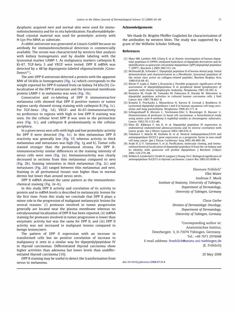
Fig. 2. Localization of DPP II protein (a–d) and of mRNA (e–h). (a) Variable degree of immunoreactivity is seen in individual nevus cells (nevus nest outlined). (b) In thin
melanomas few melanoma cells are immunoreactive. Melanoma cells in thick melanoma (c) and metastases (d) mostly show strong immunoreactivity. Immunoreactivity of
the peritumoral stroma is low. DPP II message is strongly expressed in nevus cells (e), absent in melanoma cells of thin melanoma (f) and moderate in thick melanoma (g) and
melanoma metastasis (h). Variable amount of staining in the peritumoral stroma. E: epidermis, T: tumor.
Letters to the Editor / Journal of Dermatological Science 53 (2009) 65–8470
Letters to the Editor / Journal of Dermatological Science 53 (2009) 65–84 71
dysplastic acquired nevi and normal skin were used for immu-nohistochemistry and for in situ hybridization. Paraformaldehyde-fixed cryostat material was used for proteolytic activity withH-Lys-Pro-MNA as substrate.
A rabbit antiserum was generated at Linaris GmbH because noantibody for immunohistochemical detection is commerciallyavailable. The serum was characterized by western blot analysiswith kidney homogenates and by double labeling with thelysosomal marker LAMP-1. As malignancy markers cathepsin B,Ki-67, TGF-beta 3 and VEGF were tested. DPP II mRNA wasdetected by a 48 bp digoxigenin-labeled oligonucleotide (Gene-Detect1).
The anti-DPP II antiserum detected a protein with the apparentMW of 54 kDa in homogenates (Fig. 1a) which corresponds to theweight reported for DPP II isolated from rat kidney 54 kDa [9]. Co-localization of the DPP II antiserum and the lysosomal membraneprotein LAMP-1 in melanoma was seen (Fig. 1b).
Consecutive and co-labeling of DPP II immunoreactivemelanoma cells showed that DPP II positive tumors or tumorregions rarely showed strong staining with cathepsin B (Fig. 1c).For TGF-beta- (Fig. 1d), VEGF- and Ki-67-immunoreactivityno preference to regions with high or low DPP II staining wasseen. On the cellular level DPP II was seen in the perinucleararea (Fig. 1c), and cathepsin B predominantly in the cellularperiphery.
In a given nevus nest cells with high and low proteolytic activityfor DPP II were detected (Fig. 1e). In thin melanomas DPP IIreactivity was generally low (Fig. 1f) whereas activity in thickmelanomas and metastases was high (Fig. 1g and h). Tumor cellsstained stronger than the peritumoral stroma. For DPP II-immunoreactivity similar differences in the staining intensity ofnevus cells were seen (Fig. 2a). Immunoreactivity was clearlydecreased in sections from thin melanomas compared to nevi(Fig. 2b). Staining intensities in thick melanomas (Fig. 2c) andmetastases (Fig. 2d) ranged between thin melanomas and nevi.Staining in all peritumoral tissues was higher than in normaldermis but lower than around nevus nests.
DPP II mRNA showed the same pattern as the immunohisto-chemical staining (Fig. 2e–h).
In this study DPP II activity and correlation of its activity toprotein and to mRNA levels is described in melanocytic lesions forthe first time. From this study we conclude that DPP II plays aminor role in the progression of malignant melanocytic lesions forseveral reasons: (i) proteases involved in tumor progressiongenerally are located near the plasma membrane whereas noextralysosomal localization of DPP II has been reported, (ii) mRNAstaining for proteases involved in tumor progression is lower thanenzymatic activity but was the same for DPP II, and (iii) DPP IIactivity was not increased in malignant lesions compared tobenign lesions/nevi.
The pattern of DPP II expression with an increase intransformed cells but no positive correlation of increase tomalignancy is seen in a similar way for dipeptidylpeptidase IVin thyroid carcinomas. Differentiated thyroid carcinoma showhigher activities than adenoma but lower levels than undiffer-entiated thyroid carcinoma [10].
DPP II staining may be useful to detect the transformation fromnevus to melanoma.
Acknowledgements
We thank Dr. Brigitte Pfeiffer-Guglielmi for characterization ofthe antibodies by western blots. The study was supported by agrant of the Wilhelm Schuler Stiftung.
References
[1] Maes MB, Lambeir AM, Gilany K, et al. Kinetic investigation of human dipep-tidyl peptidase II (DPPII)-mediated hydrolysis of dipeptide derivatives and itsidentification as quiescent cell proline dipeptidase (QPP)/dipeptidyl peptidase7 (DPP7). Biochem J 2005;386:315–24.
[2] McDonald JK, Schwabe C. Dipeptidyl peptidase II of bovine dental pulp. Initialdemonstration and characterization as a fibroblastic, lysosomal peptidase ofthe serine class active on collagen-related peptides. Biochim Biophys Acta1980;616:68–81.
[3] Klener P, Lojda Z, Haber J, Kvasnicka J. Possible prognostic significance of theassessment of dipeptidylpeptidase II in peripheral blood lymphocytes ofpatients with chronic lymphocytic leukemia. Neoplasma 1987;34:581–6.
[4] Komatsu M, Urade M, Yamaoka M, Fukasawa K, Harada M. Alteration indipeptidyl peptidase activities in cultured human carcinoma cells. J NatlCancer Inst 1987;78:863–8.
[5] Krepela E, Prochazka J, Mynarikova H, Karova B, Cermak J, Roubkova H.Lysosomal dipeptidyl-peptidases I and II in human squamous cell lung carci-noma and lung parenchyma. Neoplasma 1996;43:171–8.
[6] Schlagenhauff B, Klessen C, Teichmann-Dorr S, Breuninger H, Rassner G.Demonstration of proteases in basal cell carcinomas: a histochemical studyusing amino acid-4-methoxy-2-naphthyl-amides as chromogenic substrates.Cancer 1992;70:1133–40.
[7] Khin EE, Kikkawa F, Ino K, et al. Dipeptidyl peptidase IV expression inendometrial endometrioid adenocarcinoma and its inverse correlation withtumor grade. Am J Obstet Gynecol 2003;188:670–6.
[8] Tokuhara T, Adachi M, Hashida H, et al. Neutral endopeptidase/CD10 andaminopeptidase N/CD13 gene expression as a prognostic factor in non-smallcell lung cancer. Jpn J Thorac Cardiovasc Surg 2001;49:489–96.
[9] Araki H, Li Y, Yamamoto Y, et al. Purification, molecular cloning, and immu-nohistochemical localization of dipeptidyl peptidase II from the rat kidney andits identity with quiescent cell proline dipeptidase. J Biochem (Tokyo)2001;129:279–88.
[10] Kehlen A, Lendeckel U, Dralle H, Langner J, Hoang-Vu C. Biological significance ofaminopeptidase N/CD13 in thyroid carcinomas. Cancer Res 2003;63:8500–6.