RESEARCH PAPER Direct profiling of the phospholipid composition of adult Caenorhabditis elegans using whole-body imaging mass spectrometry Saira Hameed 1 & Koji Ikegami 1 & Eiji Sugiyama 1 & Shoko Matsushita 1 & Yoshishige Kimura 1 & Takahiro Hayasaka 1,3 & Yuki Sugiura 1,4 & Noritaka Masaki 1 & Michihiko Waki 1 & Isao Ohta 2 & Md Amir Hossen 1 & Mitsutoshi Setou 1 Received: 14 April 2015 /Revised: 8 July 2015 /Accepted: 20 July 2015 /Published online: 27 August 2015 # The Author(s) 2015. This article is published with open access at Springerlink.com Abstract A protocol for the direct analysis of the phospho- lipid composition in the whole body of adult soil nematode, Caenorhabditis elegans (C. elegans), was developed, which combined freeze-cracking of the exoskeletal cuticle and matrix-assisted laser desorption/ionization-imaging mass spectrometry (MALDI-IMS). Biomolecules in the m/z range from 700 to 900 were more effectively detected in the freeze- cracked than from simple frozen adult nematode bodies. Dif- ferent distribution of biomolecules was observed in a nema- tode body when the matrix was applied with a sublimation deposition method. The whole-body IMS technique was ap- plied on genetically deficient mutant C. elegans to combine whole-body lipidomics and genetics, by comparing the fatty acid compositions, especially of the phosphatidylcholine (PC) species, between the wild-type and fat-1 mutants, which lack the gene encoding an n-3 fatty acid desaturase. A significant reduction of PC(20:5/20:5) and PC(20:4/20:5) and a marked increase of PC(20:4/20:4), PC(20:3/20:4), and PC(20:3/20:3) were detected in the fat-1 mutants in pos- itive ion mode. In addition, phospholipid compositions other than PCs were analyzed in negative ion mode. A loss of a possible phosphatidylinositol (PI) with 18:0/ 20:5 and a compensative accumulation of putative PI(18:0/20:4) were detected in the fat-1 mutants. In con- clusion, the whole-body MALDI-IMS technique is useful for the profiling of multiple biomolecules in C. elegans in both intra- and inter-individual levels. Keywords Caenorhabditis elegans . Cuticle . Exoskeleton . Freeze-cracking . Matrix-assisted laser desorption/ ionization-imaging mass spectrometry . Phosphatidylcholine . Phosphatidylinositol Introduction Recent advances in mass spectrometry have enabled the direct analyses of biomolecules in tissue samples without any target- specific labeling [1, 2]. Matrix-assisted laser desorption/ ionization-imaging mass spectrometry (MALDI-IMS) can be used for the analyses of the spatial distribution of various biomolecules, which range from small metabolites to lipids, peptides, and intact proteins, in tissue sections [3–6]. The state of the art MALDI-IMS technique has been used for the inves- tigation of molecular distributions in mammalian tissues [7], including samples of diseased human tissues [8–10]. It has also been used for label-free non-targeted analyses of Published in the topical collection New Applications of Mass Spectrometry in Biomedicine with guest editors Fumio Nomura, Mitsutoshi Setou, and Toshimitsu Niwa. Electronic supplementary material The online version of this article (doi:10.1007/s00216-015-8932-7) contains supplementary material, which is available to authorized users. * Mitsutoshi Setou [email protected]1 Department of Cell Biology and Anatomy, Hamamatsu University School of Medicine, 1-20-1 Handayama, Higashi-ku, Hamamatsu, Shizuoka 431-3192, Japan 2 Research Equipment Center, Hamamatsu University School of Medicine, 1-20-1 Handayama, Higashi-ku, Hamamatsu, Shizuoka 431-3192, Japan 3 Present address: Faculty of Health Sciences, Health Innovation and Technology Center, Hokkaido University, Kita 12 Nishi 5, Kita-ku, Sapporo, Hokkaido 060-0812, Japan 4 Present address: Department of Biochemistry, School of Medicine, Keio University, Shinjuku-ku, Tokyo 160-8582, Japan Anal Bioanal Chem (2015) 407:7589–7602 DOI 10.1007/s00216-015-8932-7
Transcript
RESEARCH PAPER
Direct profiling of the phospholipid composition of adultCaenorhabditis elegans using whole-body imagingmass spectrometry
Received: 14 April 2015 /Revised: 8 July 2015 /Accepted: 20 July 2015 /Published online: 27 August 2015# The Author(s) 2015. This article is published with open access at Springerlink.com
Abstract A protocol for the direct analysis of the phospho-lipid composition in the whole body of adult soil nematode,Caenorhabditis elegans (C. elegans), was developed, whichcombined freeze-cracking of the exoskeletal cuticle andmatrix-assisted laser desorption/ionization-imaging massspectrometry (MALDI-IMS). Biomolecules in the m/z rangefrom 700 to 900 were more effectively detected in the freeze-cracked than from simple frozen adult nematode bodies. Dif-ferent distribution of biomolecules was observed in a nema-tode body when the matrix was applied with a sublimationdeposition method. The whole-body IMS technique was ap-plied on genetically deficient mutant C. elegans to combinewhole-body lipidomics and genetics, by comparing the fatty
acid compositions, especially of the phosphatidylcholine (PC)species, between the wild-type and fat-1 mutants, which lackthe gene encoding an n-3 fatty acid desaturase. A significantreduction of PC(20:5/20:5) and PC(20:4/20:5) and amarked increase of PC(20:4/20:4), PC(20:3/20:4), andPC(20:3/20:3) were detected in the fat-1 mutants in pos-itive ion mode. In addition, phospholipid compositionsother than PCs were analyzed in negative ion mode. Aloss of a possible phosphatidylinositol (PI) with 18:0/20:5 and a compensative accumulation of putativePI(18:0/20:4) were detected in the fat-1 mutants. In con-clusion, the whole-body MALDI-IMS technique is usefulfor the profiling of multiple biomolecules in C. elegansin both intra- and inter-individual levels.
Recent advances in mass spectrometry have enabled the directanalyses of biomolecules in tissue samples without any target-specific labeling [1, 2]. Matrix-assisted laser desorption/ionization-imaging mass spectrometry (MALDI-IMS) can beused for the analyses of the spatial distribution of variousbiomolecules, which range from small metabolites to lipids,peptides, and intact proteins, in tissue sections [3–6]. The stateof the art MALDI-IMS technique has been used for the inves-tigation of molecular distributions in mammalian tissues [7],including samples of diseased human tissues [8–10]. It hasalso been used for label-free non-targeted analyses of
Published in the topical collection New Applications of MassSpectrometry in Biomedicine with guest editors Fumio Nomura,Mitsutoshi Setou, and Toshimitsu Niwa.
Electronic supplementary material The online version of this article(doi:10.1007/s00216-015-8932-7) contains supplementary material,which is available to authorized users.
1 Department of Cell Biology and Anatomy, Hamamatsu UniversitySchool of Medicine, 1-20-1 Handayama, Higashi-ku,Hamamatsu, Shizuoka 431-3192, Japan
2 Research Equipment Center, Hamamatsu University School ofMedicine, 1-20-1 Handayama, Higashi-ku,Hamamatsu, Shizuoka 431-3192, Japan
3 Present address: Faculty of Health Sciences, Health Innovation andTechnology Center, Hokkaido University, Kita 12 Nishi 5, Kita-ku,Sapporo, Hokkaido 060-0812, Japan
4 Present address: Department of Biochemistry, School of Medicine,Keio University, Shinjuku-ku, Tokyo 160-8582, Japan
biomolecules in various species [11], such as microbes[12], plants [13], parasites [14], arthropods, includingcrustaceans such as the giant tiger prawn (Penaeusmonodon) [15], and insects such as the fruit fly (Dro-sophila melanogaster) [16, 17].
The so i l nematode , Caenorhabdi t i s e l egans(C. elegans), is a common model organism and is exten-sively used in life science research [18]. C. elegans has awide range of advantages for experimental research [19].The short life span of minimally 3 days and the capabilityof being frozen enable a number of iteration of experi-mental tests in a short time [18]. These allow quick for-ward genetics, combined with the ease of genetic manip-ulations [20]. The body structure with multiple organscomposed of the fixed number of cells (~1000 somaticcells) [18], with well-characterized cell fate is highlypowerful to developmental biology. Given these advan-tages in multiple directions, C. elegans has a big potentialto provide a powerful platform for “Integrating-Omics,”in which genet ics , t ranscr iptomics, proteomics,lipidomics, and metabolomics are combined to find newinsights [21], opening a new era of life sciences.C. elegans also begins to be used in applied sciences,such as drug discovery or screening [22].
Some studies have investigated the metabolomic pro-filing of genetically deficient mutant nematodes [23–28].C. elegans has a balloon-like body with high osmoticpressure that is enclosed by an exoskeleton consisting ofa tough impermeable cuticle [29]. This has hindered thedirect detection and analysis of the biomoleculescontained inside. Thus, the components of nematodebodies have been extracted in most metabolomics anal-yses. The very thick and rigid exoskeleton was thoughtto inhibit a direct “whole-body” IMS, as the cuticlelayer of plants should be bypassed using vibratome sec-tioning before the IMS analysis of molecules beneaththeir cuticles [30]. Moreover, the cryosectioning of exo-skeletal organisms requires special handling, which istime-consuming and requires well-designed equipment[16]. Thus, the development of a facile sample prepara-tion for the analysis of the biomolecules using thewhole-body IMS of C. elegans has been desired.
Our aim was to establish a facile protocol for the whole-body MALDI-IMS of adult C. elegans. To accomplish this,we combined a freeze-cracking technique with MALDI-IMSand were able to successfully analyze nematodes to vi-sualize biomolecules in an individual nematode level.We further combined the whole-body MALDI-IMS togenetics through the comparison of the wild-type andfat-1 mutants and succeeded in detecting significant dif-ferences in the fatty acid compositions of the phospha-tidylcholine (PC) and phosphatidylinositol (PI) speciesbetween the two genetically different nematode lines.
Materials and methods
Chemicals
Methanol (MeOH), ethanol (EtOH), chloroform (CHCl3), ul-trapure water, and potassium acetate (CH3COOK) were pur-chased from Wako Pure Chemical Industries (Osaka, Japan).Calibration standard peptides (human bradykinin and angioten-sin II) were purchased from Bruker Daltonics (Billerica, MA,USA). 2,5-Dihydroxybenzoic acid (DHB) was purchased fromBruker Daltonics (Billerica, MA, USA) or Sigma-Aldrich (St.Louis, MO, USA). 9-Aminoacridine hemihydrate (9-AA) waspurchased from Acros Organics (NJ, USA).
Nematodes
C. elegans strains were grown at 20 °C under standard condi-tions [18] on nematode growth medium (NGM) agar plates(0.3 % NaCl, 0.25 % Bacto Peptone, 1.5 % agar, 0.0005 %cholesterol, 1 mM CaCl2, 1 mM MgSO4, 25 mM potassiumphosphate buffer [pH 6.0]), which were seeded with the OP50Escherichia coli strain as a food source. The wild-type strain(Bristol N2) and fat-1 mutant (BX24: fat-1(wa9) IV) ofC. elegans were obtained from the Caenorhabditis GeneticsCenter (Minneapolis, MN, USA). A synchronous culture ofC. elegans was obtained by bleaching the nematodes 3 daysbefore observation. Only adult nematodes were used for theMALDI-IMS and liquid chromatography-electrospray ioniza-tion-tandem mass spectrometry (LC-ESI-MS/MS) analyses.
Sample preparation for MALDI-IMS
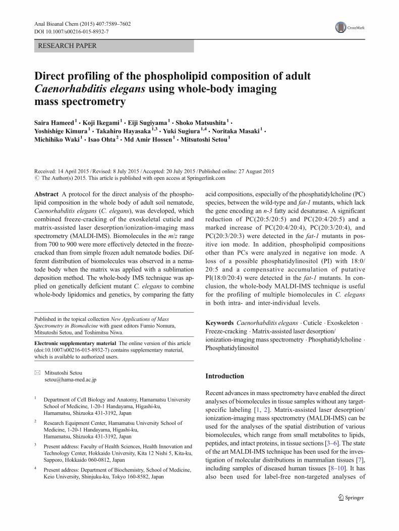
The nematodes that were grown on the surface of the NGMagar plates were harvested by thoroughly washing with water(Fig. 1a), and the live nematodes were transferred into a glasstube using a Pasteur pipette (Fig. 1b). Subsequently, water drop-lets containing live nematodes were transferred onto indium-tin-oxide (ITO)-coated glass slides (Bruker Daltonics) (Fig. 1c).The freeze-cracking method involved covering the nematodecontaining water droplets with a cover glass that was lightlypressed with a finger to immobilize the nematodes and then tomake direct contact with their surfaces (Fig. 1d). The sampleslide was then rapidly frozen on an aluminum block at liquidnitrogen temperature (−196 °C) (Fig. 1e). The aluminum blockallowed for a faster heat transfer, which generated a temperaturegradient acrossC. elegans because of their cylindrical morphol-ogy. The cover glass was quickly detached to remove thecracked cuticle exoskeletons (Fig. 1f). The sample slide wasthen dried under vacuum for ~1 h (Fig. 1g).
To compare the freeze-cracked and simple frozen nema-todes, water droplets containing live nematodes were trans-ferred to the left and right sides of an ITO-coated glass slide.The left side was used for the simple frozen nematodes,
7590 S. Hameed et al.
whereas the right was used for the freeze-cracked nematodes(Fig. 1d). Simple frozen nematodes were not subjected to anycracking treatment but were frozen and dried simultaneouslywith cracked ones (Fig. 1e–g).
Scanning electron microscopy
Scanning electron microscopy (SEM) was performed aspreviously reported [31, 32]. Simple frozen and freeze-cracked nematode specimens were coated with osmiumtetroxide (OsO4) using a plasma multi coater modelPMC-5000 (Meiwa, Japan). The SEM observation ofnematodes was performed using a Hitachi S-4800 fieldemission scanning electron microscope at an accelerationvoltage of 1.0 kV and an emission current of 10 μA. The
vacuum level in the observation chamber was 10−5–10−7 Pa, and the working distance was 8.0 mm.
Matrix application
DHB was chosen to detect PC in positive ion mode,since i t is most commonly used for MALDI-MSand MALDI-IMS with high vacuum chamber [33–35]. 9-AA was used to detect PI in negative ion mode, sinceit is most common for lipid analyses in negative ion modeof MALDI-MS and MALDI-IMS [36, 37]. To extract andco-crystallize the analytes, the nematode samples werespray-coated with DHB solution (50 mg/mL in 70 % MeOHcontaining 20 mM CH3COOK) for analyses in positiveion mode, or 9-AA solution (10 mg/mL in 70 % EtOH)
Fig. 1 Workflow for whole-bodyMALDI-IMS analyses of adultC. elegans. a, b Harvesting livenematodes from a nematodegrowth medium (NGM) agarplate into a glass tube bysuspension in water. c Seeding thenematodes onto an ITO-coatedglass slide. d Applying a coverglass on a nematode subset andpressing them for freeze-cracking(on the right side of the glassslide). The other subset of livenematodes was left untreated. eRapid freezing of the nematodeson a liquid nitrogen-cooledaluminum block. Through thisprocess, the nematodes that werecovered by the coverslip werefreeze-cracked, whereas theothers were simply frozen. fRemoving the coverslip to detachthe cuticle exoskeleton from thenematodes. g Vacuum drying ofthe nematodes on a pre-cooledaluminum block
Whole-body imaging mass spectrometry of Caenorhabditis elegans 7591
for analyses in negative ion mode on an ITO-coated glass slideusing a 0.2-mm caliber nozzle airbrush (Procon Boy FWAPlatinum; Mr. Hobby, Tokyo, Japan). Approximately 2 mLof DHB or 3 mL of 9-AA solutions were sprayed over 15 to20 min. The airbrush was moved right to left, top to bottom,and vice versa, for around 1200 times, while maintaining adistance of 10 cm between the nozzle and nematode tissuesurfaces. After the matrix application, the slide was incubatedin desiccator for around 5 min. The humidity of room wasmaintained under 25 % at 23 °C. The glass slide was observedby a microscope to confirm whether the matrix layer uniform-ly covered the nematode sample surfaces.
The nematode samples were also subjected to matrix appli-cation by sublimation deposition method, with 600 mg ofDHB sublimated at 170 °C for the deposition thickness toreach 1.5 μm, using iMLayer (Shimadzu, Japan).
MALDI-IMS
MALDI-IMS was performed using an ultraflex II TOF/TOFinstrument (Bruker Daltonics), equipped with a Smartbeam-IINd:YAG 355 nm laser, with 25 μm in raster scan pitch. Thelaser frequency was 200 Hz, and the data were acquired usingan ion source voltage of 25 kV and a reflector voltage of26.30 kV in the positive ion reflectron mode. Calibration ofthe MS was performed using DHB ([M+H]+, m/z 155.03),human bradykinin fragment 1–7 ([M+H]+, m/z 757.40), andhuman angiotensin II ([M+H]+, m/z 1046.54). In negative ionmode, the laser frequency was 100 Hz, and the data wereacquired using an ion source voltage of 20.11 kVand a reflec-tor voltage of 21.07 kV. The data acquisition areas, over whichthe spectra were measured, were set by tracing the outline ofthe well-cracked nematodes. These were characterized by theobservation of the nematode bodies and the region immedi-ately outside them. The mass spectra were acquired by aver-aging the signals from 500 laser pulses per sample measure-ment point, and the mass measurement range was set to m/z700–1000. The nematode specimens were automatically ras-ter scanned using flexControl (ver. 3 or 3.4) and flexImaging(ver. 2.1 or 4.0) software (Bruker Daltonics). The acquiredraw mass spectra were normalized to the total ion current(TIC). The images of the detected molecular ions were con-structed using the flexImaging (2.1 and 4.0) software. Sixsimple frozen and six freeze-cracked nematodes were ana-lyzed to evaluate the effectiveness of the exoskeleton removal.Four nematodes were analyzed to examine whether biomole-cules were retained in nematode bodies. Six wild-type and fat-1 mutant nematodes were analyzed in positive ion mode.Eight wild-type and fat-1 mutant nematodes were analyzedin negative ion mode. The data were presented as plots foreach nematode and the mean of the signal intensities acquiredfrom the multiple samples.
Phospholipids of the harvested wild-type and fat-1 mu-tants were extracted with Folch method [38], and theywere analyzed using LC-ESI-MS/MS with a 4000Q-TRAP triple quadrupole linear ion trap mass spectrometer(AB SCIEX, Framingham, MA, USA) equipped with anACQUITY ultra-performance liquid chromatography sys-tem (Waters, Milford, MA, USA). An ACQUITY UPLCBEH C18 column (2.1×50 mm, i.d., 1.7 mm particles;Waters) was connected to a guard column (2.1×5 mm;Waters), and the temperature of column oven was main-tained at 40 °C [9]. The mobile phase consisted of a gra-dient of two solvent mixtures. Solvent A was composed ofacetonitrile, MeOH, and water (19:19:2 v/v/v), containingformic acid (0.1 vol.%) and ammonia (0.028 vol.%). Sol-vent B was composed of isopropanol containing formicacid (0.1 vol.%) and ammonia (0.028 vol.%). A gradientelution using solvents A and B was performed at a flowrate of 0.40 mL/min. To profile the molecular species ofthe specific phospholipid classes, a precursor ion scanningfor the polar head groups of the PCs and sphingomyelins(SMs) (m/z=184) was performed using the positive iondetection mode of the 4000Q-TRAP instrument. Fragmentions were generated through col l is ion- induced-dissociation (CID) [39]. The optimal collision energywas determined by the preliminarily analysis ofPC(16:0/18:1), which was used as a standard lipid. ThePC molecular species detected were assigned using their
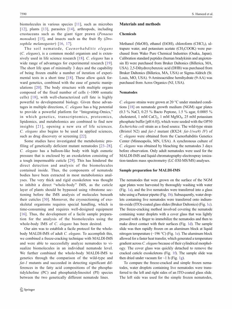
�Fig. 2 Freeze-cracked nematode bodies yield stronger signals. aScanning electron microscopy images of simple frozen and freeze-cracked C. elegans. Scale bars: 200 μm in low magnification (top),25 μm in high magnification (bottom). b, c Averaged mass spectraranging from m/z 700 to 1000 that were detected in the whole body ofb simple frozen and c freeze-cracked C. elegans. Four of the major masspeaks at m/z 796.5, 806.5, 846.6, and 868.5 were selected to depict thethermal color scale images. d Optical images and thermal color scaleimages of the four selected molecules on the nematode bodies. Scalebar: 200 μm. Color scale: deep blue, faint signal; red-purple, maximumsignal. e Quantitative signal intensities on nematode bodies. The data areshown as plots for each nematode (diamonds; n=6) and mean (red line)±SD (blue lines). The p values were calculated using a t test
Table 1 Molecular weights of the phosphatidylcholine species inC. elegans
m/z values and the relative retention times of PC molecu-lar species that were previously described [9, 34, 39], orby referring to the online database, “The Human
Metabolome Database (HMDB)” (http://www.hmdb.ca/spectra/ms/search), and the total fatty acid compositionof C. elegans [23–25] (Table 1).
Whole-body imaging mass spectrometry of Caenorhabditis elegans 7593
Further analyses of fatty acid compositions in the PC spe-cies of interest were performed by a hybrid quadrupole-Orbitrap mass spectrometer (Q Exactive; Thermo Scientific,Waltham, MA, USA), with the mass resolution of 5 ppm.Separation of the molecular species was carried out usingAgilent1100 series HPLC System (Agilent Technologies,Germany) equipped with Acclaim™ 120 C18 column (2.1×150 mm, i.d., 3 μm particles; Thermo Scientific). The temper-ature of the column oven was maintained at 50 °C. The injec-tion volume was 5.0 μL. The temperature of the sample traywas kept at 10 °C. Solvent A was composed of water, aceto-nitrile, MeOH (2:1:1, v/v/v), containing ammonium formate(5 mM) and formic acid (0.1 vol.%). Solvent B was composedof acetonitrile, isopropanol (1:9, v/v), containing ammoniumformate (5 mM) and formic acid (0.1 vol.%). A gradient elu-tion using solvents A and B was performed at a flow rate of0.30 mL/min for 50 min from the initial composition (A/B:80/20, v/v) to the final composition (A/B: 0/100 vol.%) with alinear gradient. MS and MS/MS analyses were performed inboth positive and negative ion modes. MS spectra were ac-quired in the range of m/z 700–900. MS/MS spectra near thepeak top of interested peaks were acquiredwith a targetedMS/MS method. The target mass-resolving power at m/z 200 wasset to 70,000 for both MS and MS/MS analyses. The isolationwindow for MS/MS was set to 0.4m/z. The temperature of ionsource heater was set to 350 °C, and the capillary temperaturewas at 250 °C. The ion spray voltage was set to 3.5 kV forboth ion modes. Maximum injection time was set to 100 msfor bothMS andMS/MS analyses. The automatic gain controltarget was set to 1×10⁷ for MS and 2×10⁵ for MS/MS. Thenormalized collision energy for MS/MS was set to 30 %. Ex-tracted ion chromatograms (EICs) were generated within atheoretical value of ±5 ppm for PC species of interest.
Results
Relative effectiveness of the freeze-cracking methodin the direct detection of multiple biomolecules
To evaluate the effectiveness of the exoskeleton removal, weperformed the comparative analyses of the two procedures,simple freezing and freeze-cracking, in parallel on a glass slide(Fig. 1). To effectively remove the exoskeletal cuticle ofC. elegans and expose the internal structures of the nema-todes, we placed a cover slip on the nematodes and pressedit with a finger before chilling them using a liquid nitrogen-cooled aluminum block (Fig. 1d). We evaluated the effective-ness of this procedure by comparing it with a simple freezingmethod. We observed the condition of the samples using anSEM. The surface of the freeze-cracked nematode bodieslooked highly scabrous with multiple wrinkles (Fig. 2a; right),whereas the simple frozen nematodes had a highly smooth
surface, which appeared to retain an intact exoskeletal cuticle(Fig. 2a; left). The microscopy showed that freeze-crackingdrastically changed the surface condition of the samplespecimens.
We then analyzed the freeze-cracked nematodes usingMALDI-IMS and compared the signal intensity of the biomol-ecules with those from the simple frozen nematodes (Fig. 2b,c). The averaged mass spectra obtained from the freeze-cracked nematodes had increased signal intensity throughoutthe m/z range 700–1000 when compared to the simple frozennematodes (Fig. 2b vs. Fig. 2c). In particular, the molecularions that were observed between m/z 700 and 900 were de-tected with much higher signal intensities from the freeze-cracked nematodes than those from the simple frozen nema-todes. We selected four molecules with m/z 796.5, 806.5,846.6, and 868.5, respectively (arrows in Fig. 2b, c), to visu-ally compare their signal intensities from the nematode bodies.The signal intensities of these selected molecules increased by>50 % of the maximum signal level (yellow-to-red colors) insome regions of freeze-cracked nematodes, whereas those inthe simple frozen nematodes appeared near the noise level(blue-to-cyan colors) (Fig. 2d). We performed further semi-quantitative analyses of the signal intensities of the four mol-ecules. In all four molecules, the signal intensities detectedfrom the freeze-cracked nematodes were significantly higherthan those from the simple frozen nematodes (n=6) (Fig. 2e).These results demonstrated that the freeze-cracking methodenabled the highly effective direct detection of multiple inter-nal biomolecules, which provided a facile whole-bodyMALDI-IMS.
Molecular distribution analyses in nematode bodiesby combining the freeze-cracking method with the matrixsublimation
The nematodes were pressed with a finger during thefreeze-cracking procedure (Fig. 1d). This has a potentialrisk in that the nematode bodies could be punctured dur-ing this process, which could result in the delocalizationor drift of intra-body ingredients from the nematode bodyto the surrounding area. Thus, we evaluated whether thebiomolecules were retained in the nematode bodies. Forthis evaluation, we expanded the imaging measurementareas to include the glass surface adjacent to the nematodebodies (Fig. 3a, b; insets) and compared the mass spectrafrom regions of interest (ROIs) inside and outside of thenematode bodies (Fig. 3a, b; insets). The averaged massspectrum acquired from the ROI inside of the nematodebody (Fig. 3a) has several mass peaks with m/z valuesranging from 750 to 900, whereas that taken from theROI outside of the nematode body had few significantmass peaks (Fig. 3b).
7594 S. Hameed et al.
Fig. 3 Freeze-cracking method allows analyses of molecular distributionin the nematode body. a, b Averaged mass spectra detected from theregions of interest a inside and b outside of the nematode body. Four ofthe major mass peaks at m/z 808.5, m/z 832.5, m/z 846.6, and m/z 868.5were selected to depict thermal color scale images. Insets show the outlineof nematode body (orange dotted line) and scanned area in IMS (whitedotted line). c Thermal color scale images of the selected four molecules.Scale bar: 250 μm. Color scale: deep blue, faint signal; red-purple,
maximal signal. d Quantitative signal intensities in the regions ofinterest. The data are shown as plots for each nematode (diamonds; n=4) and mean (red line)±SD (blue lines). The p values were calculatedusing a paired t test. e An averaged mass spectrum detected from freeze-cracked nematode body with DHB matrix applied by sublimation. Threeof major mass peaks at m/z 758.4, 810.4, and 895.6 were selected todepict pseudo color scale images. f Pseudo color scale images of theselected three molecules. Scale bar: 250 μm
Whole-body imaging mass spectrometry of Caenorhabditis elegans 7595
We selected four of major ion peaks, m/z values of 808.5,832.5, 846.6, and 868.5 (arrows in Fig. 3a, b), to monitorwhether or not those effectively detectable molecules aredrifted from the worm body to the outside of body. All fourof the molecular ions showed signal intensities that increasedby >50 % of the maximum from the inside area of the nema-tode body (yellow-to-red colors), whereas they appeared nearthe background level in the area outside of nematode body(blue-to-cyan colors) albeit slightly delocalized to the sur-rounding glass surface (Fig. 3c). The semi-quantitative analy-ses of nematodes demonstrated that the averaged signal inten-sities of all four of the molecules were significantly higherinside than outside the nematode bodies (n=4) (Fig. 3d). The-se results indicated that the biomolecules were retained insidenematode bodies during the freeze-cracking process.
We further sought to analyze the distribution of biomole-cules inside the nematode body with matrix applied via asublimation deposition method. The averaged mass spectrumacquired from nematode bodies provided a number of masspeaks (Fig. 3e). We selected three of major mass peaks at m/z758.4, m/z 810.4, and m/z 895.6 to observe the spatial distri-bution of those molecules in the nematode body. These threemolecules showed clearly different distribution patterns in thenematode body: the molecule with m/z 758.4 was stronglylocalized at the main body trunk (Fig. 3f; purple); the mole-cule with m/z 810.4 was detected almost throughout the nem-atode body, except in the head and tail regions (Fig. 3f; green);the molecule withm/z 895.6 was strongly detected in the headand tail regions (Fig. 3f; red).
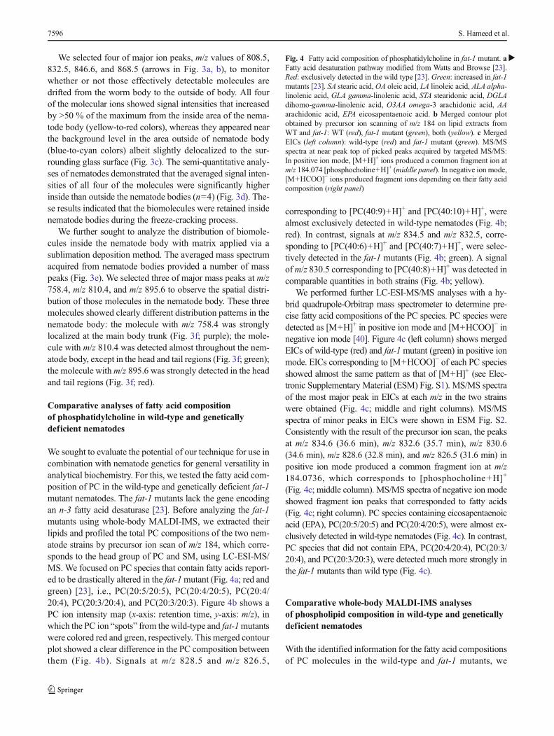
Comparative analyses of fatty acid compositionof phosphatidylcholine in wild-type and geneticallydeficient nematodes
We sought to evaluate the potential of our technique for use incombination with nematode genetics for general versatility inanalytical biochemistry. For this, we tested the fatty acid com-position of PC in the wild-type and genetically deficient fat-1mutant nematodes. The fat-1 mutants lack the gene encodingan n-3 fatty acid desaturase [23]. Before analyzing the fat-1mutants using whole-body MALDI-IMS, we extracted theirlipids and profiled the total PC compositions of the two nem-atode strains by precursor ion scan of m/z 184, which corre-sponds to the head group of PC and SM, using LC-ESI-MS/MS. We focused on PC species that contain fatty acids report-ed to be drastically altered in the fat-1mutant (Fig. 4a; red andgreen) [23], i.e., PC(20:5/20:5), PC(20:4/20:5), PC(20:4/20:4), PC(20:3/20:4), and PC(20:3/20:3). Figure 4b shows aPC ion intensity map (x-axis: retention time, y-axis: m/z), inwhich the PC ion “spots” from the wild-type and fat-1mutantswere colored red and green, respectively. This merged contourplot showed a clear difference in the PC composition betweenthem (Fig. 4b). Signals at m/z 828.5 and m/z 826.5,
corresponding to [PC(40:9)+H]+ and [PC(40:10)+H]+, werealmost exclusively detected in wild-type nematodes (Fig. 4b;red). In contrast, signals at m/z 834.5 and m/z 832.5, corre-sponding to [PC(40:6)+H]+ and [PC(40:7)+H]+, were selec-tively detected in the fat-1 mutants (Fig. 4b; green). A signalofm/z 830.5 corresponding to [PC(40:8)+H]+ was detected incomparable quantities in both strains (Fig. 4b; yellow).
We performed further LC-ESI-MS/MS analyses with a hy-brid quadrupole-Orbitrap mass spectrometer to determine pre-cise fatty acid compositions of the PC species. PC species weredetected as [M+H]+ in positive ion mode and [M+HCOO]− innegative ion mode [40]. Figure 4c (left column) shows mergedEICs of wild-type (red) and fat-1 mutant (green) in positive ionmode. EICs corresponding to [M+HCOO]− of each PC speciesshowed almost the same pattern as that of [M+H]+ (see Elec-tronic Supplementary Material (ESM) Fig. S1). MS/MS spectraof the most major peak in EICs at each m/z in the two strainswere obtained (Fig. 4c; middle and right columns). MS/MSspectra of minor peaks in EICs were shown in ESM Fig. S2.Consistently with the result of the precursor ion scan, the peaksat m/z 834.6 (36.6 min), m/z 832.6 (35.7 min), m/z 830.6(34.6 min), m/z 828.6 (32.8 min), and m/z 826.5 (31.6 min) inpositive ion mode produced a common fragment ion at m/z184.0736, which corresponds to [phosphocholine+H]+
(Fig. 4c; middle column). MS/MS spectra of negative ion modeshowed fragment ion peaks that corresponded to fatty acids(Fig. 4c; right column). PC species containing eicosapentaenoicacid (EPA), PC(20:5/20:5) and PC(20:4/20:5), were almost ex-clusively detected in wild-type nematodes (Fig. 4c). In contrast,PC species that did not contain EPA, PC(20:4/20:4), PC(20:3/20:4), and PC(20:3/20:3), were detected much more strongly inthe fat-1 mutants than wild type (Fig. 4c).
Comparative whole-body MALDI-IMS analysesof phospholipid composition in wild-type and geneticallydeficient nematodes
With the identified information for the fatty acid compositionsof PC molecules in the wild-type and fat-1 mutants, we
�Fig. 4 Fatty acid composition of phosphatidylcholine in fat-1 mutant. aFatty acid desaturation pathway modified from Watts and Browse [23].Red: exclusively detected in the wild type [23]. Green: increased in fat-1mutants [23]. SA stearic acid,OA oleic acid, LA linoleic acid, ALA alpha-linolenic acid, GLA gamma-linolenic acid, STA stearidonic acid, DGLAdihomo-gamma-linolenic acid, O3AA omega-3 arachidonic acid, AAarachidonic acid, EPA eicosapentaenoic acid. b Merged contour plotobtained by precursor ion scanning of m/z 184 on lipid extracts fromWT and fat-1: WT (red), fat-1 mutant (green), both (yellow). c MergedEICs (left column): wild-type (red) and fat-1 mutant (green). MS/MSspectra at near peak top of picked peaks acquired by targeted MS/MS:In positive ion mode, [M+H]+ ions produced a common fragment ion atm/z 184.074 [phosphocholine+H]+ (middle panel). In negative ion mode,[M+HCOO]− ions produced fragment ions depending on their fatty acidcomposition (right panel)
7596 S. Hameed et al.
Whole-body imaging mass spectrometry of Caenorhabditis elegans 7597
performed whole-body MALDI-IMS by applying the freeze-cracking method to both of the strains (Fig. 5a). The averagedmass spectra between m/z 810–880 included five mass peakswith m/z values of 864.5, 866.5, 868.5, 870.5, and 872.5(Fig. 5b), which corresponded to the K+ adducts ([M+K]+)of the following PC species: PC(20:5/20:5), PC(20:4/20:5),PC(20:4/20:4), PC(20:3/20:4), and PC(20:3/20:3), respective-ly. The IMS image of each PC species revealed similar PCcomposition differences between the wild-type and fat-1 mu-tants as were detected using LC-ESI-MS/MS (Fig. 5c).PC(20:5/20:5) and PC(20:4/20:5) had higher intensity signalsin the wild-type (cyan-to-yellow colors in the thermal colorscale) than in the fat-1mutants (blue-to-cyan colors) (Fig. 5c).In contrast, PC(20:4/20:4), PC(20:3/20:4), and PC(20:3/20:3)were more abundant in the fat-1 mutants (cyan-to-orangecolors) than in the wild type (blue-to-cyan colors) (Fig. 5c).Semi-quantitative analyses clearly demonstrated the differ-ences of the PC compositions between the wild-type and fat-1mutants (n=6) (Fig. 5d), and their statistical values are givenin Table 2. The signal intensities of PC(20:5/20:5) andPC(20:4/20:5) were higher in the wild-type than in the fat-1mutants (p<0.01 and p<0.05, respectively, t test), whereas thesignal intensities of PC(20:4/20:4), PC(20:3/20:4), andPC(20:3/20:3) were higher in the fat-1 mutants than in thewild type (p<0.01 for all cases, t test).
We also analyzed the wild-type and fat-1 mutants in nega-tive ion mode to test the effect of the fat-1 mutation on phos-pholipids composition besides PCs. The averaged mass spec-tra showed only a few peaks (Fig. 5e). The peaks atm/z 758.4,m/z 883.4, and m/z 885.4 were selected to obtain images ofthose molecules with thermal color scale (Fig. 5f). Two mol-ecules with m/z 883.4 and 885.4 were detected almost mutu-ally exclusively in wild-type and fat-1 mutant (Fig. 5f): amolecule with m/z 883.4 was detected in wild type while amolecule with m/z 885.4 being detected in fat-1 mutants(p<0.005, t test, n=8) (Fig. 5g). No significant differencewas detected for the molecule with m/z 758.4 (p>0.05, t test)(Fig. 5g). These results demonstrated that our whole-bodyMALDI-IMS technique was capable of being connected togenetics by analyzing individual strains.
Discussion
We presented a procedure for the direct analysis of the lipidcompositions of the whole body of a well-established modelorganism, C. elegans, without requiring lipid extraction ortarget-specific labeling. We also showed that whole-bodyMALDI-IMS could offer analyses of molecular distributionin the nematode body and be combined with genetics. Thekey point of our protocol was to remove the exoskeletal cuti-cle from nematode bodies, aiming to efficiently generate co-crystals of biomolecules and matrices.
Before this study, two groups tried to directly analyze thecomponents of nematodes without using extraction. One studydetected some unidentified biomolecules in larval nematodesusing time-of-flight secondary ion mass spectrometry (TOF-SIMS) [41]. This work also combined whole-body imagingwith genetics by comparing the molecular compositions ofthe wild-type and daf-2mutants [41]. TOF-SIMSwas designedfor the analysis of molecules present near the surface of speci-mens (within several nanometers) [42], and thus would requirethe removal or bypassing of the cuticle exoskeleton to analyzethe intra-body composition. That study thus only detected mol-ecules on the surface of the nematodes, although the cuticle ofL1 larvae is thinner than that of adult nematodes [43]. Anotherstudy detected manganese, which is not a biomolecule, in larvalnematodes using laser ablation-inductively coupled plasma-mass spectrometry (LA-ICP-MS) [44]. LA-ICP-MS was de-signed for the analysis of elements such as metals, and uses alaser that is >10 times stronger (>1 mJ) [45, 46] than those usedin MALDI-IMS (a few hundred μJ). The high-powered laserdegrades most organic compounds, although it enables the pen-etration of the cuticle layer and the ablation of intra-body ma-terials. Furthermore, any degraded fragments of organic com-pounds would be completely destroyed within the ICP [47].Thus, LA-ICP-MS is incapable of analyzing organic com-pounds. Our whole-body MALDI-IMS technique addressedthese problems through co-crystallization of phospholipids witha matrix in the nematode body by removing the cuticle usingfreeze-cracking.
Our technique provided a lipidomic analysis of the PCspecies in genetically deficient mutant nematodes. The resultof the comparative whole-body IMS between the wild-typeand fat-1 mutants is consistent with the fatty acid composi-tions reported previously [23–25]. The lower level of EPA-containing PCs, PC(20:5/20:5) and PC(20:4/20:5), in fat-1mutants is consistent with the finding that they lose EPA [24].
�Fig. 5 Whole-body MALDI-IMS of genetically deficient nematodes. aScanning electron microscopy images of freeze-cracked wild-type andfat-1 mutants. Scale bar: 25 μm. b Averaged mass spectra detectedfrom freeze-cracked wild-type or fat-1 mutants in positive ion mode.Five mass peaks at m/z 864.5, m/z 866.5, m/z 868.5, m/z 870.5, and m/z872.5 were selected to depict thermal color scale images. c Thermal colorscale images of the five selected PC species. Scale bar: 250 μm. Colorscale: deep blue, faint signal; red-purple, maximum signal. dQuantitativesignal intensities of the PC species in the wild-type and fat-1mutants. Thedata are shown as plots for each nematode (diamonds; n=6) and mean(red line)±SD (blue lines). The p values were calculated using a t test. eAveraged mass spectra detected from freeze-cracked wild-type or fat-1mutants in negative ion mode. Three mass peaks atm/z 758.4,m/z 883.4,and m/z 885.4 were selected to depict thermal color scale images. fThermal color scale images of the three selected mass peaks. Scale bar:250 μm. Color scale: deep blue, faint signal; red-purple, maximumsignal. g Quantitative signal intensities of the detected biomolecularspecies in the wild-type and fat-1 mutants. The data are shown as plotsfor each nematode (diamonds; n=8) and mean (red line)±SD (blue lines).The p values were calculated using a t test
7598 S. Hameed et al.
Whole-body imaging mass spectrometry of Caenorhabditis elegans 7599
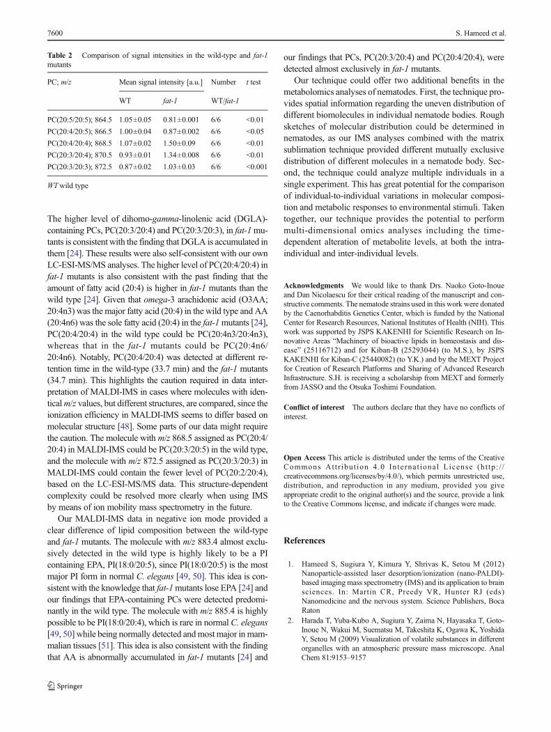
The higher level of dihomo-gamma-linolenic acid (DGLA)-containing PCs, PC(20:3/20:4) and PC(20:3/20:3), in fat-1mu-tants is consistent with the finding that DGLA is accumulated inthem [24]. These results were also self-consistent with our ownLC-ESI-MS/MS analyses. The higher level of PC(20:4/20:4) infat-1 mutants is also consistent with the past finding that theamount of fatty acid (20:4) is higher in fat-1 mutants than thewild type [24]. Given that omega-3 arachidonic acid (O3AA;20:4n3) was the major fatty acid (20:4) in the wild type and AA(20:4n6) was the sole fatty acid (20:4) in the fat-1mutants [24],PC(20:4/20:4) in the wild type could be PC(20:4n3/20:4n3),whereas that in the fat-1 mutants could be PC(20:4n6/20:4n6). Notably, PC(20:4/20:4) was detected at different re-tention time in the wild-type (33.7 min) and the fat-1 mutants(34.7 min). This highlights the caution required in data inter-pretation of MALDI-IMS in cases where molecules with iden-ticalm/z values, but different structures, are compared, since theionization efficiency in MALDI-IMS seems to differ based onmolecular structure [48]. Some parts of our data might requirethe caution. The molecule with m/z 868.5 assigned as PC(20:4/20:4) in MALDI-IMS could be PC(20:3/20:5) in the wild type,and the molecule with m/z 872.5 assigned as PC(20:3/20:3) inMALDI-IMS could contain the fewer level of PC(20:2/20:4),based on the LC-ESI-MS/MS data. This structure-dependentcomplexity could be resolved more clearly when using IMSby means of ion mobility mass spectrometry in the future.
Our MALDI-IMS data in negative ion mode provided aclear difference of lipid composition between the wild-typeand fat-1 mutants. The molecule with m/z 883.4 almost exclu-sively detected in the wild type is highly likely to be a PIcontaining EPA, PI(18:0/20:5), since PI(18:0/20:5) is the mostmajor PI form in normal C. elegans [49, 50]. This idea is con-sistent with the knowledge that fat-1mutants lose EPA [24] andour findings that EPA-containing PCs were detected predomi-nantly in the wild type. The molecule with m/z 885.4 is highlypossible to be PI(18:0/20:4), which is rare in normalC. elegans[49, 50] while being normally detected andmost major inmam-malian tissues [51]. This idea is also consistent with the findingthat AA is abnormally accumulated in fat-1 mutants [24] and
our findings that PCs, PC(20:3/20:4) and PC(20:4/20:4), weredetected almost exclusively in fat-1 mutants.
Our technique could offer two additional benefits in themetabolomics analyses of nematodes. First, the technique pro-vides spatial information regarding the uneven distribution ofdifferent biomolecules in individual nematode bodies. Roughsketches of molecular distribution could be determined innematodes, as our IMS analyses combined with the matrixsublimation technique provided different mutually exclusivedistribution of different molecules in a nematode body. Sec-ond, the technique could analyze multiple individuals in asingle experiment. This has great potential for the comparisonof individual-to-individual variations in molecular composi-tion and metabolic responses to environmental stimuli. Takentogether, our technique provides the potential to performmulti-dimensional omics analyses including the time-dependent alteration of metabolite levels, at both the intra-individual and inter-individual levels.
Acknowledgments We would like to thank Drs. Naoko Goto-Inoueand Dan Nicolaescu for their critical reading of the manuscript and con-structive comments. The nematode strains used in this work were donatedby the Caenorhabditis Genetics Center, which is funded by the NationalCenter for Research Resources, National Institutes of Health (NIH). Thiswork was supported by JSPS KAKENHI for Scientific Research on In-novative Areas “Machinery of bioactive lipids in homeostasis and dis-ease” (25116712) and for Kiban-B (25293044) (to M.S.), by JSPSKAKENHI for Kiban-C (25440082) (to Y.K.) and by the MEXT Projectfor Creation of Research Platforms and Sharing of Advanced ResearchInfrastructure. S.H. is receiving a scholarship from MEXT and formerlyfrom JASSO and the Otsuka Toshimi Foundation.
Conflict of interest The authors declare that they have no conflicts ofinterest.
Open Access This article is distributed under the terms of the CreativeCommons At t r ibut ion 4 .0 In te rna t ional License (h t tp : / /creativecommons.org/licenses/by/4.0/), which permits unrestricted use,distribution, and reproduction in any medium, provided you giveappropriate credit to the original author(s) and the source, provide a linkto the Creative Commons license, and indicate if changes were made.
References
1. Hameed S, Sugiura Y, Kimura Y, Shrivas K, Setou M (2012)Nanoparticle-assisted laser desorption/ionization (nano-PALDI)-based imagingmass spectrometry (IMS) and its application to brainsciences. In: Martin CR, Preedy VR, Hunter RJ (eds)Nanomedicine and the nervous system. Science Publishers, BocaRaton
2. Harada T, Yuba-Kubo A, Sugiura Y, Zaima N, Hayasaka T, Goto-Inoue N, Wakui M, Suematsu M, Takeshita K, Ogawa K, YoshidaY, Setou M (2009) Visualization of volatile substances in differentorganelles with an atmospheric pressure mass microscope. AnalChem 81:9153–9157
Table 2 Comparison of signal intensities in the wild-type and fat-1mutants
PC; m/z Mean signal intensity [a.u.] Number t test
3. Esquenazi E, Coates C, Simmons L, Gonzalez D, Gerwick W,Dorrestein P (2008) Visualizing the spatial distribution of second-ary metabolites produced by marine cyanobacteria and sponges viaMALDI-TOF imaging. Mol Biosyst 4:562–570
4. Thomas A, Charbonneu JL, Fournaise E, Chaurand P (2012)Sublimation of new matrix candidates for high spatial resolutionimaging mass spectrometry of lipids: enhanced information in bothpositive and negative polarities after 1,5-diaminonapthalene depo-sition. Anal Chem 84:2048–2054
5. Eberlin L, Liu X, Ferreira C, Santagata S, Agar N, Cooks R (2011)Desorption electrospray ionization thenMALDImass spectrometryimaging of lipid and protein distributions in single tissue sections.Anal Chem 83:8366–8371
6. Caprioli R, Farmer T, Gile J (1997)Molecular imaging of biologicalsamples: localization of peptides and proteins using MALDI-TOFMS. Anal Chem 69:4751–4760
7. Hayasaka T, Goto-Inoue N, Sugiura Y, Zaima N, NakanishiH, Ohishi K, Nakanishi S, Naito T, Taguchi R, Setou M(2008) Matrix-assisted laser desorption/ionization quadrupoleion trap time-of-flight (MALDI-QIT-TOF)-based imagingmass spectrometry reveals a layered distribution of phospho-lipid molecular species in the mouse retina. Rapid CommunMass Spectrom 22:3415–3426
8. Hirano H, Masaki N, Hayasaka T, Watanabe Y, Masumoto K,Nagata T, Katou F, Setou M (2014) Matrix-assisted laserdesorption/ionization imaging mass spectrometry revealed tracesof dental problem associated with dental structure. Anal BioanalChem 406:1355–1363
9. Matsumoto J, Sugiura Y, Yuki D, Hayasaka T, Goto-Inoue N,Zaima N, Kunii Y, Wada A, Yang Q, Nishiura K, Akatsu H, HoriA, Hashizume Y, Yamamoto T, Ikemoto K, Setou M, Niwa S(2011) Abnormal phospholipids distribution in the prefrontal cortexfrom a patient with schizophrenia revealed by matrix-assisted laserdesorption/ionization imaging mass spectrometry. Anal BioanalChem 400:1933–1943
10. Uchiyama Y, Hayasaka T, Masaki N, Watanabe Y, Masumoto K,Nagata T, Katou F, Setou M (2014) Imaging mass spectrometrydistinguished the cancer and stromal regions of oral squamous cellcarcinoma by visualizing phosphatidylcholine (16:0/16:1) andphosphatidylcholine (18:1/20:4). Anal Bioanal Chem 406:1307–1316
11. Vrkoslav V, Muck A, Cvacka J, Svatos A (2010) MALDI imagingof neutral cuticular lipids in insects and plants. J Am Soc MassSpectrom 21:220–231
12. Moree W, Phelan V, Wu C, Bandeira N, Cornett D, Duggan B,Dorrestein P (2012) Interkingdom metabolic transformations cap-tured by microbial imaging mass spectrometry. Proc Natl Acad SciU S A 109:13811–13816
13. Shroff R, Vergara F, Muck A, Svatos A, Gershenzon J (2008)Nonuniform distribution of glucosinolates in Arabidopsis thalianaleaves has important consequences for plant defense. Proc NatlAcad Sci U S A 105:6196–6201
14. Ferreira M, de Oliveira D, de Oliveira R, Allegretti S, Vercesi A,Catharino R (2014) Mass spectrometry imaging: a new vision indifferentiating Schistosoma mansoni strains. J Mass Spectrom 49:86–92
15. Chansela P, Goto-Inoue N, Zaima N, Sroyraya M, Sobhon P, SetouM (2012) Visualization of neuropeptides in paraffin-embedded tis-sue sections of the central nervous system in the decapod crusta-cean, Penaeus monodon, by imaging mass spectrometry. Peptides34:10–18
16. Niehoff AC, Kettling H, Pirkl A, Chiang YN, Dreisewerd K, YewJY (2014) Analysis of Drosophila lipids by matrix-assisted laserdesorption/ionization mass spectrometric imaging. Anal Chem 86:11086–11092
17. Urban P, Chang C, Wu J, Chen Y (2011) Microscale MALDI im-aging of outer-layer lipids in intact egg chambers from Drosophilamelanogaster. Anal Chem 83:3918–3925
18. Brenner S (1974) The genetics ofCaenorhabditis elegans. Genetics77:71–94
19. Corsi AK (2006) A biochemist’s guide to Caenorhabditis elegans.Anal Biochem 359:1–17
20. Fay D (2006) Genetic mapping and manipulation: chapter 1-introduction and basics. WormBook 17:1–12. http://www.wormbook.org/chapters/www_introandbasics/introandbasics.html
21. VanAssche R, Broeckx V, BoonenK,Maes E, De HaesW, SchoofsL, Temmerman L (2015) Integrating -omics: systems biology asexplored through C. elegans research. J Mol Biol. doi:10.1016/j.jmb.2015.03.015
22. O’Reilly LP, Luke CJ, Perlmutter DH, Silverman GA, Pak SC(2014) C. elegans in high-throughput drug discovery. Adv DrugDeliv Rev 69–70:247–253
23. Watts JL, Browse J (2006) Dietary manipulation implicates lipidsignaling in the regulation of germ cell maintenance in C. elegans.Dev Biol 292:381–392
24. Watts J, Browse J (2002) Genetic dissection of polyunsaturatedfatty acid synthesis in Caenorhabditis elegans. Proc Natl AcadSci U S A 99:5854–5859
25. Satouchi K, Hirano K, Sakaguchi M, Takehara H, Matsuura F(1993) Phospholipids from the free-l iving nematodeCaenorhabditis elegans. Lipids 28:837–840
26. Mahanti P, Bose N, Bethke A, Judkins JC, Wollam J, Dumas KJ,Zimmerman AM, Campbell SL, Hu PJ, Antebi A, Schroeder FC(2014) Comparative metabolomics reveals endogenous ligands ofDAF−12, a nuclear hormone receptor, regulating C. elegans devel-opment and lifespan. Cell Metab 19:73–83. doi:10.1016/j.cmet.2013.11.024
27. Tanaka T, Ikita K, Ashida T, Motoyama Y, Yamaguchi Y, SatouchiK (1996) Effects of growth temperature on the fatty acid composi-tion of the free living nematode Caenorhabditis elegans. Lipids 31:1173–1178
28. Wählby C, Conery AL, Bray MA, Kamentsky L, Larkins-Ford J,Sokolnicki KL, VeneskeyM,Michaels K, Carpenter AE, O’RourkeEJ (2014) High- and low-throughput scoring of fat mass and bodyfat distribution in C. elegans. Methods 68:492–499
29. Page AP, Johnstone IL (2007) The cuticle. In: WormBook (ed) TheC. elegans Research Community, WormBook, doi/10.1895/wormbook.1.138.1, http://www.wormbook.org
30. Jun JH, Song Z, Liu Z, Nikolau BJ, Yeung ES, Lee YJ (2010) High-spatial and high-mass resolution imaging of surface metabolites ofArabidopsis thaliana by laser desorption-ionizationmass spectrom-etry using colloidal silver. Anal Chem 82:3255–3265
31. Ohta I, Takaku Y, Suzuki H, Ishii D, Muranaka Y, Shimomura M,Hariyama T (2014) Dressing living organisms in a thin polymermembrane, the NanoSuit, for high-vacuum FE-SEM observation.Microscopy 63:295–300
32. Takaku Y, Suzuki H, Ohta I, Ishii D, Muranaka Y, Shimomura M,Hariyama T (2013) A thin polymer membrane, nano-suit, enhanc-ing survival across the continuum between air and high vacuum.Proc Natl Acad Sci U S A 110:7631–7635
33. Wei Y, Zhang Y, Lin Y, Li L, Liu J, Wang Z, Xiong S, Zhao Z(2015) A uniform 2,5-dihydroxybenzoic acid layer as a matrix forMALDI-FTICR MS-based lipidomics. Analyst 140:1298–1305
34. Sugiura Y, Konishi Y, Zaima N, Kajihara S, Nakanishi H, TaguchiR, Setou M (2009) Visualization of the cell-selective distribution ofPUFA-containing phosphatidylcholines in mouse brain by imagingmass spectrometry. J Lipid Res 50:1776–1788
35. Ellis SR, Brown SH, In Het Panhuis M, Blanksby SJ, Mitchell TW(2013) Surface analysis of lipids by mass spectrometry: more thanjust imaging. Prog Lipid Res 52:329–353
Whole-body imaging mass spectrometry of Caenorhabditis elegans 7601
36. Cerruti CD, Benabdellah F, Laprévote O, Touboul D, Brunelle A(2012) MALDI imaging and structural analysis of rat brain lipidnegative ions with 9-aminoacridine matrix. Anal Chem 84:2164–2171
37. Fuchs B, Süss R, Schiller J (2010) An update ofMALDI-TOFmassspectrometry in lipid research. Prog Lipid Res 49:450–475
38. Folch J, Lees M, Sloane Stanley GH (1957) A simple method forthe isolation and purification of total lipides from animal tissues. JBiol Chem 226:497–509
39. Taguchi R, Houjou T, Nakanishi H, Yamazaki T, Ishida M,Imagawa M, Shimizu T (2005) Focused lipidomics by tandemmass spectrometry. J Chromatogr B Analyt Technol Biomed LifeSci 823:26–36
40. Murphy RC (2015) New developments inmass spectrometry No. 4.Tandemmass spectrometry of lipids: molecular analysis of complexlipids. Royal Society of Chemistry, Cambridge
41. Geier FM, Fearn S, Bundy JG, McPhail DS (2013) ToF-SIMSanalysis of biomolecules in the model organism Caenorhabditiselegans. Surf Interface Anal 45:234–236
42. Vickerman JC, Briggs D (2013) Tof-SIMS: materials analysis bymass spectrometry, 2nd edn. IM Publications LLP, Chichester
43. Cox GN, Staprans S, Edgar RS (1981) The cuticle ofCaenorhabditis elegans. II. Stage-specific changes in ultrastructureand protein composition during postembryonic development. DevBiol 86:456–470
44. Brinkhaus SG, Bornhorst J, Chakraborty S, Wehe CA, Niehaus R,Reifschneider O, AschnerM, Karst U (2014) Elemental bioimagingof manganese uptake in C. elegans. Metallomics 6:617–621
45. Durrant SF, Ward NI (2005) Recent biological and environmentalapplications of laser ablation inductively coupled plasma massspectrometry (LA-ICP-MS). J Anal At Spectrom 20:821–829
46. Hanć A, Komorowicz I, Iskra M, Majewski W, Barałkiewicz D(2011) Application of spectroscopic techniques: ICP-OES, LA-ICP-MS and chemometric methods for studying the relationshipsbetween trace elements in clinical samples from patients with ath-erosclerosis obliterans. Anal Bioanal Chem 399:3221–3231
47. Hill SJ (2006) Inductively coupled plasma spectrometry and itsapplications, 2nd edn. Wiley-Blackwell, Hoboken
48. Petkovic M, Schiller J, Müller M, Benard S, Reichl S, Arnold K,Arnhold J (2001) Detection of individual phospholipids in lipidmixtures by matrix-assisted laser desorption/ionization time-of-flight mass spectrometry: phosphatidylcholine prevents the detec-tion of further species. Anal Biochem 289:202–216
49. Lee HC, Inoue T, Imae R, Kono N, Shirae S, Matsuda S, Gengyo-Ando K, Mitani S, Arai H (2008) Caenorhabditis elegans mboa-7,a member of the MBOAT family, is required for selective incorpo-ration of polyunsaturated fatty acids into phosphatidylinositol. MolBiol Cell 19:1174–1184
50. Imae R, Inoue T, Kimura M, Kanamori T, Tomioka NH, Kage-Nakadai E, Mitani S, Arai H (2010) Intracellular phospholipaseA1 and acyltransferase, which are involved in Caenorhabditiselegans stem cell divisions, determine the sn-1 fatty acyl chain ofphosphatidylinositol. Mol Biol Cell 21:3114–3124
51. Hicks AM, DeLong CJ, Thomas MJ, Samuel M, Cui Z (2006)Unique molecular signatures of glycerophospholipid species in dif-ferent rat tissues analyzed by tandem mass spectrometry. BiochimBiophys Acta 1761:1022–1029