LABORATORY 8 119 J PRE-LAB HOMEWORK e upcoming lab will be a case study-based exploration examining the genet- ics and molecular biology of a genetic disorder known as sickle cell anemia. In order to prepare for this lab, you will need to do some background research on sickle cell anemia and review some aspects of molecular biology that include the relationship between genes, proteins and mutations. Please review the fol- lowing resources in preparation for lab. 1. Read Part 1: Meeting the Affected Family. 2. Review the chapters in your textbook which explore DNA transcription, RNA translation and the nature of gene mutations. 3. Complete the pre-lab homework activity incomplete table showing the dif- ferent types of mutations and their predicted structural (size and shape) and functional effects on the polypeptide product of the gene. Part I. Meeting the Family Affected by Sickle Cell e beginning of the fall term at Universite Cheikh Anta Diop in Dakar, Sene- gal, was one week away, and Dr. Mohamedu Fall, a pediatrician and researcher, was excited but also apprehensive about teaching genetics for the first time. Dr. Fall sat down to a cup of coffee and began to think about how he would begin his course. He remembered that when he was in school, his genetics professor had taken the class to the clinic in Dakar and immersed them in diagnosing and treating patients on the very first day. He wanted to give his students the same engaging experience, but didn’t have time to consider it now, as he had a meeting scheduled that morning with some old friends, Chaka and Fatu Sar. Dr. Fall had treated Chaka’s daughter, Fatu, at the clinic several times for both malaria and the sickle cell anemia disorder she had inherited. Since the last time Discovering the Genetics and Molecular Biology of Sickle Cell Anemia Student Instructions FOR REVIEW ONLY. NOT FOR CLASSROOM USE.
Transcript
LABORATORY 8
119
J PRE-LAB HOMEWORK
The upcoming lab will be a case study-based exploration examining the genet-ics and molecular biology of a genetic disorder known as sickle cell anemia. In order to prepare for this lab, you will need to do some background research on sickle cell anemia and review some aspects of molecular biology that include the relationship between genes, proteins and mutations. Please review the fol-lowing resources in preparation for lab.
1. Read Part 1: Meeting the Affected Family.
2. Review the chapters in your textbook which explore DNA transcription,RNA translation and the nature of gene mutations.
3. Complete the pre-lab homework activity incomplete table showing the dif-ferent types of mutations and their predicted structural (size and shape) andfunctional effects on the polypeptide product of the gene.
Part I. Meeting the Family Affected by Sickle Cell
The beginning of the fall term at Universite Cheikh Anta Diop in Dakar, Sene-gal, was one week away, and Dr. Mohamedu Fall, a pediatrician and researcher, was excited but also apprehensive about teaching genetics for the first time. Dr. Fall sat down to a cup of coffee and began to think about how he would begin his course. He remembered that when he was in school, his genetics professor had taken the class to the clinic in Dakar and immersed them in diagnosing and treating patients on the very first day. He wanted to give his students the same engaging experience, but didn’t have time to consider it now, as he had a meeting scheduled that morning with some old friends, Chaka and Fatu Sar. Dr. Fall had treated Chaka’s daughter, Fatu, at the clinic several times for both malaria and the sickle cell anemia disorder she had inherited. Since the last time
Discovering the Genetics and Molecular Biology of Sickle Cell AnemiaStudent Instructions
FOR REVIEW O
NLY. NOT FOR C
LASSROOM USE.
120 Discovering the Genetics and Molecular Biology of Sickle Cell Anemia
he had seen Chaka, his former wife, Fatu’s mother, had passed away. Chaka’s most recent letter said that he had remarried, having met Emily Motanya, an American Peace Corps worker, in Senegal 2 years ago. They were married about a year ago and settled in Dakar. Chaka’s letter also said that Emily was African- American, having been born to parents who had immigrated to the United States from South Africa.
Just then, Chaka and little Fatu, hiding behind her father’s leg, appeared at the door. “Dr. Fall, it’s so good to see you! How are you?” asked Chaka with his usual broad smile. Dr. Fall came out from behind his desk and greeted Chaka, and then crouched down and offered his hand to the small and fragile little girl. “My goodness, it has been a long time! Look how big you’ve gotten, Fatu! How old are you now?” asked Dr. Fall. Fatu answered shyly, “Five…” as she held up a handful of little fingers. “Well you’re just so big; I thought you were at least eight!” Fatu smiled her father’s same big broad smile. “Hey, I just happen to have some toys for you. Would you like to play with them while your Daddy and I talk?” said Dr. Fall, pointing to a large toy box in the corner of his office. Fatu smiled and looked up at her dad, and Chaka smiled and nodded. Fatu happily ran over to the toy box. “I was quite glad to hear from you the other day, Chaka. I’d been looking forward to seeing you and Fatu again. What can I do for you?” “Well,” began Chaka, “the reason I contacted you, Dr. Fall, is that my wife Emily and I are considering having another child, but as you might suspect, I am concerned about sickle cell. Emily seems to think that since she wasn’t born in Africa, and since there is no history of the sickle cell in her family, that we shouldn’t worry. Dr. Fall, I don’t know how to make her understand that this is something that we do have to be concerned about.” Chaka then whispered to Dr. Fall, “I’ve seen how hard life is for Fatu, and I just don’t know if I want to bring another child into this world that would have to suffer like she does.” As Dr. Fall was listening to Chaka, an idea popped into his head. “Chaka, I wouldn’t normally ask this of a patient, but since you are such a good friend … How would you, Emily, and Fatu like to come and meet some of my students? We’re about to start our term, and I’ve been wracking my brain trying to think of a good way to show my students that those genes that they will be studying are inside real people with real lives. I’d love for them to meet you, and you could share your story with them, and then together we could learn more about the genetics of sickle cell anemia. You’d be doing me a big favor, and what better way to help Emily understand this disorder than to show her how we know about it.” “Dr. Fall, I think that is a wonderful idea!” said Chaka, happy to be of service to this man who had helped his family so much over the years. “There’s only one catch…” said Dr. Fall. “I’ll need a blood sample from
FOR REVIEW O
NLY. NOT FOR C
LASSROOM USE.
Discovering the Genetics and Molecular Biology of Sickle Cell Anemia 121
LABORATORY 8
each of you so that we can check your hemoglobin.” “That’s no problem for me, and Fatu is used to giving blood now, but Emily is not fond of needles, although I’m sure I can convince her.” “Great!” said Dr. Fall. “Here are some resources on sickle cell anemia that I’d like you and Emily to look over before meeting with the students. They are a little technical, but I think that you know enough about sickle cell to help Emily understand them.” “Thanks, Dr. Fall. When would you like us to come?”
FOR REVIEW O
NLY. NOT FOR C
LASSROOM USE.
122 Discovering the Genetics and Molecular Biology of Sickle Cell Anemia
FOR REVIEW O
NLY. NOT FOR C
LASSROOM USE.
Discovering the Genetics and Molecular Biology of Sickle Cell Anemia 123
LABORATORY 8WorksheetName ___________________________ Team Name _____________________________
J PRE-LAB HOMEWORK ACTIVITY: EXPLORING THE MOLECULAR BIOLOGY AND
EVOLUTIONARY GENETICS OF SICKLE CELL ANEMIA
Review the chapters in your textbook dealing with DNA transcription and mRNA translation and the effects that gene mutations can have on their protein products. Complete the incomplete table below showing the different types of mutations and their predicted structural (size and shape) and functional effects on the poly-peptide product of the gene.
Type of Mutation Predicted Structural Effects
Deletion, substitution or addition that results in a premature stop codon.
This would produce a smaller polypeptide. How small would depend on where the premature stop codon occurred in the gene…near the end or at the beginning. This would likely result in a substantial change in the shape of the protein and therefore would significantly alter its function.
Deletion, substitution or addition that alters the sequence of the stop codon.
Substitution of one nucleotide for another.
FOR REVIEW O
NLY. NOT FOR C
LASSROOM USE.
124 Discovering the Genetics and Molecular Biology of Sickle Cell Anemia
Deletion of one nucleotide early in the gene.
Addition of three successive nucleotides early in the gene.
Deletion of an entire codon.
A base substitution which prevents post-transcriptional removal of an intron.FOR R
EVIEW ONLY. N
OT FOR CLASSROOM U
SE.
Discovering the Genetics and Molecular Biology of Sickle Cell Anemia 125
LABORATORY 8
Note to studeNt: The student instructions are meant to provide the guidance you will need to complete the lab. Additionally, each team will be provided with one Team Lab Notebook (TLN) for each lab. Record your team’s methods, hypotheses, predictions, data, conclusions and answers to specific questions in the TLN, but consult the Student Instructions in this lab manual for guidance in conducting each lab.
Part II. Exploring the Family’s Hemoglobin
Using your knowledge of genetics and the research you did for homework on mutations, work with your team members to answer the following questions.
1. Why do you think that Dr. Fall wants a blood sample from Emily and Chaka if neither of them has sickle cell anemia? That is, what’s wrong with Emily’s contention that they need not worry about sickle cell since she was not born in Africa and there is no history of the disorder in her family?
2. What else (besides whether or not Emily is a carrier) could looking at their hemoglobin tell us about this genetic condition?
HINt: Think about what you learned from the homework worksheet table for this lab.
Discuss these questions with your team members and be prepared to share your answers with the rest of the class.
When finished, we will watch the movie Sickle Cell Anemia and Malaria from the PBS special entitled The Secret of Life. Feel free to jot down some notes about the relationship between this genetic disorder and the malaria disease as you watch the movie.
Dr. Fall introduced Emily, Chaka and Fatu to his class a week later. Chaka, with considerable help from Fatu, told the class about some of the things that they had experienced as a result of Fatu’s sickle cell anemia. They spoke of how easily and quickly Fatu fatigued, the severe pain that often accompanied this fatigue and the frequent trips to the hospital for blood transfusions. After they finished, Dr. Fall informed the class that Fatu and her dad and step-mom had been brave enough to provide blood samples for the class to study. Additionally, a sample from an anonymous donor, who is known to be homozygous for both the alpha and beta-globin genes and contains completely “normal” hemoglobin, was also provided. Dr. Fall stressed to the class that using the term “normal” only implied that this anonymous person’s globin genes were the most common forms of each gene found in the human population, and that this person shows no signs of sickle
FOR REVIEW O
NLY. NOT FOR C
LASSROOM USE.
126 Discovering the Genetics and Molecular Biology of Sickle Cell Anemia
cell anemia, or other genetic disorders related to hemoglobin. Dr. Fall then in-formed the class that the hemoglobin from each of the samples had already been extracted and purified, and is available in plastic dropper bottles.
The bottles are labeled as follows:
• Tube #1 (positive control—purified hemoglobin from an individual that is normal—It contains blood from a person known to be homozygous for both the alpha and beta globin genes and contains normal hemoglobin.)
• Tube #2 (purified hemoglobin from Emily—phenotype: normal)
• Tube #3 (purified hemoglobin from Chaka—phenotype: normal)
• Tube #4 (purified hemoglobin from Fatu—phenotype: sickle cell anemia)
• Tube #5 (negative control— your team should decide what goes in this tube!) Read over the electrophoresis procedures (next page) before you make this decision.
Dr. Fall also provided the following background reading to the class, and then asked the class to help Chaka and Emily understand the molecular causes of sickle cell anemia and to help them with their decision about having another child.
Sickle Cell Anemia
Hemoglobin is a protein consisting of four separate polypeptide chains (two alpha globin chains and two beta globin chains), often referred to as subunits. Therefore, in humans, there are two genes that code for the mature functional hemoglobin protein. One gene codes for the two alpha subunits, the other for the two beta subunits.
There are more than 300 known mutations in the alpha- and beta-globin genes (found on chromosome #11); one of these causes a disease known as sickle cell anemia. Several of these mutations result in beta or alpha subunits that are either longer or shorter than normal (the normal beta-globin subunit is known to be 146 amino acids long, and the normal alpha-globin subunit is 141 amino acids long). Others are simple base substitutions that alter the ami-no acid sequence of the alpha- or beta-globin subunits. The sickle anemia mu-tation causes hemoglobin molecules within a red blood cell to stack together into rigid rods during periods of low blood pH and oxygen concentration. This stacking causes the normally circular red blood cells to become sickle-shaped. Sickle-shaped red blood cells become stuck in small blood vessels, obstruct-ing normal blood flow.
FOR REVIEW O
NLY. NOT FOR C
LASSROOM USE.
Discovering the Genetics and Molecular Biology of Sickle Cell Anemia 127
LABORATORY 8
Reviewing Mutations
In today’s lab we will assist Dr. Fall in attempting to try to determine the nature of the mutation that is involved in sickle cell anemia and whether or not Emily is a carrier for this mutation.
At the end of today’s lab each person will be asked to write a letter to Emily and Chaka informing them of the results of today’s lab and educating/ advising them on the decision they must make with regard to whether or not they should attempt to have another child.
Before we begin attempting to determine the nature of the mutation inherited by Chaka, Fatu and possibly Emily, discuss your research on various types of mutations that can occur to a gene (assigned for homework). In each case, come to a team consensus on the predicted effect of the mutation on the structure of polypeptide product of this gene. Write your predictions in your TLN and be prepared to share them with the class.
Gel Electrophoresis
Organic molecules like DNA and polypeptides are exceedingly small entities. They are so small that even under an electron microscope, very little can be discerned about the structure of the molecule. Electrophoresis is a technique that allows researchers to explore certain physical properties of molecules that can provide information about their basic structure. For example, DNA electrophoresis is a technique that sorts DNA molecules based on their over-all size, which is reflected in the number of nucleotide pairs comprising each molecule. The sorting of DNA molecules occurs by loading these fragments into one end of a gel-like substance (agarose), which is then exposed to an electrical field. The charged phosphate groups of DNA nucleotides uniformly make each nucleotide of a DNA strand negatively charged that allows them to be pulled through the gel under the influence of the electric field. The rate of migration through the gel is determined by the length of the DNA strand. Since individual base pairs of DNA are roughly the same size, the number of base pairs in the DNA strand can then be determined by how far the strand moves through the gel relative to molecules of known size.
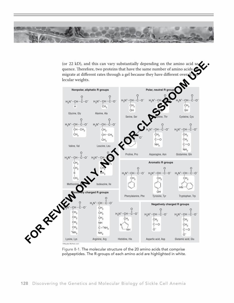
The molecular structure of proteins/polypeptides is more complex than that of DNA. First, the molecular weights of the component amino acids differ de-pending on the structure of their R-groups (Figure 8-1). Amino acids can vary in size ranging from 89–204 daltons, so polypeptides comprised of 200 amino acids would have a molecular weight in the neighborhood of 22,000 daltons
FOR REVIEW O
NLY. NOT FOR C
LASSROOM USE.
128 Discovering the Genetics and Molecular Biology of Sickle Cell Anemia
(or 22 kD), and this can vary substantially depending on the amino acid se-quence. Therefore, two proteins that have the same number of amino acids can migrate at different rates through a gel because they have different overall mo-lecular weights.
Nonpolar, aliphatic R groups Polar, neutral R groups
Positively charged R groups
Figure 8-1. The molecular structure of the 20 amino acids that comprise polypeptides. The R-groups of each amino acid are highlighted in white.
FOR REVIEW O
NLY. NOT FOR C
LASSROOM USE.
Discovering the Genetics and Molecular Biology of Sickle Cell Anemia 129
LABORATORY 8
Notice also that the R-groups of different amino acids that make up a polypep-tide can have different charges, and this too can affect the rate of migration through a gel. Finally, proteins are not linear structures like DNA but are instead folded into complex three-dimensional (3-D) configurations, and can often be comprised of multiple polypeptides bound together through various chemical interactions or bonds. The differences in the shape of a protein or polypeptide can also affect its rate of migration through a gel.
Types of Protein Electrophoresis
SDS-PAGE: One method of sorting proteins or polypeptides is called sodium dodecylsulfate-polyacrylamide gel electrophoresis, commonly referred to as SDS-PAGE (Figure 8-2). This method sorts polypeptides by molecular weight and is generally sensitive enough to resolve size differences in polypeptides of about 100 daltons (the approximate molecular weight of one amino acid) or greater.
Figure 8-2. SDS-PAGE of three polypeptides varying in overall length.
FOR REVIEW O
NLY. NOT FOR C
LASSROOM USE.
130 Discovering the Genetics and Molecular Biology of Sickle Cell Anemia
Proteins are first treated with sample preparation buffer (Laemmli) that breaks down (denatures) the protein by separating interacting polypeptides and dis-rupting its 3-D structure and then uniformly coating the protein with a nega-tive charge. Laemmli sample buffer contains a detergent, sodium dodecyl sulfate (SDS), which binds to proteins, penetrates the interior and coats the protein uni-formly with negative charge. This also effectively disrupts the majority of quater-nary, tertiary, and secondary 3-D structure of the protein. The second chemical in the sample buffer is called Tris. It functions to maintain the protein solution at a pH conducive to allow for electrophoretic separation. The sample buffer also con-tains glycerol that increases the density of the solution so that protein samples can be pipetted and added to an aqueous gel system. A dye is also added that colors the protein solution so that it can be easily tracked during electrophoresis. Finally, disulfide bonds between cysteine (cys) amino acids also contribute to a protein’s tertiary structure. If these bonds are not broken, proteins will not be completely linear and will not migrate solely based upon size. A reducing agent called DTT is also present, which breaks these bonds. The final step is to heat the mixture to 95° C for 5 minutes, completing the denaturation. At this point, all proteins are in their completely denatured state, are linear, and uniformly negatively charged. As in DNA electrophoresis, a current is then applied and the charged properties allow them to be carried through the electric field. The sieving effect of the gel is able to resolve size differences reflected by the number of amino acids comprising the respective polypeptides. So polypeptides of higher relative molecular weight (more amino acids) will now migrate through the gel slower than polypeptides of lower molecular weight (fewer amino acids). The negatively charged SDS deter-gent, which uniformly coats the denatured polypeptides, is the primary driver in the electrophoretic separation.
Native PAGE: In this method proteins are prepared in a non-reducing and non-denaturing sample buffer, which maintains the proteins’ secondary struc-ture and native charge density (see R-group charge/polarity in Figure 8-1). Unlike SDS-PAGE, where the electrophoretic mobility of proteins depends primarily on their length, in Native PAGE the mobility depends on the protein’s size combined with its overall charge and 3-D conformation. Moreover the electric charge driv-ing the electrophoresis is governed by the intrinsic charge on the protein at the pH of the running buffer. This charge will, of course, depend on the amino acid composition of the protein and the pH of the buffer solution in which the poly-peptides are dissolved. For our investigation, Native PAGE can be useful in sorting two polypeptides of the same length whose overall electrical charge is different, or where alterations in the amino acid sequence of a polypeptide influence its
FOR REVIEW O
NLY. NOT FOR C
LASSROOM USE.
Discovering the Genetics and Molecular Biology of Sickle Cell Anemia 131
LABORATORY 8
conformation and therefore rate of migration through the gel. The sample prepa-ration buffer used in Native PAGE contains Tris (which maintains a pH at which the polypeptides we are investigating have an overall negative charge), glycerol and an electrophoretic dye to track the movement of the polypeptides during electrophoresis.
In today’s inquiry, we will be running both types of protein electrophoresis in order to determine if Emily is a carrier for sickle cell anemia and also to provide some information about the possible nature of the mutation involved in sickle cell anemia. Several teams in lab will run an SDS-PAGE and several teams will run Native PAGE. Both gels will be analyzed by each team to help Chaka and Emily resolve their situation.
Note: Because polyacrylamide gel is quite expensive and carcinogenic, we will instead be simulating SDS-PAGE and Native PAGE using agarose.
Practicing Formulating a Hypothesis and Electrophoresis Prediction
Present one possible hypothesis as to the nature of the sickle cell mutation. Feel free to use one of the mutations from the table in your TLN or make one up of your own. However, be sure to specify which gene (the alpha globin or beta glo-bin) is mutated. Then predict the outcome of the electrophoresis of each of the hemoglobin samples from the four people in this scenario if your hypothesized mutation was the cause of sickle cell anemia.
▶▶ RECORD YOUR HYPOTHESES AND THE ASSOCIATED PREDICTION IN YOUR TLN BEFORE YOU BEGIN THE LAB. CHECK YOUR HYPOTHESIS AND PREDIC-TION WITH YOUR INSTRUCTOR BEFORE YOU PROCEED.
Here is one possible hypothesized mutation: If the sickle cell mutation was one that resulted in a premature stop codon early in the beta globin gene AND Emily was found to be a carrier of this mutation, then we would predict the following SDS-PAGE results.
FOR REVIEW O
NLY. NOT FOR C
LASSROOM USE.
132 Discovering the Genetics and Molecular Biology of Sickle Cell Anemia
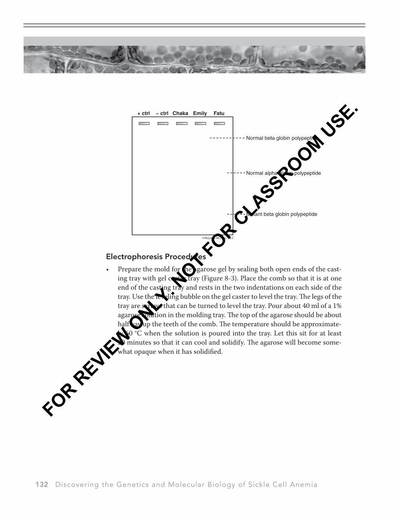
• Prepare the mold for the agarose gel by sealing both open ends of the cast-ing tray with gel caster tray (Figure 8-3). Place the comb so that it is at one end of the casting tray and rests in the two indentations on each side of the tray. Use the leveling bubble on the gel caster to level the tray. The legs of the tray are screws that can be turned to level the tray. Pour about 40 ml of a 1% agarose solution in the molding tray. The top of the agarose should be about halfway up the teeth of the comb. The temperature should be approximate-ly 60 °C when the solution is poured into the tray. Let this sit for at least 20 minutes so that it can cool and solidify. The agarose will become some-what opaque when it has solidified.
FOR REVIEW O
NLY. NOT FOR C
LASSROOM USE.
Discovering the Genetics and Molecular Biology of Sickle Cell Anemia 133
Figure 8-3. Procedure for pouring the gel using the gel caster.
• Gently pull the comb from the agarose gel, leaving distinct and open wells, and remove the casting tray from the gel caster. Transfer the gel to the elec-trophoresis unit, making sure to place the wells of the gel closest to the anode (black socket). You will use the micropipette to load the wells of the gel.
For Those Teams Running SDS-PAGE
• Prepare your hemoglobin samples by placing 2 drops of each person’s hemoglobin in separate microcentrifuge tubes. Add 1 drop of Laemmli sample buffer to each tube.
FOR REVIEW O
NLY. NOT FOR C
LASSROOM USE.
134 Discovering the Genetics and Molecular Biology of Sickle Cell Anemia
• The hemoglobin samples have already been heated to 95 °C to fully denature the polypeptides.
For Those Teams Running the Native PAGE
• Prepare your hemoglobin samples by placing 2 drops of each person’s he-moglobin in separate microcentrifuge tubes. Add 1 drop of Tris/Glycine (no SDS) sample buffer to each tube to provide the appropriate pH and density for electrophoretic separation.
• At this point the hemoglobin samples have been denatured to their second-ary structure, alpha and beta globin polypeptides have been separated.
All Teams
• Load 7 µl of the prepared hemoglobin samples into separate wells. Make sure that your samples are loaded evenly and that there are samples in the first and last wells (see Figure 8-4). Be careful not to poke a hole in the bottom of the gel while loading your samples. It is best to get the tip of the micropipette right above the well, and let the sample drop into the well below it.
+ ct
rl
− ct
rl
Chaka
Emily
Load your gels like this:
Fatu
Skip these wells
Figure 8-4. Load your gel like this.
• Pour the electrophoresis running buffer (TGS for SDS-PAGE; Tris buffer for Native PAGE) in the wells of the electrophoresis apparatus until the solution just covers the top of the gel.
• Run the gel at 55–60 volts until you get good separation (about 30–40 min-utes). Assign someone on your team to keep an eye on the gels, and do not let any bands run off the end of the gel. Take the gel out of the buffer solu-tion immediately, placing it on a light table, and interpret the results. While you are waiting for your gels to finish running, work on Week 5 of the Fruit Fly Genetics lab.
When your gel has finished running (that is, there is good separation between the bands on the gel) proceed to Part III to interpret the gel and share the news with Emily and Chaka.
FOR REVIEW O
NLY. NOT FOR C
LASSROOM USE.
Discovering the Genetics and Molecular Biology of Sickle Cell Anemia 135
LABORATORY 8
Part III. The Electrophoresis Results
The next class period, several of Dr. Fall’s students arrived early, eager to find out the results of the electrophoresis, but also a bit apprehensive about how they would share the potentially bad news with Chaka and Emily. Dr. Fall told his students that sharing both good and bad news was just part of being a doctor, and they would have to get used to it. However, he told them that what is most important is that they are careful in their analysis and to explain all aspects of the results in terms Chaka and Emily could easily understand, while being sensi-tive to their situation. “After all…” he said, “…there is reason to be optimistic even if the worst-case outcome in this situation is realized. Put yourself in Emily and Chaka’s place. What would you want to know more about with regard to making a decision about whether or not to have another child?”
For this analysis, pair up with another team that ran a different type of gel elec-trophoresis than the one your team ran. Some things to remember as you ana-lyze the gel:
• Remember that the Native PAGE can only tell you if the mutation has af-fected the shape and/or charge to mass ratio of the protein. For this gel, the upper band (closest to the well) in the positive control is the larger beta globin polypeptide.
• Remember that the SDS-PAGE gel separates polypeptide solely based on their size (reflected in the number of amino acids present in the polypeptide).
• Also remember that it is the position of the bands, NOT the color of the bands, which you should use to evaluate the gel.
Begin by Looking at the Native PAGE
1. Can you tell from the gel if Emily is a carrier for sickle cell anemia?
2. Can you tell from the gel which of the globin genes (alpha or beta) has mu-tated in this family and which persons inherited this mutation?
Now Examine the SDS-PAGE Gel
3. Can you tell from the gel which bands are the alpha and which bands are the beta polypeptides?
4. Can you tell from the gel if Emily is a carrier for sickle cell anemia?
5. Can you tell from the gel which of the globin genes (alpha or beta) has mu-tated in this family and which persons inherited this mutation?
6. What do the results of this gel tell you about how this mutation influences the length of the mutated polypeptide?
FOR REVIEW O
NLY. NOT FOR C
LASSROOM USE.
136 Discovering the Genetics and Molecular Biology of Sickle Cell Anemia
Now, Put the Results of Both Gels Together
7. Examine the following table (and in the TLN), which shows some of the common (or possible*) mutations to the globin genes that can be detected using protein gel electrophoresis. Which of the common mutations to the globin genes may have been inherited by members of this family (see Table 8-1)?
Table 8-1. Commonly found mutations to the globin genes.
Name Mutation
Hb Wayne Single nucleotide deletion in the alpha globin gene causes a frameshift, changing the codons for amino acids 139–141 and the stop codon.
Hb Grady Nine extra nucleotides are added to the alpha globin gene, which adds 3 amino acids between amino acids 118 and 119 of the polypeptide.
HbS A nucleotide substitution in the beta globin gene changes valine to glutamine at amino acid 6 of the polypeptide.
Hb Constant Spring A nucleotide substitution in the alpha globin gene changes the stop codon to the codon for glutamine.
Hb McKees Rock A nucleotide substitution in the beta globin gene changes tyrosine to a stop codon at amino acid 145 of the polypeptide.
Hb Leiden Three nucleotides are deleted from the beta globin gene resulting in amino acid 6 being deleted from the polypeptide.
*Hb Silent A nucleotide substitution in the alpha globin gene changes valine to proline at amino acid 63 of the polypeptide.
*HbL2 Mutation in the beta globin gene which prevents post translational removal of intron #2.
FOR REVIEW O
NLY. NOT FOR C
LASSROOM USE.
Discovering the Genetics and Molecular Biology of Sickle Cell Anemia 137
LABORATORY 8
J HOMEWORK
Imagine that you were a genetic counselor asked to use the results from this lab investigation to advise Chaka and Emily regarding their decision to have a child. Compose a formal letter in which you inform and advise them on their decision. Consider the following in your letter:
• Consider your audience—Emily and Chaka are not biologists, so try your best to write the letter so that it is understandable to them, and properly considers the importance and difficulty of their decision.
• What do the results of the electrophoresis experiment tell us about the kind of mutation(s) inherited by members of this family? How do these results provide evidence for Emily’s genotype with respect to the globin genes? Again, consider your audience.
• What are the relative probabilities that a child of Emily and Chaka’s could inherit each possible genotype?
• Dr. Fall suggested that even in the worst-case outcome of the protein elec-trophoresis test, there are reasons to be optimistic. What are those reasons given the results of the electrophoresis test and their relation to malaria?
• Feel free to do additional background research to inform your letter.
You may discuss this letter with your teammates, share knowledge and research resources. However EACH person on the team should write their own letter in their own words. The letter is due at next week’s lab. Consult with your instruc-tor on how to submit your letter for grading.
FOR REVIEW O
NLY. NOT FOR C
LASSROOM USE.
138 Discovering the Genetics and Molecular Biology of Sickle Cell Anemia