Discovery of a Novel Single-Stranded DNA Virus from a Sea TurtleFibropapilloma by Using Viral Metagenomics�
Terry Fei Fan Ng,1 Charles Manire,2† Kelly Borrowman,3 Tammy Langer,4‡Llewellyn Ehrhart,3 and Mya Breitbart1*
College of Marine Science, University of South Florida, 140 7th Avenue South, St. Petersburg, Florida 337011; Mote Marine Laboratory, 1600Ken Thompson Parkway, Sarasota, Florida 342362; College of Sciences, University of Central Florida, 4000 Central Florida Blvd.,
Received 16 September 2008/Accepted 23 December 2008
Viral metagenomics, consisting of viral particle purification and shotgun sequencing, is a powerful techniquefor discovering viruses associated with diseases with no definitive etiology, viruses that share limited homologywith known viruses, or viruses that are not culturable. Here we used viral metagenomics to examine virusesassociated with sea turtle fibropapillomatosis (FP), a debilitating neoplastic disease affecting sea turtlesworldwide. By means of purifying and shotgun sequencing the viral community directly from the fibropapil-loma of a Florida green sea turtle, a novel single-stranded DNA virus, sea turtle tornovirus 1 (STTV1), wasdiscovered. The single-stranded, circular genome of STTV1 was approximately 1,800 nucleotides in length.STTV1 has only weak amino acid level identities (25%) to chicken anemia virus in short regions of its genome;hence, STTV1 may represent the first member of a novel virus family. A total of 35 healthy turtles and 27 turtleswith FP were tested for STTV1 using PCR, and only 2 turtles severely afflicted with FP were positive. Theaffected turtles were systemically infected with STTV1, since STTV1 was found in blood and all major organs.STTV1 exists as a quasispecies, with several genome variants identified in the fibropapilloma of each positiveturtle, suggesting rapid evolution of this virus. The STTV1 variants were identical over the majority of theirgenomes but contained a hypervariable region with extensive divergence. This study demonstrates the potentialof viral metagenomics for discovering novel viruses directly from animal tissue, which can enhance ourunderstanding of viral evolution and diversity.
Discovering novel viruses is difficult due to the inability togrow many viruses in cell culture systems, the small size andlow nucleic acid content of viruses, and the lack of a conservedgenetic element found in all viral genomes. The most commonmethods for diagnosing viral infections use PCR, microarrays,immunologic assays, or replication in cell culture to test forspecific viruses. These methods are limited for viral discoverybecause many viruses cannot be cultured on standard cell lines,and primers and panviral microarrays require some degree ofsequence conservation in order to identify viruses. Recentstudies have demonstrated that viral particle purification andshotgun sequencing (viral metagenomics) constitute an effec-tive method for describing novel viruses, including those thatare unrelated to previously described viral families (reviewedin references 23 and 28). Viral metagenomics has been used tocharacterize viral communities present in the environment (6,8, 11, 15, 22, 24, 25, 31) and to describe viruses in mammalfeces (12, 13, 16, 69), cell culture (39, 43), respiratory tractaspirates (3), and blood (14, 40). However, to date, viruseshave not been purified directly from animal tissues for met-agenomic sequencing, due to the high background concentra-
tions of host nucleic acids. In this study, we successfully puri-fied viruses directly from a sea turtle fibropapilloma andsubsequently discovered a novel virus that could not have beenidentified using PCR with degenerate primers or a panviralmicroarray. The methods presented here are directly applica-ble to a variety of animals and tissue types, therefore signifi-cantly enhancing our ability to discover novel viruses.
Fibropapillomatosis (FP) is a debilitating neoplastic diseaseaffecting sea turtles worldwide (2, 34). Affected turtles developepithelial fibropapillomas, and some turtles develop internalfibromas in terminal cases (67). FP primarily affects green seaturtles, Chelonia mydas, but has been confirmed in all seven seaturtle species (32, 35). In some regions, such as the IndianRiver Lagoon in Florida, FP affects more than half of the greenturtle population (19, 32, 34). FP has been documented sincethe late 1930s (61) and is now widespread throughout theworld (32, 35). Since all species of marine turtles are eitherendangered or threatened (18), the additional stress of FPposes a potential threat to the future survival of sea turtles.
The etiological agent responsible for FP is unknown; how-ever, current evidence strongly supports the involvement of analphaherpesvirus, FP-associated turtle herpesvirus (FPTHV)(45, 47, 54, 55, 68). Transmission studies have demonstratedthat the infectious FP agent is less than 0.45 �m in size and canbe inactivated by organic solvents and high-speed centrifuga-tion, which is consistent with the theory of an enveloped virusas the causative agent (36, 37). Using PCR, FPTHV DNA hasbeen found in most examined fibropapillomas but not detectedin the vast majority of healthy turtles (45, 47, 54). However,extensive attempts at isolating and propagating FPTHV in cell
* Corresponding author. Mailing address: University of South Flor-ida, College of Marine Science, 140 7th Avenue South, St. Petersburg,FL 33701. Phone: (727) 553-3520. Fax: (727) 553-1189. E-mail: [email protected].
† Present address: Atlantis Resort, Paradise Island, Bahamas.‡ Present address: Volusia Marine Science Center, Ponce Inlet, FL
32127.� Published ahead of print on 30 December 2008.
2500
Dow
nloa
ded
from
http
s://j
ourn
als.
asm
.org
/jour
nal/j
vi o
n 23
Nov
embe
r 20
21 b
y 18
3.10
2.11
6.15
5.
culture have been unsuccessful (19). Since FPTHV has notbeen obtained in pure culture and shown to cause FP, it ispossible that there are other undescribed, coexisting virusesinvolved in the etiology of FP. For example, Lu et al. (48)observed a small naked virus able to induce tumor-like aggre-gates in sea turtle cell lines. Only a very short sequence wasobtained from these viruses, which had weak similarity to pap-illomaviruses (48). Another study, by Casey et al. (17), identi-fied a retrovirus in Hawaiian green sea turtles, but it is believedto be an endogenous retrovirus unrelated to FP. Therefore, theobjective of this study was to use viral metagenomics to identifyDNA viruses associated with FP in a Florida green sea turtle.
Viral particles were purified from a sea turtle fibropapillomausing a combination of size selection, density-dependent cen-trifugation, and nuclease treatment. DNA was extracted fromthe purified viruses, amplified with sequence-independent am-plification, and shotgun sequenced. A novel single-strandedDNA virus with extremely limited similarity to known viruseswas discovered. The genome of this virus, which we havenamed sea turtle tornovirus 1 (STTV1), was completely se-quenced. In addition, several closely related variants of thisvirus were identified, indicating that STTV1 circulates as aquasispecies. Here we describe the STTV1 genome sequence,prevalence among green sea turtles, and variability within theSTTV1 quasispecies.
MATERIALS AND METHODS
Sea turtle fibropapilloma sample. A wild juvenile green turtle with FP ofcategory 3 severity (7) was captured from Lake Worth Lagoon, FL, in 2006 andtransferred to the Clearwater Aquarium and later to the Mote Marine Aquariumfor rehabilitation. Severe external fibropapillomas were present, and X-raysrevealed the presence of internal fibromas. Due to its deteriorating condition,the turtle was humanely euthanized. External fibropapillomas from this turtlewere biopsied and kept at �80°C until processed.
Viral purification and DNA extraction. The external fibropapilloma was ho-mogenized in sterile SM buffer (50 mM Tris, 10 mM MgSO4, 0.1 M NaCl, pH7.5) using a mortar and pestle. Host cells were removed by centrifugation at10,000 � g at 4°C for 10 min. The supernatant was then filtered through a0.45-�m filter (Millipore, Billerica, MA) and loaded onto a cesium chloride stepgradient consisting of 1 ml each of 1.7, 1.5, and 1.2 g/ml in sterile SM buffer (14).The gradient was ultracentrifuged at 61,000 � g at 4°C for 3 h, and the viralfraction between 1.2 g/ml and 1.5 g/ml was collected. The viral fraction was thenincubated for 10 min with 0.2 volumes of chloroform. The supernatant contain-ing the virus was removed from the chloroform and incubated with 2.5 U DNaseI (Sigma-Aldrich, St. Louis, MO) at 37°C for 3 h. The DNase-treated sample wasconcentrated and washed twice with sterile SM buffer on a Microcon 30 column
(Millipore). DNase activity was then inhibited by adding EDTA to a final con-centration of 20 mM. Finally, DNA was extracted from the purified viruses usingthe QIAamp MinElute virus spin kit (Qiagen, Valencia, CA) according to themanufacturer’s instructions. To ensure the effectiveness of the viral purificationprocedure, the extracted DNA was assayed for turtle DNA contamination usingeukaryotic rRNA gene PCR (universal primer A, 5�-AACCTGGTTGATCCTGCCAGT-3�; universal primer B, 5�-TGATCCTTCTGCAGGTTCACCTAC-3�)(62) and for mitochondrial DNA contamination with conserved primers (L14841,5�-AAAAAGCTTCCATCCAACATCTCAGCATGATGAAA-3�; H15149, 5�-AAACTGCAGCCCCTCAGAATGATATTTGTCCTCA-3�) (44).
Viral metagenome library construction and sequencing. Clone libraries wereprepared for shotgun sequencing as described previously (14). First, viral DNAwas amplified with sequence-independent amplification using the GenomiPhi V2DNA amplification kit (GE Healthcare, Piscataway, NJ) according to the man-ufacturer’s instructions. This method uses isothermal strand displacement am-plification with the phi29 DNA polymerase and random hexamers to amplifytotal genomic DNA by 2 to 3 orders of magnitude (10).
The amplified DNA was then randomly fragmented, linker adapted, andfurther amplified with primers to the linkers using the GenomePlex whole-genome amplification kit (Sigma-Aldrich, St. Louis, MO) according to the man-ufacturer’s instructions. The reaction product was processed with a PCR cleanupkit (Mo Bio Laboratories, Solana Beach, CA) and then cloned into the pCR4vector using a Topo TA cloning kit (Invitrogen, Carlsbad, CA). Transformantswere screened for inserts and then sequenced by Agencourt Bioscience (Beverly,MA) using the M13 forward primer. A total of 276 transformants were se-quenced, and the sequences were trimmed for vector and read quality using theSequencher 4.7 software program (Gene Codes, Ann Arbor, MI). Trimmedmetagenomic sequences (n � 211) were assembled into contigs (contiguoussequences) using a minimum overlap of 25 bp and a minimum match percentageof 85%. All assemblies were examined manually and verified experimentallywith PCR.
Complete genome sequencing of sea turtle tornovirus 1 and its quasispeciesvariants. Both trimmed sequences and assembled contigs from the viral metage-nome were analyzed using BLASTX and TBLASTX against the GenBank non-redundant database (4, 5). Several sequences with amino-acid-level similarity tochicken anemia virus (CAV) were noted, and efforts were focused on completingthe genome sequence of this virus. PCR primers (Table 1) were designed toamplify and sequence the genomic regions between the contigs. Unless otherwisespecified, all PCRs were in a final volume of 50 �l containing 200 �M de-oxynucleoside triphosphates, 1 U of Red Taq polymerase (Sigma-Aldrich), 1�Red Taq reaction buffer, 1 �M of the specific primers, and either 5 �l of thesample DNA or 1 �l of sample DNA amplified by using Genomiphi. PCR withthe primer set STTV1-b was performed to amplify a region of 1,571 nucleotidesin order to confirm the assembly of the metagenomic sequences. PCR wasperformed using the STTV1-c inverse primers to confirm the circular nature ofthe STTV1 genome.
A series of amplicons with different sizes were observed from the STTV1-cPCR and gel electrophoresis, reflecting a hypervariable region (HVR) in thegenome that was difficult to sequence and required a different sequencing strat-egy. Two strategies were employed to sequence the HVR. The first strategyincluded rolling-circle amplification (Templiphi; GE Healthcare) with randomhexamer primers, followed by touchdown PCR using STTV1-c primers over a
TABLE 1. PCR primers used to sequence the STTV1 genome, characterize STTV1 prevalence, and determine genome polarityand strandedness
Primer name Sequence (5� to 3�) PCR conditions
STTV1-a Forward GAGTATTGCCGAGGCTCAAG 95°C for 5 min; 45 cycles of �94°C for 30 s, 58°C minus 0.2°C per cycle for30 s, 72°C for 2 min�; 72°C for 10 minSTTV1-a Reverse TGCAGAATTCTCGTTGAAGG
STTV1-b Forward AGTCAGAGAGCAGCGGATGT Same as for STTV1-aSTTV1-b Reverse ATGGTACAACCCCAAAGCGSTTV1-c Forward AAGCACCTTCCCAATACGG 95°C for 5 min; 45 cycles of �94°C for 30 s, a gradient of annealing tempSTTV1-c Reverse TTATAGGGGCAGTAGGCACG of 55.4°C, 58.1°C and 60.7°C minus 0.2°C per cycle for 30 s, and 72°C
for 2 min�; 72°C for 10 minSTTV1-d Antisense ACATCCGCTGCTCTCTGACT 95°C for 5 min; 50 cycles of �94°C for 1 min, 49°C for 1 min, 72°C for
3 min�; 72°C for 10 minSTTV1-d Sense GGGAGGATTGCTGAGATGAASTTV1-e Forward CGTGCCTACTGCCCCTATAA Same as STTV1-dSTTV1-e Reverse CCAGATAATTAGTAGCACCAGAATGASTTV1-e Forward CGTGCCTACTGCCCCTATAA 95°C for 5 min; 45 cycles of �94°C for 1 min, 54°C minus 0.2°C per cycle
for 1 min, 72°C for 3 min�; 72°C for 10 minSTTV1-f Reverse GGGAGGATTGCTGAGATGAA
VOL. 83, 2009 NOVEL VIRUS FROM A SEA TURTLE FIBROPAPILLOMA 2501
Dow
nloa
ded
from
http
s://j
ourn
als.
asm
.org
/jour
nal/j
vi o
n 23
Nov
embe
r 20
21 b
y 18
3.10
2.11
6.15
5.
gradient of annealing temperatures (Table 1), pooling of PCR products, andTopo TA cloning. The second strategy was identical to the first one with theexception that rolling-circle amplification was not used. For each tornovirus-positive turtle, 10 clones were selected and sequenced with the M13 forward andreverse primers. Each clone was sequenced at least three times. The HVRsequences were regarded as valid only if both ends of the HVR sequence alignedto the main STTV1 sequence with a total overlap of at least 80 nucleotides,excluding the primer region. Since the genomes were identical with the exceptionof the HVR, these were regarded as variants of the STTV1 quasispecies. Putativeopen reading frames (ORFs) in the genome were predicted using the NCBI ORFFinder (http://www.ncbi.nlm.nih.gov/projects/gorf) with a minimum ORF size of100 amino acids, and the genome was annotated using the SeqBuilder softwareprogram (DNAstar, Madison, WI). Repeats were analyzed using the TandemRepeats Finder database (http://tandem.bu.edu/trf/trf.html) (9) and by aligningthe sequence with itself using BLAST (http://www.proweb.org/proweb/Tools/selfblast.html). The potential nuclear localization signal was predicted usingPREdictNLS (20).
Polarity and single-stranded nature of STTV1. The polarity of the STTV1genome was determined using the strand-specific primer extension method (51).Purified virus DNA was amplified linearly for 50 cycles with either the STTV1-dsense primer or the STTV1-d antisense primer (Table 1) in a reaction mixture of50 �l. The strand-specific primer extension was repeated with the same primerfor an additional 50 cycles by adding the entire first reaction mixture to a new50-�l PCR. Five microliters of the primer extension products were then used asthe target in a PCR with the STTV1-e primers, which were internal to thestrand-specific primers (Table 1). The PCR product intensities were comparedby gel electrophoresis and ethidium bromide staining. The strand of packagedviral DNA is complementary to the strand-specific primer with a stronger PCRsignal.
The single-stranded nature of the purified STTV1 DNA was determined byrestriction enzyme digestion followed by PCR with primers flanking the restric-tion site. Two microliters of purified viral DNA was digested with 10 U of AluI(New England Biolabs, Ipswich, MA) for 60 min in the presence of 1� enzymebuffer in a total reaction of 20 �l. Five microliters of the digested product wasamplified by PCR using STTV1-e Forward and STTV1-f Reverse (Table 1),which flanked the digestion site. Resistance to digestion and subsequent ampli-fication by PCR confirmed the single-stranded nature of the packaged viralgenome.
PCR detection of STTV1 prevalence in Florida green sea turtles. To investi-gate the prevalence and tissue specificity of the newly discovered STTV1, nu-merous sample types (fibropapillomas, various internal organs, blood, and ex-ternal swabs) were examined using PCR with the STTV1-a primers (Table 1).Fibropapillomas from four captive green turtles were biopsied at Mote MarineAquarium, FL, and total DNA was extracted from the fibropapillomas using theDNeasy Blood & Tissue kit (Qiagen, Valencia CA). The fibropapilloma fromone of the turtles was positive for STTV1, and therefore, oral, conjunctival,cloacal, and unaffected skin swabs, as well as a blood sample from this turtle,were tested using the same extraction and PCR protocols.
This PCR-positive turtle was later euthanized due to deteriorating healthconditions. To further examine the distribution of STTV1 in this turtle, externalfibropapilloma, internal fibroma, blood, kidney, liver, lung, pancreas, spleen,stomach, heart, muscle, esophagus, and urinary bladder were biopsied, and DNAwas extracted and tested by PCR with the STTV1-a primers. To determine thesensitivity of this assay, PCR was performed with the STTV1-a primers on adilution series of positive control DNA. The tissues were also tested for FPTHVusing PCR with FPTHV-specific primers (GTHV1, 5�-TGTCTGGAGGTGGCGGCCACG-3�; GTHV2, 5�-GACACGCAGGCCAAAAAGCGA-3�) (55). TheFPTHV PCR conditions were 94°C for 5 min and 35 cycles of [94°C for 30s, 62°Cfor 30s, and 72°C for 30s], followed by 72°C for 5 min.
Results demonstrated that STTV1 could be detected in external swabs fromthe positive turtle; therefore, swabbing was used as a noninvasive samplingmethod to test a large number of FP and non-FP turtles for the presence ofSTTV1. Conjunctival swabs, oral swabs, fibropapilloma swabs, skin swabs, and/orblood samples were taken from a total of 17 captive and 45 wild green sea turtles.The 17 captive turtles were being kept at Mote Marine Aquarium and Clearwa-ter Marine Aquarium in Florida, and 12 of these turtles were affected by FP.Twenty-six of the wild turtles were captured as part of a routine monitoringsurvey at the Indian River Lagoon (Melbourne Beach, FL), and 15 of theseturtles had FP. Nineteen of the wild turtles were captured in Trident SubmarineBasin (Cape Canaveral, FL), and none of these turtles had FP. In addition,leeches were collected opportunistically when wild green turtles were sampled tocheck for the presence of STTV1. DNA was extracted from the swabs and
leeches using the DNeasy Blood & Tissue kit and tested by PCR with theSTTV1-a primers (Table 1).
Nucleotide sequence accession numbers. The complete genome sequences foreach of the nine STTV1 quasispecies variants have been deposited to GenBankwith the accession numbers EU867816 to EU867824.
RESULTS
Purification of viruses from fibropapilloma tissue. Using acombination of size selection, density-dependent centrifuga-tion, and nuclease treatment, viral particles were successfullypurified from the sea turtle fibropapilloma. The purified viralfraction tested negative for turtle host DNA by PCR with theeukaryotic rRNA gene PCR (62) and was highly reduced inmitochondrial DNA as assessed by universal mitochondriumPCR (44) (Fig. 1). PCR with the STTV1-e primers revealed anenrichment of STTV1 in the purified viral fraction. This dem-onstrates the efficacy of the methods described here for puri-fying viral particles away from contaminating nucleic acids inanimal tissues.
Analysis of viral metagenomic sequences and discovery of anovel sea turtle tornovirus. BLAST analysis of the 211 viralmetagenomic sequences revealed 13 sequences (6%) that as-sembled into contigs with weak amino acid similarity to CAV,therefore indicating the discovery of a new virus (STTV1).These sequences were used to design PCR primers to completethe STTV1 genome sequence and verify the assembly of themetagenomic sequences.
Eleven sequences of the viral metagenome were similar tobacteriophages, which likely infect the turtle’s normal bacteriaflora. Some of the sequences had amino acid similarity tobacteria and Plasmodium, which may be the result of imperfectviral purification or may be viral sequences that have no viralhomologs in the database. The majority of the remaining se-quences had no similarities in GenBank.
FIG. 1. Effectiveness of the virus purification procedure as deter-mined by PCR assays. “Purified viral DNA,” as indicated by arrows,was extracted from viral particles purified by filtration, density-depen-dent centrifugation, and nuclease treatment. “Total FP DNA” wasextracted from a sea turtle fibropapilloma without any purification ofviruses. The amount of host and mitochondrial DNA was greatlyreduced in the purified viral DNA, while STTV1 was enriched in thisfraction.
2502 NG ET AL. J. VIROL.
Dow
nloa
ded
from
http
s://j
ourn
als.
asm
.org
/jour
nal/j
vi o
n 23
Nov
embe
r 20
21 b
y 18
3.10
2.11
6.15
5.
Screening of fibropapillomas for STTV1 by PCR. Includingthe turtle where STTV1 was originally discovered, two out offive fibropapilloma samples tested positive for STTV1. BothSTTV1-positive turtles were severely afflicted by FP and haddeveloped extensive fibropapillomas and internal fibromas
when the samples were taken. The sensitivity of the STTV-1aPCR was determined to be approximately 10 targets.
Prevalence and tissue specificity of STTV1. In one of theSTTV1-positive turtles, STTV1 was detected in external fibro-papilloma, internal fibroma, blood, kidney, liver, lung, pan-
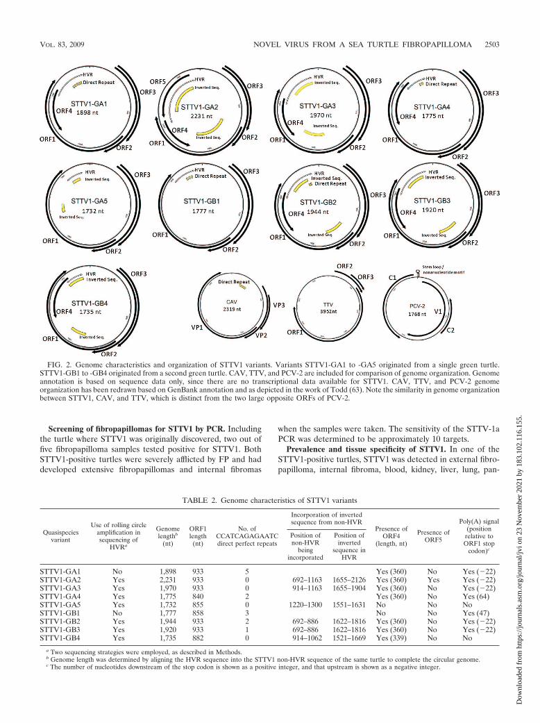
FIG. 2. Genome characteristics and organization of STTV1 variants. Variants STTV1-GA1 to -GA5 originated from a single green turtle.STTV1-GB1 to -GB4 originated from a second green turtle. CAV, TTV, and PCV-2 are included for comparison of genome organization. Genomeannotation is based on sequence data only, since there are no transcriptional data available for STTV1. CAV, TTV, and PCV-2 genomeorganization has been redrawn based on GenBank annotation and as depicted in the work of Todd (63). Note the similarity in genome organizationbetween STTV1, CAV, and TTV, which is distinct from the two large opposite ORFs of PCV-2.
TABLE 2. Genome characteristics of STTV1 variants
Quasispeciesvariant
Use of rolling circleamplification insequencing of
HVRa
Genomelengthb
(nt)
ORF1length
(nt)
No. ofCCATCAGAGAATCdirect perfect repeats
Incorporation of invertedsequence from non-HVR
Presence ofORF4
(length, nt)
Presence ofORF5
Poly(A) signal(position
relative toORF1 stop
codon)c
Position ofnon-HVR
beingincorporated
Position ofinverted
sequence inHVR
STTV1-GA1 No 1,898 933 5 Yes (360) No Yes (�22)STTV1-GA2 Yes 2,231 933 0 692–1163 1655–2126 Yes (360) Yes Yes (�22)STTV1-GA3 Yes 1,970 933 0 914–1163 1655–1904 Yes (360) No Yes (�22)STTV1-GA4 Yes 1,775 840 2 Yes (360) No Yes (64)STTV1-GA5 Yes 1,732 855 0 1220–1300 1551–1631 No No NoSTTV1-GB1 No 1,777 858 3 No No Yes (47)STTV1-GB2 Yes 1,944 933 2 692–886 1622–1816 Yes (360) No Yes (�22)STTV1-GB3 Yes 1,920 933 1 692–886 1622–1816 Yes (360) No Yes (�22)STTV1-GB4 Yes 1,735 882 0 914–1062 1521–1669 Yes (339) No No
a Two sequencing strategies were employed, as described in Methods.b Genome length was determined by aligning the HVR sequence into the STTV1 non-HVR sequence of the same turtle to complete the circular genome.c The number of nucleotides downstream of the stop codon is shown as a positive integer, and that upstream is shown as a negative integer.
VOL. 83, 2009 NOVEL VIRUS FROM A SEA TURTLE FIBROPAPILLOMA 2503
Dow
nloa
ded
from
http
s://j
ourn
als.
asm
.org
/jour
nal/j
vi o
n 23
Nov
embe
r 20
21 b
y 18
3.10
2.11
6.15
5.
creas, spleen, stomach, heart, esophagus, and urinary bladdersamples. STTV1 was not detected in the muscle sample or inan internal fibroma biopsied from the right lung. STTV1 wasalso found in external swabs collected from the conjunctiva,oral cavity, cloaca, external fibropapilloma, and unaffectedskin. In contrast, FPTHV was detected only in the fibropapil-lomas and fibromas and was not found anywhere else in theturtle. Since STTV1 could be detected in external swabs col-lected noninvasively, this method was used to screen morecaptive and wild green sea turtles for STTV1.
A total of 62 green turtles (27 with FP and 35 without FP)were swabbed and tested for STTV1. The presence of FP wasdetermined by careful inspection for fibropapillomas (7). Theonly turtles positive for STTV1 were the same two turtlesidentified previously by screening of fibropapillomas. All otherturtles were negative for STTV1 in the conjunctival swabs, oralswabs, fibropapilloma swabs, skin swabs, and/or blood samples.Interestingly, STTV1 was detected in leeches collected fromone of the wild green sea turtles captured in the Indian RiverLagoon (Florida), suggesting that leeches may be a possiblevector for the transmission of STTV1.
Sequence analysis of the sea turtle tornovirus genome andits quasispecies variants. The STTV1 genomes from the twopositive turtles were completely sequenced. In order to com-plete the STTV1 genome, it was necessary to sequence theconserved region of the genome (non-HVR) and the HVRseparately. The non-HVR was amplified with the STTV1-bprimers. Over the 1,571-nucleotide (nt) STTV1-b amplicon,the viruses from the two positive turtles differ at only 10 posi-tions (sequence divergence of 0.6%); thus, the non-HVR ofSTTV1 were almost identical.
PCR using the STTV1-c inverse primer set confirmed thecircular nature of the STTV1 genome. PCR with these primersalso identified a HVR, in which nine different PCR ampliconswere identified in the two turtles. The HVR varied in length byalmost 500 nucleotides, leading to quasispecies genome sizesranging from 1,732 to 2,231 nucleotides (Fig. 2 and Table 2).Five variants, named STTV1-GA1 to STTV1-GA5, originatedfrom the same turtle, while the other four variants (STTV1-GB1 to STTV1-GB4) were from the second turtle. The se-quence alignment of the HVR of the nine variants is shown inFig. 3a. At both ends of the HVR (positions 1507 to 1629 andpositions 2125 to 2166), variation is due only to insertions ordeletions. In the middle of the HVR (positions 1630 to 2124),variations include nucleotide insertions, deletions, and substi-tutions. Some of the variations were shared between two ormore variants.
Sequence analysis demonstrated that six of the variants in-corporated an inverted sequence from the non-HVR regioninto the middle of the HVR (Fig. 2 and Table 2). For example,STTV1-GB2 and STTV1-GB3 incorporated identical se-quences from the non-HVR into the HVR. The HVR ofSTTV1-GA2, STTV1-GA3, and STTV1-GB4 shared anotherinverted non-HVR sequence, but the length of the incorpo-rated sequence was variable. The HVR of STTV1-GA5 con-tained an inverted non-HVR sequence that was not found inthe other variants in this study. The sequences incorporatedinto the HVR were all located inside the sequence annotatedas ORF1.
ORF1 was present in all nine variants, and it spanned both
the conserved regions and HVR. Due to the addition anddeletion of nucleotides in the HVR, the length of ORF1 variedfrom 840 to 933 nt (Table 2). ORF1 was characterized by anarginine-rich region, containing 23 arginines in the first 43amino acids (53%). The arginine-rich region of ORF1 wassimilar to the arginine-rich region in other single-strandedDNA viruses, such as anelloviruses, gyroviruses, and circovi-ruses. The ORF1 arginine-rich region was located in the non-HVR region and therefore was shared by all STTV1 genomevariants. Analysis using a nuclear localization signal predictiontool revealed that the arginine-rich region in ORF1 had strongpotential as a nuclear localization signal. Other than the char-acteristic of an arginine-rich region, the remaining sequence ofthe STTV1 ORF1 had no amino acid level similarity to anyproteins in the nonredundant database.
Unlike ORF1, both ORF2 and ORF3 were located entirelywithin the non-HVR region. ORF2 was 687 nt in length, and itshared low (25%) amino acid identity with the CAV VP2protein over a region of 217 amino acids. STTV1 ORF2 had 16amino acids identical to those of the CAV pfam (04861) overa region of 40 amino acids. STTV1 ORF3 was 522 nt in length,and it had no similarity to any known proteins.
While ORF1, ORF2, and ORF3 were found in every ge-nome variant sequenced, two other putative ORFs were foundonly in some of the variants, depending on the sequence of theHVR (Fig. 2 and Table 2). ORF4, which had no similarity toany known proteins, was present in seven of the nine variants.Although the start codon of ORF4 was in the HVR, the lengthof ORF4 was typically 360 nt, except in STTV1-GB4, wherethe length was 339 nt.
ORF5 was present only in STTV1-GA2, where it was 522 ntin length. Sequence analysis showed that the first 471 nt of thisORF were the inverted repeat of a non-HVR sequence nearthe beginning of ORF1 (Fig. 2 and Table 2). Therefore, ORF5resembled ORF1 for the most part, including the arginine-richregion. ORF5 was shorter than ORF1 and had no similarity toany known protein except the arginine-rich characteristic.
A TATA box (TATAAA) was identified in the non-HVRsequence, and thus, all of the variants contain a TATA box inthe genome. The TATA box was arbitrarily designated to bethe first nucleotide of the circular genome, and it was located171 nt upstream of the start codon of ORF2. Additionally, apoly(A) signal (AATAAA) was identified in the HVR. Thepoly(A) signal was positioned 22 nt upstream of the stop codonof ORF1 in five of the variants (STTV1-GA1, -GA2, -GA3,-GB2, and -GB3 in Table 2), while it was 47 nt and 64 ntdownstream for two variants (STTV1-GB1 and -GA4). Theremaining two variants (STTV1-GA5 and -GB4) had nopoly(A) signal.
Direct repeats were recognized in the HVR sequences offour variants (Fig. 3b). Two types of direct repeats were rec-ognized: a perfect direct repeat of 13 nt in length (CCATCAGAGAATC) and a near-perfect direct repeat of 11 nt in theform of (T/C)CATCAGAGAATC. The 13-nt perfect directrepeats occurred between two to five times in different variantsand were interspaced with the 11-nt near-perfect direct repeatand/or gaps of other short sequences.
Genomic features distinguishing STTV1 quasispecies vari-ants. Nucleotide insertions, deletions, and substitutions in theHVR contribute to the array of genomic features seen in the
2504 NG ET AL. J. VIROL.
Dow
nloa
ded
from
http
s://j
ourn
als.
asm
.org
/jour
nal/j
vi o
n 23
Nov
embe
r 20
21 b
y 18
3.10
2.11
6.15
5.
FIG. 3. (a) Alignment of the nine HVR sequences. The variants STTV1-GA1 to STTV1-GA5 originated from a single green turtle, whileSTTV1-GB1 to STTV1-GB4 originated from a second turtle. Primer STTV1-B Forward (5� CAAGCACCTTCCCAATACGGC 3�) is underlined.Part of STTV1-B Reverse (5�TTATAGGGGCAGTAGGCACG 3�) is underlined, but the first 5 nt of the primer were located at positions 1 to5 in the circular genome and are not shown in the alignment. (b) Direct repeats in the HVR of the variants STTV1-GA1, STTV1-GA4,STTV1-GB1, and STTV-GB2. PR, 13-nt perfect direct repeat in the form of CCATCAGAGAATC; NPR, 11-nt near-perfect direct repeat in theform of (T/C)CATCAGAGAATC. The position of the last nucleotide shown in each variant is noted on the right.
VOL. 83, 2009 NOVEL VIRUS FROM A SEA TURTLE FIBROPAPILLOMA 2505
Dow
nloa
ded
from
http
s://j
ourn
als.
asm
.org
/jour
nal/j
vi o
n 23
Nov
embe
r 20
21 b
y 18
3.10
2.11
6.15
5.
nine different STTV1 quasispecies variants (Fig. 2 and 3a;Table 2). First, differences in the length of the HVR contributeto variation in the STTV1 genome size, which ranges from1,735 to 2,231 nt. Second, the HVR sequence generates adifferent amino acid composition and length of ORF1, whichspans between the HVR and non-HVR in each of the variants.The length of ORF1 can vary from 840 to 933 nt. Third, ORF4is found in seven of the nine variants, while ORF5 is found inone variant. Fourth, the number and pattern of direct perfectrepeats and direct near-perfect repeats differ in the HVR ofvarious variants (Fig. 3b). Fifth, various lengths of non-HVRsequences are incorporated into the HVR in inverted orienta-tion in some of the variants (Fig. 2 and Table 2). Sixth, apoly(A) signal was found in most variants upstream or down-stream of the stop codon of ORF1.
The combination of these features makes up the differentgenome characteristics of each variant (Fig. 2 and Table 2).STTV1-GA1 had a genome of 1,898 nt, with a GC content of51.8%. It contained putative ORF4 and five 13-nt perfect re-peats interspaced with three 11-nt near-perfect repeats. TheSTTV1-GA2 genome was 2,231 nt in length and was charac-terized by the presence of ORF4 and ORF5. It incorporated aninverted sequence from the non-HVR, making it the largestSTTV1 variant sequenced. No direct repeat was found in theSTTV1-GA2 genome. STTV1-GA3 (1,970 nt), STTV1-GB3(1,920 nt), and STTV1-GB4 (1,735 nt) all contained ORF4 andinverted sequence from the non-HVR but had no or only onecopy of the direct repeat sequence. STTV1-GA4 (1,775 nt)contained ORF4, as well as two 13-nt perfect repeats in-terspaced with two 11-nt near-perfect repeats. STTV1-GA5(1,732 nt) had an inverted sequence from the non-HVR butlacked ORF4. STTV1-GB1 (1,777 nt) had three 13-nt perfectrepeats interspaced with two 11-nt near-perfect repeats andalso lacked ORF4. The STTV1-GB2 genome (1,944 nt) con-tained ORF4, incorporated an inverted sequence from thenon-HVR, and had two 13-nt perfect direct repeats in-terspaced with one copy of the 11-nt near-perfect direct repeatsequence.
Polarity and strandedness of the purified STTV1. TheSTTV1 DNA packaged in the viral particle was singlestranded, since the purified STTV1 DNA treated with a re-striction enzyme could subsequently be amplified by PCR overthe restriction enzyme recognition site.
ORF1, ORF2, and ORF3 were found consistently in everySTTV1 variant, and since these three ORFs were likely to betranscribed, they were considered positive sense. Polarity ex-periments demonstrated that the viral particle contains a neg-ative-sense genome, which is the reverse complement of thesequence shown in this article.
DISCUSSION
This article demonstrates the use of viral metagenomics todiscover novel viruses directly from an animal tissue. We havesuccessfully purified virus particles against a high backgroundof host DNA (Fig. 1) and have identified and completely se-quenced the genome of a novel sea turtle virus, STTV1, fromthe purified DNA.
STTV1 possesses a single-stranded circular genome, andmost of its genome has no similarities to any known viruses.
ORF2 has weak (25%) amino acid level similarity to the VP2protein of CAV. The remainder of the genome has no aminoacid similarity to any protein, except that the arginine-richregion of ORF1 and ORF5 is similar to those of anelloviruses,gyroviruses, and circoviruses. The STTV1 genome is so uniquethat it is highly unlikely to have been discovered by degenerateprimers or panviral microarrays based on known viruses. Basedon its genome sequence, we believe that STTV1 may representa new viral genus of the Circoviridae family or possibly even anew viral family.
The genome organization of STTV1 is more similar to thatof CAV (the only member of the Gyrovirus genus) and Torqueteno virus (TTV) (a member of the floating Anellovirus genus)than to those of circoviruses (Fig. 2). The STTV1 genomeconsists of a region of direct repeats followed by short ORF2and ORF3 sequences, and ORF1 starts near the end of ORF3(Fig. 2). CAV is characterized by a similar genome organiza-tion pattern: repeat/promoter region at the noncoding part ofthe genome, followed by VP2 and VP3, with VP1 startingimmediately after VP3 (Fig. 2). Likewise, genome annotationof TTV using sequence data reveals a similar ORF organiza-tion: short ORF2 and ORF3, followed by a long ORF1 (63). Incontrast, circoviruses have a different genome organizationfrom STTV1, containing two long ORFs (V1 and C1) locatedon opposite strands (Fig. 2) (63). STTV1 also lacks the con-served stem-loop structure and the nonanucleotide motif ofcircoviruses (64). It should be noted that transcriptional stud-ies have recently updated the ORF annotations of CAV andTTV (41, 42), but the discussion here concerns only annotationbased on sequence data, since there is no transcriptional datafrom STTV1 to compare with at this time. There are currentlytwo families of single-stranded DNA (ssDNA) animal viruses,namely, Circoviridae and Parvoviridae, as well as the floatinganellovirus genus. The discovery of STTV1 provides a linkbetween anelloviruses and gyroviruses (Circoviridae), sinceSTTV1 shares some genomic features with both groups ofviruses. STTV1 may therefore be an important evolutionaryintermediate that can enhance our understanding of the evo-lution of ssDNA viruses.
ORF1, ORF2, and ORF3 are found consistently in everySTTV1 variant (Fig. 2) and are located on the opposite strandfrom the strand packaged in viral particles, suggesting a neg-ative-sense genome organization. ORF4 and ORF5 are foundonly in some variants, and they are on the opposite strand ofORF1 to ORF3. Further studies are needed to determine if allthe ORFs are transcribed, which will reveal whether STTV1 isa negative-sense or ambisense virus. Comparatively, TTV andCAV also have three ORFs that are arranged in negative-senseorganization, while members of the Circovirus genus have anambisense genome organization (Fig. 2).
STTV1 ORF1 has an arginine-rich region similar to that inother ssDNA viruses, such as anelloviruses, gyroviruses, andcircoviruses (49, 50). STTV1 ORF1 contains 23 arginine resi-dues in the first 43 amino acids (53%). In comparison, ORF1of the anellovirus TTV contains 44 arginines over the first 82amino acids (54%), VP1 of the gyrovirus CAV contains 23arginines over the first 53 amino acids (43%), and ORF C1 ofporcine circovirus type 2 (PCV-2) contains 17 arginines overthe first 40 amino acids (43%). Interestingly, an arginine-richregion can be identified in almost all of the anelloviruses,
2506 NG ET AL. J. VIROL.
Dow
nloa
ded
from
http
s://j
ourn
als.
asm
.org
/jour
nal/j
vi o
n 23
Nov
embe
r 20
21 b
y 18
3.10
2.11
6.15
5.
gyroviruses, and circoviruses, but it is not normally found inother ssDNA viruses, such as parvoviruses, geminiviruses, ino-viruses, nanoviruses, and microviruses. The arginine-rich re-gion in ORF1 of the STTV1 genome has strong potential as anuclear localization signal. For herpes simplex virus type 1 (38)and PCV-2 (46), an arginine-rich region is responsible fornuclear localization. The arginine-rich motif in TTV was orig-inally suggested to be a nuclear localization signal, but subse-quent expression studies have not confirmed this prediction(53).
In the affected turtles, numerous STTV1 genome variantsare present at any given time, demonstrating that STTV1 cir-culates as quasispecies. The variants are identical over themajority (80%) of their genome, with the exception of anextremely divergent HVR. Cloning and sequencing of theHVR using STTV1-c PCR revealed that all of the clonedsequences are unique. Therefore, the STTV1 genome variantshave not been sequenced exhaustively yet, and the presentstudy shows only representative variants of the quasispecies.Additional sequencing effort will likely reveal more STTV1variants with different HVR sequences.
The STTV1 quasispecies contain high sequence diversity,which can be the result of multiple infections or of havingdescended from a single variant. If the latter is true, this levelof divergence and rate of mutation are consistent with those ofother small ssDNA viruses (27). Similarly high sequence diver-sity is present among other ssDNA viruses, including anellovi-ruses, gyroviruses, geminiviruses, and parvoviruses (26, 33, 49,58–60, 65). Recent studies have shown that these ssDNA vi-ruses can reach mutation rates comparable to those of RNAviruses, whose polymerase lacks proofreading ability (27). Thehigh level of sequence divergence among the STTV1 quasispe-cies supports the theory that evolutionary rates in viruses arecontrolled not only by polymerase fidelity but also by genomicarchitecture and replication speed (27). It will be intriguing toinvestigate how the STTV1 quasispecies generates such highlevels of sequence divergence. The function of ORF1 is un-known, but the arginine-rich region and analogy to the genomeorganization of CAV suggest that it may be the capsid protein.In a culture-based study of CAV (58), a HVR in the capsidprotein (VP1) led to CAV genome variants with increasedrates of replication or spread of infection. Since ORF1 ofSTTV1 also includes a HVR, further studies should investigatethe effects of this sequence divergence on virus replication.
Of the 62 turtles (27 with FP and 35 without FP) examinedin this study, STTV1 was present in only two of the turtles.Both STTV1-positive turtles had severe FP, including bothexternal fibropapillomas and internal fibromas. One of theSTTV1-positive turtles also had regrowth of fibropapillomasafter surgical removal. Since STTV1 was not found in themajority of the FP turtles, it does not appear to be the caus-ative agent of FP. However, STTV1 may be involved in acoinfection with the FPTHV in some terminal cases, since bothaffected turtles were positive for FPTHV and STTV1. Inter-estingly, while FPTHV was found only in fibropapilloma tissue,affected turtles were systemically infected with STTV1, whichcould be detected in the fibropapilloma and in the blood,conjunctiva, oral cavity, unaffected skin, and many internalorgans. The role of STTV1 in sea turtles is currently unknown.It is possible that STTV1 is a commensal virus in sea turtles,
that STTV1 negatively impacts the sea turtle’s immune system(similar to CAV in chickens [1]), or that STTV1 is an oppor-tunistic pathogen in these turtles since they were severely af-flicted with FP. Future studies will be needed to examine therole of STTV1 in FP progression or immune status.
Virus purification and shotgun sequencing (viral met-agenomics) directly from tissues have a number of advantagesover other virus discovery methods. Since viral metagenomicsdoes not rely upon similarity to known viral genomes, thismethod can be used to describe a whole new spectrum of viralgenomes that could not be identified through degenerate PCRor panviral microarrays (66). Compared to studies in whichviruses were replicated in cell culture prior to shotgun se-quencing (39, 43), viral metagenomics performed directly withanimal tissue samples circumvents the limitation that manyviruses cannot be grown in cell culture or that appropriate celllines are unavailable. Direct metagenomic sequencing withoutthe initial virus purification step can be useful in identifyingpotential viral pathogens (such as those in references 21, 30,and 52); however, viral sequences represented less than 0.05%of the entire sequencing effort in these studies, compared tothe 6% viral sequence recovery rate in this study. In addition,digital transcript subtraction, a downstream application forsubtracting host sequence after direct metagenomic sequenc-ing (30), will not be applicable for virus discovery in the ma-jority of animals for which a genome sequence is not available.Compared to rolling-circle amplification, which selects for vi-ruses with circular genomes (56, 57), the method describedhere can be used to detect viruses with both linear and circulargenomes. Representational difference analysis discovers vi-ruses utilizing DNA hybridization between tester nucleic acidsfrom affected tissue and driver nucleic acids from uninfectedtissue, but it requires a large amount of both infected andunaffected materials (23), which are not always available, es-pecially for humans, endangered species, or animals involvedin unexpected mortality events.
Viral metagenomics is a promising technique for virus dis-covery, enabling the identification of viruses with limited ho-mology to known viruses. For example, the STTV1 genomedescribed here is extremely different from genomes of anypreviously described viruses. Currently, most known single-stranded DNA animal viruses have been identified in samplesfrom birds and mammals using PCR with degenerate primers.The use of degenerate primers for viral discovery tends to bemost successful with viruses from closely related hosts, limitingthe potential for discovery of novel viruses in other classes ofanimals. STTV1 is only the second single-stranded DNA virusknown in reptiles (the other is snake parvovirus [29]), and it isclearly different from other known ssDNA viruses. Since viralmetagenomics can be used to discover viruses from many tissuetypes from a wide range of animals, this approach has thepotential to significantly enhance the fields of virus discoveryand infectious disease monitoring.
ACKNOWLEDGMENTS
This project was funded by grants to M.B. from the Florida SeaTurtle Grants Program (08-003R) and the Alfred P. Sloan Foundation(BR-4772). T.F.F.N. was funded by the Gulf Oceanographic Charita-ble Trust Fellowship and the William and Elsie Knight OceanographicFellowship.
VOL. 83, 2009 NOVEL VIRUS FROM A SEA TURTLE FIBROPAPILLOMA 2507
Dow
nloa
ded
from
http
s://j
ourn
als.
asm
.org
/jour
nal/j
vi o
n 23
Nov
embe
r 20
21 b
y 18
3.10
2.11
6.15
5.
We thank Eric Anderson, Dean Bagley, Meghan Koperski, andKelly Martin for assistance with sampling.
REFERENCES
1. Adair, B. M. 2000. Immunopathogenesis of chicken anemia virus infection.Dev. Comp. Immunol. 24:247–255.
2. Aguirre, A. A., G. H. Balazs, B. Zimmerman, and T. R. Spraker. 1994.Evaluation of Hawaiian green turtles (Chelonia-Mydas) for potential patho-gens associated with fibropapillomas. J. Wildl. Dis. 30:8–15.
3. Allander, T., M. T. Tammi, M. Eriksson, A. Bjerkner, A. Tiveljung-Lindell,and B. Andersson. 2005. Cloning of a human parvovirus by molecular screen-ing of respiratory tract samples. Proc. Natl. Acad. Sci. USA 102:12891–12896.
4. Altschul, S. F., W. Gish, W. Miller, E. W. Myers, and D. J. Lipman. 1990.Basic local alignment search tool. J. Mol. Biol. 215:403–410.
5. Altschul, S. F., T. L. Madden, A. A. Schaffer, J. H. Zhang, Z. Zhang, W.Miller, and D. J. Lipman. 1997. Gapped BLAST and PSI-BLAST: a newgeneration of protein database search programs. Nucleic Acids Res. 25:3389–3402.
6. Angly, F. E., B. Felts, M. Breitbart, P. Salamon, R. A. Edwards, C. Carlson,A. M. Chan, M. Haynes, S. Kelley, H. Liu, J. M. Mahaffy, J. E. Mueller, J.Nulton, R. Olson, R. Parsons, S. Rayhawk, C. A. Suttle, and F. Rohwer. 2006.The marine viromes of four oceanic regions. PLoS Biol. 4:2121–2131.
7. Balazs, G. H. 1991. Current status of fibropapillomas in the Hawaiian greenturtle, Chelonia mydas, p. 47–57. In G. H. Balazs and S. Pooley (ed.),Research plan for marine turtle fibropapilloma. U.S. Department of Com-merce NOAA Technical Memorandum NMFS-SWFSC-157. National Oce-anic and Atmospheric Administration, Washington, DC.
8. Bench, S. R., T. E. Hanson, K. E. Williamson, D. Ghosh, M. Radosovich, K.Wang, and K. E. Wommack. 2007. Metagenomic characterization of Ches-apeake Bay virioplankton. Appl. Environ. Microbiol. 73:7629–7641.
9. Benson, G. 1999. Tandem repeats finder: a program to analyze DNA se-quences. Nucleic Acids Res. 27:573–580.
10. Blanco, L., A. Bernad, J. M. Lazaro, G. Martin, C. Garmendia, and M.Salas. 1989. Highly efficient DNA synthesis by the phage phi 29 DNApolymerase. J. Biol. Chem. 264:8935–8940.
11. Breitbart, M., B. Felts, S. Kelley, J. M. Mahaffy, J. Nulton, P. Salamon, andF. Rohwer. 2004. Diversity and population structure of a near-shore marine-sediment viral community. Proc. R. Soc. Lond. B Biol. Sci. 271:565–574.
12. Breitbart, M., M. Haynes, S. Kelly, R. Edwards, B. Felts, J. Mahaffy, J. E.Mueller, J. Nulton, S. Rayhawk, B. Rodriguez-Brito, P. Salamon, and F.Rohwer. 2008. Viral diversity and dynamics in an infant’s gut. Res. Microbiol.159:367–373.
13. Breitbart, M., I. Hewson, B. Felts, J. M. Mahaffy, J. Nulton, P. Salamon, andF. Rohwer. 2003. Metagenomic analyses of an uncultured viral communityfrom human feces. J. Bacteriol. 185:6220–6223.
14. Breitbart, M., and F. Rohwer. 2005. Method for discovering novel DNAviruses in blood using viral particle selection and shotgun sequencing. Bio-Techniques 39:729–736.
15. Breitbart, M., P. Salamon, B. Andresen, J. M. Mahaffy, A. M. Segall, D.Mead, F. Azam, and F. Rohwer. 2002. Genomic analysis of unculturedmarine viral communities. Proc. Natl. Acad. Sci. USA 99:14250–14255.
16. Cann, A. J., S. E. Fandrich, and S. Heaphy. 2005. Analysis of the viruspopulation present in equine faeces indicates the presence of hundreds ofuncharacterized virus genomes. Virus Genes 30:151–156.
17. Casey, R. N., S. L. Quackenbush, T. M. Work, G. H. Balazs, P. R. Bowser,and J. W. Casey. 1997. Evidence for retrovirus infections in green turtlesChelonia mydas from the Hawaiian islands. Dis. Aquat. Org. 31:1–7.
18. CITES Secretariat. 2008. Convention on international trade in endangeredspecies of wild fauna and flora. Appendix I. CITES Secretariat, Geneva,Switzerland. www.cites.org.
19. Coberley, S. S. 2002. Ph.D. thesis. The role of herpesviruses in marine turtlediseases. University of Florida, Gainesville, FL.
20. Cokol, M., R. Nair, and B. Rost. 2000. Finding nuclear localization signals.EMBO Rep. 1:411–415.
21. Cox-Foster, D. L., S. Conlan, E. C. Holmes, G. Palacios, J. D. Evans, N. A.Moran, P. L. Quan, T. Briese, M. Hornig, D. M. Geiser, V. Martinson, D.vanEngelsdorp, A. L. Kalkstein, A. Drysdale, J. Hui, J. H. Zhai, L. W. Cui,S. K. Hutchison, J. F. Simons, M. Egholm, J. S. Pettis, and W. I. Lipkin.2007. A metagenomic survey of microbes in honey bee colony collapsedisorder. Science 318:283–287.
22. Culley, A. I., A. S. Lang, and C. A. Suttle. 2006. Metagenomic analysis ofcoastal RNA virus communities. Science 312:1795–1798.
23. Delwart, E. L. 2007. Viral metagenomics. Rev. Med. Virol. 17:115–131.24. Desnues, C., B. Rodriguez-Brito, S. Rayhawk, S. Kelley, T. Tran, M. Haynes,
H. Liu, M. Furlan, L. Wegley, B. Chau, Y. J. Ruan, D. Hall, F. E. Angly, R. A.Edwards, L. L. Li, R. V. Thurber, R. P. Reid, J. Siefert, V. Souza, D. L.Valentine, B. K. Swan, M. Breitbart, and F. Rohwer. 2008. Biodiversity andbiogeography of phages in modern stromatolites and thrombolites. Nature452:340–345.
25. Dinsdale, E. A., R. A. Edwards, D. Hall, F. Angly, M. Breitbart, J. M. Brulc,M. Furlan, C. Desnues, M. Haynes, L. Li, L. McDaniel, M. A. Moran, K. E.
Nelson, C. Nilsson, R. Olson, J. Paul, B. R. Brito, Y. Ruan, B. K. Swan, R.Stevens, D. L. Valentine, R. V. Thurber, L. Wegley, B. A. White, and F.Rohwer. 2008. Functional metagenomic profiling of nine biomes. Nature452:629–632.
26. Duffy, S., and E. C. Holmes. 2008. Phylogenetic evidence for rapid rates ofmolecular evolution in the single-stranded DNA begomovirus Tomato yel-low leaf curl virus. J. Virol. 82:957–965.
27. Duffy, S., L. A. Shackelton, and E. C. Holmes. 2008. Rates of evolutionarychange in viruses: patterns and determinants. Nat. Rev. Genet. 9:267–276.
28. Edwards, R. A., and F. Rohwer. 2005. Viral metagenomics. Nat. Rev. Mi-crobiol. 3:504–510.
29. Farkas, S. L., Z. Zadori, M. Benko, S. Essbauer, B. Harrach, and P. Tijssen.2004. A parvovirus isolated from royal python (Python regius) is a memberof the genus Dependovirus. J. Gen. Virol. 85:555–561.
30. Feng, H. C., J. L. Taylor, P. V. Benos, R. Newton, K. Waddell, S. B. Lucas,Y. Chang, and P. S. Moore. 2007. Human transcriptome subtraction by usingshort sequence tags to search for tumor viruses in conjunctival carcinoma.J. Virol. 81:11332–11340.
31. Fierer, N., M. Breitbart, J. Nulton, P. Salamon, C. Lozupone, R. Jones, M.Robeson, R. A. Edwards, B. Felts, S. Rayhawk, R. Knight, F. Rohwer, andR. B. Jackson. 2007. Metagenomic and small-subunit rRNA analyses revealthe genetic diversity of bacteria, archaea, fungi, and viruses in soil. Appl.Environ. Microbiol. 73:7059–7066.
32. Foley, A. M., B. A. Schroeder, A. E. Redlow, K. J. Fick-Child, and W. G. Teas.2005. Fibropapillomatosis in stranded green turtles (Chelonia mydas) fromthe eastern United States (1980–98): trends and associations with environ-mental factors. J. Wildl. Dis. 41:29–41.
33. Ge, L. M., J. T. Zhang, X. P. Zhou, and H. Y. Li. 2007. Genetic structure andpopulation variability of tomato yellow leaf curl China virus. J. Virol. 81:5902–5907.
34. Greenblatt, R. J., S. L. Quackenbush, R. N. Casey, J. Rovnak, G. H. Balazs,T. M. Work, J. W. Casey, and C. A. Sutton. 2005. Genomic variation of thefibropapilloma-associated marine turtle herpesvirus across seven geographicareas and three host species. J. Virol. 79:1125–1132.
35. Herbst, L. 1994. Fibropapillomatosis of marine turtles. Annu Rev. Fish Dis.4:389–425.
36. Herbst, L. H., E. R. Jacobson, R. Moretti, T. Brown, J. P. Sundberg, andP. A. Klein. 1995. Experimental transmission of green turtle fibropapilloma-tosis using cell-free tumor extracts. Dis. Aquat. Org. 22:1–12.
37. Herbst, L. H., R. Moretti, T. Brown, and P. A. Klein. 1996. Sensitivity of thetransmissible green turtle fibropapillomatosis agent to chloroform and ul-tracentrifugation conditions. Dis. Aquat. Org. 25:225–228.
38. Hibbard, M. K., and R. M. Sandrigoldin. 1995. Arginine-rich regions suc-ceeding the nuclear localization region of the herpes simplex virus type 1regulatory protein ICP27 are required for efficient nuclear localization andlate gene expression. J. Virol. 69:4656–4667.
39. Jones, M. S., B. Harrach, R. D. Ganac, M. M. A. Gozum, W. P. dela Cruz,B. Riedel, C. Pan, E. L. Delwart, and D. P. Schnurr. 2007. New adenovirusspecies found in a patient presenting with gastroenteritis. J. Virol. 81:5978–5984.
40. Jones, M. S., A. Kapoor, V. V. Lukashov, P. Simmonds, F. Hecht, and E.Delwart. 2005. New DNA viruses identified in patients with acute viralinfection syndrome. J. Virol. 79:8230–8236.
41. Kamada, K., A. Kuroishi, T. Kamahora, P. Kabat, S. Yamaguchi, and S.Hino. 2006. Spliced mRNAs detected during the life cycle of Chicken anemiavirus. J. Gen. Virol. 87:2227–2233.
42. Kamahora, T., S. Hino, and H. Miyata. 2000. Three spliced mRNAs of TTvirus transcribed from a plasmid containing the entire genome in COS1 cells.J. Virol. 74:9980–9986.
43. Kapoor, A., J. Victoria, P. Simmonds, C. Wang, R. W. Shafer, R. Nims, O.Nielsen, and E. Delwart. 2008. A highly divergent picornavirus in a marinemammal. J. Virol. 82:311–320.
44. Kocher, T. D., W. K. Thomas, A. Meyer, S. V. Edwards, S. Paabo, F. X.Villablanca, and A. C. Wilson. 1989. Dynamics of mitochondrial DNA evo-lution in animals: amplification and sequencing with conserved primers.Proc. Natl. Acad. Sci. USA 86:6196–6200.
45. Lackovich, J. K., D. R. Brown, B. L. Homer, R. L. Garber, D. R. Mader, R. H.Moretti, A. D. Patterson, L. H. Herbst, J. Oros, E. R. Jacobson, S. S. Curry,and A. P. Klein. 1999. Association of herpesvirus with fibropapillomatosis ofthe green turtle Chelonia mydas and the loggerhead turtle Caretta caretta inFlorida. Dis. Aquat. Org. 37:89–97.
46. Liu, Q. G., S. K. Tikoo, and L. A. Babiuk. 2001. Nuclear localization of theORF2 protein encoded by porcine circovirus type 2. Virology 285:91–99.
47. Lu, Y., Y. Wang, Q. Yu, A. A. Aguirre, G. H. Balazs, V. R. Nerurkar, and R.Yanagihara. 2000. Detection of herpesviral sequences in tissues of greenturtles with fibropapilloma by polymerase chain reaction. Arch. Virol. 145:1885–1893.
48. Lu, Y. N., A. A. Aguirre, T. M. Work, G. H. Balazs, V. R. Nerurkar, and R.Yanagihara. 2000. Identification of a small, naked virus in tumor-like aggre-gates in cell lines derived from a green turtle, Chelonia mydas, with fibro-papillomas J. Virol. Methods 86:25–33.
49. Nishizawa, T., H. Okamoto, F. Tsuda, T. Aikawa, Y. Sugai, K. Konishi, Y.
2508 NG ET AL. J. VIROL.
Dow
nloa
ded
from
http
s://j
ourn
als.
asm
.org
/jour
nal/j
vi o
n 23
Nov
embe
r 20
21 b
y 18
3.10
2.11
6.15
5.
Akahane, M. Ukita, T. Tanaka, Y. Miyakawa, and M. Mayumi. 1999. Qua-sispecies of TT virus (TTV) with sequence divergence in hypervariable re-gions of the capsid protein in chronic TTV infection. J. Virol. 73:9604–9608.
50. Noteborn, M. H. M., G. F. Deboer, D. J. Vanroozelaar, C. Karreman, O.Kranenburg, J. G. Vos, S. H. M. Jeurissen, R. C. Hoeben, A. Zantema, G.Koch, H. Vanormondt, and A. J. Vandereb. 1991. Characterization of clonedchicken anemia virus DNA that contains all elements for the infectiousreplication cycle. J. Virol. 65:3131–3139.
51. Okamoto, H., M. Ukita, T. Nishizawa, J. Kishimoto, Y. Hoshi, H. Mizuo, T.Tanaka, Y. Miyakawa, and M. Mayumi. 2000. Circular double-strandedforms of TT virus DNA in the liver. J. Virol. 74:5161–5167.
52. Palacios, G., J. Druce, L. Du, T. Tran, C. Birch, T. Briese, S. Conlan, P.Quan, J. Hui, J. Marshall, J. F. Simons, M. Egholm, C. D. Paddock, W.Shieh, C. S. Goldsmith, S. R. Zaki, M. Catton, and W. I. Lipkin. 2008. A newarenavirus in a cluster of fatal transplant-associated diseases. N. Engl.J. Med. 358:991–998.
53. Qiu, J. M., L. Kakkola, F. Cheng, C. Y. Ye, M. Soderlund-Venermo, M.Hedman, and D. J. Pintel. 2005. Human circovirus TT virus genotype 6expresses six proteins following transfection of a full-length clone. J. Virol.79:6505–6510.
54. Quackenbush, S. L., R. N. Casey, R. J. Murcek, T. A. Paul, T. M. Work, C. J.Limpus, A. Chaves, L. duToit, J. V. Perez, A. A. Aguirre, T. R. Spraker, J. A.Horrocks, L. A. Vermeer, G. H. Balazs, and J. V. Casey. 2001. Quantitativeanalysis of herpesvirus sequences from normal tissue and fibropapillomas ofmarine turtles with real-time PCR. Virology 287:105–111.
55. Quackenbush, S. L., T. M. Work, G. H. Balazs, R. N. Casey, J. Rovnak, A.Chaves, L. duToit, J. D. Baines, C. R. Parrish, P. R. Bowser, and J. W. Casey.1998. Three closely related herpesviruses are associated with fibropapillo-matosis in marine turtles. Virology 246:392–399.
56. Rector, A., R. Tachezy, K. Van Doorslaer, T. MacNamara, R. D. Burk, J. P.Sundberg, and M. Van Ranst. 2005. Isolation and cloning of a papillomavi-rus from a North American porcupine by using multiply primed rolling-circleamplification: the Erethizon dorsatum papillomavirus type 1. Virology 331:449–456.
57. Rector, A., R. Tachezy, and M. Van Ranst. 2004. A sequence-independentstrategy for detection and cloning of circular DNA virus genomes by usingmultiply primed rolling-circle amplification. J. Virol. 78:4993–4998.
58. Renshaw, R. W., C. Soine, T. Weinkle, P. H. Oconnell, K. Ohashi, S. Watson,B. Lucio, S. Harrington, and K. A. Schat. 1996. A hypervariable region inVP1 of chicken infectious anemia virus mediates rate of spread and celltropism in tissue culture. J. Virol. 70:8872–8878.
59. Shackelton, L. A., and E. C. Holmes. 2006. Phylogenetic evidence for therapid evolution of human B19 erythrovirus. J. Virol. 80:3666–3669.
60. Shackelton, L. A., C. R. Parrish, U. Truyen, and E. C. Holmes. 2005. Highrate of viral evolution associated with the emergence of carnivore parvovirus.Proc. Natl. Acad. Sci. USA 102:379–384.
61. Smith, G., and C. Coates. 1938. Fibro-epithelial growths of the skin in largemarine turtles, Chelonia mydas (Linnaeus). Zoologica 23:93–98.
62. Sogin, M. L. 1990. Amplification of ribosomal RNA genes for molecularevolution studies, p. 307–314. In M. A. Innis, D. H. Gelfand, J. J. Sninsky,and T. J. White (ed.), PCR protocols: a guide to methods and applications.Academic Press, San Diego, CA.
63. Todd, D. 2000. Circoviruses: immunosuppressive threats to avian species: areview. Avian Pathol. 29:373–394.
64. Todd, D., J. H. Weston, D. Soike, and J. A. Smyth. 2001. Genome sequencedeterminations and analyses of novel circoviruses from goose and pigeon.Virology 286:354–362.
65. Umemura, T., Y. Tanaka, K. Kiyosawa, H. J. Alter, and J. W. K. Shih. 2002.Observation of positive selection within hypervariable regions of a newlyidentified DNA virus (SEN virus). FEBS Lett. 510:171–174.
66. Wang, D., L. Coscoy, M. Zylberberg, P. C. Avila, H. A. Boushey, D. Ganem,and J. L. DeRisi. 2002. Microarray-based detection and genotyping of viralpathogens. Proc. Natl. Acad. Sci. USA 99:15687–15692.
67. Work, T. M., G. H. Balazs, R. A. Rameyer, and R. A. Morris. 2004. Retro-spective pathology survey of green turtles Chelonia mydas with fibropapillo-matosis in the Hawaiian Islands, 1993–2003. Dis. Aquat. Org. 62:163–176.
68. Yu, Q. G., N. J. Hu, Y. N. Lu, V. R. Nerurkar, and R. Yanagihara. 2001.Rapid acquisition of entire DNA polymerase gene of a novel herpesvirusfrom green turtle fibropapilloma by a genomic walking technique. J. Virol.Methods 91:183–195.
69. Zhang, T., M. Breitbart, W. H. Lee, J. Q. Run, C. L. Wei, S. W. L. Soh, M. L.Hibberd, E. T. Liu, F. Rohwer, and Y. J. Ruan. 2006. RNA viral communityin human feces: prevalence of plant pathogenic viruses. PLoS Biol. 4:108–118.
VOL. 83, 2009 NOVEL VIRUS FROM A SEA TURTLE FIBROPAPILLOMA 2509