Discovery of multiple hidden allosteric sites by combining Markov state models and experiments Gregory R. Bowman a,b,1 , Eric R. Bolin c , Kathryn M. Hart a,b , Brendan C. Maguire b , and Susan Marqusee a,b,1 a Department of Molecular and Cell Biology, b Institute for Quantitative Biosciences, and c Biophysics Graduate Program, University of California, Berkeley, CA 94720 Edited by Robert L. Baldwin, Stanford University, Stanford, CA, and approved January 27, 2015 (received for review September 16, 2014) The discovery of drug-like molecules that bind pockets in proteins that are not present in crystallographic structures yet exert allosteric control over activity has generated great interest in designing pharmaceuticals that exploit allosteric effects. However, there have only been a small number of successes, so the therapeutic potential of these pockets—called hidden allosteric sites—remains unclear. One challenge for assessing their utility is that rational drug design approaches require foreknowledge of the target site, but most hidden allosteric sites are only discovered when a small molecule is found to stabilize them. We present a means of decou- pling the identification of hidden allosteric sites from the discov- ery of drugs that bind them by drawing on new developments in Markov state modeling that provide unprecedented access to microsecond- to millisecond-timescale fluctuations of a protein’s structure. Visualizing these fluctuations allows us to identify po- tential hidden allosteric sites, which we then test via thiol labeling experiments. Application of these methods reveals multiple hid- den allosteric sites in an important antibiotic target—TEM-1 β-lac- tamase. This result supports the hypothesis that there are many as yet undiscovered hidden allosteric sites and suggests our method- ology can identify such sites, providing a starting point for future drug design efforts. More generally, our results demonstrate the power of using Markov state models to guide experiments. thiol labeling | antibiotic resistance | molecular dynamics A hidden allosteric site is a binding pocket that is not present in the crystal structure of a protein, but becomes available as the protein fluctuates and is capable of controlling the protein’s function by communicating with the active site (Fig. 1) (1). Ligands that bind these sites exert control over the protein’s function by perturbing the ensemble of structures the protein adopts (2, 3). Such sites could have unknown biological functions and serve as valuable targets for drug design, particularly for proteins that are currently considered undruggable because known structures lack pockets that are suitable for drug design. Unfortunately, it has been difficult to explore either of these possibilities because identifying hidden allosteric sites and mole- cules that bind them remains a profound challenge. For example, most structure-function studies focus on a single representative structure of a protein and give little hint as to where hidden allo- steric sites might occur or what sort of molecules might bind them. Detecting hidden allosteric sites experimentally is difficult because most of the available methods couple the identification of such sites with the drug discovery process. Given the diffi- culties inherent to drug design, this coupling likely produces many false negatives, leaving hidden allosteric sites undiscovered. For example, high-throughput screening can reveal hidden allosteric sites (4, 5) but failure to identify allosteric modulators does not disprove the existence of allosteric sites. Tethering provides a site- directed screen that is useful for specifically searching for allosteric sites but will still suffer from false negatives if the library being screened does not include small molecules that will bind an allo- steric site tightly enough (6, 7). Understanding the full ensemble of structures a protein can adopt would overcome these limitations (8), but such an understanding remains elusive. For instance, room temperature crystallography and NMR have the potential to reveal alternative structures containing hidden allosteric sites, but further developments are required to make such measure- ments routine for any given protein target (9–14). A number of computational techniques have been developed to aid in the discovery of hidden allosteric sites. For example, there are a variety of methods for understanding how informa- tion flows from one region of a protein to another (15–19), as well as for identifying potential binding pockets (20–24). More recent work has accounted for both of these ingredients (25, 26). Although these methods are important developments, many are only applicable to small proteins operating on fast timescales. Therefore, new approaches are needed for addressing many bi- ologically relevant systems. Furthermore, these methods only partially decouple the discovery of hidden allosteric sites from the identification of allosteric ligands because identifying a compound that binds to a predicted site is still the primary means of testing computationally predicted allosteric sites. Here, we use a combination of computation and experiment to identify hidden allosteric sites without requiring the simultaneous discovery of ligands that bind and modulate them. Decoupling the discovery of allosteric sites from the identification of allosteric ligands should facilitate drug discovery by providing more in- formation to base design decisions on. For example, rather than performing blind screens, computationally generated structures of potential allosteric sites can be used as a starting point for rational design. The locations of potential hidden allosteric sites can also be used to direct tethering screens, potentially providing a less resource-intensive means of discovering hidden allosteric Significance Rational drug design efforts typically focus on identifying inhibitors that bind to protein active sites. Pockets that are not present in crystallographic structures yet can exert allosteric (i.e., long-range) control over distant active sites present an exciting alternative. However, identifying these hidden allo- steric sites is extremely challenging because one usually has to simultaneously find a small molecule that binds to and sta- bilizes the open conformation of the pocket. Here, we present a means of combining advances in computer modeling—using Markov state models to capture long timescale dynamics— with biophysical experiments to identify hidden allosteric sites without requiring the simultaneous discovery of drug-like compounds that bind them. Using this technology, we discover multiple hidden allosteric sites in a single protein. Author contributions: G.R.B., E.R.B., K.M.H., B.C.M., and S.M. designed research; G.R.B., E.R.B., K.M.H., and B.C.M. performed research; G.R.B., E.R.B., K.M.H., B.C.M., and S.M. contributed new reagents/analytic tools; G.R.B., E.R.B., K.M.H., B.C.M., and S.M. analyzed data; and G.R.B., E.R.B., K.M.H., B.C.M., and S.M. wrote the paper. The authors declare no conflict of interest. This article is a PNAS Direct Submission. 1 To whom correspondence may be addressed. Email: [email protected] or [email protected]. This article contains supporting information online at www.pnas.org/lookup/suppl/doi:10. 1073/pnas.1417811112/-/DCSupplemental. 2734–2739 | PNAS | March 3, 2015 | vol. 112 | no. 9 www.pnas.org/cgi/doi/10.1073/pnas.1417811112
Transcript
Discovery of multiple hidden allosteric sites bycombining Markov state models and experimentsGregory R. Bowmana,b,1, Eric R. Bolinc, Kathryn M. Harta,b, Brendan C. Maguireb, and Susan Marquseea,b,1
aDepartment of Molecular and Cell Biology, bInstitute for Quantitative Biosciences, and cBiophysics Graduate Program, University of California, Berkeley,CA 94720
Edited by Robert L. Baldwin, Stanford University, Stanford, CA, and approved January 27, 2015 (received for review September 16, 2014)
The discovery of drug-like molecules that bind pockets in proteinsthat are not present in crystallographic structures yet exert allostericcontrol over activity has generated great interest in designingpharmaceuticals that exploit allosteric effects. However, therehave only been a small number of successes, so the therapeuticpotential of these pockets—called hidden allosteric sites—remainsunclear. One challenge for assessing their utility is that rationaldrug design approaches require foreknowledge of the target site,but most hidden allosteric sites are only discovered when a smallmolecule is found to stabilize them. We present a means of decou-pling the identification of hidden allosteric sites from the discov-ery of drugs that bind them by drawing on new developmentsin Markov state modeling that provide unprecedented access tomicrosecond- to millisecond-timescale fluctuations of a protein’sstructure. Visualizing these fluctuations allows us to identify po-tential hidden allosteric sites, which we then test via thiol labelingexperiments. Application of these methods reveals multiple hid-den allosteric sites in an important antibiotic target—TEM-1 β-lac-tamase. This result supports the hypothesis that there are many asyet undiscovered hidden allosteric sites and suggests our method-ology can identify such sites, providing a starting point for futuredrug design efforts. More generally, our results demonstrate thepower of using Markov state models to guide experiments.
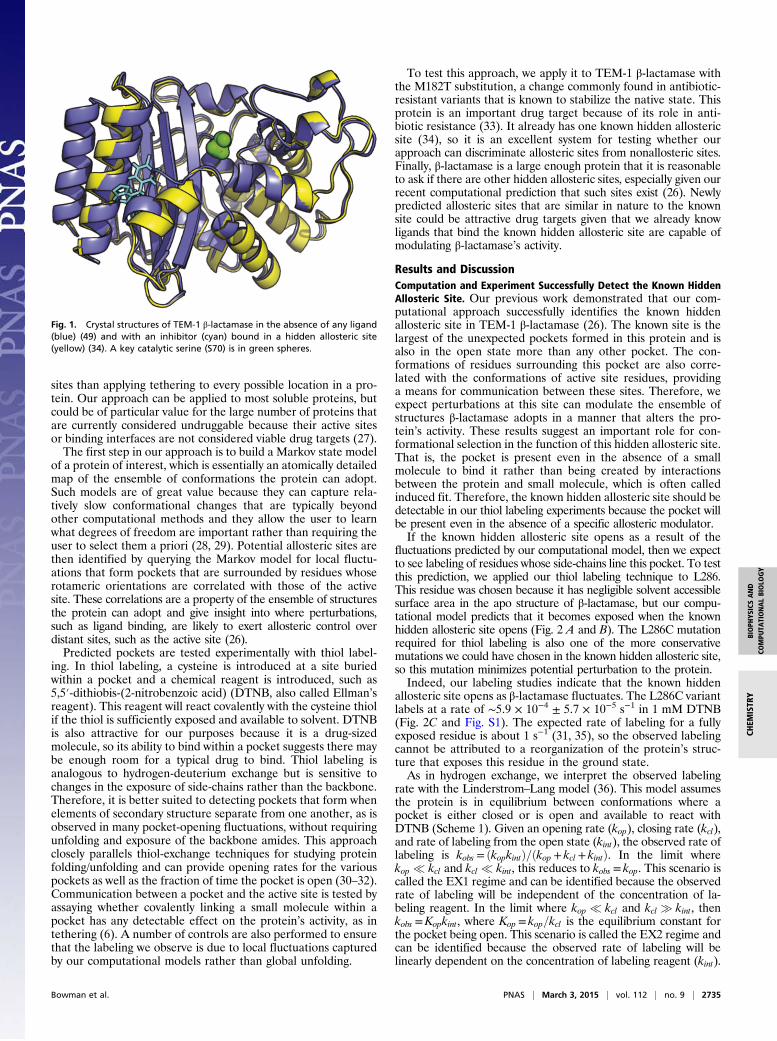
Ahidden allosteric site is a binding pocket that is not presentin the crystal structure of a protein, but becomes available as
the protein fluctuates and is capable of controlling the protein’sfunction by communicating with the active site (Fig. 1) (1).Ligands that bind these sites exert control over the protein’sfunction by perturbing the ensemble of structures the proteinadopts (2, 3). Such sites could have unknown biological functionsand serve as valuable targets for drug design, particularly forproteins that are currently considered undruggable becauseknown structures lack pockets that are suitable for drug design.Unfortunately, it has been difficult to explore either of thesepossibilities because identifying hidden allosteric sites and mole-cules that bind them remains a profound challenge. For example,most structure-function studies focus on a single representativestructure of a protein and give little hint as to where hidden allo-steric sites might occur or what sort of molecules might bind them.Detecting hidden allosteric sites experimentally is difficult
because most of the available methods couple the identificationof such sites with the drug discovery process. Given the diffi-culties inherent to drug design, this coupling likely producesmany false negatives, leaving hidden allosteric sites undiscovered.For example, high-throughput screening can reveal hidden allostericsites (4, 5) but failure to identify allosteric modulators does notdisprove the existence of allosteric sites. Tethering provides a site-directed screen that is useful for specifically searching for allostericsites but will still suffer from false negatives if the library beingscreened does not include small molecules that will bind an allo-steric site tightly enough (6, 7). Understanding the full ensemble ofstructures a protein can adopt would overcome these limitations(8), but such an understanding remains elusive. For instance,
room temperature crystallography and NMR have the potentialto reveal alternative structures containing hidden allosteric sites,but further developments are required to make such measure-ments routine for any given protein target (9–14).A number of computational techniques have been developed
to aid in the discovery of hidden allosteric sites. For example,there are a variety of methods for understanding how informa-tion flows from one region of a protein to another (15–19), aswell as for identifying potential binding pockets (20–24). Morerecent work has accounted for both of these ingredients (25, 26).Although these methods are important developments, many areonly applicable to small proteins operating on fast timescales.Therefore, new approaches are needed for addressing many bi-ologically relevant systems. Furthermore, these methods onlypartially decouple the discovery of hidden allosteric sites fromthe identification of allosteric ligands because identifying acompound that binds to a predicted site is still the primary meansof testing computationally predicted allosteric sites.Here, we use a combination of computation and experiment to
identify hidden allosteric sites without requiring the simultaneousdiscovery of ligands that bind and modulate them. Decoupling thediscovery of allosteric sites from the identification of allostericligands should facilitate drug discovery by providing more in-formation to base design decisions on. For example, rather thanperforming blind screens, computationally generated structuresof potential allosteric sites can be used as a starting point forrational design. The locations of potential hidden allosteric sitescan also be used to direct tethering screens, potentially providinga less resource-intensive means of discovering hidden allosteric
Significance
Rational drug design efforts typically focus on identifyinginhibitors that bind to protein active sites. Pockets that are notpresent in crystallographic structures yet can exert allosteric(i.e., long-range) control over distant active sites present anexciting alternative. However, identifying these hidden allo-steric sites is extremely challenging because one usually has tosimultaneously find a small molecule that binds to and sta-bilizes the open conformation of the pocket. Here, we presenta means of combining advances in computer modeling—usingMarkov state models to capture long timescale dynamics—with biophysical experiments to identify hidden allosteric siteswithout requiring the simultaneous discovery of drug-likecompounds that bind them. Using this technology, we discovermultiple hidden allosteric sites in a single protein.
Author contributions: G.R.B., E.R.B., K.M.H., B.C.M., and S.M. designed research; G.R.B.,E.R.B., K.M.H., and B.C.M. performed research; G.R.B., E.R.B., K.M.H., B.C.M., and S.M.contributed new reagents/analytic tools; G.R.B., E.R.B., K.M.H., B.C.M., and S.M. analyzeddata; and G.R.B., E.R.B., K.M.H., B.C.M., and S.M. wrote the paper.
sites than applying tethering to every possible location in a pro-tein. Our approach can be applied to most soluble proteins, butcould be of particular value for the large number of proteins thatare currently considered undruggable because their active sitesor binding interfaces are not considered viable drug targets (27).The first step in our approach is to build a Markov state model
of a protein of interest, which is essentially an atomically detailedmap of the ensemble of conformations the protein can adopt.Such models are of great value because they can capture rela-tively slow conformational changes that are typically beyondother computational methods and they allow the user to learnwhat degrees of freedom are important rather than requiring theuser to select them a priori (28, 29). Potential allosteric sites arethen identified by querying the Markov model for local fluctu-ations that form pockets that are surrounded by residues whoserotameric orientations are correlated with those of the activesite. These correlations are a property of the ensemble of structuresthe protein can adopt and give insight into where perturbations,such as ligand binding, are likely to exert allosteric control overdistant sites, such as the active site (26).Predicted pockets are tested experimentally with thiol label-
ing. In thiol labeling, a cysteine is introduced at a site buriedwithin a pocket and a chemical reagent is introduced, such as5,5′-dithiobis-(2-nitrobenzoic acid) (DTNB, also called Ellman’sreagent). This reagent will react covalently with the cysteine thiolif the thiol is sufficiently exposed and available to solvent. DTNBis also attractive for our purposes because it is a drug-sizedmolecule, so its ability to bind within a pocket suggests there maybe enough room for a typical drug to bind. Thiol labeling isanalogous to hydrogen-deuterium exchange but is sensitive tochanges in the exposure of side-chains rather than the backbone.Therefore, it is better suited to detecting pockets that form whenelements of secondary structure separate from one another, as isobserved in many pocket-opening fluctuations, without requiringunfolding and exposure of the backbone amides. This approachclosely parallels thiol-exchange techniques for studying proteinfolding/unfolding and can provide opening rates for the variouspockets as well as the fraction of time the pocket is open (30–32).Communication between a pocket and the active site is tested byassaying whether covalently linking a small molecule within apocket has any detectable effect on the protein’s activity, as intethering (6). A number of controls are also performed to ensurethat the labeling we observe is due to local fluctuations capturedby our computational models rather than global unfolding.
To test this approach, we apply it to TEM-1 β-lactamase withthe M182T substitution, a change commonly found in antibiotic-resistant variants that is known to stabilize the native state. Thisprotein is an important drug target because of its role in anti-biotic resistance (33). It already has one known hidden allostericsite (34), so it is an excellent system for testing whether ourapproach can discriminate allosteric sites from nonallosteric sites.Finally, β-lactamase is a large enough protein that it is reasonableto ask if there are other hidden allosteric sites, especially given ourrecent computational prediction that such sites exist (26). Newlypredicted allosteric sites that are similar in nature to the knownsite could be attractive drug targets given that we already knowligands that bind the known hidden allosteric site are capable ofmodulating β-lactamase’s activity.
Results and DiscussionComputation and Experiment Successfully Detect the Known HiddenAllosteric Site. Our previous work demonstrated that our com-putational approach successfully identifies the known hiddenallosteric site in TEM-1 β-lactamase (26). The known site is thelargest of the unexpected pockets formed in this protein and isalso in the open state more than any other pocket. The con-formations of residues surrounding this pocket are also corre-lated with the conformations of active site residues, providinga means for communication between these sites. Therefore, weexpect perturbations at this site can modulate the ensemble ofstructures β-lactamase adopts in a manner that alters the pro-tein’s activity. These results suggest an important role for con-formational selection in the function of this hidden allosteric site.That is, the pocket is present even in the absence of a smallmolecule to bind it rather than being created by interactionsbetween the protein and small molecule, which is often calledinduced fit. Therefore, the known hidden allosteric site should bedetectable in our thiol labeling experiments because the pocket willbe present even in the absence of a specific allosteric modulator.If the known hidden allosteric site opens as a result of the
fluctuations predicted by our computational model, then we expectto see labeling of residues whose side-chains line this pocket. To testthis prediction, we applied our thiol labeling technique to L286.This residue was chosen because it has negligible solvent accessiblesurface area in the apo structure of β-lactamase, but our compu-tational model predicts that it becomes exposed when the knownhidden allosteric site opens (Fig. 2 A and B). The L286C mutationrequired for thiol labeling is also one of the more conservativemutations we could have chosen in the known hidden allosteric site,so this mutation minimizes potential perturbation to the protein.Indeed, our labeling studies indicate that the known hidden
allosteric site opens as β-lactamase fluctuates. The L286C variantlabels at a rate of ∼5.9 × 10⁻⁴ ± 5.7 × 10⁻⁵ s−1 in 1 mM DTNB(Fig. 2C and Fig. S1). The expected rate of labeling for a fullyexposed residue is about 1 s−1 (31, 35), so the observed labelingcannot be attributed to a reorganization of the protein’s struc-ture that exposes this residue in the ground state.As in hydrogen exchange, we interpret the observed labeling
rate with the Linderstrom–Lang model (36). This model assumesthe protein is in equilibrium between conformations where apocket is either closed or is open and available to react withDTNB (Scheme 1). Given an opening rate (kop), closing rate (kcl),and rate of labeling from the open state (kint), the observed rate oflabeling is kobs = ðkopkintÞ=ðkop + kcl + kintÞ. In the limit wherekop � kcl and kcl � kint, this reduces to kobs = kop. This scenario iscalled the EX1 regime and can be identified because the observedrate of labeling will be independent of the concentration of la-beling reagent. In the limit where kop � kcl and kcl � kint, thenkobs =Kopkint, where Kop = kop=kcl is the equilibrium constant forthe pocket being open. This scenario is called the EX2 regime andcan be identified because the observed rate of labeling will belinearly dependent on the concentration of labeling reagent (kint).
Fig. 1. Crystal structures of TEM-1 β-lactamase in the absence of any ligand(blue) (49) and with an inhibitor (cyan) bound in a hidden allosteric site(yellow) (34). A key catalytic serine (S70) is in green spheres.
Bowman et al. PNAS | March 3, 2015 | vol. 112 | no. 9 | 2735
To determine whether the observed rate of labeling is pro-viding information about the opening rate or the fraction of timea pocket is open or exposed, we measured the rate of labelingwith varying concentrations of DTNB. Fig. 2D shows that thelabeling rate is independent of [DTNB], which is consistent withthe EX1 regime. Therefore, we conclude that the observed rateof labeling captures the opening rate of this pocket.
Pockets Are Clearly Distinguishable from Nonpockets. Our experi-mental approach might give false positives if the cysteine muta-tions cause significant destabilization of the protein. For example,introducing a cysteine could globally destabilize the protein suchthat labeling occurs directly from global unfolding rather thantransient exposure of the pocket within the native state ensemble.If this was true for the L286C variant, we would expect the la-beling rate to approximate the rate of global unfolding becauselabeling is in the EX1 regime.To test whether labeling is due to global unfolding, we de-
termined the unfolding rate of our cysteine variant and com-pared it with the measured labeling rate (Table S1). Followingprevious work on the unfolding of β-lactamase (37, 38), wemeasured the unfolding rate of the L286C variant by monitoringthe change in the circular dichroism (CD) signal as a function ofthe final urea concentration (Fig. 3). Extrapolating back to 0 Murea (the labeling conditions), we find that the rate of unfoldingis about 20-fold smaller than the observed rate of labeling. There-fore, labeling must be due to a fluctuation across a barrier fromthe native state that is lower than the barrier to global unfolding.As a control, we created cysteine variants at buried sites not
predicted to form a pocket. Residues L190 and I260 are bothburied in the ligand-free structure of β-lactamase, and our modelpredicts that there are no pockets that expose these residuesto drug-sized molecules. Consistent with this prediction, we donot observe any labeling of cysteines at these positions over the
course of a 12-h labeling reaction. Therefore, we conclude thatthese residues remain buried in the native-state ensemble andthat introducing a cysteine does not cause a local destabilizationthat creates an unpredicted pocket or local unfolding. This re-sult, in combination with the lack of observed labeling for thetwo endogenous cystines in the protein that are oxidized in adisulfide bond, also confirms that the labeling we observe forother residues is not due to a reaction with the two cysteines thatnaturally form a disulfide in β-lactamase. The fact that ourcomputational model successfully discriminates where labelingwill and will not occur also adds significant weight to our con-clusion that labeling is due to the formation of a pocket ratherthan a large-scale unfolding event.Given the proximity of the known hidden allosteric site to two
of the four tryptophan residues in β-lactamase, we reasoned thatopening of this pocket may expose these tryptophans to solventand lead to a change in the protein’s fluorescence. Indeed,opening of this pocket in our computational model increases thesolvent accessible surface area of Trp229’s side-chain from 36%in the ligand-free structure to 69 ± 9% when the pocket is open.The solvent accessible surface area of Trp290’s side-chain in-creases from 43% in the ligand-free structure to 85 ± 8% whenthe pocket is open. Because pocket opening precedes globalunfolding and might be on the pathway to global unfolding, wehypothesized that monitoring unfolding by fluorescence shoulddetect pocket opening and yield a faster rate than monitoringunfolding by CD. To test this prediction experimentally, wemeasured the rate of change in fluorescence of the L286C var-iant as a function of the final urea concentration and used linearextrapolation to find the rate of change in the absence ofdenaturant. This procedure yielded a rate of 5.5 × 10⁻⁴ ± 2.4 ×10⁻⁴ s−1, in reasonable agreement with the rate of labeling withDTNB of 5.9 × 10⁻⁴ ± 5.7 × 10⁻⁵ s−1. The fact that these ratesare 20-fold larger than the rate of unfolding demonstrates thatlabeling precedes unfolding and, therefore, occurs from a rarestate on the native side of the rate-limiting barrier to unfolding.Interestingly, this state appears to be distinct from previouslycharacterized high-energy states on the unfolded side of the rate-limiting barrier to unfolding that were also detected by fluores-cence (37, 38).
A
C D
B
Fig. 2. Thiol labeling of the known hidden allosteric site. (A and B) Surfacerepresentations of the closed and open states of the known hidden allostericsite, respectively. L286 (yellow) is only visible in the open state. A key cata-lytic serine (S70) is shown in green as a reference point. (C) Labeling of L286Cin 1 mM DTNB. (D) The dependence of the labeling rate of L286C on theconcentration of the labeling reagent (DTNB) with error bars representingthe SD from three replicates.
Fig. 3. Thiol labeling is not due to unfolding. Log of the unfolding rate ofL286C as monitored by CD for different urea concentrations with a linear fit(black line) used for extrapolating back to the unfolding rate at 0 M urea.The labeling rate (yellow circle) is considerably faster than unfolding, so itmust correspond to a fluctuation within the native state.
2736 | www.pnas.org/cgi/doi/10.1073/pnas.1417811112 Bowman et al.
There Is Communication Between the Known Allosteric Site and ActiveSite. We also exploited our thiol labeling to test whether there iscommunication between the pockets we detect and the activesite, as indicated by correlations in the ensemble of structuresβ-lactamase adopts. We previously predicted that almost anyhidden binding pocket should also serve as an allosteric site dueto coupling of many residues to different portions of the activesite (26). To test whether a given pocket communicates with theactive site, we measured the activity of proteins with and withoutTNB (one half of DTNB) covalently bound within the pocket.A measurable change in activity would demonstrate that there iscommunication, although it should not be used as a quantitativemeasure of potential inhibition because other molecules that bindthe same site could be more potent inhibitors or even enhance theprotein’s activity (39). Using this approach, we find that the spe-cific activity of L286C is reduced from 361 ± 29 to 97 ± 6 nmolproduct/μg/min. These results suggest that there is communicationbetween the site of modification and the active site. AlthoughDTNB is a drug-sized molecule (SI Materials and Methods), TNB issignificantly smaller than typical drugs and has not been optimizedfor binding this hidden allosteric site. Therefore, it is entirely pos-sible that an allosteric modulator specifically designed to bind thissite could be a much stronger β-lactamase inhibitor. Identifying suchnoncovalent inhibitors (or activators) would serve as the ultimateverification of the existence of our hidden allosteric sites andremains an important future direction. Although we have not yetdiscovered new molecules that bind the hidden allosteric sitesrevealed by our approach, we note that the allosteric inhibitor dis-covered by Horn et al. (34) demonstrates that it is possible for smallmolecules to bind such hidden allosteric sites strongly enough tostabilize the open form of a pocket and alter an enzyme’s activity.As a control, we tested whether thiol labeling of residues that
our computational model predicts should have little communica-tion with the active site alters β-lactamase’s activity. Specifically,we chose to perform thiol labeling of A150 because it is a surfaceresidue that is available for labeling and surrounding residues haveweak correlations with the active site in our computational model.Complete labeling of the A150C variant has a negligible effect onthe protein’s activity (Table 1), consistent with our prediction thatit does not communicate with the active site.
Discovery of Previously Unidentified Hidden Allosteric Sites. Now wecan begin testing whether our model successfully predicts novelhidden allosteric sites. Toward this end, we chose to focus onpockets that our simulations predict will expose residues thatare completely buried in the static, apo-crystal structure. Suchpockets are the most amenable to our thiol labeling experiments,although there may be other pockets that are equally potent al-losteric sites but lack residues with this differential exposure.
Residues appropriate for our thiol labeling experiments wereselected by examining the fraction of each residue’s surface areathat is typically accessible to probes of varying sizes (Fig. 4). Spe-cifically, we chose probe radii of 0.14, 0.24, 0.34, 0.44, and 0.54 nm.This range mimics molecules like water at the smallest scale andmore drug-like molecules at the largest scale. We calculated theaccessible surface area for the side-chain of each residue for a givenprobe size as follows: (i) computationally roll a sphere with thegiven probe radius across the surface of a representative structurefor each state in the Markov model and calculate the accessiblesurface area of every residue’s side-chain; (ii) calculate the averageaccessible surface area of each side-chain by taking the averageacross all states, weighted by their equilibrium populations; and(iii) divide the result for each residue by the total possible ac-cessible surface area of its side-chain. One result of this analysis isthat the fluctuations β-lactamase undergoes make basically everyresidue’s side-chain accessible to water, as previously observed inother proteins (40). However, many residues are not accessibleto larger molecules. We selected residues that are accessible toprobes with radii of at least 0.34 nm because this is consistent withthe size of DTNB and residues that we have already shown tolabel are accessible at this probe size, whereas residues that wehave shown do not label are not. Of the residues that meet thiscriterion, we selected the smallest residues possible to minimizethe perturbation caused by mutating to a cysteine. Based on thesecriteria, residues A232 and A249 point into the most promisingpocket. Residue S203 also points into a second pocket.We performed thiol labeling of A232C to test our first predicted
pocket (Fig. 5). Fig. 5 C and D shows that the A232C variant labelsat a rate of 3.6 × 10⁻³ ± 1.0 × 10⁻³ s−1 independent of the con-centration of labeling reagent, and therefore this is the rate at whichthe residue becomes exposed. The rate of unfolding is also 200-foldslower than the rate of labeling (Table S1), so the observed labelingis not due to global unfolding. Complete labeling of the proteinreduces the activity of the protein by 1.5-fold (Table 1). In additionto our labeling experiments, we again reasoned that opening of thispocket could lead to a change in fluorescence by exposing Trp229 tosolvent. Indeed, opening of this pocket in our computational modelincreases the solvent accessible surface area of Trp229’s side-chainfrom 36% in the ligand-free structure to 56 ± 12% when the pocketis open. Experimentally monitoring unfolding by fluorescence, asdescribed previously, yielded a rate of 1.9 × 10⁻³ ± 1.2 × 10⁻³ s−1, inreasonable agreement with the DTNB labeling rate for this variant.We also performed separate experiments on an A249C variant. Theside-chain of this residue points into the same pocket but has lessexposure than residue 232 because it is not exposed in the exactsame set of structural states where this pocket is open as residue 232.Indeed, we observe labeling of the A249C variant at a rate fivefoldless than the A232 variant (Table S1), consistent with the residue atposition 249 being exposed on opening of the pocket. Based on all of
Table 1. Specific activities (nmol product/μg protein/min) oflabeled and unlabeled proteins
The reduction in activity on labeling demonstrates that there is commu-nication between the proposed allosteric sites and the active site. Thepockets are (1) the known allosteric site, (2) the first predicted site, and (3)the second predicted site.
A B
Fig. 4. Residues selected for labeling in each pocket. (A) Ribbon diagram ofβ-lactamase highlighting residues in the known hidden allosteric site (L286,yellow), the first predicted site (A232, red), and the second predicted site(S203, magenta). A key catalytic serine (S70) is shown in green. (B) Averagepercent of residues’ surface area that is accessible to a variety of probe sizes.L190 and I260 are buried, whereas A150 is on the surface.
Bowman et al. PNAS | March 3, 2015 | vol. 112 | no. 9 | 2737
the results for these two variants, we conclude that this site is ahidden allosteric site, consisting of an unexpected pocket with theability to communicate with the active site. An allosteric modulatorof this site would need to have a greater effect on activity but, asexplained before, this is entirely possible.We also tested the second predicted pocket via thiol labeling of
S203C. This residue labels at a rate of 1.5 × 10⁻² ± 3.4 × 10⁻³ s−1,again independent of the concentration of labeling reagent (Fig.S2). This rate is 50-fold faster than the rate of global unfolding(Table S1), so it captures the rate of exposure of the residue.There is also communication between this site and the active site,as demonstrated by an ∼1.5-fold reduction in activity on labeling.Therefore, we conclude that this second predicted site is also ahidden allosteric site.
ConclusionsWe developed an approach that combines computation andexperiment to detect hidden allosteric sites arising from theensemble of structures a protein can adopt. Importantly, ourapproach does not require the simultaneous discovery of small
molecules that bind and modulate these sites, so our method-ology can be used to guide subsequent drug design efforts.Using this approach, we have demonstrated that a single
protein—TEM-1 β-lactamase—accommodates multiple hiddenallosteric sites. This result is surprising because TEM-1 β-lacta-mase has been studied extensively without observing these sites.Furthermore, there may even be other hidden allosteric sites inthis single protein that are not amenable to the experimentalmethodology we describe here.Our results suggest there are many as yet undiscovered hidden
allosteric sites and that our techniques should provide a means ofdetecting them. Once discovered, these allosteric sites can thenbe targeted with rational drug design or followed up on to dis-cover their biological relevance. In the case of TEM-1, the hid-den allosteric sites we discovered could be valuable targets forantibiotic development.These results lay an important foundation for future work on
hidden allosteric sites. For example, an important next step willbe to discover new allosteric modulators that bind the hidden al-losteric sites revealed by our methodology. Furthermore, our resultsdemonstrate the value of our advanced computational methodsand argue for further developments to make an even more quan-titative comparison between computation and experiment.
Materials and MethodsSimulations were run with Gromacs (41, 42), and the Markov state model wasbuilt with MSMBuilder (43, 44), as described previously (26). This particularmodel provides a statistically reliable description of dynamics on tens of mi-crosecond timescales. Further details are given in SI Materials and Methods.Accessible surface areas were measured with Gromacs and a Voronoi-basedmethod (45). Structures were visualized with PyMOL (46).
TEM-1 β-lactamase with the M182T stabilizing mutation and all cysteinevariants of this background sequence were purified from the periplasmicfraction of BL21(DE3) cells as described in previous work (47) and SI Materialsand Methods. We measured enzyme activities following previous work (48),as described in SI Materials and Methods. Cysteine mutations were introducedwith Quik-Change mutagenesis. Thiol labeling experiments were run with30 μM protein and varying concentrations of DTNB (also called Ellman’s re-agent) in 20 mM Tris, pH 8.0. The time course for labeling was monitored byfollowing the absorbance at 412 nm in a Cary 100Bio UV-Vis spectropho-tometer (27 °C) after starting the reaction via manual mixing. Rates wereinterpreted using the Linderstrom–Lang model (36). Unfolding rates and equi-librium melts were monitored by circular dichroism, as described in SI Materialsand Methods. Further details are given in SI Materials and Methods.
ACKNOWLEDGMENTS. We thank Norbert Lindow, Daniel Baum, and Hans-Christian Hege for helpful discussion on pocket detection. This work wasfunded by National Institutes of Health Grant R01-GM050945. G.R.B. holdsa Career Award at the Scientific Interface from the Burroughs WellcomeFund and was also supported by the Miller Institute.
1. Hardy JA, Wells JA (2004) Searching for new allosteric sites in enzymes. Curr Opin
Struct Biol 14(6):706–715.2. Motlagh HN, Wrabl JO, Li J, Hilser VJ (2014) The ensemble nature of allostery. Nature
508(7496):331–339.3. Gunasekaran K, Ma B, Nussinov R (2004) Is allostery an intrinsic property of all dy-
namic proteins? Proteins 57(3):433–443.4. Lundqvist T, et al. (2007) Exploitation of structural and regulatory diversity in gluta-
mate racemases. Nature 447(7146):817–822.5. Ceccarelli DF, et al. (2011) An allosteric inhibitor of the human Cdc34 ubiquitin-con-
jugating enzyme. Cell 145(7):1075–1087.6. Erlanson DA, et al. (2000) Site-directed ligand discovery. Proc Natl Acad Sci USA
97(17):9367–9372.7. Arkin MR, et al. (2003) Binding of small molecules to an adaptive protein-protein
interface. Proc Natl Acad Sci USA 100(4):1603–1608.8. Wrabl JO, et al. (2011) The role of protein conformational fluctuations in allostery,
function, and evolution. Biophys Chem 159(1):129–141.9. Fischer M, Coleman RG, Fraser JS, Shoichet BK (2014) Incorporation of protein flexi-
bility and conformational energy penalties in docking screens to improve ligand
discovery. Nat Chem 6(7):575–583.10. Fraser JS, et al. (2009) Hidden alternative structures of proline isomerase essential for
catalysis. Nature 462(7273):669–673.
11. Manley G, Loria JP (2012) NMR insights into protein allostery. Arch Biochem Biophys519(2):223–231.
12. Wand AJ (2013) The dark energy of proteins comes to light: Conformational entropyand its role in protein function revealed by NMR relaxation. Curr Opin Struct Biol23(1):75–81.
13. Boehr DD, McElheny D, Dyson HJ, Wright PE (2006) The dynamic energy landscape ofdihydrofolate reductase catalysis. Science 313(5793):1638–1642.
14. Henzler-Wildman K, Kern D (2007) Dynamic personalities of proteins. Nature450(7172):964–972.
15. Hilser VJ, Dowdy D, Oas TG, Freire E (1998) The structural distribution of cooperativeinteractions in proteins: Analysis of the native state ensemble. Proc Natl Acad Sci USA95(17):9903–9908.
18. Süel GM, Lockless SW, Wall MA, Ranganathan R (2003) Evolutionarily conservednetworks of residues mediate allosteric communication in proteins. Nat Struct Biol10(1):59–69.
A
C D
B
Fig. 5. Thiol labeling of the first predicted hidden allosteric site. (A and B)Surface representations of the closed and open states of the known hiddenallosteric site, respectively. A232 (red) is only visible in the open state.(C) Labeling of residue A232C in 1 mM DTNB. (D) The dependence of thelabeling rate on the concentration of the labeling reagent (DTNB) with errorbars representing the SD from three replicates.
2738 | www.pnas.org/cgi/doi/10.1073/pnas.1417811112 Bowman et al.
19. Feher VA, Durrant JD, Van Wart AT, Amaro RE (2014) Computational approaches tomapping allosteric pathways. Curr Opin Struct Biol 25:98–103.
20. Eyrisch S, Helms V (2007) Transient pockets on protein surfaces involved in protein-protein interaction. J Med Chem 50(15):3457–3464.
21. Johnson DK, Karanicolas J (2013) Druggable protein interaction sites are more pre-disposed to surface pocket formation than the rest of the protein surface. PLOSComput Biol 9(3):e1002951.
22. Schmidtke P, Bidon-Chanal A, Luque FJ, Barril X (2011) MDpocket: Open-source cavitydetection and characterization on molecular dynamics trajectories. Bioinformatics27(23):3276–3285.
23. Frembgen-Kesner T, Elcock AH (2006) Computational sampling of a cryptic drugbinding site in a protein receptor: Explicit solvent molecular dynamics and inhibitordocking to p38 MAP kinase. J Mol Biol 359(1):202–214.
24. Schames JR, et al. (2004) Discovery of a novel binding trench in HIV integrase. J MedChem 47(8):1879–1881.
25. Grant BJ, et al. (2011) Novel allosteric sites on Ras for lead generation. PLoS ONE6(10):e25711.
26. Bowman GR, Geissler PL (2012) Equilibrium fluctuations of a single folded proteinreveal a multitude of potential cryptic allosteric sites. Proc Natl Acad Sci USA 109(29):11681–11686.
27. Hopkins AL, Groom CR (2002) The druggable genome. Nat Rev Drug Discov 1(9):727–730.
28. Bowman GR, Huang X, Pande VS (2010) Network models for molecular kinetics andtheir initial applications to human health. Cell Res 20(6):622–630.
29. Chodera JD, Noé F (2014) Markov state models of biomolecular conformational dy-namics. Curr Opin Struct Biol 25:135–144.
30. Bernstein R, Schmidt KL, Harbury PB, Marqusee S (2011) Structural and kinetic map-ping of side-chain exposure onto the protein energy landscape. Proc Natl Acad SciUSA 108(26):10532–10537.
31. Sridevi K, Udgaonkar JB (2002) Unfolding rates of barstar determined in native andlow denaturant conditions indicate the presence of intermediates. Biochemistry 41(5):1568–1578.
32. Jha SK, Marqusee S (2014) Kinetic evidence for a two-stage mechanism of proteindenaturation by guanidinium chloride. Proc Natl Acad Sci USA 111(13):4856–4861.
33. Bush K (2013) Proliferation and significance of clinically relevant β-lactamases. Ann NY Acad Sci 1277:84–90.
34. Horn JR, Shoichet BK (2004) Allosteric inhibition through core disruption. J Mol Biol336(5):1283–1291.
35. Jocelyn PC (1987) Spectrophotometric assay of thiols. Methods Enzymol 143:44–67.36. Berger A, Linderstrom-Lang K (1957) Deuterium exchange of poly-DL-alanine in
aqueous solution. Arch Biochem Biophys 69:106–118.37. Vanhove M, Raquet X, Frère JM (1995) Investigation of the folding pathway of the
TEM-1 beta-lactamase. Proteins 22(2):110–118.38. Kather I, Jakob RP, Dobbek H, Schmid FX (2008) Increased folding stability of TEM-1
beta-lactamase by in vitro selection. J Mol Biol 383(1):238–251.39. Sadowsky JD, et al. (2011) Turning a protein kinase on or off from a single allosteric
site via disulfide trapping. Proc Natl Acad Sci USA 108(15):6056–6061.40. Damjanovi�c A, García-Moreno B, Lattman EE, García AE (2005) Molecular dynamics
study of water penetration in staphylococcal nuclease. Proteins 60(3):433–449.41. Van Der Spoel D, et al. (2005) GROMACS: Fast, flexible, and free. J Comput Chem
26(16):1701–1718.42. Pronk S, et al. (2013) GROMACS 4.5: A high-throughput and highly parallel open
source molecular simulation toolkit. Bioinformatics 29(7):845–854.43. Bowman GR, Huang X, Pande VS (2009) Using generalized ensemble simulations and
Markov state models to identify conformational states. Methods 49(2):197–201.44. Beauchamp KA, et al. (2011) MSMBuilder2: Modeling conformational dynamics at the
picosecond to millisecond scale. J Chem Theory Comput 7(10):3412–3419.45. Lindow N, Baum D, Hege H-C (2011) Voronoi-based extraction and visualization of
molecular paths. IEEE Trans Vis Comput Graph 17(12):2025–2034.46. Schrödinger LLC (2002) The PyMOL Molecular Graphics System, Version 1.6.0.0.47. Hanes MS, Ratcliff K, Marqusee S, Handel TM (2010) Protein-protein binding affinities
by pulse proteolysis: Application to TEM-1/BLIP protein complexes. Protein Sci 19(10):1996–2000.
48. O’Callaghan CH, Morris A, Kirby SM, Shingler AH (1972) Novel method for detectionof beta-lactamases by using a chromogenic cephalosporin substrate. AntimicrobAgents Chemother 1(4):283–288.
49. Wang X, Minasov G, Shoichet BK (2002) Evolution of an antibiotic resistance enzymeconstrained by stability and activity trade-offs. J Mol Biol 320(1):85–95.
Bowman et al. PNAS | March 3, 2015 | vol. 112 | no. 9 | 2739