Nevada State Health Division Discovery of Radiation and Radiation Safety Discovery of Radiation X-ray radiation discovered November 8, 1895 by Wilhelm Konrad Roentgen, German Physicist. Noticed fluorescence on a piece of cardboard coated with barium platincyanide using a Crookes Cathode ray tube. Days later placed his hand between the tube and the coated cardboard and saw his bones depicted on the cardboard, first fluoroscopic screen. Seven weeks later made the first radiograph of his wife’s hand on a photographic plate. Radiation discovered in Uranium salts on March 1, 1896 by Antoine-Heneri Becquerel, French scientist. Placed uranium salts on photographic plates in a drawer, developed the plates and saw dark spots.
Transcript
Nevada State Health Division
Discovery of Radiation and Radiation Safety
Discovery of Radiation X-ray radiation discovered November 8, 1895 by Wilhelm Konrad Roentgen, German Physicist. Noticed fluorescence on a piece of cardboard coated with barium platincyanide using a Crookes Cathode ray tube. Days later placed his hand between the tube and the coated cardboard and saw his bones depicted on the cardboard, first fluoroscopic screen. Seven weeks later made the first radiograph of his wife’s hand on a photographic plate.
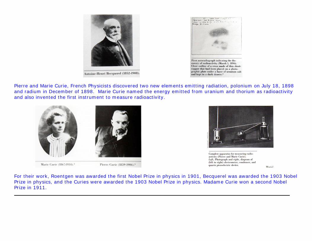
Radiation discovered in Uranium salts on March 1, 1896 by Antoine-Heneri Becquerel, French scientist. Placed uranium salts on photographic plates in a drawer, developed the plates and saw dark spots.
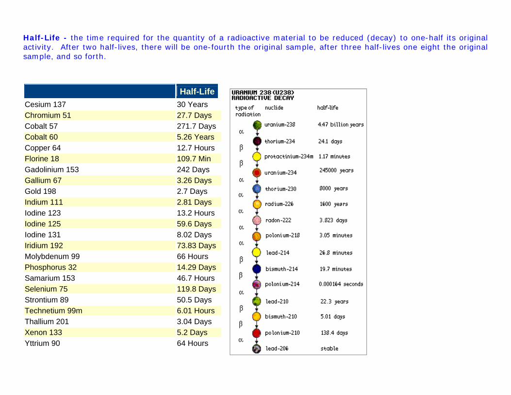
Pierre and Marie Curie, French Physicists discovered two new elements emitting radiation, polonium on July 18, 1898 and radium in December of 1898. Marie Curie named the energy emitted from uranium and thorium as radioactivity and also invented the first instrument to measure radioactivity.
For their work, Roentgen was awarded the first Nobel Prize in physics in 1901, Becquerel was awarded the 1903 Nobel Prize in physics, and the Curies were awarded the 1903 Nobel Prize in physics. Madame Curie won a second Nobel Prize in 1911. _________________________________________________________________________________________________________
X-ray Tubes
Johann Heinrich Geissler (1814-1879) German inventor and glass blower invented the mercury air pump that produced a higher vacuum than had ever been attained at the time (1875) in a cold cathode tube.
Sir William Crookes (1832-1919) improved on Geissler's method of evacuating vessels and during 1875 achieved vacuums of about 1/100 mm of mercury. An electrical discharge through the highly rarified air in his tubes showed the existence of cathode rays culminating in the discovery of the electron. Roentgen was using a Crookes tube when he discovered x-rays in 1895.
William David Coolidge 1873-1975, American physical chemist, Massachusetts Institute of Technology, 1896. He joined the General Electric Company in 1905 and served as director of its research laboratory (1932-40) and as vice president and director of research (1940-44). He invented the first hot cathode X-ray tube (Coolidge X-ray tube) with a tungsten heating element that revolutionized the field of radiology and is the is the basis for today’s X-ray.
What is radiation? Radiation is energy released from unstable atoms in the form of waves or particles. Radiation is all around us and comes from the earth and outer space and is classified as natural and man-made. Natural radiation accounts for approximately 82% of total exposure and 18% from man-made sources.
The average annual whole body radiation exposure per individual is 360 millirems (mR). 1. Non-Ionizing Electromagnetic Radiation
a. Radio b. Microwaves c. Infra Red (heat) d. Visible Light
e. Ultra Violet 2. Ionizing Electromagnetic Radiation
a. X-rays b. Gamma Rays c. Cosmic Rays 3. Ionizing Atomic Particle Radiation
a. Beta Rays b. Alpha Rays c. Neutrons
Alpha Radiation – slow speed and low penetrating particles, can be stopped by paper and skin, no hazard if external to the body, high hazard if internal to the body (lung and gastrointestinal tissue), a large number of ionizations in small areas causing damage to tissue and cells. Beta Radiation – high-energy electrons, fast moving, travel through 10 feet of air and penetrate very thin layers of materials (aluminum foil and skin tissue), can be stopped by metal and plastics, hazard is internal and external to the body.
Gamma/X-ray Radiation – high-energy light, electromagnetic wave or photons, no weight, travels at the speed of light (186,000 miles/second) in a vacuum, Gamma rays are emitted from the nuclei of certain radioactive elements, X-rays are a stream of fast moving electrons that suddenly undergo a reduction of speed, man made in with an X-ray tube. Stopped by thick walls of cement, lead or steel. The hazard to the body is internal and external. Neutron Radiation - Neutrons and protons are particles found in the nucleus, or center, of atoms. Neutrons interact by collisions with atoms and transfer energy during these collisions. Fissioning of uranium, the bombarding of isotopes of uranium, the splitting of the uranium atom which causes a chain reaction and releases energy, heat and more neutrons. Sources of neutrons are nuclear reactors, spent fuel rods, atomic weapons, accelerators, combining alpha-emitting isotopes with beryllium produces a neutron source, etc. Shielding depends on the energy of the neutron. High- speed neutrons are slowed by collisions with hydrogenous materials such as water, paraffin, or concrete and slow neutrons are captured by secondary shielding materials such as boron or cadmium.
The Atom - Atoms are made up of three subatomic particles: protons, neutrons, and electrons. The protons and neutrons are packed together in the nucleus at the center of the atom (see Figure 1). The electrons orbit the nucleus. The number of protons in the nucleus determines what material (element) the atom is. Isotopes – While all atoms of the same element have the same number of protons, it is possible for atoms of one element to have different numbers of neutrons. Atoms of the same element with different numbers of neutrons are called isotopes.
Radioactive Decay - When the nucleus of a radioactive isotope gives up its extra energy, that energy is called ionizing radiation. Ionizing radiation may take the form of alpha particles, beta particles, or gamma rays. Ionizing radiation is of concern because it may cause adverse health effects. The process of emitting the radiation is called radioactive decay.
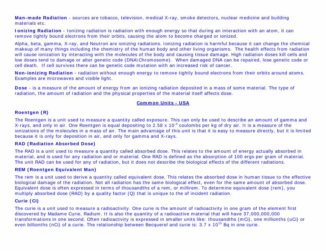
Half-Life - the time required for the quantity of a radioactive material to be reduced (decay) to one-half its original activity. After two half-lives, there will be one-fourth the original sample, after three half-lives one eight the original sample, and so forth.
Half-Life Cesium 137 30 Years Chromium 51 27.7 Days Cobalt 57 271.7 Days Cobalt 60 5.26 Years Copper 64 12.7 Hours Florine 18 109.7 Min Gadolinium 153 242 Days Gallium 67 3.26 Days Gold 198 2.7 Days Indium 111 2.81 Days Iodine 123 13.2 Hours Iodine 125 59.6 Days Iodine 131 8.02 Days Iridium 192 73.83 Days Molybdenum 99 66 Hours Phosphorus 32 14.29 Days Samarium 153 46.7 Hours Selenium 75 119.8 Days Strontium 89 50.5 Days Technetium 99m 6.01 Hours Thallium 201 3.04 Days Xenon 133 5.2 Days Yttrium 90 64 Hours
Man-made Radiation - sources are tobacco, television, medical X-ray, smoke detectors, nuclear medicine and building materials etc.
Ionizing Radiation - Ionizing radiation is radiation with enough energy so that during an interaction with an atom, it can remove tightly bound electrons from their orbits, causing the atom to become charged or ionized.
Alpha, beta, gamma, X-ray, and Neutron are ionizing radiations. Ionizing radiation is harmful because it can change the chemical makeup of many things including the chemistry of the human body and other living organisms. The health effects from radiation will cause ionization by interacting with the molecules of the body and causing tissue damage. High radiation doses kill cells and low doses tend to damage or alter genetic code (DNA\Chromosome). When damaged DNA can be repaired, lose genetic code or cell death. If cell survives there can be genetic code mutation with an increased risk of cancer.
Non-ionizing Radiation - radiation without enough energy to remove tightly bound electrons from their orbits around atoms. Examples are microwaves and visible light.
Dose - is a measure of the amount of energy from an ionizing radiation deposited in a mass of some material. The type of radiation, the amount of radiation and the physical properties of the material itself affects dose.
Common Units - USA
Roentgen (R)
The Roentgen is a unit used to measure a quantity called exposure. This can only be used to describe an amount of gamma and X-rays, and only in air. One Roentgen is equal depositing to 2.58 x 10⁻⁴ coulombs per kg of dry air. It is a measure of the ionizations of the molecules in a mass of air. The main advantage of this unit is that it is easy to measure directly, but it is limited because it is only for deposition in air, and only for gamma and X-rays.
RAD (Radiation Absorbed Dose)
The RAD is a unit used to measure a quantity called absorbed dose. This relates to the amount of energy actually absorbed in material, and is used for any radiation and or material. One RAD is defined as the absorption of 100 ergs per gram of material. The unit RAD can be used for any of radiation, but it does not describe the biological effects of the different radiations.
REM (Roentgen Equivalent Man)
The rem is a unit used to derive a quantity called equivalent dose. This relates the absorbed dose in human tissue to the effective biological damage of the radiation. Not all radiation has the same biological effect, even for the same amount of absorbed dose. Equivalent dose is often expressed in terms of thousandths of a rem, or millirem. To determine equivalent dose (rem), you multiply absorbed dose (RAD) by a quality factor (Q) that is unique to the of incident radiation.
Curie (Ci)
The curie is a unit used to measure a radioactivity. One curie is the amount of radioactivity in one gram of the element first discovered by Madame Curie, Radium. It is also the quantity of a radioactive material that will have 37,000,000,000 transformations in one second. Often radioactivity is expressed in smaller units like: thousandths (mCi), one millionths (uCi) or even billionths (nCi) of a curie. The relationship between Becquerel and curie is: 3.7 x 10¹⁰ Bq in one curie.
Common Units - SI - International Standard
Gray (Gy)
The gray is a unit used to measure a quantity called absorbed dose. This relates to the amount of energy actually absorbed in some material, and is used for any of radiation and any material. One gray is equal to one joule of energy deposited in one kg of a material. The unit gray can be used for any of radiation, but it does not describe the biological effects of the different radiations. Absorbed dose is often expressed in terms of hundredths of a gray, or centi-grays. One gray is equivalent to 100 RAD.
Sievert (Sv)
The Sievert is a unit used to derive a quantity called equivalent dose. This relates the absorbed dose in human tissue to the effective biological damage of the radiation. Not all radiation has the same biological effect, even for the same amount of absorbed dose. Equivalent dose is often expressed in terms of millionths of a Sievert, or micro-Sievert. To determine equivalent dose (Sv), you multiply absorbed dose (Gy) by a quality factor (Q) that is unique to the incident radiation. One Sievert is equivalent to 100 rem.
Becquerel (Bq)
The Becquerel is a unit used to measure a radioactivity. One Becquerel is that quantity of a radioactive material that will have 1 transformation in one second. Often radioactivity is expressed in larger units like: thousands (kBq), millions (MBq) or even billions (GBq) of a Becquerel. As a result of having one Becquerel being equal to one transformation per second, there are 3.7 x 10¹⁰ Bq in one curie.
Radiation Effects Chronic Dose Effects - are those resulting from many small doses of radiation delivered over a large period of time. Biological repair occurs during the exposure period and effects that appear will take years to develop. Continual exposure over many years may produce observable effects. 1. Low energy (<50KeV): These effects are known to appear as skin cancer, cataracts or other surface effects. Normally the damage does not become apparent until as little as six months or as much as ten to twenty years after exposure.
2. High energy (>50KeV): The biological damage produced by this radiation may include genetic mutations, various organ cancers (such as bone or thyroid) and leukemia in a person that received a radiation dose over a long period of time.
Acute Dose Effects –are those resulting from a high dose of x-radiation in a short period of time (approximately 100 rad or more). Biological repair mechanisms do not have time to function before effects appear, within days or weeks.
1. Low energy (<50KeV): These effects typically appear as erythema, epilation, pigmentation, dermatitis, ulceration and other skin-surface effects on the upper extremities of the body. These are slow to heal and can lead to cancer. Amputation of one or more fingers is sometimes required.
2. High energy (>50KeV): This radiation can penetrate deep into the body and cause damage to the lining of the gastrointestinal tract, blood-forming cells in the bone marrow, and the central nervous system. These effects can lead to death.
Somatic Effects - are effects from some agent, like radiation that are seen in the individual who receives the agent.
Genetic Effects - are effects from some agent, which are seen in the offspring of the individual who received the agent. The agent must be encountered pre-conception.
Stochastic Effects - are effects that occur on a random basis with its effect being independent of the size of dose. The effect typically has no threshold and is based on probabilities, with the chances of seeing the effect increasing with dose. Cancer is thought to be a stochastic effect.
Non-stochastic Effect - are effects that can be related directly to the dose received. The effect is more severe with a higher dose, i.e., the burn gets worse as dose increases. It typically has a threshold, below which the effect will not occur. A skin burn from radiation is a non-stochastic effect.
Radiation sickness (syndrome) The complex of symptoms characterizing the disease known as radiation injury, resulting from excessive exposure (greater than 200 rads or 2 gray) of the whole body (or large part) to ionizing radiation. The earliest of these symptoms are nausea, fatigue, vomiting, and diarrhea, which may be followed by loss of hair (epilation), hemorrhage, inflammation of the mouth and throat, and general loss of energy. In severe cases, where the radiation exposure has been approximately 1,000 rad (10 gray) or more, death may occur within two to four weeks. Those who survive 6 weeks after the receipt of a single large dose of radiation to the whole body may generally be expected to recover.
Instruments to Measure Radiation
Ion Chamber - The most common is a gas filled radiation detector. This instrument works on the principle that as radiation passes through air or a specific gas, ionization of the molecules in the air occur. When a high voltage is placed between two areas of the gas filled space, the positive ions will be attracted to the negative side of the detector (the cathode) and the free electrons will travel to the positive side (the anode). These charges are collected by the anode and cathode, which then form a very small current in the wires going to the detector. By placing a very sensitive current measuring device between the wires from the cathode and anode, the small current measured and displayed as a signal. The more radiation that enters the chamber, the more current will be displayed by the instrument.
Many types of gas-filled detectors exist, but the two most common are the ion chamber used for measuring large amounts of radiation and the Geiger-Muller or GM detector used to measure very small amounts of radiation.
Scintillation Detector - The second most common type instrument. The basic principle behind this instrument is the use of a special material that glows or “scintillates” when radiation interacts with it. The most common type of material is a type of salt called sodium-iodide. The light produced from the scintillation process interacts with device called a photo multiplier tube. Electrons are released from the photo multiplier tube into a dynode plate that produces another set of electron pulses that are detected and displayed by a special instrument.
X-ray Devices By accelerating electrons with a high voltage and allowing them to collide with a metal target produce X-rays. The X-rays are produced when the electrons are suddenly decelerated upon collision with the metal target (anode); the x-rays are called Bremsstrahlung or "braking radiation". X-rays are filtered and collimated to improve image quality, reduce the radiation dose to the patient and medical personnel. X-ray Tube - consists of a glass bulb or a metal ceramic envelop under vacuum. Inside the evacuated area, a cathode comprises the filament wire and the anode, the target with a high melting point. An electrical current drives electrons through the low resistance filament wire. In that way, the filament will get hot and electrons are emitted. Due to the connected high voltage between cathode and anode, the emitting electrons are accelerated in the direction of the target. X-rays are produced when these electrons strike the target (which is normally made of tungsten due to its high melting point). To prevent damage of the target (anode), the tube is cooled by fluid or air and has rotating anode that accelerates to 9000 – 10,000 rpm’s. The product of mA, time and kVp determines the radiation output from the X-ray tube (patient and staff radiation exposure). About 99.8% of the energy produced in an X-ray tube is changed to heat. Only 0.2% is converted to X-rays.
Collimator – a device made of a highly absorbing material such as lead that focuses or limits the X-ray beam in a particular direction and area.
Half Value Layer The thickness of any specified material necessary to reduce the X-ray beam intensity to one-half its original value. The penetrating ability of an X-ray beam (quality) is dependent on the kVp selected for the exposure.
The half-value layer provides important information about the energy characteristics of the X-ray beam. If the half-value layer for a given x-ray beam is low or soft, then the X-ray beam contains more low energy and less penetrating radiation. Low-energy radiation increases the radiation dose to the patient and decreases image detail and causes fogging on film. When the half-value layer is high or hardened, the X-ray beam contains more high energy and is highly penetrating. There is less radiation dose to the patient and increased image detail on the film. It is important to have a physicist evaluate all x-ray equipment on a regular basis, typically once a year, and measure half-value layer as part of that testing.
Fluoroscopic Devices
Early fluoroscopic procedures produced low intensity visual images. It requires the radiologist to adapt their eyes to the dark to view the image. In the late 1940’s scientists invented the x-ray image intensifier, which brightened fluoroscopic images.
Commercial x-ray systems with image intensifiers were introduced in the mid 1950s. The x-ray image intensifier enabled the radiologist to visualize the output image without dark adaptation.
In a fluoroscopy system, the image intensifier is located opposite the x-ray tube. The X-ray image intensifier in the fluoroscopic
imaging system converts the x-ray spectrum transmitted through the patient into a highly visible image. It converts the x-ray photons into light photons at the image intensifier input phosphor, the visible light photons are then converted into electrons at the photocathode, accelerating and focusing the electrons through use of electrodes, and converting the electrons back into visible light at the output phosphor. The intensity of the final image is several thousand times brighter than the initial image created at the input phosphor.
An x-ray image intensifier has two major functions: (a) to intercept the x-ray photons and convert them into visible light photons
and (b) to amplify or intensify this light signal. The image intensifier creates a large gain (or intensification) in luminance at the output screen compared with that at the input screen. The output screen image can be viewed with closed-circuit monitor and recorded on film or digitally.
Automatic Exposure Control (AEC) measures the dose of radiation that strikes the X-ray film behind the patient, and turns the X-ray system off when the predetermined dose for that screen-film combination has been reached. This assures that only the smallest required dose is administered. Automatic Brightness Control (ABC) - When imaging the output (image densities) to the monitor will vary over a very wide range, depending on the kVp and mA used and the thickness/density of the object. The ABC automatically corrects for this when needed to achieve increased image quality and also decreases the total radiation dose to the patient. Pulsed Fluoroscopy - the X-ray beam output is not continuous; it is delivered in pulses that follow in rapid succession. This reduces the amount of time during which radiation is released. The resulting radiation-free gaps in the imaging process are filled with the last stored digital image until a new and more current image is available. The short X-ray pulses mean that the dose is significantly reduced; additionally, image definition is increased. Pulsing can take place either by using a pulse control at the X-ray generator, or with a grid-controlled X-ray tube; however, the grid control leads to a lower level of radiation exposure. If the radiation exposure time to the body can be reduced, this leads to a decrease in the total dose to the patient.
X-ray Film, Intensifying Screens and Cassettes
X-ray Film - consists of a base (polyester plastic) and Emulsion (silver halide gelatin). The film base is coated on both sides for general radiography and single side coated for special exams, studies and image capture devices.
Intensifying Screens - consists of microscopic crystals of phosphor (fluorescent material) coated on plastic or cardboard bases. When exposed to radiation the screen phosphors fluoresce either blue or green light depending on the speed of the film and cause the film to be exposed. Only 2% of the image on the X-ray film is caused by direct radiation exposure and the rest of the image on film (98%) is caused by light from the intensifying screens. By using intensifying screens the amount of radiation needed to obtain an X-ray image is reduced by 30% to 60%.
Film Cassettes - are light tight containers made out of plastic or metal that sandwich the film between the intensifying screens.
Radiation Safety
Radiation Safety starts with ALARA "AS LOW AS REASONABLY ACHIEVABLE". Every effort should be made to reduce exposure to radiation since any amount is potentially harmful. Time, distance, and shielding are three practical methods to minimize radiation exposure.
TIME - The dose of radiation received is directly proportional to the amount of time spent in a radiation field. Thus, reducing the time by one-half will reduce the radiation dose by one-half.
DISTANCE - Radiation exposure decreases rapidly as the distance between the worker and the radiation source increases. Maximizing distance represents one the simplest and most effective methods for reducing radiation exposure to workers.
Inverse Square Law - This law states that the amount of radiation at a given distance from a point source varies inversely with the square of the distance. Any point source that spreads its influence equally in all directions without a limit to its range will obey the inverse square law.
For example, doubling the distance from a radiation source will reduce the dose to one-fourth of its original value, and increasing the distance by a factor of three will reduce the dose to one-ninth of its original value. For example, if the dose rate at one foot from a source is 20 mR/hr, then the dose rate at two feet (twice the distance) will be 5 mR/hr.
SHIELDING
Shielding material between a worker and the radiation source decreases exposure. Materials with high densities and high atomic numbers are the most effective shielding choice for protection from X-rays. Thus, substances such as lead, concrete, and steel are very practical shielding materials because of the abundance of atoms and electrons that can interact with the photon. The amount of shielding necessary to reduce the radiation intensity to a desired level can be calculated from the half-value layer of the material.
Personal Monitoring Devices (dosimeters) or film badges are required for workers who may receive 10% of the maximum dose of external radiation (500 millirems (mR) per quarter (3-month period). Film badges must be worn during any procedure involving radiation sources. When not worn film badges should be stored away from all sources of radiation in an area where they will not be exposed to excessive heat, sunlight, or moisture.
Safety Rules: Mobile X-ray and Fluoroscopic Equipment
A. Vacate the room if possible.
B. If in the room, stand as far away as possible from the patient, the X-ray tube and the useful beam. Wear a protective lead apron with a film badge placed on the collar. It is suggested that when a fluoroscope is in use wear a wrap-around apron, thyroid collar and leaded glasses.
C. When a patient must be held in position for radiography a mechanical supporting or restraining devices should be used. If staff must hold the patient that individual shall be protected with appropriate shielding devices, such as protective lead gloves and an apron. The individual should be so positioned that no part of his body will be struck by the useful beam and that his body is as far as possible from the edge of the useful beam.
D. During fluoroscopy care should be taken to limit the useful beam to the smallest area consistent with clinical requirements and to align the X-ray beam with the patient.
E. Gonadal shielding should be used for the patient when appropriate.
F. Use the maximum source-skin distance consistent with the conditions of the examination when using a fluoroscope.
G. Follow all instructions given by the X-ray technologist.
Radiation and Pregnancy
A. A woman should declare her pregnancy to a supervisor or management.
B. Vacate the room if possible. If in the room wear a wrap-around apron, thyroid collar, lead glasses, don’t hold patients or film and stand as far away as possible from the patient. Suggest wearing two film badges, one on the apron collar and the other on the low abdomen at the waist behind the lead apron.
C. The facility is responsible for assuring that the duties of a female staff member will not result in a deep dose equivalent in excess of 0.1 rem (100 mR) during the entire pregnancy.
THE MOST RADIOSENSITIVE PERIOD FOR THE EMBRYO/FETUS IS FROM EIGHT TO FIFTEEN (8-15) WEEKS GESTATION AGE.
A major factor in reducing unnecessary radiation exposure to staff and patients is to replace old X-ray equipment with new digital equipment. The benefits and advantages are: decreased radiation exposure to patients and staff, increased image quality and diagnostic information, no repeat of exams and films due to poor image density quality, transfer of patient exam results, images and information over networks and phone lines to physicians and medical facilities, archiving of information in minimal space, and retrieving perfect originals at the touch of a few keys.
Occupational Dose Limits: Whole body (Total Effective Dose Equivalent) = 5 rem/year, Sum of individual organs or tissue = 50 rem/year, Eye dose (lens of eye) = 15 rem/year, Skin or any extremity = 50 rem/year, Dose to embryos of declared pregnant woman = 0.5 rem for the entire pregnancy, Minors = 10% of above limits. Member of public = 0.1 rem/year. NAC 459.558 Personnel monitoring. All persons who are associated with the operation of an X-ray system are subject to the occupational exposure limits and the requirements for the determination of the doses which are stated in NAC 459.365. When protective clothing or devices are worn on portions of the body and a monitoring device or devices are required, at least one device must be utilized as follows: 1. When an apron is worn, the monitoring device must be worn at the collar outside the apron. 2. The dose to the whole body, based on the maximum dose attributed to any one critical organ, which are the gonads, the blood forming organs, head and trunk or lens of the eye, must be recorded in the reports required by NAC 459.3655. If more than one device is used and a record is made of the data, each dose must be identified with the area where the device was worn on the body. Exposure of a personnel monitoring device to indicate deceptively a dose delivered to a person is prohibited. NAC 459.570 Fluoroscopic X-ray systems: Exposure rate limits. 1. The exposure measured at the point where the center of the useful beam enters the patient must not exceed 10 roentgens (100 millisieverts) per minute, except during recording of fluoroscopic images or when provided with optional high level control. 2. When provided with optional high level control, the equipment must not be operable at any combination of tube potential and current which will result in an exposure rate, measured at the point where the center of the useful beam enters the patient, in excess of: (a) Five roentgens (50 millisieverts) per minute if the high level control is not activated; and (b) Twenty roentgens (200 millisieverts) per minute if the high level control is activated and the unit was manufactured on or after May 19, 1995. � Special means of activation of high level controls, such as additional pressure applied continuously by the operator, will be required to avoid accidental use. A continuous signal audible to the fluoroscopist must indicate activation and use of the high level control. NAC 459.574 Fluoroscopic X-ray systems: Indication of potential and current; source-skin distance; exceptions for fluoroscopy imaging system. 1. During fluoroscopy and cinefluorography, X-ray tube potential and current must be continuously indicated. 2. Except as otherwise provided in subsection 3, the source to skin distance must not be less than: (a) Thirty-eight centimeters on stationary fluoroscopes installed after February 28, 1980;
(b) Thirty-five and five-tenths centimeters on stationary fluoroscopes which are in operation before February 28, 1980; (c) Thirty centimeters on all mobile fluoroscopes; and (d) Twenty centimeters for image-intensified fluoroscopes used for specific surgical application. The users' operating manual must provide precautionary measures to be followed during the use of this device. 3. A fluoroscopy imaging system, including a small format type and miniature C-arm type, used to perform low power, X-ray image intensified fluoroscopy on extremities must: (a) Be operated only by a licensed practitioner of the healing arts. (b) Possess a positive, nonremovable means to ensure a source-skin distance during operation of not less than 9 centimeters, unless a different distance is approved by the Food and Drug Administration. (c) Be clearly labeled as for use only on extremities. (d) Bear a certification label that includes: (1) The statement "This product is in conformity with the performance standards for diagnostic X-ray systems and their major components set forth in 21 C.F.R. § 1020"; and (2) If the Food and Drug Administration grants a variance from any performance standards for diagnostic X-ray systems and their major components set forth in 21 C.F.R. § 1020, a statement of the variance and the identification number assigned to the variance by the Food and Drug Administration. (e) Include an operating manual that contains: (1) Any special instructions that may be necessary because of the unique features of the system, including, without limitation, special instructions concerning exposure rates, safety procedures and precautions; and (2) Recommended machine settings for representative sample fluoroscopic examinations for which the system is designed, including data on skin and tabletop exposures resulting from these settings. NAC 459.339 Precautionary procedures: Conditions requiring individual monitoring of external and internal occupational doses. Each licensee and registrant shall monitor exposures from sources of radiation at levels sufficient to demonstrate compliance with the limits for occupational doses specified in NAC 459.10 to NAC 459.950, inclusive. As a minimum: 1. Each licensee and registrant shall monitor occupational exposure to radiation from licensed and unlicensed sources under the control of the licensee or registrant and shall supply and require the use of personnel monitoring equipment by: (a) Adults who are likely to receive in 1 year, from sources of radiation external to the body, a dose in excess of 10 percent of the limits specified in NAC 459.325; (b) Minors who are likely to receive in 1 year, from sources of radiation external to the body, a deep-dose equivalent in excess of 0.1 rem (1 millisievert), a lens dose equivalent in excess of 0.15 rem (1.5 millisieverts), or a shallow-dose equivalent to the skin or extremities in excess of 0.5 rem (5 millisieverts); (c) Women who have declared their pregnancy and are likely to receive, during the entire pregnancy, from sources of radiation external to the body, a deep-dose equivalent in excess of 0.1 rem (1 millisievert); and (d) Any person entering a high or very high radiation area. NAC 459.321 Development, implementation and review of program for protection against radiation; establishment of constraint on air emissions to environment of radioactive material. 1. Each licensee and registrant shall:
(a) Develop, document and carry out a program for protection against radiation commensurate with the scope of its licensed or registered activities and sufficient to ensure compliance with the provisions of NAC 459.010 to NAC 459.950, inclusive. (b) Use, to the extent practicable, procedures and engineering controls, based upon sound principles of protection against radiation, to achieve occupational doses and doses to members of the public that are as low as is reasonably achievable. (c) Review, at intervals not to exceed 12 months, the content and implementation of the program for protection against radiation. 2. A licensee or registrant shall, to achieve doses to members of the public that are as low as is reasonably achievable pursuant to paragraph (b) of subsection 1, establish a constraint on air emissions to the environment of radioactive material, excluding radon 222 and its decay products, such that the individual member of the public likely to receive the highest dose from such emissions will not be expected to receive a total effective dose equivalent in excess of 10 millirems (0.1 millisievert). 3. A licensee or registrant that causes, permits or is otherwise responsible for air emissions of radioactive material to the environment that exceed the constraint established pursuant to subsection 2 shall: (a) Submit to the Division the report required by NAC 459.371; and (b) Promptly take appropriate corrective action to prevent any recurrence. NAC 459.3635 Records of program for protection against radiation. 1. Each licensee and registrant shall maintain records of its program for protection against radiation required pursuant to NAC 459.321, including: (a) The provisions of the program; and (b) The results of audits and other reviews of the content and implementation of the program. 2. The licensee or registrant shall retain the records required by paragraph (a) of subsection 1 until the Division terminates each license or registration requiring the record. The licensee or registrant shall retain each record required by paragraph (b) of subsection 1 for at least 3 years after the record is made. NAC 459.3665 Records of results from individual monitoring. 1. Each licensee and registrant shall maintain records of doses received by all persons for whom monitoring is required pursuant to NAC 459.339, and records of doses received by persons during planned special exposures, accidents and emergency conditions. These records must include, when applicable: (a) The deep-dose equivalent to the whole body, lens dose equivalent, shallow-dose equivalent to the skin and shallow-dose equivalent to the extremities; (b) The estimated intake of radionuclides; (c) The committed effective dose equivalent assigned to the intake of radionuclides; (d) The specific information used to calculate the committed effective dose equivalent pursuant to NAC 459.3275 and, when required, pursuant to NAC 459.339; (e) The total effective dose equivalent, when required pursuant to NAC 459.3255; and (f) The total of the deep-dose equivalent and the committed dose to the organ receiving the highest total dose. 2. The licensee or registrant shall make entries of the records specified in this section at intervals not to exceed 1 year. 3. The licensee or registrant shall maintain the records required pursuant to this section on a record of occupational exposure for a monitoring period, in accordance with the instructions for that form provided by the Division.
4. The licensee or registrant shall maintain the records of doses to an embryo with the records of doses to the woman carrying the embryo who has declared her pregnancy. The records of the declaration of pregnancy, including the estimated date of conception, must also be maintained, but may be maintained separately from the records regarding doses. 5. The licensee or registrant shall retain each form or record required by this section until the Division authorizes its disposal.
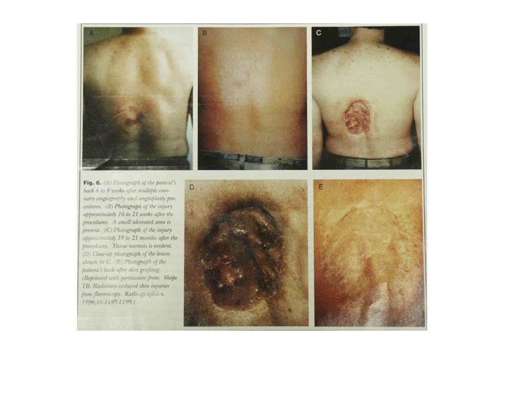
Radiation Induced Burns From Fluoroscopy Patient had two cardiac catheterizations within a 5-month interval. A total of 247 minutes of fluoroscopy and 2700 cinefluorographic images performed. Within 24 hours of the second procedure developed pain and skin erythema. The area ulcerated and necrosed over the next 5 months. Patient had extensive skin grafting to close wound.
Radiation Exposure to the U.S. Population
In the following table, the first column shows the sources of radiation. The second column shows an estimate of the number of people exposed to that source. For natural sources, the entire United States population is assumed to be exposed. The third column provides a measurement of the average dose (in units of millirems) to those exposed (number in column 2). The last column averages the total dose from the specific source over the entire U. S. population. For natural sources, the third and fourth columns are identical.
Exposure Source Population Exposed
(millions) 230
Average Dose Equivalent to Exposed
Population (millirems/year)
Average Dose Equivalent to U.S.
Population (millirems/year)
Natural
Radon 230 200 200
Other 230 100 100
Occupational 0.93 230 0.9
Nuclear Fuel Cycle1 - - - - - - 0.05
Consumer Products:
Tobacco 2 50 - - - - - -
Other 120 5 - 30 5 - 13
Environment 25 0.6 0.06
Medical:
Diagnostic X-rays3 - - - - - - 39
Nuclear medicine4 - - - - - - 14
Approximate Total 230 - - - 360
1 Collective dose to regional population within 50 miles of each facility.
2 Difficult to determine a whole body dose equivalent. However, the dose to a portion of the lungs is estimated to be 16,000 millirems/year.
3 Number of persons unknown. However, 180 million examinations performed with an average dose of 50 millirems per examination.
4 Number of persons unknown. However, 7.4 million examinations performed with an average dose of 430 millirems per examination.
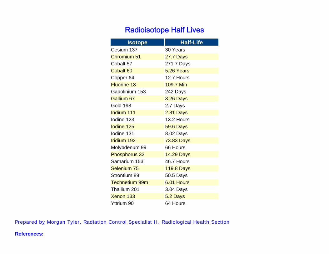
Radioisotope Half Lives Isotope Half-Life
Cesium 137 30 Years Chromium 51 27.7 Days Cobalt 57 271.7 Days Cobalt 60 5.26 Years Copper 64 12.7 Hours Fluorine 18 109.7 Min Gadolinium 153 242 Days Gallium 67 3.26 Days Gold 198 2.7 Days Indium 111 2.81 Days Iodine 123 13.2 Hours Iodine 125 59.6 Days Iodine 131 8.02 Days Iridium 192 73.83 Days Molybdenum 99 66 Hours Phosphorus 32 14.29 Days Samarium 153 46.7 Hours Selenium 75 119.8 Days Strontium 89 50.5 Days Technetium 99m 6.01 Hours Thallium 201 3.04 Days Xenon 133 5.2 Days Yttrium 90 64 Hours
Prepared by Morgan Tyler, Radiation Control Specialist II, Radiological Health Section References:
The Fundamentals of X-ray and Radium Physics, Joseph Selman, M.D. Radiology “ An Illustrated History”, Ronald L. Eisenberg The Health Physics and Radiological Health Handbook, Bernard Shleien ASRT, Radiation Safety In Fluoroscopy, Teresa G. Norris University of Michigan Health Physics Web Site HyperPhysics, Department of Physics and Astronomy, Carl R. (Rod) Nave Amersham Health, Medcyclopaedia, http://www.medcyclopaedia.com, Amersham Health) Conference of Radiation Control Program Directors, Inc. (CRCPD) Columbia University Medical Center, Washington Heights, Manhattan, New York. U. S. Nuclear Regulatory Commission, (NRC) University Of Nevada, EHS, Myung Chul Jo, CHP, (www.ehs.unr.edu/rso/radprin.htm)