+ All Categories
Home > Documents > Disease Causing Organisms in Procambarus cla- rkii and...

Disease Causing Organisms in Procambarus cla- rkii and...

Date post: 29-May-2018
Category:
Upload: ngokhanh
View: 217 times
Download: 0 times
Share this document with a friend
19
6 Egypt. J. Vet. Sci. Vol. 47, No.1, pp. 63-81 (2016) * Corresponding author: WafaaTawfik Abbas, Assistant Professor, 00201001125171. Email: [email protected] T Disease Causing Organisms in Procambarus cla- rkii and Gambusia affinis with Emphasis on their Role in Biomonitoring of Aquatic Pollution W.S. Soliman, Wafaa T. Abbas * , Taghreed, B. Ibrahim, Amany, M. Kenawy and M.Y. Elgendy Department of Hydrobiology, Veterinary Research Division, National Research Centre, Cairo, Egypt. HE HEALTH status of red swamp crayfish, Procambarus clarkii and mosquito fish, Gambusia affinis collected from Elmansoria canal, Giza, Egypt was investigated. The canal is known to receive lofty loads of pollutants from diverse anthropogenic sources. 113 bacterial isolates were obtained from the investigated fish specimens. Isolates were phenotypically identified as, Aeromonas hydrophila 26.54%, Vibrio parahaemolyticus 21.23%, Pseudomonas fluorescens 14.15%, E. coli 10.61%, Citrobacter sp. 7.96%, Enterobacter sp. 8.84%, Staphylococcus sp. 4.42% and Micrococcus sp. 6.19%. High gill infestations with Centrocestus sp. encysted metacercariae were noticed in mosquito fish. No parasitic infestations were recorded in crayfish. Challenge experiment confirmed the pathogenicity of Aeromonas hydrophila isolates. The water analysis revealed high heavy metals levels with values, Ni 0.71, Pb 0.34 and Cd 0.2 ppm while Zn and Cu were in normal values. Metals analysis in crayfish and mosquito fish tissues denoted bioaccumulation. Crayfish muscles showed, Ni >Zn >Cu >Pb>Cd while their levels in mosquito fish demonstrated, Ni >Zn>Pb>Cd>Cu. Proliferative, degenerative and necrotic alterations were evident in histological sections. Results suggest that both crayfish and mosquito fish can serve as carriers for some fish disease pathogenic agents and a convenient tool for biomonitoring aquatic pollution. Keywords: Bacteria, Parasites, Pollution, Crayfish, Mosquito fish. Pathogenic microorganisms distribute wildly in the aquatic environment especially in polluted habitats. There is close relationship between emergence of aquatic animal diseases and coexistence of diverse pollutants in the aquatic environment (Elgendy et al., 2015a). Polluted water deteriorates fish host defenses allowing increased opportunities for microbial agents to affect fish populations (Arkoosh et al., 1998). Moreover, some pollutants and wastes are nutritious and cause eutrophication increasing bacterial load and algae in water as well as induce critical oxygen deficiencies (Ansari et al., 2011). Accordingly,
Transcript

6 Egypt. J. Vet. Sci. Vol. 47, No.1, pp. 63-81 (2016)

*Corresponding author: WafaaTawfik Abbas, Assistant Professor, 00201001125171.

Email: [email protected]

T

Disease Causing Organisms in Procambarus cla-

rkii and Gambusia affinis with Emphasis on their

Role in Biomonitoring of Aquatic Pollution

W.S. Soliman, Wafaa T. Abbas*, Taghreed, B. Ibrahim,

Amany, M. Kenawy and M.Y. Elgendy

Department of Hydrobiology, Veterinary Research Division,

National Research Centre, Cairo, Egypt.

HE HEALTH status of red swamp crayfish, Procambarus

clarkii and mosquito fish, Gambusia affinis collected from

Elmansoria canal, Giza, Egypt was investigated. The canal is known

to receive lofty loads of pollutants from diverse anthropogenic

sources. 113 bacterial isolates were obtained from the investigated

fish specimens. Isolates were phenotypically identified as, Aeromonas

hydrophila 26.54%, Vibrio parahaemolyticus 21.23%, Pseudomonas

fluorescens 14.15%, E. coli 10.61%, Citrobacter sp. 7.96%,

Enterobacter sp. 8.84%, Staphylococcus sp. 4.42% and Micrococcus

sp. 6.19%. High gill infestations with Centrocestus sp. encysted

metacercariae were noticed in mosquito fish. No parasitic infestations

were recorded in crayfish. Challenge experiment confirmed the

pathogenicity of Aeromonas hydrophila isolates. The water analysis

revealed high heavy metals levels with values, Ni 0.71, Pb 0.34 and

Cd 0.2 ppm while Zn and Cu were in normal values. Metals analysis

in crayfish and mosquito fish tissues denoted bioaccumulation.

Crayfish muscles showed, Ni >Zn >Cu >Pb>Cd while their levels in

mosquito fish demonstrated, Ni >Zn>Pb>Cd>Cu. Proliferative,

degenerative and necrotic alterations were evident in histological

sections. Results suggest that both crayfish and mosquito fish can

serve as carriers for some fish disease pathogenic agents and a

convenient tool for biomonitoring aquatic pollution.

Keywords: Bacteria, Parasites, Pollution, Crayfish, Mosquito fish.

Pathogenic microorganisms distribute wildly in the aquatic environment

especially in polluted habitats. There is close relationship between emergence of

aquatic animal diseases and coexistence of diverse pollutants in the aquatic

environment (Elgendy et al., 2015a). Polluted water deteriorates fish host

defenses allowing increased opportunities for microbial agents to affect fish

populations (Arkoosh et al., 1998). Moreover, some pollutants and wastes are

nutritious and cause eutrophication increasing bacterial load and algae in water

as well as induce critical oxygen deficiencies (Ansari et al., 2011). Accordingly,

W.S. SOLIMAN et al.

Egypt. J. Vet. Sci. Vol. 47, No.1 (2016)

64

the early detection of these pollutants strongly assists to restrict the spread of

many detrimental microbes to aquatic animals and/or human beings (Kuklina

et al., 2014).

Procambarus clarkii and Gambusia affinis are two models used largely in

biomonitoring studies. They are extremely adaptable aquatic organisms endure

wide range of critical environmental conditions (Abdelghany, 2002). Both are

obstinately introduced into many non-indigenous aquatic habitats in the aim to

solve some troubles. They feed on variety of food resources including algae,

detritus, gastropods as well as numerous invertebrates (Whitledge and Rabeni

1997, Rincon et al. 2002). Meanwhile they are also preyed upon by various fish

species, aquatic birds and mammals (Holdich, 2002) and can play prominent

roles in water ecosystem, the burrowing activities of crayfish help to create

appropriate habitats for many other small aquatic organisms (Pintor and Soluk,

2006). Furthermore, crayfish can biologically control many snails which

represent important vectors as well as intermediate host of numerous pathogenic

agents (Fishar, 2006).Confirmatory previous reports highlighted their significant

role in the limitation of some parasitic infestations endemic to aquatic habitats

(Haddaway et al., 2012 and Du Preez, 2013).

Interestingly, crayfish does not migrate and usually localize in their habitats

(Banks and Brown, 2002). Furthermore, it has a long life span extending up to 2

years with continuous contact with water and sediment as well as tolerate

polluted environments consequently it can act as good biomonitors for aquatic

pollution since it accumulates respective elements in their tissues (Moss et al.,

2010).

On the other hand, mosquito fish, Gambusia affinis, are willfully introduced

in many countries as a bio-control for mosquito larvae especially in African

countries. Its aggressive feeding habits on the eggs and larvae of fish have been

accused for the decline of a number of fish species (Rincon et al., 2002).

Additionally, these fish constantly cause finnipping for other cohabitant fish

subsequently stress them and potentiate their infection by lots of opportunistic

bacterial and fungal agents (Lloyd, 1990).

Among pollutants, heavy metals are of particular concern due to their toxicity

and competence to bio-accumulates in aquatic ecosystems (Miller et al.,2003)

subsequently, metals affect human causing chronic toxicity and possibly cancer

(Mohamed et al., 2016 and Zhao et al., 2014).

The present study aimed to assess the health status of two important aquatic

organisms, Procambarus clarkii and Gambusia affinis collected from Elmansoria

canal, Abo-Rawash, Giza, Egypt. This was performed in bio-monitoring studies

to investigate the effect of water pollution on the existence of bacterial and

parasitic agents as well as to demonstrate the accompanied histopathological

alterations.

DISEASE CAUSING ORGANISMS IN PROCAMBARUSCLARKII …

Egypt. J. Vet. Sci. Vol. 47, No.1 (2016)

65

Material and Methods

Area of study and sampling

The present study was conducted in Elmansoria canal, a small branch from

the River Nile, Abo-Rawash area, Giza, Egypt, during 2015 summer season.

This branch receives high load of pollutants come from different anthropogenic

sources. 50 samples of each red swamp crayfish, Procambarus clarkii (45-65 g)

and mosquito fish, Gambusia affinis (4-5 g) were collected by fishermen. The

specimens were transported alive in plastic bags containing water and supplied

with oxygen, within the minimum time of delay to Hydrobiology Department,

National Research Centre, Egypt. Water samples were collected early in the

morning in sterile bottles from the subsurface layer of different three points

around the selected site.

Bacteriological examination

Swabs from gills, hepatopancreas and hemolymph were aseptically obtained

from crayfish specimens according to (Lucíaet al., 2003).On the other hand,

regarding mosquito fish, loopfuls were retrieved from gills and kidneys. Inoculi

were further enriched in tryptic soy broth then smeared onto agar media, Brain

heart infusion agar (BHI) (Oxoid), Tryptic soy agar (TSA) (Oxoid), Aeromonas

and Pseudomonas specific agar media (Oxoid). The inoculated plates were

incubated at 25 oC for 24 to 48 h. Water samples obtained from Elmansoria canal

were also analyzed microbiologically. Representative numbers of the different

colonial types detected on the media were collected from plates and streaked on

TSA for purification and identification according to Buller (2004).

Identification of isolates

Identification of pure bacterial isolates was performed by studying their

morphological and biochemical characteristics according to schemes

demonstrated by Bergey,s Manual of Systemic Bacteriology (1982) using

traditional as well as commercial API 20 E and API NE systems following the

criteria described in (Buller, 2004).

Parasitological examination

All crayfish and mosquito fish samples were thoroughly investigated for

external parasites by visual inspection via naked eye. Furthermore, wet smears

from the cephalothoracic cavity and gills of crayfish and skin and gills of

mosquito fish were freshly examined, fixed with methanol, stained by 10%

Giemsa stain and examined under the bright field microscope to identify the

presence of any external protozoan parasites. Small pieces of gills, liver,

hepatopancreas and muscles from both crayfish and mosquito fish were

compressed between two glass slides (compressorium) and examined under the

binocular dissecting microscope for the presence of encysted metacercariae

(Pritchard and Kruse, 1982).

W.S. SOLIMAN et al.

Egypt. J. Vet. Sci. Vol. 47, No.1 (2016)

66

Experimental infection

Randomly selected A. hydrophila isolate was used for challenge experiment.1.24 × 10

7 CFU/ml inoculi were prepared according to (Elsherry, 2004) then injected into

three different separated species, crayfish, mosquito fish and Oreochromis niloticus. Experimental animals were initially adapted to the wet lab conditions, kept in glass aquaria supplemented with sufficient water for an entire period of two weeks before the onset of challenge experiment. Each group contained 10 fish of the same species. Three other control groups, one for each species, were injected with phosphate buffer saline. Crayfish were injected into the haemolymph while mosquito fish and tilapia fish were challenged using the intra peritoneal route. All fishes were observed for two weeks after challenge with A. hydrophila. Dead fishes were processed microbiologically to re-isolate A. hydrophila in the aim to confirm the specificity of pathogenicity.

Water quality examination

Conductivity and pH of water samples were measured on spot while collecting the samples using digital portable apparatus, (pH meter model HI 8314 and digital conductivity meter HI2300 Hanna Ins. Romania). After acidification of water samples, phosphate, ammonia and nitrate were determined by colorimetric methods in the lab (APHA, 1995).

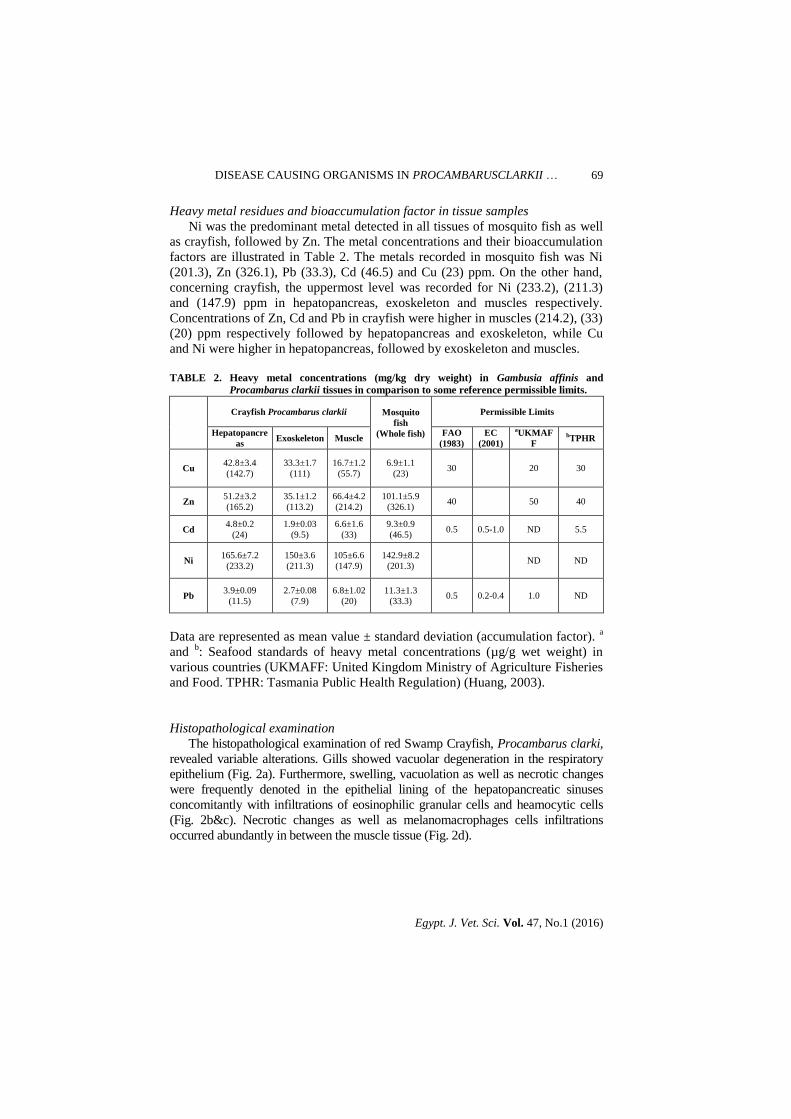
Heavy metals analysis

Water samples Water samples were acidified by concentrated nitric acid (5ml/L) and heavy

metals (Cu, Zn, Cd, Ni, and Pb) were detected in one pooled sample by the atomic absorption spectrophotometer (Perkin-Elmer 3110, USA) (APHA, 1995).

Crayfish and mosquito fish

The same metals were detected in the tissue samples. Crayfish samples were dissected, hepatopancreas, muscles and exoskeleton were dried in an oven (120

oC). On the other hand, mosquito fish was excavated and dried as a whole.

Dried tissues were grounded in a ceramic mortar, 0.5g of it were digested using concentrated nitric acid and the heavy metals concentrations were measured using the atomic absorption spectrophotometer (Perkin-Elmer 3110, USA) (Riyahi, 2000).

Accumulation factor (AF)

Accumulation factor (AF) was calculated according to the following equation:

AF = Concentration of the heavy metal in the organ (mg/kg)/concentration of the heavy metal in water (mg/L) (Authman et al., 2013).

Histopathological examination

Small portions of gills, hepatopancreas and muscles were fixed in Davidson’s fixative for 48 hrs, then dehydrated in ascending grades of alcohol and cleared in xylene. The fixed tissues were embedded in paraffin wax and sectioned at 5 microns. Sections were stained with Hematoxylin and Eosin method (Bernet et al., 1999), examined microscopically and photographed by using a microscopic camera.

DISEASE CAUSING ORGANISMS IN PROCAMBARUSCLARKII …

Egypt. J. Vet. Sci. Vol. 47, No.1 (2016)

67

Results

Clinical examination

Investigated crayfish were lethargic, had some blistering on the end of the

telson with necrosis and erosion on the tail region. Some specimens also

demonstrated congestion and enlargement of hepatopancreas. On the other hand,

mosquito fish specimens showed no specific signs except for erosions of skin

and fins. Some specimens demonstrated petechial hemorrhages on the external

body surfaces (Fig. 1a&b).

Bacteriological examination

Total number of 113 bacterial isolates was retrieved from investigated fish

specimens, 72 isolates from crayfish and 41 isolates from mosquito fish.

Retrieved isolates were further identified as, Aeromonas hydrophila26.54%,

Vibrio parahaemolyticus 21.23%, Pseudomonas fluorescens 14.15%, E. coli

10.61%, Citrobacter sp. 7.96%, Enterobacter sp. 8.84%, Staphylococcus sp.

4.42% and Micrococcus sp. (6.19%). Concerning water samples, the total

bacterial count was 2.5X10⁶, additionally, similar bacterial isolates were also

detected in water samples collected from the canal.

Parasitological examination

Parasitological examination of mosquito fish demonstrated high frequency of

Centrocestus sp. encysted metacercariae in gills with 75% infection rate, 2-5

cysts per gill filament (Fig. 1c). On the other hand, crayfish showed no parasitic

infestations (Fig. 1d).

Fig. 1. Mosquito-fish, Gambusia affinis (a). Red Swamp Crayfish, Procambarus clarkii (b). Freshly

compressed gills of Gambusia affinis showing infections with Centrocestus sp. (Heterophydae)

encysted metacercariae (c). Freshly prepared gills of Procambarus clarkii showing absence of

parasitic infestations (d).

W.S. SOLIMAN et al.

Egypt. J. Vet. Sci. Vol. 47, No.1 (2016)

68

Experimental infection

Challenge experiment indicated the pathogenicity of A. hydrophila isolates.

60%, 100% and 80% of, crayfish, mosquito fish and tilapia fish respectively

were died during the 2 weeks experimental period. Succumbed fish showed

petechial hemorrhages on the external body surfaces and different degrees of

erosion. Marked congestion of kidney was noticed in tilapia and mosquito fish.

Some fish died without exhibiting any signs. A. hydrophila was re-isolated from

all succumbed fishes.

Water analysis

Physicochemical water quality measures

The average recorded values for pH, EC, nitrate, ammonia and phosphate

exhibited normal values when compared with the some international permissible

limits (Table 1).

Heavy metals in water

Analysis of heavy metals in water samples demonstrated higher concentrations

of some metals over the permissible limits, the uppermost concentration was

detected for Ni (0.71ppm) and the lowest one was Cd (0.2ppm). On the other hand,

Zn and Cu values were within normal standards (0.3ppm) (Table 1).

TABLE 1. Physicochemical and heavy metals analysis of water samples collected

From Elmansoria canal in comparison to some reference permissible limits.

Parameter Site Permissible

limits References

pH 7.29 6-9 Egyptian Environmental Law No. 4 (1994)

EC (dS/m) 0.4 0.7-3

PO4 (ppm) 3.0 5 Egyptian Environmental Law No. 4 (1994)

NH3 (ppm) 1.5 3 Egyptian Environmental Law No. 4 (1994)

up to 12 WHO, 2003

NO3 (ppm) 7.3 40 Egyptian Environmental Law No. 4 (1994)

Cu (ppm) 0.3

1 Egyptian law No. 48 (1982)

1.5 Egyptian Environmental Law No. 4 (1994)

0.009 USEPA (2006)

Zn (ppm) 0.31

1 Egyptian law No. 48 (1982)

5 Egyptian Environmental Law No. 4 (1994)

0.12 USEPA (2006)

Cd (ppm) 0.2

0.01 FAO (1983), Egyptian law No. 48 (1982)

0.05 Egyptian Environmental Law No. 4 (1994)

0.00025 USEPA (2009)

Ni (ppm) 0.71

0.01 Egyptian law No. 48 (1982)

0.1 Egyptian Environmental Law No. 4 (1994)

0.052 USEPA (2006)

Pb (ppm) 0.34 0.05 FAO (1983), Egyptian law No. 48 (1982)

0.0025 USEPA (2006)

DISEASE CAUSING ORGANISMS IN PROCAMBARUSCLARKII …

Egypt. J. Vet. Sci. Vol. 47, No.1 (2016)

69

Heavy metal residues and bioaccumulation factor in tissue samples

Ni was the predominant metal detected in all tissues of mosquito fish as well

as crayfish, followed by Zn. The metal concentrations and their bioaccumulation

factors are illustrated in Table 2. The metals recorded in mosquito fish was Ni

(201.3), Zn (326.1), Pb (33.3), Cd (46.5) and Cu (23) ppm. On the other hand,

concerning crayfish, the uppermost level was recorded for Ni (233.2), (211.3)

and (147.9) ppm in hepatopancreas, exoskeleton and muscles respectively.

Concentrations of Zn, Cd and Pb in crayfish were higher in muscles (214.2), (33)

(20) ppm respectively followed by hepatopancreas and exoskeleton, while Cu

and Ni were higher in hepatopancreas, followed by exoskeleton and muscles.

TABLE 2. Heavy metal concentrations (mg/kg dry weight) in Gambusia affinis and

Procambarus clarkii tissues in comparison to some reference permissible limits.

Crayfish Procambarus clarkii Mosquito

fish

(Whole fish)

Permissible Limits

Hepatopancre

as Exoskeleton Muscle

FAO

(1983)

EC

(2001)

aUKMAF

F bTPHR

Cu 42.8±3.4

(142.7)

33.3±1.7

(111)

16.7±1.2

(55.7)

6.9±1.1

(23) 30 20 30

Zn 51.2±3.2

(165.2)

35.1±1.2

(113.2)

66.4±4.2

(214.2)

101.1±5.9

(326.1) 40 50 40

Cd 4.8±0.2

(24)

1.9±0.03

(9.5)

6.6±1.6

(33)

9.3±0.9

(46.5) 0.5 0.5-1.0 ND 5.5

Ni 165.6±7.2

(233.2)

150±3.6

(211.3)

105±6.6

(147.9)

142.9±8.2

(201.3) ND ND

Pb 3.9±0.09

(11.5)

2.7±0.08

(7.9)

6.8±1.02

(20)

11.3±1.3

(33.3) 0.5 0.2-0.4 1.0 ND

Data are represented as mean value ± standard deviation (accumulation factor). a

and b: Seafood standards of heavy metal concentrations (µg/g wet weight) in

various countries (UKMAFF: United Kingdom Ministry of Agriculture Fisheries

and Food. TPHR: Tasmania Public Health Regulation) (Huang, 2003).

Histopathological examination

The histopathological examination of red Swamp Crayfish, Procambarus clarki,

revealed variable alterations. Gills showed vacuolar degeneration in the respiratory

epithelium (Fig. 2a). Furthermore, swelling, vacuolation as well as necrotic changes

were frequently denoted in the epithelial lining of the hepatopancreatic sinuses

concomitantly with infiltrations of eosinophilic granular cells and heamocytic cells

(Fig. 2b&c). Necrotic changes as well as melanomacrophages cells infiltrations

occurred abundantly in between the muscle tissue (Fig. 2d).

W.S. SOLIMAN et al.

Egypt. J. Vet. Sci. Vol. 47, No.1 (2016)

70

Fig. 2. Histopathological alterations noticed in Procambarus clarkii: Gill tissues

showing slight degenerative changes and heamocytic cells infiltrations

H&E, X400 (a). Hepatopancreatic sinuses showing vacuolar degeneration

with heamocytic and eosinophilic granular cells infiltrations in the cell

membrane lining H&E, X400 (b). Hepatopancreatic sinuses showing severe

necrotic changes H&E X400 (c). Necrotic changes and melanomacrophages

cells infiltrations in muscle tissue H&E, X400 (d).

On the other hand, regarding mosquito fish, Gambusia affinis, gills were

congested (Fig. 3a & b). Additionally, proliferative, degenerative and necrotic

changes were commonly detected in the respiratory epithelium. Moreover,

parasitic sections and cysts were noticed frequently in between gill tissues (Fig.

3c, d, e & f).

Discussion

Even though the hazards of water contamination on the health status and

survivability of aquatic animals are well acknowledged, it remains a matter of

interest due to its detrimental outcomes on both fish and human beings.

Maintaining favorable water quality measures is critical for aquatic animal

health. Hence keen monitoring via bioindicators can provide valuable data for

assessment of environmental status (Burger, 2006).

DISEASE CAUSING ORGANISMS IN PROCAMBARUSCLARKII …

Egypt. J. Vet. Sci. Vol. 47, No.1 (2016)

71

Fig. 3. Histopathological alterations noticed in Gambusia affinis: Gill tissue showing,

congestion, degenerative, necrotic changes in the respiratory epithelium and

hyperplasia H&E, X400 (a & b). Parasitic sections and cysts in between the

gill tissues H&E, X400 (c, d, e & f).

A substantial body of scientific evidence has been published on the close

relationship between pollution and microbial infections in aquatic animals.

Infections are common in aquatic animals especially those exposed to

unfavorable environmental conditions (Sinderman, 1995, Elgendy et al., 2013

and Moustafa et al., 2014).

Bacteriological examination demonstrated that A. hydrophila was the most

predominant bacterial isolates 26.54%. The ubiquitous nature of this pathogen in

aquatic environment may clarify this relatively high prevalence. Losses

attributed to this microorganism in fish and shellfish have been recorded

W.S. SOLIMAN et al.

Egypt. J. Vet. Sci. Vol. 47, No.1 (2016)

72

worldwide (Tulsidas et al., 2008 and Elgendy, 2015b). A. hydrophila are

frequently retrieved from both diseased and apparently healthy individuals,

interestingly, even both types cause problems and mortalities in many farmed

aquatic species (Quaglio et al., 2006a).

Furthermore, Vibrio parahaemolyticus recorded 21.23%. Previous reports

have showed that several Vibrio sp. Were frequently involved in infections as

well as mortalities affecting crayfish including, V. mimicus and V. cholera

(Thune et al., 1991 and Wong et al., 1995).Vibrios infections are commonly

associated with no characteristic signs (Eaves and Ketterer,1994).Moreover,

vibrios can do infections in many cultured as well as wild aquatic animals

predisposed susceptible by underlying poor conditions (Edgerton et al.,

2002,Quaglioet al., 2006b,Moustafa et al., 2015 and Elgendy et al., 2015a).

Results also demonstrated the recovery of other bacterial species including

Pseudomonas fluorescens14.15%, E. coli 10.61%, Citrobacter sp. 7.96%,

Enterobacter sp. 8.84 %, Staphylococcus sp. 4.42 % and Micrococcus sp.

(6.19%). Results are in concordance with previous reports discussing microbial

infections in crayfish denoting that many microorganisms are frequently detected

including, Coxiella cheraxi, Acinetobacter, Nocardia, Bacillus, Spiroplasma,

Citrobacter, Corynebacterium, Flavobacterium, Micrococcus, Pseudomonads,

Staphylococcus and several Vibrio species (Edgerton et al., 2002, Quaglio et al.,

2006b and Jiravanichpaisal et al., 2009). Many bacterial infections can colonize

the external exoskeleton and haemolymph of crayfish with the competence to

impact this species in its natural habitat when their existence combined with

unfavorable environmental conditions (David et al., 2005).The lofty loads of

discharges from sewage and agriculture drainages being thrown into the

Elmansoryia canal are loaded with many bacterial species.

Pathogenicity studies confirmed the virulence of isolated bacteria not only for

crayfish and mosquito fish but also for tilapia fish. Challenge experiment

confirmed that crayfish can acts as a reservoir for many pathogenic

microorganisms threatening cohabitant fish species. Wong et al. (1995)

demonstrated that Vibrio mimicus retrieved from the hemolymph of crayfish

suffering from asymptomatic bacteraemia were found to be primary pathogens,

able to produce disease in unstressed, healthy aquatic animals. On the other

hand, the finnipping habits of mosquito fish for other cohabitant fish species may

potentiate transmission as well as establishment of many microbial infections

(Lloyd, 1990).

Parasitological examination demonstrated absence of parasitic infestations in

crayfish specimens which may be relevant to their potential resistance against

some infections. On the other hand, mosquito fish were found to be vulnerable to

Centrocestus infections. Similar infestations with Centrocestus sp. were reported

in many fish species including Gambusia affinis (Nishigori, 1924 and Mitchell

et al., 2005). The detections of Centrocestussp.in mosquito fish despite the

existence of crayfish in their environment may be related to the limited

DISEASE CAUSING ORGANISMS IN PROCAMBARUSCLARKII …

Egypt. J. Vet. Sci. Vol. 47, No.1 (2016)

73

competence of crayfish to consume some thick-shelled snails (Hofkinet al.,

1992) as well as may be also linked to the host specificity of parasites (Bauer et

al., 2000). Previous studies have reported that the snail intermediate hosts of

Centrocestus sp. have high potentials to endure harsh environmental conditions

as well as numerous insecticides (Mitchell et al., 2005).

Earlier reports have confirmed the marked effect of invasive predator aquatic

species, like crayfish, on the diversity and abundance of parasitic communities of

cohabitant native species through interference with their pathway from

intermediate to definitive hosts, (Holmes, 1979 and Poulin et al., 2011).Several

reports have signified the potential role of crayfish in the biological control of

some epidemic diseases, in particular, infestations that include snails in their life

cycle like, schistosomiasis, fascioliasis and heterophyid sp. (Mkoji et al., 1995,

1999 and Khalil & Sleem, 2011) through attacking and consuming parasites

hosts (Holmes 1979, Poulinet al., 2011 and Pulkkinen et al., 2013)

Results of water analysis revealed high concentrations of some heavy metals

in the investigated water samples which may be claimed to the surplus

discharges coming from sewage, agriculture and industrial activities near

Elmansoria canal. The majority of evaluated metals were found in higher

concentrations in water, Ni>Pb>Cd while Zn and Cu were within normal values.

Nickel was the uppermost detected metals (0.7 ppm), extremely higher than the

Egyptian environmental laws, 1982 & 1994 and the permissible limits USEPA

(2009).Many industrial activities can contribute largely to amplify its levels

including, stainless steel, ceramics, batteries and nickel plating (Al-Attar, 2007).

This was consequently followed by the high concentration of nickel and its

accumulation factor in both mosquito fish and crayfish tissues. Concentrations of

Cd and Pb (0.2 and 0.34 ppm respectively) were also higher than the permissible

limits, which probably be relevant to the presence of industrial and sewage waste

water discharges in Elmansouria canal.

Results confirmed the ability of crayfish and mosquito fish to accumulate

heavy metals since tissue concentrations were several times higher than the

detected metal levels in water signifying the value of both fish species in

biomonitoring studies. Although Zinc concentration in water (0.31mg/l) was not

exceeding the permissible levels, it recorded high tissues concentrations and

accumulation factor, especially in crayfish muscles. Zinc is an essential metal for

metabolic reactions and its high tissues concentrations is independent on its

water levels (Jakimska et al., 2011).Additionally, previous reports have also

confirmed that crayfish has considerable ability to manipulate elevated Zn levels

through their metabolic processes (Alcorlo et al., 2006).

The tendency of heavy metals to bioaccumulate in the investigated crayfish

tissues varied. Some metals, Pb and Cd, bioaccumulated mainly in muscles in

comparison to their levels in other organs in concordance with previous studies

reporting muscles as the most organ for heavy metals bioaccumulation (Higueras

et al., 2006).These metals are not involved in crayfish metabolism hence it

W.S. SOLIMAN et al.

Egypt. J. Vet. Sci. Vol. 47, No.1 (2016)

74

amplifies in tissues with longer exposure to elevated metal levels in the

surrounding aquatic environment (Alcorlo et al., 2006).

In accordance with previous studies, other organs including, exoskeleton,

hepatopancreas and gills have been also found to bioaccumulate metals from

aquatic environments. In the current study, Ni was found to maximize more in

crayfish exoskeleton that may be alleged as an attempt to eliminate out metals

from the body through excretion (Macheviciene, 2002).The detected high

concentration of Cu in crayfish hepatopancreas may also reflect the storage

capacity of this organ for Cu (Bagatto and Alikhan, 1987). The hepatopancreas

concentrates metals from the digestive tract and haemolymph, consequently store

them in its intracellular vacuoles (Roldan and Shivers, 1987).

Generally, the indiscriminate disposal of industrial wastes, domestic sewage,

agriculture drainages and other human activities into aquatic resources have long

been considered a potential source for entrance of an assortment of heavy metals

into aquatic ecosystems (Rajeshkumar & Munuswamy, 2011 and Maceda-

Veigaet al., 2012).

Histopathological studies demonstrated variable degenerative, proliferative

and infiltrative changes in histopathological sections. These alterations may be

relevant to the damaging effects of the detected metals as well as may also be

linked to the detected microbial infections. Similar alterations were noticed in

previous studies (Moustafa et al., 2015, Elgendy et al., 2013 and Elgendy et al.,

2015b). The long exposure of aquatic animals to environmental pollutants, heavy

metals in particular, influences their capability to defend against microbial

infections by reducing the competence of their external and internal defense

mechanisms including phagocytic activity of leukocytes and antibody synthesis

(Sinderman, 1995). Moreover, majority of metals cause coagulation and

precipitation changes of fish mucus, primary defense mechanism of aquatic

animals, as well as cytological damages to the gills (Burton et al., 1972). These

injuries diminish gas exchange with tissue hypoxia and in some cases cause high

mortalities. Moreover, gill lesions reduce the resistance against infectious agents

and open portals of entry for establishing pathogenic bacteria especially when

combined with unfavorable quality measures typical to that noticed in

Elmansoria canal. On the other hand, we also argue that the elevated metals

levels potentially created unsuitable conditions for existence of some

ectoparasitic infestations in the investigated specimens in concordance with

(Kuperman, 1992). Data extracted from our study clearly support the value of crayfish and

mosquito fish in biomonitoring aquatic pollution. Results also demonstrated that

these aquatic animals may acts as a significant reservoir of many microbial

infections threatening fish as well as human beings. Hence keen monitoring via

bioindicators can provide valuable data for assessment of environmental status

which will support maintaining favorable water quality measures for aquatic

animal health.

DISEASE CAUSING ORGANISMS IN PROCAMBARUSCLARKII …

Egypt. J. Vet. Sci. Vol. 47, No.1 (2016)

75

References

Abdelghany, A.E. (2002) Studies on partial and complete replacements of fish meal with

Gambusia meal in diets for the common carp, Cyprinus carpio. Egyptian Journal of

Aquatic Biology and Fisheries, 6, 141-765.

Al-Attar, A.M. (2007) The influences of nickel exposure on selected physiological

parameters and gill structure in the teleost fish, Oreochromis niloticus. Journal of

Biological Sciences, 7, 77-85.

Alcorlo, P., Otero, M., Crehuet, M., Baltanás, A. and Montes, C. (2006) The use of the

red swamp crayfish (Procambarus clarkii, Girard) as indicator of the bioavailability

of heavy metals in environmental monitoring in the River Guadiamar (SW,

Spain). Science of the Total Environment, 366, 380-390.

Ansari, A.A., Sarvajeet, A.A., Lanza, S.G. and Rast, G.R. (2011) Eutrophication:

causes, consequences and control, Ed. No. 504.45 EUT. The Netherlands. Springer.

APHA(1995) Standard methods for the examination of water and wastewater 19th

ed. Washington, DC, USA, American Public Health Association.

Arkoosh, M.R., Casillas, E., Clemons, E., Kagley, A.N., Olson, R., Reno, P. and

Stein, J.E. (1998) Effect of pollution on fish diseases: potential impacts on salmonid

populations. Journal of Aquatic Animal Health, 10, 182-190.

Authman, M.M., Abbas, H.H. and Abbas, W.T. (2013) Assessment of metal status in

drainage canal water and their bioaccumulation in Oreochromis niloticus fish in

relation to human health. Environmental monitoring and assessment, 185, 891-907.

Bagatto, G. and Alikhan, M.A. (1987) Copper, cadmium, and nickel accumulation in

crayfish populations near copper-nickel smelters at Sudbury, Ontario, Canada. Bulletin

of Environmental Contamination and Toxicology, 38, 540-545.

Banks, P.D. and Brown, K.M. (2002) Hydrocarbon effects on fouling assemblages: the

importance of taxonomic differences, seasonal, and tidal variation. Marine

Environmental Research, 53, 311-326.

Bauer, A., Trouvé, S., Grégoire, A., Bollache, L. and Cézilly, F. (2000) Differential

influence of Pomphorhynchus laevis (Acanthocephala) on the behaviour of native and

invader gammarid species. International Journal for Parasitology, 30, 1453-1457.

Bergey,s Manual of Systemic Bacteriology (1982) Williams & Wilkins, Baltimore,

London,Vol.1.

Bernet, D., Schmidt, H., Meier, W., Burkhardt‐Holm, P. and Wahli, T. (1999)

Histopathology in fish: proposal for a protocol to assess aquatic pollution. Journal of

Fish Diseases, 22, 25-34.

Buller, N.B. (2004) Interpretation of biochemical identification tests and sets. Bacteria

from fish and other aquatic animals, a practical identification manual, 117-136.

W.S. SOLIMAN et al.

Egypt. J. Vet. Sci. Vol. 47, No.1 (2016)

76

Burger, J. (2006) Bioindicators: types, development, and use in ecological assessment

and research. Environmental Bioindicators, 1, 22-39.

Burton, D.T., Jones, A.H. and Cairns Jr, J. (1972) Acute zinc toxicity to rainbow trout

(Salmogairdneri): confirmation of the hypothesis that death is related to tissue

hypoxia. Journal of the Fisheries Board of Canada, 29, 1463-1466.

David, W.C., William, B., Angelo, D., Susan, A.M. and Keven, R.C. (2005) Compendium

of Methods for the Microbiological Examination of Foods.4th ed., pp. 289-636.

Du Preez, L.H. (2013) Polystomatidae (Monogenea) of southern African Anura:

Polystoma channingi n. sp. parasitic in two closely related Cacosternum

species. African Zoology, 48, 64-71.

Eaves, L.E. and Ketterer, P.J. (1994) Mortalities in red claw crayfish Cherax quadricarinatus

associated with systemic Vibrio mimicus infection. Diseases of Aquatic

Organisms, 19, 233-237.

Edgerton, B.F., Evans, L.H., Stephens, F.J. and Overstreet, R.M. (2002) Synopsis of

freshwater crayfish diseases and commensal organisms. Aquaculture, 206, 57-135.

Elgendy, M.Y. (2013) Epizootiological and molecular studies on the common septicemic

bacterial diseases of some saltwater fishes. Ph.D. Thesis, Cairo University, Egypt.

Elgendy, M.Y., Moustafa, M., Gaafar, A.Y. and Borhan, T. (2015b) Impacts of

extreme cold water conditions and some bacterial infections on earthen-pond cultured

Nile tilapia, Oreochromis niloticus. Research Journal of Pharmaceutical, Biological

and Chemical Sciences, 6, 136-145.

Elgendy, M.Y., Soliman, W.S., Hassan, H.A., Kenawy, A.M. and Liala, A.M. (2015a) Effect of Abrupt Environmental Deterioration on the Eruption of Vibriosis in Mari-

Cultured Shrimp, Penaeus indicus, in Egypt. Fisheries and Aquatic Science.10, 146-

158.

Elsherry, Y.M.(2004)Role of crayfish in transmission of fish diseases, M.V.SC, Fish

Diseases and Management, Assiut University, Egypt.

European Commission Regulation EC 2001. No. 466/2001 of 8 March 2001. Official

Journal of the European Communities, 1.77/1.

FAO (1983) Compilation of legal limits for hazardous substances in fish and fishery

products. Food and Agriculture Organization (FAO) Fishery Circular, No. 464, pp. 5-

100.

Fishar, M.R.(2006)Red swamp crayfish Procambarus clarkii in River Nile, Egypt case

study. Biodiversity Monitoring and Assessment Project (BioMap), Nature

Conservation Sector, Egyptian Environmental Affairs Agency, Ministry of State for

Environmental Affairs.

Haddaway, N.R., Wilcox, R.H., Heptonstall, R.E., Griffiths, H.M., Mortimer, R.J.,

Christmas, M. and Dunn, A.M. (2012) Predatory functional response and prey

DISEASE CAUSING ORGANISMS IN PROCAMBARUSCLARKII …

Egypt. J. Vet. Sci. Vol. 47, No.1 (2016)

77

choice identify predation differences between native/invasive and parasitised/

unparasitised crayfish. PloS one, 7, e32229.

Higueras, P., Oyarzun, R., Lillo, J., Sánchez-Hernández, J.C., Molina, J.A., Esbrí,

J.M. and Lorenzo, S. (2006) The Almadén district (Spain): anatomy of one of the

world's largest Hg-contaminated sites. Science of the Total Environment, 356, 112-

124.

Hofkin, B V., Hofinger, D.M., Koech, D.K. and Loker, E.S. (1992) Predation of

Biomphalaria and non-target molluscs by the crayfish Procambarus clarkii:

implications for the biological control of schistosomiasis. Annals of Tropical

Medicine and Parasitology, 86, 663-670.

Holdich, D.M. (2002) Distribution of crayfish in Europe and some adjoining countries. Bulletin

Français de la Pêcheet de la Pisciculture, 367, 611-650.

Holmes, J.C. (1979) Parasite populations and host community structure. In: Host-parasite

interfaces (ed. Nickol, B.). Academic Press, New York, 27-46.

Huang, W.B. (2003) Heavy metal concentrations in the common benthic fishes caught

from the coastal waters of Eastern Taiwan. Journal of Food and Drug Analysis, 11,

324-329.

Jakimska, A., Konieczka, P., Skóra, K. and Namiesnik, J. (2011) Bioaccumulation of

metals in tissues of marine animals, part II: metal concentrations in animal

tissues. Polish Journal of Environmental Studies, 20, 1127-1146.

Jiravanichpaisal, P., Roos, S., Edsman, L., Liu, H. and Söderhäll, K. (2009) A highly

virulent pathogen, Aeromonas hydrophila, from the freshwater crayfish Pacifastacus

leniusculus. Journal of Invertebrate Pathology, 101, 56-66.

Khalil, M.T. and Sleem, S.H. (2011) Can the freshwater crayfish eradicate

schistosomiasis in Egypt and Africa? Journal of American Science, 7, 457-462.

Kuklina, I., Kouba, A., Buřič, M., Horká, I., Ďuriš, Z. and Kozák, P. (2014) Accumulation of heavy metals in crayfish and fish from selected Czech

reservoirs. Bio Med Research International, 1-9.

Kuperman, B.I. (1992) Fish parasites as bioindicators of the pollution of bodies of

water. Parazitologiia, 26, 479-482.

Lloyd, L. (1990) Native Fishes as alternatives to the exotic fish, Gambusia for insect

control. In: Introduced and translocated fishes and their ecological effects (Ed. D.A.

Pollard) pp. 115-122 Bureau of Rural Resources Proceedings No. 8 (Australian

Government Publishing Service, Canberra).

Lucía, O., Débora, P. and Catalina, R.(2003)Centro de Investigaciones Biológicasdel

Noroeste, Apdo. Postal 128, La Paz, B.C.S. Mexico.

Maceda-Veiga, A., Monroy, M. and de Sostoa, A. (2012) Metal bioaccumulation in the

Mediterranean barbel (Barbus meridionalis) in a Mediterranean River receiving

W.S. SOLIMAN et al.

Egypt. J. Vet. Sci. Vol. 47, No.1 (2016)

78

effluents from urban and industrial wastewater treatment plants. Ecotoxicology and

Environmental Safety, 76, 93-101.

Mackeviciene, G. (2002) Bioaccumulation of heavy metals in noble crayfish (Astacus

astacus L.) tissues under aquaculture conditions. Ekologija (Vilnius), 2, 79-82.

Miller, J.R., Lechler, P.J.and Bridge, G. (2003) Mercury contamination of alluvial

sediments within the Essequibo and Mazaruni river basins, Guyana. Water, Air, and

Soil Pollution, 148, 139-166.

Mitchell, A.J., Overstreet, R.M., Goodwin, A.E. and Brandt, T.M. (2005) Spread of

an exotic fish-gill trematode: a far-reaching and complex problem. Fisheries, 30, 11-

16.

Mkoji, G.M., Hofkin, B.V., Kuris, A.M., Stewart-Oaten, A., Mungai, B.N. and

Kihara, J.H. (1999) Impact of the crayfish Procambarus clarkii on Schistosoma

haematobium transmission in Kenya. The American Journal of Tropical Medicine and

Hygiene, 61, 751-759.

Mkoji, G.M., Hofkins, B.V., Ouma, J.H., Kuris, A.M., Koech, D.K. and Loker, E.S.

(1995) Research on biological control of schistosome-transmitting snails in

Kenya. Proceedings from a status of research on medical malacology in relation to

schistosomiasis in Africa, 265-274.

Mohamed, S.A., Elshal, M.F., Kumosani, T.A., Mal, A.O., Ahmed, Y.M., Almulaiky,

Y.Q., Asseri, A. and Zamzami, M.A. (2016) Heavy Metal Accumulation is

Associated with Molecular and Pathological Perturbations in Liver of Variolalouti

from the Jeddah Coast of Red Sea. International Journal of Environmental Research

and Public Health, 13, 342.

Moss, J.C., Hardaway, C.J., Richert, J.C. and Sneddon, J. (2010) Determination of

cadmium copper, iron, nickel, lead and zinc in crawfish [Procambrus clarkii] by

inductively coupled plasma optical emission spectrometry: a study over the 2009

season in Southwest Louisiana. Microchemical Journal, 95, 5-10.

Moss, R.H., Edmonds, J.A., Hibbard, K.A., Manning, M.R., Rose, S.K., Van Vuuren,

D.P. (2010) The next generation of scenarios for climate change research and

assessment. Nature, 463, 747-756.

Moustafa, M., Eissa, A.E., Laila, A.M., Gaafar, A.Y., Abumourad, I.M. and Elgendy,

M.Y. (2015) Investigations into the Potential Causes of Mass Kills in Mari-Cultured

Gilthead Sea Bream, Sparus aurata, at Northern Egypt. Research Journal of

Pharmaceutical, Biological and Chemical Sciences, 6, 466-477.

Moustafa, M., Eissa, A.E., Laila, A.M., Gaafar, A.Y., Abumourad, I.M.K. and

Elgendy, M.Y. (2014) Mass mortalities in mari-cultured European sea bass

(Dicentrarchus labrax) at Northern Egypt. Research Journal of Pharmaceutical,

Biological and Chemical Sciences, 5, 95-109.

Nishigori, M. (1924) On a new trematode Stamnosoma formosanum n. sp. and its

development. Taiwan Igakkai Zasshi, 234, 181-228.

DISEASE CAUSING ORGANISMS IN PROCAMBARUSCLARKII …

Egypt. J. Vet. Sci. Vol. 47, No.1 (2016)

79

Pintor, L.M. and Soluk, D.A. (2006) Evaluating the non-consumptive, positive effects of

a predator in the persistence of an endangered species. Biological Conservation, 130,

584-591.

Poulin, R., Paterson, R.A., Townsend, C.R., Tompkins, D.M. and Kelly, D.W. (2011)

Biological invasions and the dynamics of endemic diseases in freshwater

ecosystems. Freshwater Biology, 56, 676-688.

Pritchard, M.H. and Kruse, G.O. (1982) “The collection and Preservation of Animal

Parasites”. Technical Bulletin No. 1. The Harold W. Manter Laboratory, University

of Nebraska Press, 141 p.

Pulkkinen, K., Ruokonen, T.J., Mykrä, M., Tambe, G., Karjalainen, J. and

Hämäläinen, H. (2013) Indirect effects of invasive crayfish on native fish

parasites. Ecosphere, 4, 1-9.

Quaglio, F., Morolli, C., Galuppi, R., Bonoli, C., Marcer, F., Nobile, L., De Luise, G.

and Tampieri, M.P. (2006a) Preliminary investigations of disease-causing organisms

in the white-clawed crayfish Austropotamobius pallipes complex from streams of

northern Italy. Bulletin Francais de la Pecheet de la Pisciculture, 380-381, 1271-

1290.

Quaglio, F., Morolli, C., Galuppi, R., Tampieri, M.P., Bonoli, C., Marcer, F.,

Rotundo, G. and Germinara, G.S. (2006b) Sanitary-pathological examination of red

swamp crayfish (Procambarus clarkii, Girard 1852) in the Reno Valley. Freshwater

Crayfish, 15, 1-10.

Rajeshkumar, S. and Munuswamy, N. (2011) Impact of metals on histopathology and

expression of HSP 70 in different tissues of Milk fish (Chanos chanos) of

Kaattuppalli Island, South East Coast, India. Chemosphere, 83, 415-421.

Rincón, P.A., Correas, A.M., Morcillo, F., Risueño, P. and Lobón‐Cerviá, J. (2002)

Interaction between the introduced eastern mosquito fish and two autochthonous

Spanish tooth carps. Journal of Fish Biology, 61, 1560-1585.

Riyahi, A.R. (2000) Determination of Pb and Cd content and rang of this metals in

different tissues of fishes of Haraz River. Daneshvar. 31, 127-134.

Roldan, B.M. and Shivers, R.R. (1987) The uptake and storage of iron and lead in cells

of the crayfish (Orconectes propinquus) hepatopancreas and antennal gland. Comparative

Biochemistry and Physiology Part C: Comparative Pharmacology, 86, 201-214.

Sindermann, C.J. (1995) Ocean pollution: effects on living resources and humans. CRC

press. Inc.

Thune, R.L., Hawke, J.P. and Siebeling, R.J. (1991) Vibriosis in the red swamp

crawfish. Journal of Aquatic Animal Health, 3, 188-191.

Tulsidas, H., Ong, Y.Y. and Chan, K.C. (2008) Aeromonas hydrophila bacteraemia and

portal pyaemia. Singapore Med Case Report J, 49, 346-348.

W.S. SOLIMAN et al.

Egypt. J. Vet. Sci. Vol. 47, No.1 (2016)

80

USEPA (United States Environmental Protection Agency) (2006) National Recommended

Water Quality Criteria. Office of Water, Office of Science and Technology (4304T).

Whitledge, G.W. and Rabeni, C.F. (1997) Energy sources and ecological role of

crayfishes in an Ozark stream: insights from stable isotopes and gut

analysis. Canadian Journal of Fisheries and Aquatic Sciences, 54, 2555-2563.

WHO (World Health Organization) (2003) Ammonia in Drinking-water, background

document for development of WHO Guidelines for Drinking-water Quality, 2nd ed.

Vol. 2.Health criteria and other supporting information. World Health Organization,

Geneva.

Wong, F.Y.K., Fowler, K. and Desmarchelier, P.M. (1995) Vibriosis due to Vibrio

mimicus in Australian freshwater crayfish. Journal of Aquatic Animal Health, 7, 284-

291.

Zhao, Q., Wang, Y., Cao, Y., Chen, A., Ren, M., Ge, Y., Yu, Z., Wan, S., Hu, A., Bo,

Q. and Ruan, L. (2014) Potential health risks of heavy metals in cultivated topsoil

and grain, including correlations with human primary liver, lung and gastric cancer, in

Anhui province, Eastern China. Science of the Total Environment, 470, 340-347.

(Received 8/10/ 2016;

accepted 30/10/ 2016)

DISEASE CAUSING ORGANISMS IN PROCAMBARUSCLARKII …

Egypt. J. Vet. Sci. Vol. 47, No.1 (2016)

81

الكائنات المسببة لألمراض في إستاكوزا المياة العذبة

(Procambarus clarkii ) و أسماك الجامبوزيا(Gambusia

affinis )مع التأكيد على دورها في الرصد البيولوجي للتلوث المائى

وفاء توفيق عباس، تغريد برهان الدين إبراهيم، أمانى ،وليد صالح الدين سليمان

ح يوسف الجندىدمحم قناوى وممدو

.مصر -القاهرة -المركز القومى للبحوث -شعبة البحوث البيطرية -قسم األحياء المائية

(Procambarus clarkia)تمت دراسة الحالة الصحية العامة إلستاكوزا المياة العذبة

والتي تم تجميعها من ترعة ( Gambusia affinis)وأسماك الجامبوزيا

حيث تتعرض هذة الترعة للعديد من الملوثات من .، مصرالمنصورية بالجيزة

وقد تم . عزلة بكتيرية من عينات األسماك 111تم الحصول على . مصادر متنوعة

اإليروموناس هايدروفيال : تحديد أنواع هذة العزالت ونسب تواجدها كاألتى

، السيدوموناس (٪41.41)، الفيبريو باراهيموليتيكس (45.62٪)

، (٪5..6)، الستروباكتر (٪16.51)، اإليكوالى (٪12.16)فلوروسنس

(. ٪.5.1)والميكروكوكس ( ٪2.24)، اإلستافيلوكوكس (٪2...)اإلنتيروباكتر

كما لوحظ وجود إصابات طفيلية عالية بالحويصالت اليرقية لطفيل

بينما لم تسجل الدراسة أي إصابات . السينتروسيستسفى خياشيم أسماك الجامبوزيا

وقد أكدت تجربة إعادة العدوى بأنواع البكتيريا . إستاكوزا المياة العذبةطفيلية في

. على التأثير المرضي لعزالت اإليروموناس هايدروفيال(تجربة التحدى)المعزولة

و قد أظهرت نتائج تحليل عينات المياه من هذة المنطقة وجود نسب عالية من بعض

، (6.12)، الرصاص (6.61)النيكل : المعادن الثقيلة حيث كانت كاألتى

بينما كانت نسب تواجد عنصرى الزنك . جزء في المليون( 6.4)والكادميوم

و أكد تحليل المعادن الثقيلة فى أنسجة كالً من إستاكوزا . والنحاس في القيم الطبيعية

و كان . المياة العذبة و أسماك الجامبوزيا على التراكم البيولوجى لهذة المعادن

< النيكل: د المعادن الثقيلة فى عضالت إستاكوزا المياة العذبة كاألتىترتيب وجو

الكادميوم في حين كان ترتيبها فى أنسجة أسماك < الرصاص< النحاس< الزنك

وأظهرت . النحاس< الكادميوم< الرصاص< الزنك< النيكل: الجامبوزيا كاألتى

فى أنسجة األسماك النتائج أيضاً وجود بعض التغيرات الهستوباثولوجية الواضحة

وتخلص هذة الدراسة إلى أن كالً من إستاكوزا المياة العذبة و . موضع الدراسة

أسماك الجامبوزيا يمكن أن تكون ناقلة لبعض أمراض األسماك كما أن لها دوراً

.هاماً فى الرصد البيولوجي للتلوث المائي

-كوزا المياة العذبة إستا -التلوث -الطفيليات -البكتيريا :الكلمات الدالة

.الجامبوزيا


Recommended