Disease Mechanisms of C9ORF72 Repeat Expansions Tania F. Gendron and Leonard Petrucelli Department of Neuroscience, Mayo Clinic Florida, Jacksonville, Florida 32224 Correspondence: [email protected]G 4 C 2 repeat expansions within the C9ORF72 gene are the most common genetic cause of amyotrophic lateral sclerosis (ALS) and frontotemporal dementia (FTD). These bidirection- ally transcribed expansions lead to (1) the accumulation of sense G 4 C 2 and antisense G 2 C 4 repeat-containing RNA, (2) the production of proteins of repeating dipeptides through un- conventional translation of these transcripts, and (3) decreased C9ORF72 mRNA and protein expression. Consequently, there is ample opportunity for the C9ORF72 mutation to give rise to a spectrum of clinical manifestations, ranging from muscle weakness and atrophy to changes in behavior and cognition. It is thus somewhat surprising that investigations of these three seemingly disparate events often converge on similar putative pathological mech- anisms. This review aims to summarize the findings and questions emerging from the field’s quest to decipher how C9ORF72 repeat expansions cause the devastating diseases collec- tively referred to as “c9ALS/FTD.” A myotrophic lateral sclerosis (ALS), the most common form of motor neuron disease (MND), is characterized by the progressive de- generation of upper and lower motor neurons, leading to muscle weakness, atrophy, and spas- ticity. In addition, ALS involves several nonmo- tor systems and subcortical structures (Lowe 1994). In fact, cognitive and behavioral impair- ments reminiscent of frontotemporal dementia (FTD) are present in up to 50% of ALS patients (Lomen-Hoerth et al. 2003). FTD, second only to Alzheimer’s disease as a cause of dementia in patients under 65 (Neary et al. 1998), encom- passes a group of disorders neuropathologically characterized by degeneration of the frontal and temporal lobes. Most commonly, patients present with behavioral variant FTD, which is marked by changes in personality and behavior. Other syndromes falling under the FTD um- brella are categorized by changes in language function. The heterogeneous nature of FTD is further underscored by the fact that a propor- tion of patients with FTD develop ALS (Snow- den et al. 2002). A better understanding for the basis of the clinical overlap between ALS and FTD came about in 2006 with the discovery that TDP-43, an RNA/DNA-binding protein, forms inclusions in the central nervous system (CNS) of the majority of ALS cases and the most common pathological subtype of FTD, frontotemporal lobar degeneration with TDP- 43-positive inclusions (FTLD-TDP) (Arai et al. 2006; Neumann et al. 2006). Relatively soon thereafter, another major discovery connecting Editor: Stanley B. Prusiner Additional Perspectives on Prion Diseases available at www.perspectivesinmedicine.org Copyright # 2018 Cold Spring Harbor Laboratory Press; all rights reserved; doi: 10.1101/cshperspect.a024224 Cite this article as Cold Spring Harb Perspect Med 2018;8:a024224 1 www.perspectivesinmedicine.org on January 29, 2022 - Published by Cold Spring Harbor Laboratory Press http://perspectivesinmedicine.cshlp.org/ Downloaded from
Transcript
Disease Mechanisms of C9ORF72 RepeatExpansions
Tania F. Gendron and Leonard Petrucelli
Department of Neuroscience, Mayo Clinic Florida, Jacksonville, Florida 32224
G4C2 repeat expansions within the C9ORF72 gene are the most common genetic cause ofamyotrophic lateral sclerosis (ALS) and frontotemporal dementia (FTD). These bidirection-ally transcribed expansions lead to (1) the accumulation of sense G4C2 and antisense G2C4
repeat-containing RNA, (2) the production of proteins of repeating dipeptides through un-conventional translation of these transcripts, and (3) decreased C9ORF72 mRNA and proteinexpression. Consequently, there is ample opportunity for the C9ORF72 mutation to give riseto a spectrum of clinical manifestations, ranging from muscle weakness and atrophy tochanges in behavior and cognition. It is thus somewhat surprising that investigations ofthese three seemingly disparate events often converge on similar putative pathological mech-anisms. This review aims to summarize the findings and questions emerging from the field’squest to decipher how C9ORF72 repeat expansions cause the devastating diseases collec-tively referred to as “c9ALS/FTD.”
Amyotrophic lateral sclerosis (ALS), the mostcommon form of motor neuron disease
(MND), is characterized by the progressive de-generation of upper and lower motor neurons,leading to muscle weakness, atrophy, and spas-ticity. In addition, ALS involves several nonmo-tor systems and subcortical structures (Lowe1994). In fact, cognitive and behavioral impair-ments reminiscent of frontotemporal dementia(FTD) are present in up to 50% of ALS patients(Lomen-Hoerth et al. 2003). FTD, second onlyto Alzheimer’s disease as a cause of dementia inpatients under 65 (Neary et al. 1998), encom-passes a group of disorders neuropathologicallycharacterized by degeneration of the frontaland temporal lobes. Most commonly, patientspresent with behavioral variant FTD, which is
marked by changes in personality and behavior.Other syndromes falling under the FTD um-brella are categorized by changes in languagefunction. The heterogeneous nature of FTD isfurther underscored by the fact that a propor-tion of patients with FTD develop ALS (Snow-den et al. 2002). A better understanding for thebasis of the clinical overlap between ALS andFTD came about in 2006 with the discoverythat TDP-43, an RNA/DNA-binding protein,forms inclusions in the central nervous system(CNS) of the majority of ALS cases and themost common pathological subtype of FTD,frontotemporal lobar degeneration with TDP-43-positive inclusions (FTLD-TDP) (Arai et al.2006; Neumann et al. 2006). Relatively soonthereafter, another major discovery connecting
Editor: Stanley B. Prusiner
Additional Perspectives on Prion Diseases available at www.perspectivesinmedicine.org
Copyright # 2018 Cold Spring Harbor Laboratory Press; all rights reserved; doi: 10.1101/cshperspect.a024224
Cite this article as Cold Spring Harb Perspect Med 2018;8:a024224
1
ww
w.p
ersp
ecti
vesi
nm
edic
ine.
org
on January 29, 2022 - Published by Cold Spring Harbor Laboratory Presshttp://perspectivesinmedicine.cshlp.org/Downloaded from
ALS and FTD was made: G4C2 hexanucleotiderepeat expansions in chromosome 9 open read-ing frame 72 (C9ORF72) were identified as themost common known genetic cause of ALS andFTD (DeJesus-Hernandez et al. 2011; Rentonet al. 2011). C9ORF72 expansions account for�10%–50% of familial ALS, 5%–7% of spora-dic ALS, 12%–25% of familial FTD, and 6%–7% of sporadic FTD (Majounie et al. 2012;Cruts et al. 2013; van der Zee et al. 2013), andare also found in related clinical phenotypes,including Alzheimer’s disease and parkinson-ism (Cooper-Knock et al. 2014a; also see Gha-semi and Brown 2017 and Gijselinck et al.2017).
Clinical heterogeneity is associated with theC9ORF72 expansion, with the majority of af-fected patients displaying symptoms of eitherALS or FTD or mixed features of both (referredto as FTD-MND). There is also great variabilityamong patients with regard to age of diseaseonset and duration (Boeve et al. 2012; Byrneet al. 2012; Chio et al. 2012; Cooper-Knocket al. 2012; Hsiung et al. 2012; Mahoney et al.2012; Simon-Sanchez et al. 2012; Snowden et al.2012). The molecular basis for this clinical var-iation, seen both among and within families, isnot yet known. Upon first consideration, expan-sion length would seem a likely cause. Althoughthe lower limit for pathological G4C2 ex-pansions is not yet definitely established, un-affected individuals typically have two to 30repeats, whereas tens to even thousands of re-peats can be present in patients with ALS, FTD,and FTD-MND (DeJesus-Hernandez et al.2011; Renton et al. 2011; Beck et al. 2013; vanBlitterswijk et al. 2013; Dols-Icardo et al. 2014).Repeat length not only varies among familymembers, including monozygotic twins, butalso differs among tissues of the same individualowing to somatic instability (Beck et al. 2013;Buchman et al. 2013; van Blitterswijk et al.2013; Dols-Icardo et al. 2014; Waite et al. 2014;Fratta et al. 2015; Gijselinck et al. 2015; Nordinet al. 2015). At present, the influence of repeatlength on clinical phenotype has not been re-solved (van Blitterswijk et al. 2013; Benussi etal. 2014; Dols-Icardo et al. 2014; Cooper-Knocket al. 2015a; Nordin et al. 2015). Some studies
found that repeat size had no effect on clinicalpresentation (van Blitterswijk et al. 2013; Gijse-linck et al. 2015; Nordin et al. 2015), whereasothers report that patients with ALS harbor ahigher number of repeats than FTD patients(Dols-Icardo et al. 2014; Suh et al. 2015). Thereported effects of repeat size on age at diseaseonset, age at death, and disease duration havealso been discordant (Beck et al. 2013; van Blit-terswijk et al. 2013; Benussi et al. 2014; Dols-Icardo et al. 2014; Gijselinck et al. 2015; Nordinet al. 2015; Suh et al. 2015), perhaps because ofthe different methodologies used to estimaterepeat length, as well as differences in the tissuesanalyzed and the age of patients at their collec-tion, factors that may influence repeat size.
Neuropathologically, all ALS, FTD, andFTD-MND cases caused by the C9ORF72 repeatexpansion, collectively referred to as c9ALS/FTD, have TDP-43 pathology consisting of theabnormal accumulation of neuronal and oligo-dendroglial TDP-43 inclusions in the frontaland temporal cortex, hippocampus, and pyra-midal motor system (Mackenzie et al. 2014).The underlying cause of TDP-43 pathology inC9ORF72 repeat expansion carriers, and insporadic forms of ALS and FTD for that matter,nevertheless remains unknown. In additionto TDP-43 pathology, a characteristic findingof c9ALS/FTD is the presence of TDP-43-negative, p62-positive neuronal inclusions inthe cerebellum, hippocampus, and neocortexregions (Pikkarainen et al. 2010; Al-Sarrajet al. 2011). These inclusions, also immuno-reactive for ubiquitin and select ubiquitin-binding proteins, most notably ubiquilin-2(Brettschneider et al. 2012), contain dipeptiderepeat (DPR) proteins synthesized from senseand antisense expanded repeat-containing tran-scripts (r(G4C2)exp and r(G2C4)exp) throughrepeat associated non-ATG (RAN) translation.r(G4C2)exp and r(G2C4)exp also form nuclearRNA foci throughout the CNS, another consis-tent feature of c9ALS/FTD. Both RAN transla-tion and foci formation are thought to con-tribute to disease pathogenesis. Furthermore,because repeat expansions cause a decrease inC9ORF72 mRNA and protein expression, a lossof C9ORF72 function may too be involved.
T.F. Gendron and L. Petrucelli
2 Cite this article as Cold Spring Harb Perspect Med 2018;8:a024224
ww
w.p
ersp
ecti
vesi
nm
edic
ine.
org
on January 29, 2022 - Published by Cold Spring Harbor Laboratory Presshttp://perspectivesinmedicine.cshlp.org/Downloaded from
Each of these events and their possible contri-bution to disease are discussed below, as arepotential biomarkers and therapeutic strategiesfor these devastating diseases.
C9ORF72 LOSS OF FUNCTION
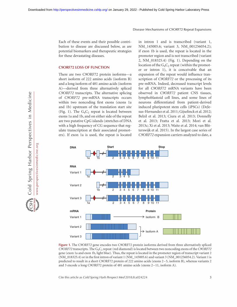
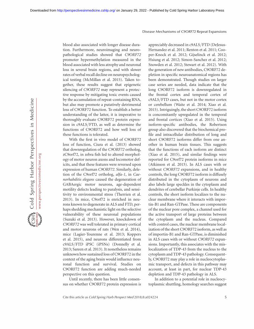
There are two C9ORF72 protein isoforms—ashort isoform of 222 amino acids (isoform B)and a long isoform of 481 amino acids (isoformA)—derived from three alternatively splicedC9ORF72 transcripts. The alternative splicingof C9ORF72 pre-mRNA transcripts occurswithin two noncoding first exons (exons 1aand 1b) upstream of the translation start site(Fig. 1). The G4C2 repeat is located betweenexons 1a and 1b, and on either side of the repeatare two putative CpG islands (stretches of DNAwith a high frequency of CG sequence that reg-ulate transcription at their associated promot-ers). If exon 1a is used, the repeat is located
in intron 1 and is transcribed (variant 1,NM_145005.6; variant 3, NM_001256054.2);if exon 1b is used, the repeat is located in thepromoter region and is not transcribed (variant2, NM_018325.4) (Fig. 1). Depending on thelocation of the G4C2 repeat (within the promot-er or intron 1), it is conceivable that anexpansion of the repeat would influence tran-scription of C9ORF72 or the processing of itspre-mRNA. Indeed, decreased transcript levelsfor all C9ORF72 mRNA variants have beenobserved in C9ORF72 patient CNS tissues,lymphoblastoid cell lines, and some lines ofneurons differentiated from patient-derivedinduced pluripotent stem cells (iPSCs) (DeJe-sus-Hernandez et al. 2011; Gijselinck et al. 2012;Belzil et al. 2013; Ciura et al. 2013; Donnellyet al. 2013; Fratta et al. 2013; Mori et al.2013c; Xi et al. 2013; Waite et al. 2014; van Blit-terswijk et al. 2015). In the largest case series ofC9ORF72 expansion carriers analyzed to date, a
1b1a 2 3 4 85 6 7 9 1110
DNA
1a 2 3 4 5
1b 2 3 4 85 6 7 9 1110
1a 2 3 4 85 6 7 9 1110
RNA
Variant 1
Variant 2
Variant 3
mRNA
Variant 1
Variant 2
Variant 3
Protein
Isoform B
Isoform A
StopStart
Figure 1. The C9ORF72 gene encodes two C9ORF72 protein isoforms derived from three alternatively splicedC9ORF72 transcripts. The G4C2 repeat (red diamond) is located between two noncoding exons of the C9ORF72gene (exon 1a and exon 1b, light blue). Thus, the repeat is located in the promoter region of transcript variant 2(NM_018325.4) or in the first intron of variant 1 (NM_145005.6) and variant 3 (NM_001256054.2). Variant 1 ispredicted to result in a short C9ORF72 protein of 222 amino acids (exons 2–5, isoform B), whereas variants 2and 3 encode a long C9ORF72 protein of 481 amino acids (exons 2–11, isoform A).
Disease Mechanisms of C9ORF72 Repeat Expansions
Cite this article as Cold Spring Harb Perspect Med 2018;8:a024224 3
ww
w.p
ersp
ecti
vesi
nm
edic
ine.
org
on January 29, 2022 - Published by Cold Spring Harbor Laboratory Presshttp://perspectivesinmedicine.cshlp.org/Downloaded from
significant decrease in variants 1 and 2, but notvariant 3, was observed in the frontal cortex andcerebellum of expansion carriers comparedwith patients without the expansion or controlsubjects (van Blitterswijk et al. 2015). Notably,an association between variant 1 and survivalafter disease onset was found, suggesting thathigher C9ORF72 levels may have beneficialeffects. This study additionally reported thatlevels of transcripts containing intron 1 (andthus the repeat) were elevated in expansion car-riers (van Blitterswijk et al. 2015), in agreementwith previous reports (Donnelly et al. 2013;Mori et al. 2013c; Sareen et al. 2013; Zu et al.2013; Haeusler et al. 2014; Liu et al. 2014).
The reduced expression of C9ORF72 mRNAin c9ALS/FTD patients may be caused by sev-eral factors, including the structure of G4C2 re-peat DNA and RNA. G4C2 DNA forms G-quad-ruplex structures (stacks of planar tetramersconsisting of four guanines connected byHoogsteen hydrogen bonds), whereas G4C2 re-peat RNA forms G-quadruplex structures andhairpins that can bind to repeat DNA to formRNA–DNA hybrids (Fratta et al. 2012; Reddyet al. 2013; Haeusler et al. 2014; Su et al. 2014).Using in vitro transcription assays of G4C2 re-peats of varying lengths (three to 70 repeats),Haeusler et al. observed that the generation ofthese distinct polymorphic DNA and RNAstructures causes, in a repeat length-dependentmanner, the abortive transcription of the repeat.It is thus noteworthy that, in the frontal cortexof C9ORF72 expansion carriers, there is an in-crease in transcripts containing sequences up-stream of the intronic repeat but not sequencesdownstream from the repeat, suggesting thatsome repeat-containing transcripts are indeedtruncated in c9ALS/FTD brain tissues (vanBlitterswijk et al. 2015).
Epigenetic processes have also been associ-ated with decreased C9ORF72 expression (Xiet al. 2013, 2014, 2015a,b; Belzil et al. 2013,2014; Liu et al. 2014; Russ et al. 2014; Gijselincket al. 2015; He et al. 2015; McMillan et al. 2015),in line with studies showing that expandedrepeats in microsatellite expansion disordersdysregulate mRNA expression through changesin DNA and histone methylation (Sutcliffe et al.
1992; Greene et al. 2007; Al-Mahdawi et al.2008; Todd et al. 2010). Trimethylated histones(H3K9me3, H3K27me3, H3K79me3, andH4K20me3) are known to repress gene expres-sion (Barski et al. 2007). Chromatin immuno-precipitation studies revealed that trimethylatedhistones bound strongly to expanded G4C2 re-peats but not to nonpathogenic repeats in thefrontal cortex and cerebellum, and this bindingis associated with decreased C9ORF72 mRNAlevels in c9ALS/FTD patients (Belzil et al.2013). A causal effect between histone trimeth-ylation and decreased C9ORF72 expression wassupported by the findings that treatment ofc9ALS/FTD-derived fibroblasts with a DNAand histone-demethylating agent not only de-creased trimethylated histone binding toC9ORF72 but also increased C9ORF72 mRNAexpression (Belzil et al. 2013). In addition tohistone methylation, repeat and CpG island hy-permethylation may decrease C9ORF72 mRNAexpression in c9ALS/FTD. Methylation of therepeat was found to occur in virtually all indi-viduals with alleles of more than 90 repeats andassociated with reduced C9ORF72 expression(Xi et al. 2015a,b). The CpG island 50 of therepeat was also hypermethylated in an expan-sion-specific manner, albeit only in a propor-tion of carriers (Xi et al. 2013, 2014; Belzil et al.2014; Liu et al. 2014; Russ et al. 2014). None-theless, Liu et al. (2014) show that, for thesubset of cases for which this CpG island washypermethylated, transcriptional silencing ofC9ORF72 was observed. Somewhat counterin-tuitively, rather than being detrimental, epige-netic silencing of the mutant C9ORF72 allelemay be protective, as it was shown to associatewith decreased accumulation of intronic repeat-containing RNA, RNA foci formation, andRAN translation. To test this hypothesis further,the relationship between C9ORF72 promoterhypermethylation and clinical features was in-vestigated (Russ et al. 2014). C9ORF72 hyper-methylation did not significantly differ betweenALS and FTD cases, nor did it predict age ofonset. Nonetheless, in c9FTD cases, C9ORF72hypermethylation in both the cerebellum andperipheral blood associated with later age atdeath, and C9ORF72 hypermethylation in the
T.F. Gendron and L. Petrucelli
4 Cite this article as Cold Spring Harb Perspect Med 2018;8:a024224
ww
w.p
ersp
ecti
vesi
nm
edic
ine.
org
on January 29, 2022 - Published by Cold Spring Harbor Laboratory Presshttp://perspectivesinmedicine.cshlp.org/Downloaded from
blood also associated with longer disease dura-tion. Furthermore, neuroimaging and neuro-pathological studies showed that C9ORF72promoter hypermethylation measured in theblood associated with less atrophy and neuronalloss in several brain regions, and with slowerrates of verbal recall decline on neuropsycholog-ical testing (McMillan et al. 2015). Taken to-gether, these results suggest that epigeneticsilencing of C9ORF72 may represent a protec-tive response by mitigating toxic events causedby the accumulation of repeat-containing RNA,but also may promote a putatively detrimentalloss of C9ORF72 function. To establish a betterunderstanding of the latter, it is imperative tothoroughly evaluate C9ORF72 protein expres-sion in c9ALS/FTD, as well as determine thefunctions of C9ORF72 and how well loss ofthese functions is tolerated.
With the first in vivo model of C9ORF72loss of function, Ciura et al. (2013) showedthat downregulation of the C9ORF72 ortholog,zC9orf72, in zebra fish led to altered morphol-ogy of motor neuron axons and locomotor def-icits, and that these features were reversed uponexpression of human C9ORF72. Similarly, dele-tion of the C9orf72 ortholog, alfa-1, in Cae-norhabditis elegans caused the degeneration ofGABAergic motor neurons, age-dependentmotility defects leading to paralysis, and sensi-tivity to environmental stress (Therrien et al.2013). In mice, C9orf72 is enriched in neu-rons known to degenerate in ALS and FTD, per-haps shedding mechanistic light on the selectivevulnerability of these neuronal populations(Suzuki et al. 2013). However, knockdown ofC9ORF72 was well tolerated in primary corticaland motor neurons of rats (Wen et al. 2014),mice (Lagier-Tourenne et al. 2013; Kopperset al. 2015), and neurons differentiated fromc9ALS/FTD iPSC (iPSNs) (Donnelly et al.2013; Sareen et al. 2013). It nonetheless remainsunknown how sustained loss of C9ORF72 in thecontext of the aging brain would influence neu-ronal function and survival. Studies onC9ORF72 function are adding much-neededperspective on this question.
Until recently, there has been little consen-sus on whether C9ORF72 protein expression is
appreciably decreased in c9ALS/FTD (DeJesus-Hernandez et al. 2011; Renton et al. 2011; Coo-per-Knock et al. 2012; Gijselinck et al. 2012;Hsiung et al. 2012; Simon-Sanchez et al. 2012;Snowden et al. 2012; Stewart et al. 2012). Withthe generation of new antibodies, C9ORF72 de-pletion in specific neuroanatomical regions hasbeen demonstrated. Though studies on largercase series are needed, data indicate that thelong C9ORF72 isoform is downregulated inthe frontal cortex and temporal cortex ofc9ALS/FTD cases, but not in the motor cortexor cerebellum (Waite et al. 2014; Xiao et al.2015). Intriguingly, the short C9ORF72 isoformis concomitantly upregulated in the temporaland frontal cortices (Xiao et al. 2015). Usingisoform-specific antibodies, the Robertsongroup also discovered that the biochemical pro-file and intracellular distribution of long andshort C9ORF72 isoforms differ from one an-other in human brain tissues. This suggeststhat the functions of each isoform are distinct(Xiao et al. 2015), and similar findings werereported for C9orf72 protein isoforms in mice(Atkinson et al. 2015). In ALS cases with orwithout C9ORF72 expansions, and in healthycontrols, the long C9ORF72 isoform is diffuselydistributed in the cytoplasm of neurons andalso labels large speckles in the cytoplasm anddendrites of cerebellar Purkinje cells. In healthycontrols, the short isoform localizes to the nu-clear membrane where it interacts with impor-tin-B1 and Ran-GTPase. These are componentsof the nuclear pore complex, a channel used forthe active transport of large proteins betweenthe cytoplasm and the nucleus. Comparedwith control cases, the nuclear membrane local-ization of the short C9ORF72 isoform, as well asof importin-B1 and Ran-GTPase, is diminishedin ALS cases with or without C9ORF72 expan-sions. Importantly, this associates with the mis-localization of TDP-43 from the nucleus to thecytoplasm and TDP-43 pathology. Consequent-ly, C9ORF72 may play a role in nucleocytoplas-mic transport, and defects in this pathway mayaccount, at least in part, for nuclear TDP-43depletion and TDP-43 pathology in ALS.
In addition to a potential role in nucleocy-toplasmic shuttling, homology searches suggest
Disease Mechanisms of C9ORF72 Repeat Expansions
Cite this article as Cold Spring Harb Perspect Med 2018;8:a024224 5
ww
w.p
ersp
ecti
vesi
nm
edic
ine.
org
on January 29, 2022 - Published by Cold Spring Harbor Laboratory Presshttp://perspectivesinmedicine.cshlp.org/Downloaded from
that C9ORF72 is a member of the DENN (dif-ferentially expressed in normal and neoplasia)-like superfamily (Zhang et al. 2012; Levine et al.2013). DENN domain proteins are highly con-served Rab-GEFs, GDP/GTP exchange factorsthat activate Rab-GTPases, the master regula-tors for intracellular membrane trafficking (Hu-tagalung and Novick 2011). Protein traffickingthrough the endosomal system is required forsorting and degrading proteins through au-tophagy or the ubiquitin–proteasome system(UPS) (Korolchuk et al. 2010). In neuronal-like cell lines, primary neurons, and humanspinal cord sections, C9ORF72 colocalizesand/or interacts with Rab-GTPases implicatedin endosomal transport and autophagy (Farget al. 2014). When C9ORF72 was knockeddown, endocytosis was impaired and autopha-gosome formation dysregulated. Studies alsosuggest that C9ORF72 interacts with ubiqui-lin-2, a member of the ubiquilin family thatregulates the degradation of ubiquitinated pro-teins, and with heterogeneous nuclear ribonu-cleoproteins (hnRNP) A1 and hnRNPA2/B1(Farg et al. 2014). hnRNPs, which includeTDP-43, are RNA-binding proteins involvedin many aspects of mRNA metabolism. Al-though hnRNP A1 and hnRNP A2/B1 residepredominantly in the nucleus, exposure tostress can trigger their recruitment to cytoplas-mic stress granules, which assemble to facilitatecell survival by triaging RNAs not required forcoping with the stress (Guil et al. 2006).
Additional studies are needed to fully deci-pher the potential role of C9ORF72 in nucleo-cytoplasmic transport, stress granule dynamics,and autophagy. However, with regard to thelatter, it is conceivable that perturbations inC9ORF72 function contribute to the increasedsensitivity of c9ALS/FTD iPSNs to chloroquineand 3-MA, two inhibitors of autophagy (Al-meida et al. 2013). This increased sensitivity sug-gests that proper autophagic processing wascompromised, which is further supported bythe finding that levels of p62, a known substrateof the autophagy pathway, were significantlyhigher in iPSNs from C9ORF72 repeat expansioncarriers compared with iPSNs from noncarriers(Almeida et al. 2013). Nevertheless, it is not yet
known whether loss of C9ORF72 function and/or the presence of DPR proteins, which have alsobeen linked to defects in protein degradation(May et al. 2014; Zhang et al. 2014; Yamakawaet al. 2015), cause the enhanced vulnerability ofc9ALS/FTD iPSNs to autophagy inhibitors.
TOXICITY MEDIATED THROUGH (G4C2)exp
OR (G2C4)exp TRANSCRIPTS
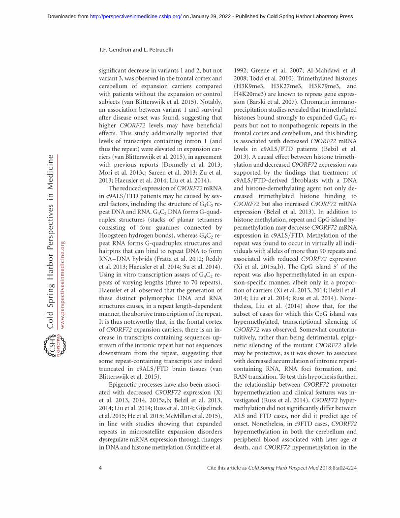
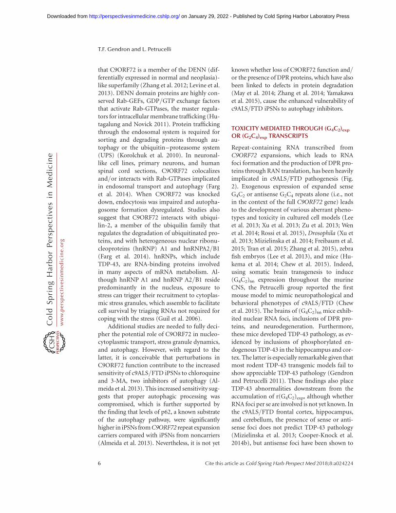
Repeat-containing RNA transcribed fromC9ORF72 expansions, which leads to RNAfoci formation and the production of DPR pro-teins through RAN translation, has been heavilyimplicated in c9ALS/FTD pathogenesis (Fig.2). Exogenous expression of expanded senseG4C2 or antisense G2C4 repeats alone (i.e., notin the context of the full C9ORF72 gene) leadsto the development of various aberrant pheno-types and toxicity in cultured cell models (Leeet al. 2013; Xu et al. 2013; Zu et al. 2013; Wenet al. 2014; Rossi et al. 2015), Drosophila (Xu etal. 2013; Mizielinska et al. 2014; Freibaum et al.2015; Tran et al. 2015; Zhang et al. 2015), zebrafish embryos (Lee et al. 2013), and mice (Hu-kema et al. 2014; Chew et al. 2015). Indeed,using somatic brain transgenesis to induce(G4C2)66 expression throughout the murineCNS, the Petrucelli group reported the firstmouse model to mimic neuropathological andbehavioral phenotypes of c9ALS/FTD (Chewet al. 2015). The brains of (G4C2)66 mice exhib-ited nuclear RNA foci, inclusions of DPR pro-teins, and neurodegeneration. Furthermore,these mice developed TDP-43 pathology, as ev-idenced by inclusions of phosphorylated en-dogenous TDP-43 in the hippocampus and cor-tex. The latter is especially remarkable given thatmost rodent TDP-43 transgenic models fail toshow appreciable TDP-43 pathology (Gendronand Petrucelli 2011). These findings also placeTDP-43 abnormalities downstream from theaccumulation of r(G4C2)exp, although whetherRNA foci per se are involved is not yet known. Inthe c9ALS/FTD frontal cortex, hippocampus,and cerebellum, the presence of sense or anti-sense foci does not predict TDP-43 pathology(Mizielinska et al. 2013; Cooper-Knock et al.2014b), but antisense foci have been shown to
T.F. Gendron and L. Petrucelli
6 Cite this article as Cold Spring Harb Perspect Med 2018;8:a024224
ww
w.p
ersp
ecti
vesi
nm
edic
ine.
org
on January 29, 2022 - Published by Cold Spring Harbor Laboratory Presshttp://perspectivesinmedicine.cshlp.org/Downloaded from
associate with nuclear loss of TDP-43 in c9ALSmotor neurons (Cooper-Knock et al. 2015b).Yet another possibility is that TDP-43 pathologydevelops in (G4C2)66 mice as a consequenceof DPR protein expression and subsequentdownstream events. Because the expression ofexpanded repeats in (G4C2)66 mice and theabove-mentioned models give rise to the accu-mulation of repeat-containing RNA, RNA foci,and RAN-translated DPR proteins, it is difficultto definitively ascribe the emergence of c9ALS/FTD-associated phenotypes to a given event. Inthe sections below, the neurotoxic potential me-diated specifically by RNA toxicity or DPR pro-teins is further discussed.
RNA TOXICITY
RNA-mediated toxicity, first described for myo-tonic dystrophy type 1, is a putative pathome-chanism involved in various microsatellite ex-pansion disorders (Belzil et al. 2012; Gendron
et al. 2014). It is believed to arise from the ab-normal interaction between essential RNA-binding proteins and repeat expansion RNAtranscripts, and the sequestration of these pro-teins into nuclear RNA foci. In c9ALS/FTD,r(G4C2)exp and r(G2C4)exp foci are detectedthroughout the CNS, being present mostly inneurons but also in astrocytes, oligodendro-cytes, and microglia (DeJesus-Hernandez et al.2011; Donnelly et al. 2013; Gendron et al. 2013;Lagier-Tourenne et al. 2013; Lee et al. 2013;Mizielinska et al. 2013; Zu et al. 2013; Cooper-Knock et al. 2014b, 2015b). Foci are also presentin c9ALS/FTD lymphoblastoid cell lines (La-gier-Tourenne et al. 2013; Cooper-Knock et al.2014b), peripheral blood leukocytes (Zu et al.2013), human neurons differentiated directlyfrom fibroblasts (iNeurons) or from iPSC (Al-meida et al. 2013; Donnelly et al. 2013; Sareenet al. 2013; Su et al. 2014), and fibroblasts (Al-meida et al. 2013; Donnelly et al. 2013; Lagier-Tourenne et al. 2013; Cooper-Knock et al.
1b1a 2 3 4 85 6 7 9 1110
RAN translated proteins
5′(GGGGCC)n(GA)n
(GP)n(GR)n
5′(GGCCCC)n
DNA
RNA
Sense
Antisense(PR)n
(PG)n(PA)n
DPR protein pathologyRNA foci
Figure 2. The repeat expansion in C9ORF72 is bidirectionally transcribed and can lead to foci formation andRAN translation. Sense and antisense RNA transcribed from the C9ORF72 repeat expansion accumulate as fociin the nucleus of cells and also serve as templates for RAN translation, thereby resulting in the production of sixdipeptide repeat proteins that aggregate into neuronal inclusions.
Disease Mechanisms of C9ORF72 Repeat Expansions
Cite this article as Cold Spring Harb Perspect Med 2018;8:a024224 7
ww
w.p
ersp
ecti
vesi
nm
edic
ine.
org
on January 29, 2022 - Published by Cold Spring Harbor Laboratory Presshttp://perspectivesinmedicine.cshlp.org/Downloaded from
2014b). In fact, foci are detected in fibroblasts ofboth symptomatic and asymptomatic carriersof the C9ORF72 expansion (Lagier-Tourenneet al. 2013; Cooper-Knock et al. 2014b).
Though inconsistencies exist among stud-ies, the percentage of cells bearing sense or an-tisense foci appears to vary among neuroana-tomical regions and could thus potentiallyinfluence disease phenotype. Mizielinska andcolleagues report that the highest burden offoci was consistently found in the frontal cortex,the region that suffers the greatest neuronal lossin FTLD. However, a separate study observed ahigher frequency of foci in the cerebellum ofc9ALS/FTD cases compared with the frontalcortex and temporal lobes (Lee et al. 2013).That foci burden inversely correlated with ageof onset in a cohort of eight c9FTD cases is ofinterest, yet needs validation in a larger series ofcases (Mizielinska et al. 2013). Also intriguing,but in need of confirmation in additional cases,was the observation that, in three c9ALS pa-tients, RNA foci were present in a higher pro-portion of motor neurons of the ventral horncompared with cerebellar granule cells; con-versely, in a patient who first presented withFTD, more foci were detected in the cerebellumthan in the ventral horn (Cooper-Knock et al.2014b).
Heterogeneity in clinical phenotype amongC9ORF72 repeat expansion carriers might beinfluenced not only by regional differences infoci frequency but also by the differential bind-ing and/or sequestration of select RNA-bindingproteins in various neuroanatomical regions.Indeed, irrespective of whether the binding ofproteins to r(G4C2)exp or r(G2C4)exp causestheir sequestration into foci, a potentially re-versible event (Cooper-Knock et al. 2014b),these abnormal interactions are likely to ad-versely influence their function. It is thus notsurprising that the transcriptome in fibroblasts(Donnelly et al. 2013; Lagier-Tourenne et al.2013), iPSNs (Donnelly et al. 2013; Sareenet al. 2013), laser-captured motor neurons(Cooper-Knock et al. 2015a), the motor cortex(Donnelly et al. 2013), the frontal cortex (Pru-dencio et al. 2015), and especially the cerebel-lum (Prudencio et al. 2015) of C9ORF72 repeat
expansion carriers differs from that in respectivemodels/tissues from control subjects, ostensi-bly as a consequence of aberrant binding of re-peat-containing transcripts to proteins that reg-ulate gene expression and splicing. In line withthis notion, prediction studies for regulators ofalternative splicing events in c9ALS brain tissuesidentified hnRNP H (Prudencio et al. 2015), aprotein binder of r(G4C2) previously implicatedin r(G4C2)exp toxicity (Lee et al. 2013; Cooper-Knock et al. 2014b). Additional members of thehnRNP family have been identified in screens todetermine protein binders of r(G4C2) andr(G2C4), as have many other proteins. Theseinclude hnRNP A3, hnRNP A1, serine–argi-nine-rich splicing factor 1 (SF2), serine–argi-nine-rich splicing factor 2 (SC35), Pur a, Aly/REF export factor (ALYREF), eIF2a, eIF2b, nu-cleolin, ADARB2, and RanGAP1 (Almeida et al.2013; Donnelly et al. 2013; Mori et al. 2013b;Reddy et al. 2013; Sareen et al. 2013; Xu et al.2013; Cooper-Knock et al. 2014b, 2015b;Haeusler et al. 2014; Rossi et al. 2015). The in-teractions between G4C2-repeat RNA and someof these proteins are discussed in more detailbelow.
The binding of nucleolin, a principal com-ponent of the nucleolus, to r(G4C2) when in aG-quadruplex structure (Haeusler et al. 2014)implicates nucleolar stress as a potential patho-mechanism of c9ALS/FTD. In lymphoblastoidcell lines, fibroblasts, and iPSNs from c9ALSpatients, the nucleolus appeared more fracturedand the nucleolin more dispersed throughoutthe nucleus compared with cells from ALS con-trols. In the c9ALS/FTD motor cortex, nucleo-lin frequently colocalized with r(G4C2)exp foci,and there was evidence of impaired rRNA pro-cessing. In accordance with chronic nucleolarstress, an increase in processing bodies (P bod-ies), which are composed of ribonucleoproteincomplexes involved in the degradation of un-translated mRNA, were significantly increasedin c9ALS iPSNs (Haeusler et al. 2014).
The binding of r(G4C2) to mRNA exportadapters, such as ALYREF, may provide a meansby which r(G4C2)exp makes its way from thenucleus to the cytoplasm (Cooper-Knock et al.2014b). It has also been speculated that the
T.F. Gendron and L. Petrucelli
8 Cite this article as Cold Spring Harb Perspect Med 2018;8:a024224
ww
w.p
ersp
ecti
vesi
nm
edic
ine.
org
on January 29, 2022 - Published by Cold Spring Harbor Laboratory Presshttp://perspectivesinmedicine.cshlp.org/Downloaded from
binding of (G4C2)exp pre-mRNA to hnRNP A3,which is also involved in mRNA export (Maet al. 2002), would result in the export of r(G4-
C2)exp to the cytoplasm where it could be RANtranslated (Mori et al. 2013b). Consistent withthis, there is a significant reduction of intranu-clear hnRNPA3 staining in the hippocampus ofc9ALS/FTD patients along with the formationof cytoplasmic and intranuclear hnRNP A3 in-clusions (Mori et al. 2013c). Pur a, shown totarget specific mRNA molecules to sites of den-dritic translation (Johnson et al. 2006), may alsoinfluence the localization of r(G4C2)exp or viceversa. For instance, in cultured cell models,(G4C2)31 markedly affected the distribution ofPur a and its binding partner, fragile X mentalretardation proteins, leading to their accumula-tion in stress granules (Rossi et al. 2015). It isalso notable that Pura formed inclusions in theeyes of (G4C2)30-expressing flies, and that eyedegeneration was suppressed upon Pur a over-expression. These data suggest that binding ofPur a by r(G4C2)exp causes a toxic loss of Pur afunction, a theory that is strengthened by thefact that depletion of Pur a was sufficient toreduce N2A cell viability, which was also rescuedby Pur a overexpression (Xu et al. 2013).
In a screen for r(G4C2) binders, the Roth-stein and Sattler groups identified 19 proteinsand initially focused their attention onADARB2, a member of the ADAR family ofproteins involved in RNA editing. ADARB2 ab-normally accumulated in the nucleus and colo-calized with r(G4C2)exp foci in iPSNs and themotor cortex of c9ALS patients (Donnellyet al. 2013). The resulting loss of ADARB2 func-tion is believed to have contributed to theheightened vulnerability of c9ALS iPSNs to glu-tamate as knockdown of ADARB2 in controliPSNs enhanced their susceptibility to gluta-mate toxicity to levels comparable to those ob-served in c9ALS iPSNs (Donnelly et al. 2013).Furthermore, knockdown of ADARB2 de-creased the number of c9ALS iPSNs with RNAfoci by half, suggesting that ADARB2 is involvedin the formation or maintenance of sense RNAfoci, and that the interaction between ADARB2and (G4C2)exp RNA plays a role in RNA-medi-ated toxicity. To determine which of the remain-
ing 19 protein binders of r(G4C2) modifyr(G4C2)-mediated neurodegeneration, geneticscreens were performed in Drosophila thatexpress 30 repeats and develop RNA foci butshow no detectable signs of RAN translation(Zhang et al. 2015). This led to the identifica-tion of RanGAP as a potent suppressor of G4C2
repeat toxicity in fly eyes and motor neurons.RanGAP, the fly ortholog of human RanGAP1, isa key regulator of nucleocytoplasmic transport.Confirming that G4C2 repeat toxicity in Droso-phila involves disrupted nucleocytoplasmictransport, a decrease in nuclear import was ob-served in (G4C2)30-expressing flies, and enhanc-ing nuclear import or suppressing nuclear ex-port of proteins suppressed (G4C2)30-mediatedneurodegeneration. Importantly, RanGAP1and components of the nuclear pore complexwere mislocalized in c9ALS iPSNs and motorcortex, and c9ALS iPSNs showed signs of im-paired nuclear import and disrupted TDP-43localization. These defects, as well as fociformation, were rescued in c9ALS iPSNs byASOs targeting G4C2 repeat RNA, suggestingthat sense r(G4C2)exp causes nucleocytoplasmictransport deficits. It would thus appear that ab-errations in the short C9ORF72 protein isoformand G4C2 RNA-mediated toxicity converge on asimilar mechanism of toxicity. Remarkably, thissame pathway has been implicated in poly(PR)and poly(GR)-mediated toxicity, as discussedbelow.
RAN TRANSLATION
DPR Protein Pathology: A NeuropathologicalHallmark of c9ALS/FTD
RAN translation is a noncanonical form oftranslation discovered by the Ranum groupwhile investigating the microsatellite expansiondisorders myotonic dystrophy type 1 and spi-nocerebellar ataxia type 8 (Zu et al. 2011). RANtranslation can occur across an entire expandedrepeat without frameshifting or prematurelystopping, despite the absence of an ATG startcodon. In addition, because RAN translationcan initiate in multiple frames, various productscan be synthesized from a given transcript.In c9ALS/FTD, both sense r(G4C2)exp and
Disease Mechanisms of C9ORF72 Repeat Expansions
Cite this article as Cold Spring Harb Perspect Med 2018;8:a024224 9
ww
w.p
ersp
ecti
vesi
nm
edic
ine.
org
on January 29, 2022 - Published by Cold Spring Harbor Laboratory Presshttp://perspectivesinmedicine.cshlp.org/Downloaded from
antisense r(G2C4)exp are RAN translated, lead-ing to the synthesis of poly(GA), poly(GP),and poly(GR) proteins from r(G4C2)exp, andpoly(PR), poly(PG), and poly(PA) proteinsfrom r(G2C4)exp (Fig. 2) (Ash et al. 2013; Gen-dron et al. 2013; Mann et al. 2013; Mori et al.2013a,c; Zu et al. 2013). Immunohistochemicalanalyses using multiple antibodies against thevarious DPR proteins have demonstrated highlyspecific staining of cytoplasmic inclusions, nu-clear inclusions, or dystrophic neurites inc9ALS/FTD patients (Ash et al. 2013; Gendronet al. 2013; Mackenzie et al. 2013, 2015; Mannet al. 2013; Mori et al. 2013a,c; Zu et al. 2013;Zhang et al. 2014; Schipper et al. 2015; Schludiet al. 2015). These inclusions, which are presentin neurons throughout the CNS (Ash et al.2013; Mackenzie et al. 2013) and in ependymalcells of the spinal cord central canal and lateralventricles (Schludi et al. 2015), can containmultiple DPR proteins (Mori et al. 2013a), arepositive for p62 (Mann et al. 2013; Mori et al.2013a), and, at least in the case of poly(GA)inclusions, contain filaments (Zhang et al.2014). The majority of DPR protein inclusionsare devoid of TDP-43, yet inclusions positive forboth poly(GA) and TDP-43 have been observedon rare occasions (Mackenzie et al. 2013; Moriet al. 2013c). In such instances, poly(GA)formed the core of the inclusion and was sur-rounded by TDP-43, suggesting that DPR pro-tein aggregation precedes TDP-43 pathology(Mackenzie et al. 2013; Mori et al. 2013c).
Poly(GA), poly(GP), and poly(GR) inclu-sions are numerous in neocortical regions, thehippocampus, and the cerebellum but are infre-quently observed in the lower motor neurons ofthe spinal cord (Ash et al. 2013; Mackenzie et al.2013, 2015; Mann et al. 2013; Mori et al.2013a,c; Gomez-Deza et al. 2015; Schludi et al.2015). Compared with these DPR proteins, in-clusions of poly(PR) and poly(PA), which areuniquely RAN translated from antisense re-peats, are far less abundant (Gendron et al.2013; Mann et al. 2013; Mori et al. 2013a; Mac-kenzie et al. 2015; Schludi et al. 2015), perhapsbecause RAN translation is less efficient fromantisense r(G2C4)exp or because r(G2C4)exp lev-els in the cytoplasm are relatively low. Alterna-
tively, poly(PR) and/or poly(PA) proteins maybe inherently more toxic than the other DPRproteins, or their biochemical properties (e.g.,half-life and solubility) may not be conducive totheir aggregation. With regard to the latter, alarger proportion of total poly(GP) is solublein c9ALS/FTD cerebellar homogenates, where-as a larger proportion of total poly(GA) is in-soluble (Gendron et al. 2015), consistent withpoly(GA) inclusions being more frequent thanthose composed of poly(GP) (Mackenzie et al.2015). Investigating these possibilities will shedlight on the paucity of poly(PR) and poly(PA)inclusions in c9ALS/FTD, whether soluble DPRproteins may be toxic entities, and will also offermechanistic insight on the factors that governRAN translation in c9ALS/FTD. Studies to dateshow that cerebellar poly(GP) and poly(GA)expression levels associate with C9ORF72transcript variant 3, the pre-mRNA of whichcontains the expanded repeat that serves as atemplate for RAN translation, but do not asso-ciate with repeat length (Gendron et al. 2015).
DPR Protein Pathology and Clinical/Neuropathological Correlations
The low abundance of DPR protein inclusionsin the spinal cord, which shows marked motorneuron loss in c9ALS, renders the contributionof RAN translation to disease pathogenesis un-clear (Ash et al. 2013; Gendron et al. 2013; Zuet al. 2013; Gomez-Deza et al. 2015; Schludiet al. 2015). In addition, reports on clinicaland neuropathological correlations with DPRprotein pathology have been conflicting, per-haps because different methods to estimateDPR protein pathology were used (e.g., semi-quantitative versus quantitative measures), dif-ferent neuroanatomical regions and DPR pro-teins were evaluated, and the staging of casesalso differed (i.e., clinically versus neuropatho-logically) (Mackenzie et al. 2013, 2015; Mannet al. 2013; Davidson et al. 2014; Schludi et al.2015). Although Mackenzie and colleaguesfound TDP-43 pathology to closely parallelneurodegeneration, they detected only moder-ate associations between the amount of poly(GA)dystrophic neurites and degeneration in the
T.F. Gendron and L. Petrucelli
10 Cite this article as Cold Spring Harb Perspect Med 2018;8:a024224
ww
w.p
ersp
ecti
vesi
nm
edic
ine.
org
on January 29, 2022 - Published by Cold Spring Harbor Laboratory Presshttp://perspectivesinmedicine.cshlp.org/Downloaded from
frontal cortex, and between total poly(GA) pa-thology and disease onset (Mackenzie et al.2013, 2015). Moreover, they observed no otherrelationship between DPR protein pathologyand clinical phenotype or neurodegenerationin their cohort of 35 cases representing the clin-ical spectrum associated with the C9ORF72mutation (Mackenzie et al. 2013, 2015). Like-wise, the Mann group found no significant dif-ferences in the pattern or frequency of poly(GA)inclusions among FTD, ALS, and FTD-MNDcases caused by C9ORF72 repeat expansions,nor did they observe differences in the extentof TDP-43 pathology between FTD patientsbearing an expansion and those who did not(Davidson et al. 2014). Conversely, the Edbauergroup discovered that, in a cohort of 14C9ORF72 expansion carriers, poly(PR) aggre-gates, despite being present in very low frequen-cy, were more common in the hippocampus ofFTLD cases compared with MND cases, andthat poly(GA) inclusions in cerebellar granulecells were significantly more abundant in FTLDcases compared with MND or FTLD-MNDcases (Schludi et al. 2015). Finally, taking a de-parture from traditional immunohistochemicalapproaches and instead using immunoassays toquantitatively measure poly(GP) and poly(GA)levels in various brain regions from 55C9ORF72 mutation carriers, the Petrucelligroup observed that poly(GP) levels in the cer-ebellum were significantly lower in patients withALS compared with patients with FTLD orFTLD-MND. Furthermore, although cerebellarpoly(GP) did not associate with age of diseaseonset or survival after onset in this large cohort,it did associate with cognitive impairment in 15c9ALS patients for whom neuropsychologicaldata were available (Gendron et al. 2015). Takentogether, the latter two studies implicate cere-bellar abnormalities as a contributor to the neu-ropathological and clinical heterogeneity asso-ciated with C9ORF72 repeat expansions. Giventhat DPR protein expression is highest in thecerebellum (Gendron et al. 2015), that robusttranscriptome changes occur in the cerebellumof c9ALS patients (Prudencio et al. 2015), thatcerebellar atrophy is reported in C9ORF72 ex-pansion carriers (Mahoney et al. 2012; Sha et al.
2012; Whitwell et al. 2012), and that the grow-ing body of neuroimaging and clinical evidencesupports cerebellar involvement in cognitiveand affective regulation (Koziol et al. 2014),more thorough investigations on the role ofthe cerebellum in c9ALS/FTD pathogenesisare warranted. In addition, the findings abovecollectively suggest that DPR protein patholo-gy/expression may influence clinical features ofC9ORF72 repeat expansion despite a lack of as-sociation with TDP-43 pathology or neuro-degeneration. Along this line of thought, thediscovery of a c9FTD kindred with early intel-lectual disability and extensive poly(GA) inclu-sions but little, if any, TDP-43 pathology couldindicate that DPR proteins are harmful or, at thevery least, that clinical symptoms can manifestin C9ORF72 expansion carriers prior to TDP-43 pathology (Proudfoot et al. 2014). This isfurther supported by three C9ORF72 mutationcarriers who developed fairly rapid cognitivedecline but died prematurely because of un-related illness; these patients had abundantpoly(GA) pathology but only sparse TDP-43pathology (Baborie et al. 2015). Despite themarked poly(GA) pathology in these patients,it must nonetheless be kept in mind that theirclinical features could also have been causedby C9ORF72 loss of function and/or RNA tox-icity. The same is true for findings emergingfrom the above-described correlation studiesbetween DPR protein pathology and clinicalphenotypes. Nevertheless, findings from severalgroups that specifically examined DPR proteinsin a variety of models provide compelling evi-dence that certain DPR proteins are toxic (Zuet al. 2013; Kwon et al. 2014; May et al. 2014;Mizielinska et al. 2014; Wen et al. 2014; Zhanget al. 2014; Freibaum et al. 2015; Jovicic et al.2015; Tao et al. 2015; Tran et al. 2015; Yamakawaet al. 2015; Yang et al. 2015).
In Vitro and In Vivo Models of DPR ProteinToxicity
To distinguish between repeat RNA and DPRprotein toxicity, the Isaacs group used an ele-gant strategy to generate either “pure repeats,”which could form foci and be RAN translated,
Disease Mechanisms of C9ORF72 Repeat Expansions
Cite this article as Cold Spring Harb Perspect Med 2018;8:a024224 11
ww
w.p
ersp
ecti
vesi
nm
edic
ine.
org
on January 29, 2022 - Published by Cold Spring Harbor Laboratory Presshttp://perspectivesinmedicine.cshlp.org/Downloaded from
or “RNA-only” repeats, which formed foci butwere not RAN translated because of stop codonsinterrupting the repeats. Expression of the purerepeats in the Drosophila eye caused eye degen-eration not observed when RNA-only repeatswere expressed. Similarly, the expression ofpure repeats, but not RNA-only repeats, in adultneurons caused early lethality of flies (Mizielin-ska et al. 2014). These in vivo experiments dem-onstrated that the C9ORF72 repeat expansionlikely causes toxicity through the productionof DPR proteins in this model. The Gao groupreached a similar conclusion using their Droso-phila model, which expressed a C9ORF72 mini-gene that contained 160 G4C2 repeats flankedby human intronic and exonic sequences. Al-though abundant nuclear RNA foci formed inthe neurons and glia of these flies, DPR proteinlevels were extremely low and virtually no tox-icity ensued (Tran et al. 2015). By comparingpoly(GP) protein levels between their (G4C2)160
flies and the Isaacs fly model that expressed 36highly toxic G4C2 repeats in the context of apoly(A)þ mRNA, the Gao group found pol-y(GP) levels to be �100-fold higher in(G4C2)36 flies than in (G4C2)160 flies, presum-ably because the poly(A) tail allowed (G4C2)36
to be efficiently exported to the cytoplasm forRAN translation. Furthermore, the Gao groupobserved that the modest toxicity that emergedin (G4C2)160 flies when subjected to a highertemperature correlated with an increase inDPR protein production but not in RNA foci(Tran et al. 2015). These data suggest that themarked and mild toxicity observed in flies ex-pressing (G4C2)36 and (G4C2)160, respectively,was caused by DPR proteins. In fact, the Gaogroup postulated that nuclear foci are neutralintermediates or possibly even protective, asthey prevent the export of r(G4C2)exp to thecytoplasm and subsequent RAN translation.
To determine which DPR proteins were tox-ic in Drosophila, Mizielinska et al. took advan-tage of “protein-only” constructs that use alter-native codons to those found within the G4C2
repeat to express poly(GR), poly(PR), poly(GA),or poly(PA). When these DPR proteins wereexpressed in the eye, only the arginine-contain-ing DPR proteins were toxic. When expressed in
adult neurons, (GR)100 and (PR)100 caused asubstantial decrease in survival, (GA)100 causeda late-onset reduction in survival, and (PA)100
had no effect (Mizielinska et al. 2014). Severalother groups have shown poly(PR) and/or pol-y(GR) to be toxic in Drosophila (Wen et al. 2014;Freibaum et al. 2015; Yang et al. 2015). For in-stance, the Trotti group demonstrated that(PR)50, but not (GA)50 or (PA)50, caused severeneurodegeneration in the fly eye and early le-thality when expressed in motor neurons (Wenet al. 2014). This group also showed that exog-enous poly(PR) proteins dramatically decreasedprimary cortical neuron survival, with the riskof death being considerably increased if nuclearaggregates were present. Interestingly, the Trottigroup found poly(PR) aggregates to be very sta-ble and to remain present even after the neuronsharboring them had died (Wen et al. 2014).Together, these findings suggest that poly(GR)and poly(PR) contribute to neurodegeneration;because both of these DPR proteins are argi-nine-rich, they may even share pathologicalmechanisms. Multiple studies provide evidencethat one such mechanism may involve impairedRNA biogenesis and nucleolar stress (Kwonet al. 2014; Wen et al. 2014; Jovicic et al. 2015;Tao et al. 2015; Yang et al. 2015), and otherssuggest these arginine-rich proteins impair thedynamics of RNA granules (i.e., stress granulesand P bodies) (Wen et al. 2014; Tao et al. 2015;Yamakawa et al. 2015). Another likely mecha-nism of disease has emerged through geneticmodifier screens using yeast and Drosophilamodels—impaired nucleocytoplasmic trans-port (Freibaum et al. 2015; Jovicic et al. 2015).Using codon-optimized constructs to express agiven DPR protein without the use of G4C2 re-peat sequences, the Gitler group found that ar-ginine-rich DPR proteins, especially poly(PR),were toxic in yeast. Subsequent unbiased screensto identify genetic modifiers that suppressed orenhanced poly(PR) toxicity uncovered genesinvolved in nucleocytoplasmic transport; theseincluded members of the karyopherin family ofnuclear import proteins, components of thenuclear pore complex, and regulators of theRAN-GTPase cycle, which generates the energyneeded to power nuclear import. Notably, up-
T.F. Gendron and L. Petrucelli
12 Cite this article as Cold Spring Harb Perspect Med 2018;8:a024224
ww
w.p
ersp
ecti
vesi
nm
edic
ine.
org
on January 29, 2022 - Published by Cold Spring Harbor Laboratory Presshttp://perspectivesinmedicine.cshlp.org/Downloaded from
regulating karyopherin expression rescued pol-y(PR) toxicity (Jovicic et al. 2015). A geneticscreen to find modifiers of (G4C2)58 toxicity inDrosophila similarly led to the identification ofgenes encoding proteins with nucleocytoplas-mic transport functions, including several com-ponents of the nuclear pore complex and themachinery that coordinates the import of nu-clear proteins and the export of nuclear RNA(Freibaum et al. 2015). With regard to the latter,cells of (G4C2)58-expressing Drosophila andc9FTD iPSNs showed evidence of nuclear RNAretention, consistent with defects in RNA ex-port. Overall, there is a compelling body ofwork supporting the toxicity of arginine-richDPR proteins.
Based on data from multiple models inves-tigating poly(GA) (in the absence of the con-founding contribution of RNA toxicity or theexpression of other DPR proteins), a case infavor of poly(GA) toxicity is also mounting(May et al. 2014; Mizielinska et al. 2014; Zhanget al. 2014; Jovicic et al. 2015; Yamakawa et al.2015). When expressed in cultured cell lines,rodent primary neurons, and P10 mice, poly(GA)formed soluble and insoluble high-molecular-weight species, as well as inclusions positive forp62 and ubiquitin, the majority of which werecytoplasmic, although nuclear inclusions werealso observed (May et al. 2014; Zhang et al.2014; Schludi et al. 2015; Yamakawa et al.2015). Zhang et al. found that expression ofGFP-(GA)50 induced caspase 3 activation andincreased extracellular lactate dehydrogenase(LDH) levels in HEK293T cells. Although Mayet al. failed to see similar poly(GA)-inducedtoxicity in their HEK293FT cultures, bothgroups found that poly(GA) proteins were neu-rotoxic to primary rodent neuronal cultures. Asin cultured cells, poly(GA) proteins formedp62- and ubiquitin-positive inclusions in neu-ronal cultures, which was associated with theactivation of caspase 3, DNA fragmentation,impaired neurite outgrowth or branching, andenhanced LDH release (May et al. 2014; Zhanget al. 2014). Poly(GA) expression was also asso-ciated with increased p62 expression (May et al.2014) and the accumulation of ubiquitinatedproteins (Zhang et al. 2014), which suggests
impaired activity of the UPS. Consistent withthis finding, Zhang et al. found that proteasomeactivity decreased in neurons expressing GFP-(GA)50, which, in turn, led to activation of en-doplasmic reticulum (ER) stress, the inhibitionof which provided rescue from poly(GA)-in-duced neurotoxicity. Importantly, mRNA levelsof ER stress markers ATF4 and CHOP were sig-nificantly increased in the frontal cortex ofc9ALS cases compared with sporadic ALS cases(Zhang et al. 2014), and c9ALS iPSNs exhibitedan increased vulnerability to tunicamycin, anER stress inducer (Haeusler et al. 2014), offeringcompelling evidence that UPS dysfunction andER stress may be pathogenic mechanisms inc9ALS/FTD.
Offering additional support that the UPS isinfluenced by poly(GA) proteins, Yamakawa etal. reported that poly(GA), as well as poly(GP)and poly(GR), caused UPS dysfunction in cul-tured cells and also rendered cells more vulner-able to UPS inhibition. Furthermore, throughan unbiased approach to identify poly(GA)-interacting proteins, May et al. found that pro-teasomal subunits and other ubiquitin-relatedproteins (e.g., ubiquilin 1 and 2) were enrichedin the poly(GA) interactome. Another interact-ing partner of interest is Unc119, a proteinrequired for the maintenance of the nervoussystem architecture in C. elegans that, when mu-tated, causes movement, sensory, and behavio-ral abnormalities in this model (Maduro et al.2000; Knobel et al. 2001). When coexpressedwith poly(GA) in HEK293 cells or primaryneurons, Unc119 colocalized with poly(GA) in-clusions and became insoluble, in contrast to itsnormal diffuse cytoplasmic localization. As thesequestration of Unc119 could cause its lossof function and contribute to neurotoxicity,this potential pathomechanism was examined.Knocking down Unc119 in primary neuronsled to dendritic withering, akin to that observ-ed upon poly(GA) expression, and neuronaldeath. Furthermore, Unc119 overexpressionwas able to partially reduce toxicity associatedwith poly(GA) expression, again suggestingthat Unc119 loss of function contributes topoly(GA)-induced neurotoxicity. It is thus ofinterest that inclusions of Unc119 were ob-
Disease Mechanisms of C9ORF72 Repeat Expansions
Cite this article as Cold Spring Harb Perspect Med 2018;8:a024224 13
ww
w.p
ersp
ecti
vesi
nm
edic
ine.
org
on January 29, 2022 - Published by Cold Spring Harbor Laboratory Presshttp://perspectivesinmedicine.cshlp.org/Downloaded from
served in tandem with a loss of cytosolicUnc119 staining in the brain of C9ORF72expansion carriers. Overall, multiple lines ofevidence suggest that poly(GA) proteins, themost abundantly expressed DPR protein inc9FTD/ALS, are detrimental.
c9ALS/FTD THERAPEUTICS ANDBIOMARKERS
The substantial advances made toward elucidat-ing how C9ORF72 repeat expansions contributeto c9ALS/FTD provide insight into potentialtherapeutics and diagnostics. Based on the as-sumption that r(G4C2)exp and r(G2C4)exp are atthe crux of c9ALS/FTD pathogenesis, as theyare required for foci formation and RAN trans-lation, strategies that target these transcripts andresult in their neutralization or degradationcould effectively block early culprits of disease.It is therefore promising that several groups haveidentified C9ORF72-targeting antisense oligo-nucleotides (ASOs) that mitigate r(G4C2)exp
foci formation, gene expression alterations, en-hanced susceptibility to excitotoxicity, and/ornucleocytoplasmic transport deficits in c9ALS/FTD fibroblasts, iPSN, and/or (G4C2)30-ex-pressing Drosophila (Donnelly et al. 2013; La-gier-Tourenne et al. 2013; Sareen et al. 2013;Zhang et al. 2015). Boding well for the potentialuse of ASOs to treat c9ALS/FTD, intrathecaladministration of ASOs against SOD1 was welltolerated and showed no significant adverseevents in a phase 1 study, and ASOs are movingtoward a phase 3 trial for spinal muscular atro-phy (Miller et al. 2013; Reddy and Miller 2015;Wirth et al. 2015). In addition to ASOs, smallmolecules offer an attractive approach for tar-geting r(G4C2)exp and r(G2C4)exp given phar-macological advantages, such as their smallmolecular weight, which is important forblood–brain barrier permeability. Indeed, theDisney and Petrucelli groups recently identifiedsmall molecules that bind the hairpin structureof r(G4C2)exp in vitro and in cells, and thatsignificantly decrease RAN translation andfoci formation in cultured cells overexpressingr(G4C2)66 and iNeurons derived from C9ORF72repeat expansion carriers (Su et al. 2014). On an
encouraging note, data from the Petrucelli(G4C2)66 mouse model imply that therapeuticstargeting G4C2 repeat RNA to mitigate fociformation and RAN translation should alsoattenuate TDP-43-mediated toxicity (Chewet al. 2015).
Should findings in yeast and Drosophila thatimplicate defective nucleocytoplasmic trans-port as a disease mechanism bear out in mam-malian models and patient-derived cells/tis-sues, this pathway would offer additionalprospective therapeutic targets for c9ALS/FTD. It is thus noteworthy that TMPyP4, aporphyrin compound that destabilizes RNAG-quadruplex structures and that blocks invitro interactions between r(G4C2)8 and itsprotein-binding partners (Zamiri et al. 2014),was found to similarly reduce the affinity ofRanGAP1 for r(G4C2)10 (Zhang et al. 2015).Moreover, TMPyP4, as well as a small-moleculeinhibitor of nuclear export, rescued nuclear im-port defects and eye neurodegeneration in(G4C2)30-expressing Drosophila (Zhang et al.2015).
For all c9ALS/FTD therapeutic strategiesbeing investigated, there are considerations tokeep in mind. Among them, their effect onC9ORF72 mRNA expression should be carefullymonitored. Although the exact contributionof C9ORF72 loss of function must still be deci-phered, causing a further decrease in C9ORF72expression by a purportedly protective inter-vention could have adverse effects, especiallyin light of the finding that higher levels ofC9ORF72 variant 1 are associated with pro-longed survival after disease onset (van Blitter-swijk et al. 2015). Therapeutics that increaseC9ORF72 expression may reverse harmfuleffects that could occur as a consequence ofC9ORF72 loss of function, although it wouldbe important that they not do so at the expenseof increasing r(G4C2)exp and r(G2C4)exp. Be-cause epigenetic processes mediate the decreasein C9ORF72 mRNA expression in c9ALS/FTD,they may offer a unique opportunity for thera-peutic development; however, caution is neededgiven findings suggesting that transcriptionalsilencing of C9ORF72 through CpG islandhypermethylation is protective by virtue of de-
T.F. Gendron and L. Petrucelli
14 Cite this article as Cold Spring Harb Perspect Med 2018;8:a024224
ww
w.p
ersp
ecti
vesi
nm
edic
ine.
org
on January 29, 2022 - Published by Cold Spring Harbor Laboratory Presshttp://perspectivesinmedicine.cshlp.org/Downloaded from
creasing RNA foci formation and RAN transla-tion (Belzil et al. 2013, 2014; Xi et al. 2013, 2014,2015a,b; Liu et al. 2014; Russ et al. 2014; Gijse-linck et al. 2015; He et al. 2015; McMillan et al.2015).
To increase the probability of success in de-veloping efficient therapeutic interventions,biomarkers and pharmacodynamic measuresof disease activity and drug action must be iden-tified. These biomarkers could encompass epi-genetic, RNA, or protein signatures unique toC9ORF72 repeat expansion carriers that can beeasily detected (e.g., in cerebrospinal fluid[CSF], blood, skin, and other biofluids or pe-ripheral tissues). One such potential biomarkerrecently came to light with the discovery thatpoly(GP) DPR proteins are detectable inthe CSF of c9ALS patients but not sporadicALS patients (Su et al. 2014). This discoverycould not only facilitate the identification ofC9ORF72 repeat expansion carriers in thecourse of diagnostic workups, but may alsopave the way in determining whether changesin DPR protein levels in CSF correlate with clin-ical phenotype or disease severity and progres-sion. Importantly, CSF DPR proteins, especiallypoly(GP) proteins that are synthesized fromboth sense and antisense transcripts, may proveuseful as a pharmacodynamic biomarker to as-sess the efficacy of r(G4C2)exp- or r(G2C4)exp-targeting therapies. The existence of DPR pro-teins in CSF also raises the possibility of theirpropagation from neuron to neuron, a phe-nomenon that is becoming more widely recog-nized in neurodegenerative diseases. WhetherDPR proteins are released from cells by regulat-ed processes or as a result of degenerating neu-rons, and whether pathology may be propagat-ed in this fashion are questions that are worthyof investigation.
CONCLUSION
Since the relatively recent discovery of C9ORF72repeat expansions, an impressive and fruitfuleffort has been put toward investigating themechanisms of disease. The normal functionsof C9ORF72 are being pieced together, the in-teracting partners of r(G4C2)exp and r(G2C4)exp
identified, the toxic potential of DPR proteinsevaluated, and many new models developed. Inaddition to these studies, therapeutic approach-es are being explored and biomarkers sought.Nevertheless, the exact pathomechanisms ofC9ORF72 expansions remain unknown, andmuch work lies ahead. Indeed, it is critical toconfirm findings from experimental models inlarge series of patient-derived cells and tissues.It is nonetheless interesting to note that certainabnormalities, such as nucleolar stress, RNAdysregulation, nucleocytoplasmic transportdeficits, and impaired protein degradation,have been uncovered by studies investigatingRNA and DPR protein toxicity, as well asC9ORF72 loss of function. Also presentlyobscure, despite recent clues, is how the samerepeat expansion, albeit with varying repeatlengths, causes a wide spectrum of clinical phe-notypes. Overall, much about c9ALS/FTD re-mains to be untangled, but should the currentpace of research on these devastating diseasescontinue, our understanding of c9ALS/FTD isexpected to grow swiftly.
ACKNOWLEDGMENTS
This work is supported by the National Insti-tutes of Health/National Institute of Neurolog-ical Disorders and Stroke [R21NS089979(T.F.G.), R21NS084528 (L.P.), R01NS088689(L.P.), R01NS063964 (L.P.), R01NS077402(L.P.), P01NS084974 (L.P.)], Department of De-fense [ALSRP AL130125 (L.P.)], Mayo ClinicFoundation (L.P.), ALS Association (T.F.G.),Target ALS (L.P.), and the Robert Packard Cen-ter for ALS Research at Johns Hopkins (L.P.).
REFERENCES�Reference is also in this collection.
Al-Mahdawi S, Pinto RM, Ismail O, Varshney D, Lymperi S,Sandi C, Trabzuni D, Pook M. 2008. The Friedreichataxia GAA repeat expansion mutation induces compa-rable epigenetic changes in human and transgenicmouse brain and heart tissues. Hum Mol Genet 17:735–746.
Almeida S, Gascon E, Tran H, Chou HJ, Gendron TF,Degroot S, Tapper AR, Sellier C, Charlet-Berguerand N,Karydas A, et al. 2013. Modeling key pathological features
Disease Mechanisms of C9ORF72 Repeat Expansions
Cite this article as Cold Spring Harb Perspect Med 2018;8:a024224 15
ww
w.p
ersp
ecti
vesi
nm
edic
ine.
org
on January 29, 2022 - Published by Cold Spring Harbor Laboratory Presshttp://perspectivesinmedicine.cshlp.org/Downloaded from
of frontotemporal dementia with C9ORF72 repeat ex-pansion in iPSC-derived human neurons. Acta Neuro-pathol 126: 385–399.
Al-Sarraj S, King A, Troakes C, Smith B, Maekawa S, BodiI, Rogelj B, Al-Chalabi A, Hortobagyi T, Shaw CE.2011. p62 positive, TDP-43 negative, neuronal cyto-plasmic and intranuclear inclusions in the cerebellumand hippocampus define the pathology of C9orf72-linked FTLD and MND/ALS. Acta Neuropathol 122:691–702.
Arai T, Hasegawa M, Akiyama H, Ikeda K, Nonaka T, MoriH, Mann D, Tsuchiya K, Yoshida M, Hashizume Y, et al.2006. TDP-43 is a component of ubiquitin-positive tau-negative inclusions in frontotemporal lobar degenerationand amyotrophic lateral sclerosis. Biochem Biophys ResCommun 351: 602–611.
Ash PE, Bieniek KF, Gendron TF, Caulfield T, Lin WL,Dejesus-Hernandez M, van Blitterswijk MM, Jansen-West K, Paul JW III, Rademakers R, et al. 2013. Uncon-ventional translation of C9ORF72 GGGGCC expansiongenerates insoluble polypeptides specific to c9FTD/ALS.Neuron 77: 639–646.
Atkinson RA, Fernandez-Martos CM, Atkin JD, VickersJC, King AE. 2015. C9ORF72 expression and cellularlocalization over mouse development. Acta NeuropatholCommun 3: 59.
Baborie A, Griffiths TD, Jaros E, Perry R, McKeith IG, BurnDJ, Masuda-Suzukake M, Hasegawa M, Rollinson S,Pickering-Brown S, et al. 2015. Accumulation of dipep-tide repeat proteins predates that of TDP-43 in fronto-temporal lobar degeneration associated with hexanucleo-tide repeat expansions in C9ORF72 gene. NeuropatholAppl Neurobiol 41: 601–612.
Barski A, Cuddapah S, Cui K, Roh TY, Schones DE, Wang Z,Wei G, Chepelev I, Zhao K. 2007. High-resolution pro-filing of histone methylations in the human genome. Cell129: 823–837.
Beck J, Poulter M, Hensman D, Rohrer JD, Mahoney CJ,Adamson G, Campbell T, Uphill J, Borg A, Fratta P,et al. 2013. Large C9orf72 hexanucleotide repeat expan-sions are seen in multiple neurodegenerative syndromesand are more frequent than expected in the UK popula-tion. Am J Hum Genet 92: 345–353.
Belzil VV, Gendron TF, Petrucelli L. 2012. RNA-mediatedtoxicity in neurodegenerative disease. Mol Cell Neurosci:S1044–S7431.
Belzil VV, Bauer PO, Prudencio M, Gendron TF, Stetler CT,Yan IK, Pregent L, Daughrity L, Baker MC, RademakersR, et al. 2013. Reduced C9orf72 gene expression inc9FTD/ALS is caused by histone trimethylation, an epi-genetic event detectable in blood. Acta Neuropathol 126:895–905.
Belzil VV, Bauer PO, Gendron TF, Murray ME, Dickson D,Petrucelli L. 2014. Characterization of DNA hypermeth-ylation in the cerebellum of c9FTD/ALS patients. BrainRes 1584: 15–21.
Benussi L, Rossi G, Glionna M, Tonoli E, Piccoli E, FostinelliS, Paterlini A, Flocco R, Albani D, Pantieri R, et al. 2014.C9ORF72 hexanucleotide repeat number in frontotem-poral lobar degeneration: A genotype-phenotype corre-lation study. J Alzheimers Dis 38: 799–808.
Boeve BF, Boylan KB, Graff-Radford NR, Dejesus-Hernan-dez M, Knopman DS, Pedraza O, Vemuri P, Jones D, LoweV, Murray ME, et al. 2012. Characterization of frontotem-poral dementia and/or amyotrophic lateral sclerosisassociated with the GGGGCC repeat expansion inC9ORF72. Brain 135: 765–783.
Brettschneider J, Van Deerlin VM, Robinson JL, Kwong L,Lee EB, Ali YO, Safren N, Monteiro MJ, Toledo JB, ElmanL, et al. 2012. Pattern of ubiquilin pathology in ALS andFTLD indicates presence of C9ORF72 hexanucleotideexpansion. Acta Neuropathol 123: 825–839.
Buchman VL, Cooper-Knock J, Connor-Robson N, Higgin-bottom A, Kirby J, Razinskaya OD, Ninkina N, Shaw PJ.2013. Simultaneous and independent detection ofC9ORF72 alleles with low and high number of GGGGCCrepeats using an optimised protocol of Southern blothybridisation. Mol Neurodegener 8: 12.
Byrne S, Elamin M, Bede P, Shatunov A, Walsh C, Corr B,Heverin M, Jordan N, Kenna K, Lynch C, et al. 2012.Cognitive and clinical characteristics of patients withamyotrophic lateral sclerosis carrying a C9orf72 repeatexpansion: A population-based cohort study. LancetNeurol 11: 232–240.
Chew J, Gendron TF, Prudencio M, Sasaguri H, Zhang YJ,Castanedes-Casey M, Lee CW, Jansen-West K, Kurti A,Murray ME, et al. 2015. C9ORF72 repeat expansions inmice cause TDP-43 pathology, neuronal loss, and behav-ioral deficits. Science 348: 1151–1154.
Chio A, Borghero G, Restagno G, Mora G, Drepper C, Tray-nor BJ, Sendtner M, Brunetti M, Ossola I, Calvo A,et al. 2012. Clinical characteristics of patients with fami-lial amyotrophic lateral sclerosis carrying the patho-genic GGGGCC hexanucleotide repeat expansion ofC9ORF72. Brain 135: 784–793.
Ciura S, Lattante S, Le Ber I, Latouche M, Tostivint H, BriceA, Kabashi E. 2013. Loss of function of C9orf72 causesmotor deficits in a zebrafish model of amyotrophic lateralsclerosis. Ann Neurol 74: 180–187.
Cooper-Knock J, Hewitt C, Highley JR, Brockington A,Milano A, Man S, Martindale J, Hartley J, Walsh T, Gels-thorpe C, et al. 2012. Clinico-pathological features inamyotrophic lateral sclerosis with expansions inC9ORF72. Brain 135: 751–764.
Cooper-Knock J, Shaw PJ, Kirby J. 2014a. The wideningspectrum of C9ORF72-related disease; genotype/pheno-type correlations and potential modifiers of clinical phe-notype. Acta Neuropathol 127: 333–345.
Cooper-Knock J, Walsh MJ, Higginbottom A, Robin High-ley J, Dickman MJ, Edbauer D, Ince PG, Wharton SB,Wilson SA, Kirby J, et al. 2014b. Sequestration of multipleRNA recognition motif-containing proteins by C9orf72repeat expansions. Brain 137: 2040–2051.
Cooper-Knock J, Bury JJ, Heath PR, Wyles M, Higginbot-tom A, Gelsthorpe C, Highley JR, Hautbergue G, RattrayM, Kirby J, et al. 2015a. C9ORF72 GGGGCC expandedrepeats produce splicing dysregulation which correlateswith disease severity in amyotrophic lateral sclerosis.PLoS ONE 10: e0127376.
Cooper-Knock J, Higginbottom A, Stopford MJ, Highley JR,Ince PG, Wharton SB, Pickering-Brown S, Kirby J, Haut-bergue GM, Shaw PJ. 2015b. Antisense RNA foci in themotor neurons of C9ORF72-ALS patients are associated
T.F. Gendron and L. Petrucelli
16 Cite this article as Cold Spring Harb Perspect Med 2018;8:a024224
ww
w.p
ersp
ecti
vesi
nm
edic
ine.
org
on January 29, 2022 - Published by Cold Spring Harbor Laboratory Presshttp://perspectivesinmedicine.cshlp.org/Downloaded from
with TDP-43 proteinopathy. Acta Neuropathol 130: 63–75.
Cruts M, Gijselinck I, Van Langenhove T, van der Zee J, VanBroeckhoven C. 2013. Current insights into the C9orf72repeat expansion diseases of the FTLD/ALS spectrum.Trends Neurosci 36: 450–459.
Davidson YS, Barker H, Robinson AC, Thompson JC, HarrisJ, Troakes C, Smith B, Al-Saraj S, Shaw C, Rollinson S,et al. 2014. Brain distribution of dipeptide repeat proteinsin frontotemporal lobar degeneration and motor neu-rone disease associated with expansions in C9ORF72.Acta Neuropathol Commun 2: 70.
DeJesus-Hernandez M, Mackenzie IR, Boeve BF, Boxer AL,Baker M, Rutherford NJ, Nicholson AM, Finch NA,Flynn H, Adamson J, et al. 2011. Expanded GGGGCChexanucleotide repeat in noncoding region of C9ORF72causes chromosome 9p-linked FTD and ALS. Neuron 72:245–256.
Dols-Icardo O, Garcia-Redondo A, Rojas-Garcia R, San-chez-Valle R, Noguera A, Gomez-Tortosa E, Pastor P,Hernandez I, Esteban-Perez J, Suarez-Calvet M, et al.2014. Characterization of the repeat expansion size inC9orf72 in amyotrophic lateral sclerosis and frontotem-poral dementia. Hum Mol Genet 23: 749–754.
Donnelly CJ, Zhang PW, Pham JT, Haeusler AR, Mistry NA,Vidensky S, Daley EL, Poth EM, Hoover B, Fines DM,et al. 2013. RNA toxicity from the ALS/FTD C9ORF72expansion is mitigated by antisense intervention. Neuron80: 415–428.
Farg MA, Sundaramoorthy V, Sultana JM, Yang S, AtkinsonRA, Levina V, Halloran MA, Gleeson PA, Blair IP, Soo KY,et al. 2014. C9ORF72, implicated in amytrophic lateralsclerosis and frontotemporal dementia, regulates endo-somal trafficking. Hum Mol Genet 23: 3579–3595.
Fratta P, Mizielinska S, Nicoll AJ, Zloh M, Fisher EM, Par-kinson G, Isaacs AM. 2012. C9orf72 hexanucleotiderepeat associated with amyotrophic lateral sclerosis andfrontotemporal dementia forms RNA G-quadruplexes.Sci Rep 2: 1016.
Fratta P, Poulter M, Lashley T, Rohrer JD, Polke JM, Beck J,Ryan N, Hensman D, Mizielinska S, Waite AJ, et al. 2013.Homozygosity for the C9orf72 GGGGCC repeat expan-sion in frontotemporal dementia. Acta Neuropathol 126:401–409.
Fratta P, Polke JM, Newcombe J, Mizielinska S, Lashley T,Poulter M, Beck J, Preza E, Devoy A, Sidle K, et al. 2015.Screening a UK amyotrophic lateral sclerosis cohortprovides evidence of multiple origins of the C9orf72expansion. Neurobiol Aging 36: 546 e541–547.
Freibaum BD, Lu Y, Lopez-Gonzalez R, Kim NC, Almeida S,Lee KH, Badders N, Valentine M, Miller BL, Wong PC,et al. 2015. GGGGCC repeat expansion in C9orf72compromises nucleocytoplasmic transport. Nature 525:129–133.
Gendron TF, Petrucelli L. 2011. Rodent models of TDP-43proteinopathy: Investigating the mechanisms of TDP-43-mediated neurodegeneration. J Mol Neurosci 45: 486–499.
Gendron TF, Bieniek KF, Zhang YJ, Jansen-West K, Ash PE,Caulfield T, Daughrity L, Dunmore JH, Castanedes-Casey M, Chew J, et al. 2013. Antisense transcripts ofthe expanded C9ORF72 hexanucleotide repeat form
nuclear RNA foci and undergo repeat-associated non-ATG translation in c9FTD/ALS. Acta Neuropathol 126:829–844.
Gendron TF, Belzil VV, Zhang YJ, Petrucelli L. 2014. Mech-anisms of toxicity in C9FTLD/ALS. Acta Neuropathol127: 359–376.
Gendron TF, van Blitterswijk M, Bieniek KF, Daughrity LM,Jiang J, Rush BK, Pedraza O, Lucas JA, Murray ME, De-saro P, et al. 2015. Cerebellar c9RAN proteins associatewith clinical and neuropathological characteristics ofC9ORF72 repeat expansion carriers. Acta Neuropathol130: 559–573.
� Ghasemi M, Brown RH Jr. 2017. Genetics of amyotrophiclateral sclerosis. Cold Spring Harb Perspect Med doi:10.1101/cshperspect.a024125.
Gijselinck I, Van Langenhove T, van der Zee J, Sleegers K,Philtjens S, Kleinberger G, Janssens J, Bettens K, VanCauwenberghe C, Pereson S, et al. 2012. A C9orf72 pro-moter repeat expansion in a Flanders-Belgian cohortwith disorders of the frontotemporal lobar degenera-tion-amyotrophic lateral sclerosis spectrum: A gene iden-tification study. Lancet Neurol 11: 54–65.
Gijselinck I, Van Mossevelde S, van der Zee J, Sieben A,Engelborghs S, De Bleecker J, Ivanoiu A, Deryck O, Ed-bauer D, Zhang M, et al. 2015. The C9orf72 repeat sizecorrelates with onset age of disease, DNA methylationand transcriptional downregulation of the promoter.Mol Psychiatry 21: 1112–1124.
� Gijselinck I, Cruts M, Van Broeckhoven C. 2017. The genet-ics of C9orf72 expansions. Cold Spring Harb Perspect Meddoi: 10.1101/cshperspect.a026757.
Gomez-Deza J, Lee YB, Troakes C, Nolan M, Al-Sarraj S,Gallo JM, Shaw CE. 2015. Dipeptide repeat protein in-clusions are rare in the spinal cord and almost absentfrom motor neurons in C9ORF72 mutant amyotrophiclateral sclerosis and are unlikely to cause their degener-ation. Acta Neuropathol Commun 3: 38.
Greene E, Mahishi L, Entezam A, Kumari D, Usdin K. 2007.Repeat-induced epigenetic changes in intron 1 of thefrataxin gene and its consequences in Friedreich ataxia.Nucleic Acids Res 35: 3383–3390.
Guil S, Long JC, Caceres JF. 2006. hnRNP A1 relocalizationto the stress granules reflects a role in the stress response.Mol Cell Biol 26: 5744–5758.
He J, Tang L, Benyamin B, Shah S, Hemani G, Liu R, Ye S, LiuX, Ma Y, Zhang H, et al. 2015. C9orf72 hexanucleotiderepeat expansions in Chinese sporadic amyotrophic lat-eral sclerosis. Neurobiol Aging 36: 2660 e2661–2668.
Hsiung GY, Dejesus-Hernandez M, Feldman HH, Sengdy P,Bouchard-Kerr P, Dwosh E, Butler R, Leung B, Fok A,Rutherford NJ, et al. 2012. Clinical and pathological fea-tures of familial frontotemporal dementia caused byC9ORF72 mutation on chromosome 9p. Brain 135:709–722.
Hukema RK, Riemslagh FW, Melhem S, van der Linde HC,Severijnen L, Edbauer D, Maas A, Charlet-Berguerand N,Willemsen R, van Swieten JC. 2014. A new inducibletransgenic mouse model for C9orf72-associated
Disease Mechanisms of C9ORF72 Repeat Expansions
Cite this article as Cold Spring Harb Perspect Med 2018;8:a024224 17
ww
w.p
ersp
ecti
vesi
nm
edic
ine.
org
on January 29, 2022 - Published by Cold Spring Harbor Laboratory Presshttp://perspectivesinmedicine.cshlp.org/Downloaded from
GGGGCC repeat expansion supports a gain-of-functionmechanism in C9orf72 associated ALS and FTD. ActaNeuropathol Commun 2: 166.
Hutagalung AH, Novick PJ. 2011. Role of Rab GTPases inmembrane traffic and cell physiology. Physiol Rev 91:119–149.
Johnson EM, Kinoshita Y, Weinreb DB, Wortman MJ,Simon R, Khalili K, Winckler B, Gordon J. 2006. Roleof Pur a in targeting mRNA to sites of translation inhippocampal neuronal dendrites. J Neurosci Res 83:929–943.
Jovicic A, Mertens J, Boeynaems S, Bogaert E, Chai N, Ya-mada SB, Paul JW 3rd, Sun S, Herdy JR, Bieri G, et al.2015. Modifiers of C9orf72 dipeptide repeat toxicity con-nect nucleocytoplasmic transport defects to FTD/ALS.Nat Neurosci 18: 1226–1229.
Knobel KM, Davis WS, Jorgensen EM, Bastiani MJ. 2001.UNC-119 suppresses axon branching in C. elegans. De-velopment 128: 4079–4092.
Koppers M, Blokhuis AM, Westeneng HJ, Terpstra ML, Zun-del CA, Vieira de Sa R, Schellevis RD, Waite AJ, Blake DJ,Veldink JH, et al. 2015. C9orf72 ablation in mice does notcause motor neuron degeneration or motor deficits. AnnNeurol 78: 426–438.
Korolchuk VI, Menzies FM, Rubinsztein DC. 2010. Mech-anisms of cross-talk between the ubiquitin-proteasomeand autophagy-lysosome systems. FEBS Lett 584: 1393–1398.
Koziol LF, Budding D, Andreasen N, D’Arrigo S, BulgheroniS, Imamizu H, Ito M, Manto M, Marvel C, Parker K, et al.2014. Consensus paper: The cerebellum’s role in move-ment and cognition. Cerebellum 13: 151–177.
Kwon I, Xiang S, Kato M, Wu L, Theodoropoulos P, Wang T,Kim J, Yun J, Xie Y, McKnight SL. 2014. Poly-dipeptidesencoded by the C9orf72 repeats bind nucleoli, impedeRNA biogenesis, and kill cells. Science 345: 1139–1145.
Lagier-Tourenne C, Baughn M, Rigo F, Sun S, Liu P, Li HR,Jiang J, Watt AT, Chun S, Katz M, et al. 2013. Targeteddegradation of sense and antisense C9orf72 RNA foci astherapy for ALS and frontotemporal degeneration. ProcNatl Acad Sci 110: E4530–4539.
Lee YB, Chen HJ, Peres JN, Gomez-Deza J, Attig J, StalekarM, Troakes C, Nishimura AL, Scotter EL, Vance C, et al.2013. Hexanucleotide repeats in ALS/FTD form length-dependent RNA foci, sequester RNA binding proteins,and are neurotoxic. Cell Rep 5: 1178–1186.
Levine TP, Daniels RD, Gatta AT, Wong LH, Hayes MJ. 2013.The product of C9orf72, a gene strongly implicated inneurodegeneration, is structurally related to DENNRab-GEFs. Bioinformatics 29: 499–503.
Liu EY, Russ J, Wu K, Neal D, Suh E, McNally AG, Irwin DJ,Van Deerlin VM, Lee EB. 2014. C9orf72 hypermethyla-tion protects against repeat expansion-associated pathol-ogy in ALS/FTD. Acta Neuropathol 128: 525–541.
Lomen-Hoerth C, Murphy J, Langmore S, Kramer JH, Ol-ney RK, Miller B. 2003. Are amyotrophic lateral sclerosispatients cognitively normal? Neurology 60: 1094–1097.
Lowe J. 1994. New pathological findings in amyotrophiclateral sclerosis. J Neurol Sci 124: 38–51.
Ma AS, Moran-Jones K, Shan J, Munro TP, Snee MJ, HoekKS, Smith R. 2002. Heterogeneous nuclear ribonucleo-
protein A3, a novel RNA trafficking response element-binding protein. J Biol Chem 277: 18010–18020.
Mackenzie IR, Arzberger T, Kremmer E, Troost D, Lorenzl S,Mori K, Weng SM, Haass C, Kretzschmar HA, Edbauer D,et al. 2013. Dipeptide repeat protein pathology inC9ORF72 mutation cases: Clinico-pathological correla-tions. Acta Neuropathol 126: 859–879.
Mackenzie IR, Frick P, Neumann M. 2014. The neuropathol-ogy associated with repeat expansions in the C9ORF72gene. Acta Neuropathol 127: 347–357.
Mackenzie IR, Frick P, Grasser FA, Gendron TF, Petrucelli L,Cashman NR, Edbauer D, Kremmer E, Prudlo J, TroostD, et al. 2015. Quantitative analysis and clinico-patho-logical correlations of different dipeptide repeat proteinpathologies in C9ORF72 mutation carriers. Acta Neuro-pathol 130: 845–861.
Maduro MF, Gordon M, Jacobs R, Pilgrim DB. 2000. TheUNC-119 family of neural proteins is functionally con-served between humans, Drosophila and C. elegans.J Neurogenet 13: 191–212.
Mahoney CJ, Beck J, Rohrer JD, Lashley T, Mok K, Shake-speare T, Yeatman T, Warrington EK, Schott JM, Fox NC,et al. 2012. Frontotemporal dementia with the C9ORF72hexanucleotide repeat expansion: Clinical, neuroana-tomical and neuropathological features. Brain 135:736–750.
Majounie E, Renton AE, Mok K, Dopper EG, Waite A, Rol-linson S, Chio A, Restagno G, Nicolaou N, Simon-San-chez J, et al. 2012. Frequency of the C9orf72 hexanucleo-tide repeat expansion in patients with amyotrophiclateral sclerosis and frontotemporal dementia: A cross-sectional study. Lancet Neurol 11: 323–330.
Mann DM, Rollinson S, Robinson A, Bennion Callister J,Thompson JC, Snowden JS, Gendron T, Petrucelli L, Ma-suda-Suzukake M, Hasegawa M, et al. 2013. Dipeptiderepeat proteins are present in the p62 positive inclusionsin patients with frontotemporal lobar degeneration andmotor neurone disease associated with expansions inC9ORF72. Acta Neuropathol Commun 1: 68.
May S, Hornburg D, Schludi MH, Arzberger T, Rentzsch K,Schwenk BM, Grasser FA, Mori K, Kremmer E, Banzhaf-Strathmann J, et al. 2014. C9orf72 FTLD/ALS-associatedGly-Ala dipeptide repeat proteins cause neuronal toxicityand Unc119 sequestration. Acta Neuropathol 128: 485–503.
McMillan CT, Russ J, Wood EM, Irwin DJ, Grossman M,McCluskey L, Elman L, Van Deerlin V, Lee EB. 2015.C9orf72 promoter hypermethylation is neuroprotective:Neuroimaging and neuropathologic evidence. Neurology84: 1622–1630.
Miller TM, Pestronk A, David W, Rothstein J, Simpson E,Appel SH, Andres PL, Mahoney K, Allred P, Alexander K,et al. 2013. An antisense oligonucleotide against SOD1delivered intrathecally for patients with SOD1 familialamyotrophic lateral sclerosis: a phase 1, randomised,first-in-man study. Lancet Neurol 12: 435–442.
Mizielinska S, Lashley T, Norona FE, Clayton EL, Ridler CE,Fratta P, Isaacs AM. 2013. C9orf72 frontotemporal lobardegeneration is characterised by frequent neuronal senseand antisense RNA foci. Acta Neuropathol 126: 845–857.
et al. 2014. C9orf72 repeat expansions cause neurodegen-eration in Drosophila through arginine-rich proteins. Sci-ence 345: 1192–1194.
Mori K, Arzberger T, Grasser FA, Gijselinck I, May S,Rentzsch K, Weng SM, Schludi MH, van der Zee J, CrutsM, et al. 2013a. Bidirectional transcripts of the expandedC9orf72 hexanucleotide repeat are translated into aggre-gating dipeptide repeat proteins. Acta Neuropathol 126:881–893.
Mori K, Lammich S, Mackenzie IR, Forne I, Zilow S,Kretzschmar H, Edbauer D, Janssens J, Kleinberger G,Cruts M, et al. 2013b. hnRNP A3 binds to GGGGCCrepeats and is a constituent of p62-positive/TDP43-neg-ative inclusions in the hippocampus of patients withC9orf72 mutations. Acta Neuropathol 125: 413–423.
Mori K, Weng SM, Arzberger T, May S, Rentzsch K,Kremmer E, Schmid B, Kretzschmar HA, Cruts M, VanBroeckhoven C, et al. 2013c. The C9orf72 GGGGCC re-peat is translated into aggregating dipeptide-repeat pro-teins in FTLD/ALS. Science 339: 1335–1338.
Neary D, Snowden JS, Gustafson L, Passant U, Stuss D,Black S, Freedman M, Kertesz A, Robert PH, AlbertM, et al. 1998. Frontotemporal lobar degeneration: Aconsensus on clinical diagnostic criteria. Neurology 51:1546–1554.
Neumann M, Sampathu DM, Kwong LK, Truax AC, Mic-senyi MC, Chou TT, Bruce J, Schuck T, Grossman M,Clark CM, et al. 2006. Ubiquitinated TDP-43 in fronto-temporal lobar degeneration and amyotrophic lateralsclerosis. Science 314: 130–133.
Nordin A, Akimoto C, Wuolikainen A, Alstermark H, Jons-son P, Birve A, Marklund SL, Graffmo KS, Forsberg K,Brannstrom T, et al. 2015. Extensive size variability of theGGGGCC expansion in C9orf72 in both neuronal andnon-neuronal tissues in 18 patients with ALS or FTD.Hum Mol Genet 24: 3133–3142.
Pikkarainen M, Hartikainen P, Alafuzoff I. 2010. Ubi-quitinated p62-positive, TDP-43-negative inclusions incerebellum in frontotemporal lobar degeneration withTAR DNA binding protein 43. Neuropathology 30: 197–199.
Proudfoot M, Gutowski NJ, Edbauer D, Hilton DA, Ste-phens M, Rankin J, Mackenzie IR. 2014. Early dipeptiderepeat pathology in a frontotemporal dementia kindredwith C9ORF72 mutation and intellectual disability. ActaNeuropathol 127: 451–458.
Prudencio M, Belzil V, Batra R, Ross CA, Gendron TF, Pre-gent L, Murray ME, Overstreet K, Piazza-Johnston A,Desaro P, et al. 2015. Distinct brain transcriptome profilesin C9orf72-associated and sporadic ALS. Nat Neurosci18: 1175–1182.
Reddy K, Zamiri B, Stanley SY, Macgregor RB Jr, Pearson CE.2013. The disease-associated r(GGGGCC)n repeat fromthe C9orf72 gene forms tract length-dependent uni- andmultimolecular RNA G-quadruplex structures. J BiolChem 288: 9860–9866.
Renton AE, Majounie E, Waite A, Simon-Sanchez J, Rol-linson S, Gibbs JR, Schymick JC, Laaksovirta H, vanSwieten JC, Myllykangas L, et al. 2011. A hexanucleotide
repeat expansion in C9ORF72 is the cause of chromo-some 9p21-linked ALS-FTD. Neuron 72: 257–268.
Rossi S, Serrano A, Gerbino V, Giorgi A, Di Francesco L,Nencini M, Bozzo F, Schinina ME, Bagni C, Cestra G,et al. 2015. Nuclear accumulation of mRNAs underliesG4C2-repeat-induced translational repression in a cel-lular model of C9orf72 ALS. J Cell Sci 128: 1787–1799.
Russ J, Liu EY, Wu K, Neal D, Suh E, Irwin DJ, McMillan CT,Harms MB, Cairns NJ, Wood EM, et al. 2014. Hyper-methylation of repeat expanded C9orf72 is a clinicaland molecular disease modifier. Acta Neuropathol 129:39–52.
Sareen D, O’Rourke JG, Meera P, Muhammad AK, Grant S,Simpkinson M, Bell S, Carmona S, Ornelas L, SahabianA, et al. 2013. Targeting RNA foci in iPSC-derived motorneurons from ALS patients with a C9ORF72 repeat ex-pansion. Sci Transl Med 5: 208ra149.
Schipper LJ, Raaphorst J, Aronica E, Baas F, de Haan R, deVisser M, Troost D. 2015. Prevalence of brain and spinalcord inclusions, including dipeptide repeat proteins, inpatients with the C9ORF72 hexanucleotide repeat expan-sion: A systematic neuropathological review. NeuropatholAppl Neurobiol 42: 547–560.
Schludi MH, May S, Grasser FA, Rentzsch K, Kremmer E,Kupper C, Klopstock T, Arzberger T, Edbauer D. 2015.Distribution of dipeptide repeat proteins in cellularmodels and C9orf72 mutation cases suggests link totranscriptional silencing. Acta Neuropathol 130: 537–555.
Sha SJ, Takada LT, Rankin KP, Yokoyama JS, Rutherford NJ,Fong JC, Khan B, Karydas A, Baker MC, DeJesus-Her-nandez M, et al. 2012. Frontotemporal dementia dueto C9ORF72 mutations: Clinical and imaging features.Neurology 79: 1002–1011.
Simon-Sanchez J, Dopper EG, Cohn-Hokke PE, HukemaRK, Nicolaou N, Seelaar H, de Graaf JR, de Koning I,van Schoor NM, Deeg DJ, et al. 2012. The clinical andpathological phenotype of C9ORF72 hexanucleotide re-peat expansions. Brain 135: 723–735.
Snowden JS, Rollinson S, Thompson JC, Harris JM, Stop-ford CL, Richardson AM, Jones M, Gerhard A, David-son YS, Robinson A, et al. 2012. Distinct clinical andpathological characteristics of frontotemporal dementiaassociated with C9ORF72 mutations. Brain 135: 693–708.
Stewart H, Rutherford NJ, Briemberg H, Krieger C, Cash-man N, Fabros M, Baker M, Fok A, Dejesus-HernandezM, Eisen A, et al. 2012. Clinical and pathological featuresof amyotrophic lateral sclerosis caused by mutation in theC9ORF72 gene on chromosome 9p. Acta Neuropathol123: 409–417.
Su Z, Zhang Y, Gendron TF, Bauer PO, Chew J, Yang WY,Fostvedt E, Jansen-West K, Belzil VV, Desaro P, et al. 2014.Discovery of a biomarker and lead small molecules totarget r(GGGGCC)-associated defects in c9FTD/ALS.Neuron 83: 1043–1050.
Suh E, Lee EB, Neal D, Wood EM, Toledo JB, Rennert L,Irwin DJ, McMillan CT, Krock B, Elman LB, et al. 2015.Semi-automated quantification of C9orf72 expansion
Disease Mechanisms of C9ORF72 Repeat Expansions
Cite this article as Cold Spring Harb Perspect Med 2018;8:a024224 19
ww
w.p
ersp
ecti
vesi
nm
edic
ine.
org
on January 29, 2022 - Published by Cold Spring Harbor Laboratory Presshttp://perspectivesinmedicine.cshlp.org/Downloaded from
size reveals inverse correlation between hexanucleotiderepeat number and disease duration in frontotemporaldegeneration. Acta Neuropathol 130: 363–372.
Sutcliffe JS, Nelson DL, Zhang F, Pieretti M, Caskey CT, SaxeD, Warren ST. 1992. DNA methylation represses FMR-1transcription in fragile X syndrome. Hum Mol Genet 1:397–400.
Suzuki N, Maroof AM, Merkle FT, Koszka K, Intoh A, Arm-strong I, Moccia R, Davis-Dusenbery BN, Eggan K. 2013.The mouse C9ORF72 ortholog is enriched in neuronsknown to degenerate in ALS and FTD. Nat Neurosci 16:1725–1727.
Tao Z, Wang H, Xia Q, Li K, Jiang X, Xu G, Wang G, Ying Z.2015. Nucleolar stress and impaired stress granule forma-tion contribute to C9orf72 RAN translation-induced cy-totoxicity. Hum Mol Genet 24: 2426–2441.
Therrien M, Rouleau GA, Dion PA, Parker JA. 2013.Deletion of C9ORF72 results in motor neuron degener-ation and stress sensitivity in C. elegans. PLoS ONE 8:e83450.
Todd PK, Oh SY, Krans A, Pandey UB, Di Prospero NA, MinKT, Taylor JP, Paulson HL. 2010. Histone deacetylasessuppress CGG repeat-induced neurodegeneration viatranscriptional silencing in models of fragile X tremorataxia syndrome. PLoS Genet 6: e1001240.
Tran H, Almeida S, Moore J, Gendron TF, Chalasani U, Lu Y,Du X, Nickerson JA, Petrucelli L, Weng Z, et al. 2015.Differential toxicity of nuclear RNA foci versus dipeptiderepeat proteins in a Drosophila model of C9ORF72 FTD/ALS. Neuron 87: 1207–1214.
van Blitterswijk M, Dejesus-Hernandez M, Niemantsver-driet E, Murray ME, Heckman MG, Diehl NN, BrownPH, Baker MC, Finch NA, Bauer PO, et al. 2013. Associ-ation between repeat sizes and clinical and pathologicalcharacteristics in carriers of C9ORF72 repeat expansions(Xpansize-72): A cross-sectional cohort study. LancetNeurol 12: 978–988.
van Blitterswijk M, Gendron TF, Baker MC, DeJesus-Her-nandez M, Finch NA, Brown PH, Daughrity LM, MurrayME, Heckman MG, Jiang J, et al. 2015. Novel clinicalassociations with specific C9ORF72 transcripts in pa-tients with repeat expansions in C9ORF72. Acta Neuro-pathol 130: 863–876.
van der Zee J, Gijselinck I, Dillen L, Van Langenhove T,Theuns J, Engelborghs S, Philtjens S, Vandenbulcke M,Sleegers K, Sieben A, et al. 2013. A pan-European study ofthe C9orf72 repeat associated with FTLD: Geographicprevalence, genomic instability, and intermediate re-peats. Hum Mutat 34: 363–373.
Waite AJ, Baumer D, East S, Neal J, Morris HR, Ansorge O,Blake DJ. 2014. Reduced C9orf72 protein levels infrontal cortex of amyotrophic lateral sclerosis and fron-totemporal degeneration brain with the C9ORF72 hex-anucleotide repeat expansion. Neurobiol Aging 35:1779.e5–1779.e13.
Wen X, Tan W, Westergard T, Krishnamurthy K, Markan-daiah SS, Shi Y, Lin S, Shneider NA, Monaghan J, PandeyUB, et al. 2014. Antisense proline-arginine RAN dipep-tides linked to C9ORF72-ALS/FTD form toxic nuclearaggregates that initiate in vitro and in vivo neuronaldeath. Neuron 84: 1213–1225.
Whitwell JL, Weigand SD, Boeve BF, Senjem ML, GunterJL, Dejesus-Hernandez M, Rutherford NJ, Baker M,Knopman DS, Wszolek ZK, et al. 2012. Neuroimag-ing signatures of frontotemporal dementia genetics:C9ORF72, tau, progranulin and sporadics. Brain 135:794–806.
Wirth B, Barkats M, Martinat C, Sendtner M, GillingwaterTH. 2015. Moving towards treatments for spinal muscu-lar atrophy: Hopes and limits. Expert Opin Emerg Drugs20: 353–356.
Xi Z, Zinman L, Moreno D, Schymick J, Liang Y, Sato C,Zheng Y, Ghani M, Dib S, Keith J, et al. 2013. Hyper-methylation of the CpG island near the G4C2 repeat inALS with a C9orf72 expansion. Am J Hum Genet 92: 981–989.
Xi Z, Rainero I, Rubino E, Pinessi L, Bruni AC, Maletta RG,Nacmias B, Sorbi S, Galimberti D, Surace EI, et al. 2014.Hypermethylation of the CpG-island near the C9orf72G4C2-repeat expansion in FTLD patients. Hum Mol Ge-net 23: 5630–5637.
Xi Z, van Blitterswijk M, Zhang M, McGoldrick P, McLeanJR, Yunusova Y, Knock E, Moreno D, Sato C, McKeeverPM, et al. 2015a. Jump from pre-mutation to patho-logic expansion in C9orf72. Am J Hum Genet 96: 962–970.
Xi Z, Zhang M, Bruni AC, Maletta RG, Colao R, Fratta P,Polke JM, Sweeney MG, Mudanohwo E, Nacmias B, et al.2015b. The C9orf72 repeat expansion itself is methylatedin ALS and FTLD patients. Acta Neuropathol 129: 715–727.
Xiao S, MacNair L, McGoldrick P, McKeever PM, McLeanJR, Zhang M, Keith J, Zinman L, Rogaeva E, Robertson J.2015. Isoform-specific antibodies reveal distinct subcel-lular localizations of C9orf72 in amyotrophic lateral scle-rosis. Ann Neurol 78: 568–583.
Xu Z, Poidevin M, Li X, Li Y, Shu L, Nelson DL, Li H,Hales CM, Gearing M, Wingo TS, et al. 2013. Expand-ed GGGGCC repeat RNA associated with amyotrophiclateral sclerosis and frontotemporal dementia causesneurodegeneration. Proc Natl Acad Sci 110: 7778–7783.
Yamakawa M, Ito D, Honda T, Kubo K, Noda M, NakajimaK, Suzuki N. 2015. Characterization of the dipeptide re-peat protein in the molecular pathogenesis of c9FTD/ALS. Hum Mol Genet 24: 1630–1645.
Yang D, Abdallah A, Li Z, Lu Y, Almeida S, Gao FB.2015. FTD/ALS-associated poly(GR) protein impairsthe Notch pathway and is recruited by poly(GA) intocytoplasmic inclusions. Acta Neuropathol 130: 525–535.
Zamiri B, Reddy K, Macgregor RB Jr, Pearson CE. 2014.TMPyP4 porphyrin distorts RNA G-quadruplex struc-tures of the disease-associated r(GGGGCC)n repeat ofthe C9orf72 gene and blocks interaction of RNA-bindingproteins. J Biol Chem 289: 4653–4659.
Zhang D, Iyer LM, He F, Aravind L. 2012. Discovery of novelDENN proteins: Implications for the evolution of eu-karyotic intracellular membrane structures and humandisease. Front Genet 3: 283.
RAN-translated proteins cause neurotoxicity by inducingER stress. Acta Neuropathol 128: 505–524.
Zhang K, Donnelly CJ, Haeusler AR, Grima JC, MachamerJB, Steinwald P, Daley EL, Miller SJ, Cunningham KM,Vidensky S, et al. 2015. The C9orf72 repeat expansiondisrupts nucleocytoplasmic transport. Nature 525: 56–61.
Zu T, Gibbens B, Doty NS, Gomes-Pereira M, Huguet A,Stone MD, Margolis J, Peterson M, Markowski TW, In-
gram MA, et al. 2011. Non-ATG-initiated translation di-rected by microsatellite expansions. Proc Natl Acad Sci108: 260–265.
Zu T, Liu Y, Banez-Coronel M, Reid T, Pletnikova O, Lewis J,Miller TM, Harms MB, Falchook AE, Subramony SH,et al. 2013. RAN proteins and RNA foci from antisensetranscripts in C9ORF72 ALS and frontotemporal demen-tia. Proc Natl Acad Sci 110: E4968–E4977.
Disease Mechanisms of C9ORF72 Repeat Expansions
Cite this article as Cold Spring Harb Perspect Med 2018;8:a024224 21
ww
w.p
ersp
ecti
vesi
nm
edic
ine.
org
on January 29, 2022 - Published by Cold Spring Harbor Laboratory Presshttp://perspectivesinmedicine.cshlp.org/Downloaded from