Diseases of Asian seabass or Barramundi, Lates calcarifer Bloch By Susan Gibson-Kueh School of Veterinary and Biomedical Sciences Division of Health Sciences, Murdoch University South Street, Murdoch WA 6150 Email: [email protected]
Transcript
Diseases of Asian seabass or Barramundi,
Lates calcarifer Bloch
By Susan Gibson-KuehSchool of Veterinary and Biomedical Sciences
Division of Health Sciences, Murdoch UniversitySouth Street, Murdoch WA 6150 Email: [email protected]
Aims of present study• Study the major diseases impacting the
culture of L. calcarifer to improve diagnosis & control
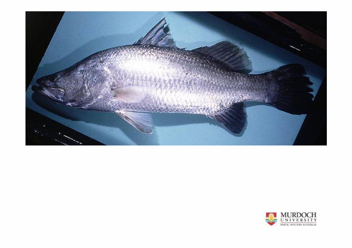
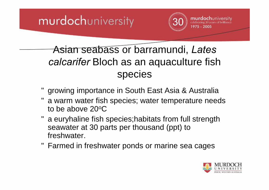
Asian seabass or barramundi, Lates calcarifer Bloch as an aquaculture fish
species• growing importance in South East Asia & Australia • a warm water fish species; water temperature needs
to be above 20oC • a euryhaline fish species;habitats from full strength
seawater at 30 parts per thousand (ppt) to freshwater.
• Farmed in freshwater ponds or marine sea cages
Hatchery culture of L. calcarifer• Eggs hatch in <1 day• fed live feed as rotifers & artemia from 2 days old. • weaned onto an artificial feed at ~3 weeks old. • Very specialized and labour intensive due to the need
for live feed production. • larvae rearing stage and the broodstock gonadal
maturation phase need to be carried out in saline water.
Grow-out of L. calcarifer
• Typically grows to 350g in six months & 2 kg in 2 years
• Grow-out cages size typically squares of between 2-4m wide & 2m deep
• Large circular cages 10-15m diameter & 10m deep
Background on research
• 12 years experience as fish pathologist & aquatic animal health veterinarian in Singapore
• Familiar with the range of diseases seen in cultured fish species including L. calcarifer
• Some diseases not well described
Materials used in research
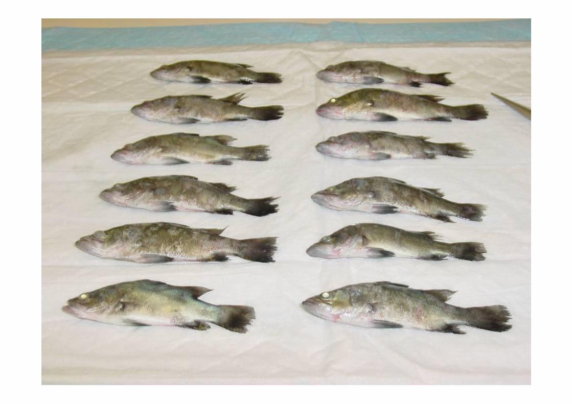
• L. calcarifer fish samples from – Singapore where I used to work– Fisheries from WA – not many barra farms here!– An established Indonesian farm– Sampled over a 3 month period in 2008 from 16
nurseries in Vietnam
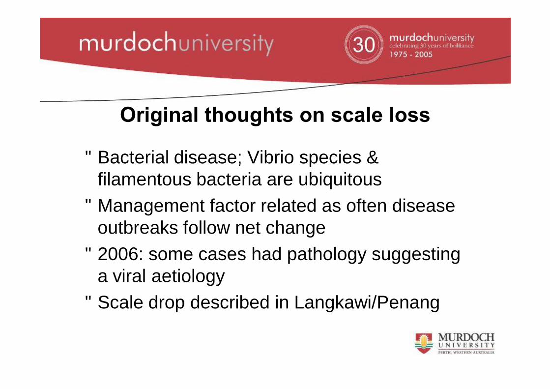
Original thoughts on scale loss
• Bacterial disease; Vibrio species & filamentous bacteria are ubiquitous
• Management factor related as often disease outbreaks follow net change
• 2006: some cases had pathology suggesting a viral aetiology
• Scale drop described in Langkawi/Penang
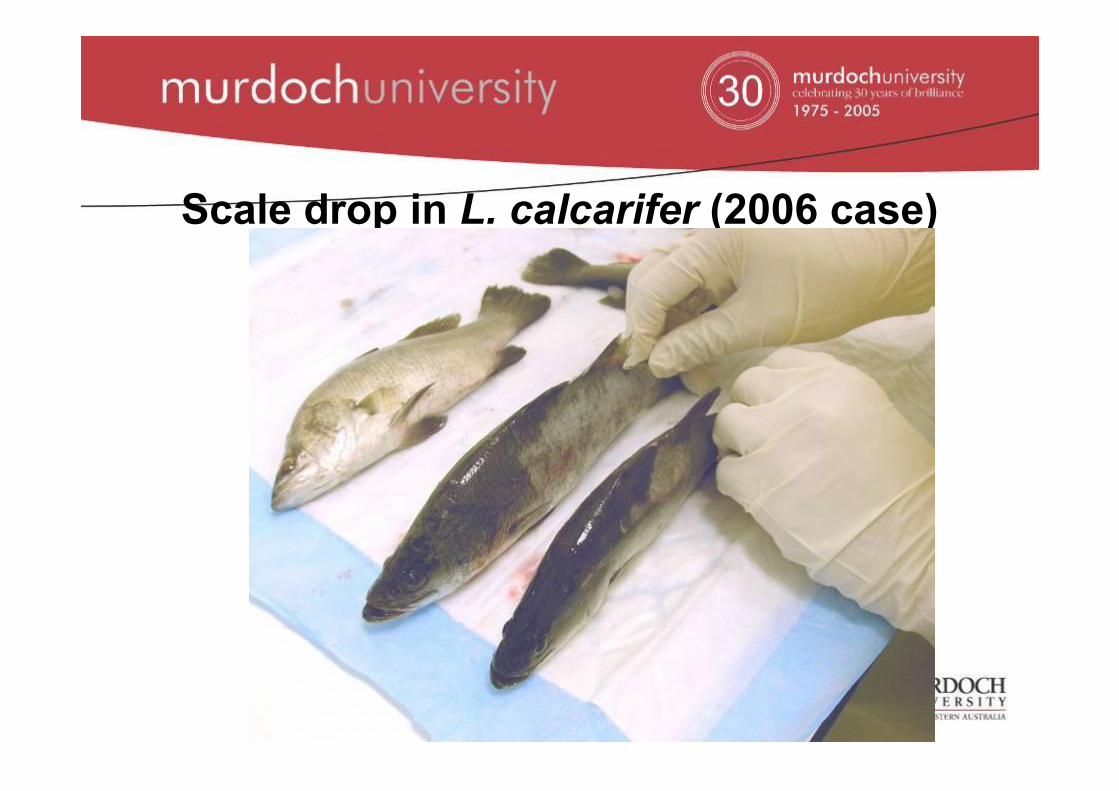
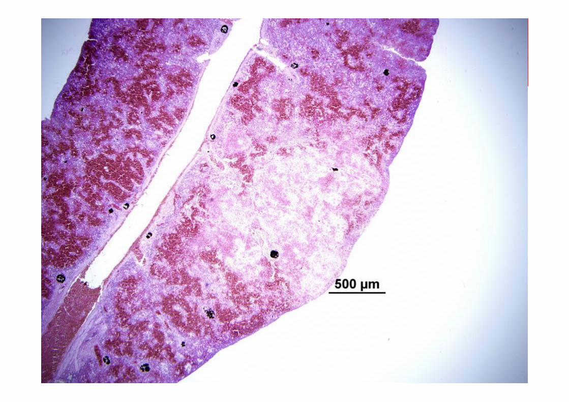
Scale drop in L. calcarifer (2006 case)
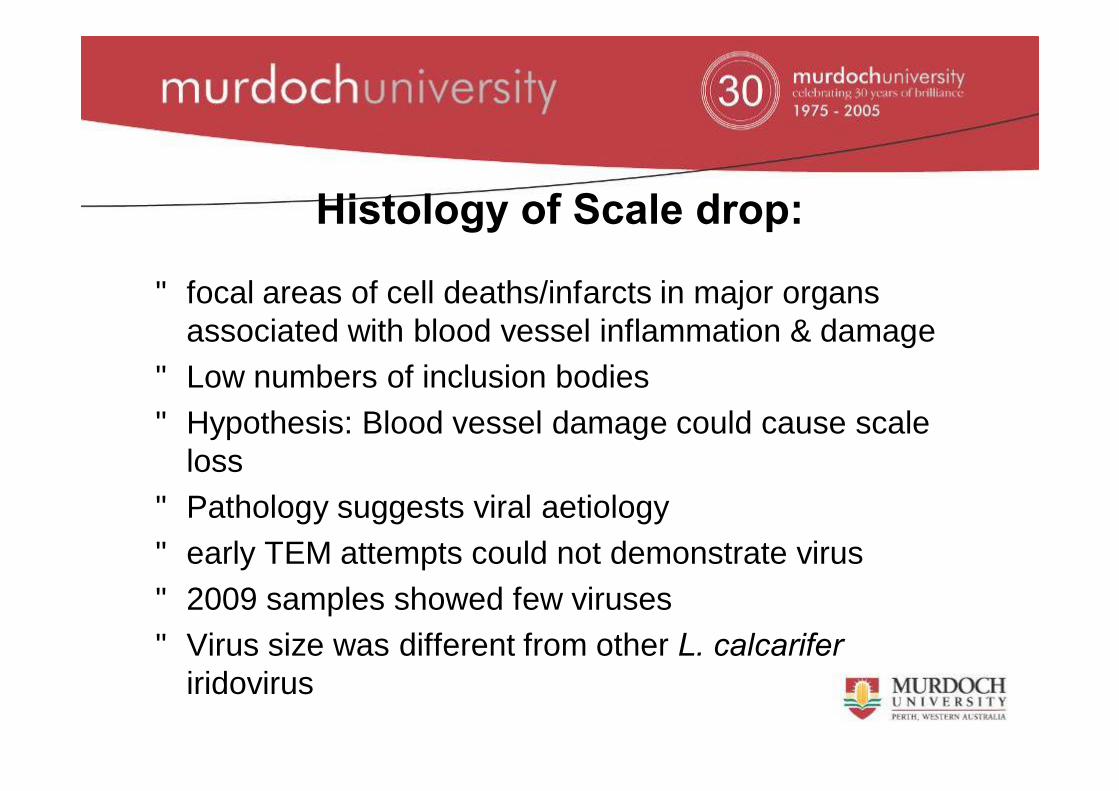
• focal areas of cell deaths/infarcts in major organs associated with blood vessel inflammation & damage
• Low numbers of inclusion bodies• Hypothesis: Blood vessel damage could cause scale
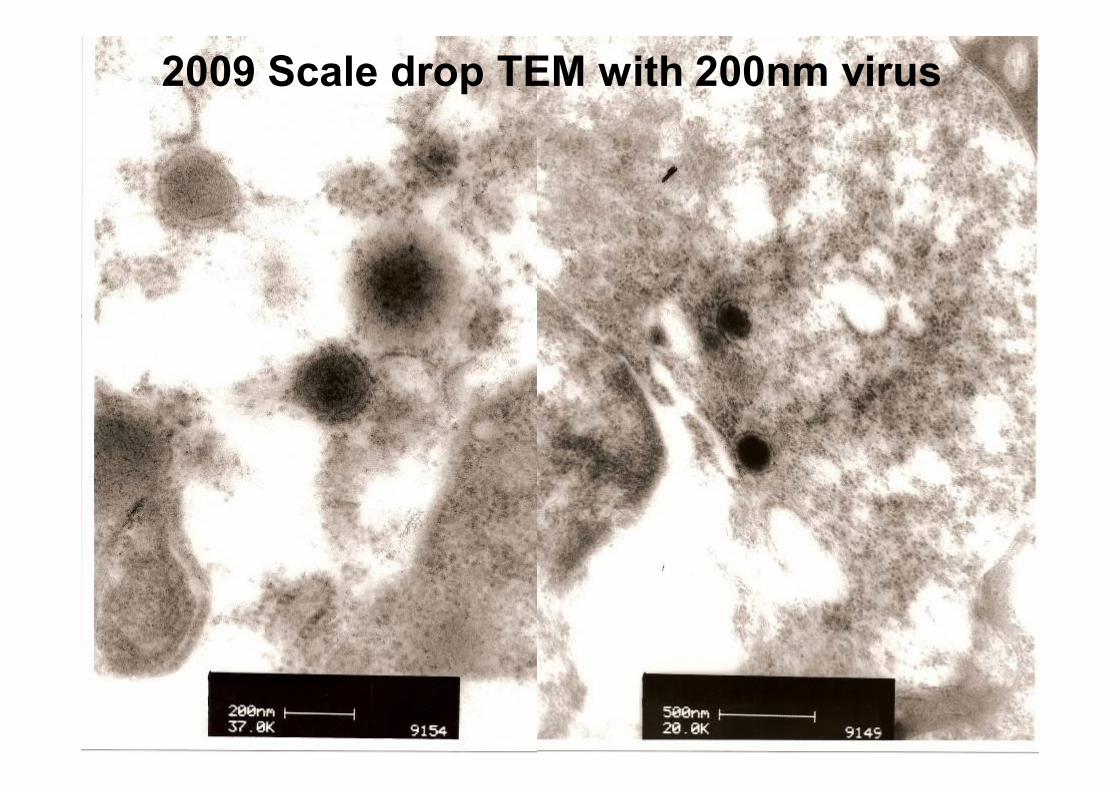
loss• Pathology suggests viral aetiology • early TEM attempts could not demonstrate virus• 2009 samples showed few viruses• Virus size was different from other L. calcarifer
iridovirus
Histology of Scale drop:
2009 Scale drop TEM with 200nm virus
Wanted to compare my scale drop virus to other L calcarifer iridovirus cases I have already encountered.
Systemic iridovirus in L. calcarifer from Singapore
• Not often observed in Singapore• No Singapore materials for TEM but wax
blocks available• Dug out wax block tissues for TEM
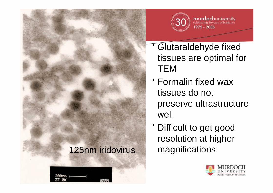
• Glutaraldehyde fixed tissues are optimal for TEM
• Formalin fixed wax tissues do not preserve ultrastructure well
• Difficult to get good resolution at higher magnifications125nm iridovirus
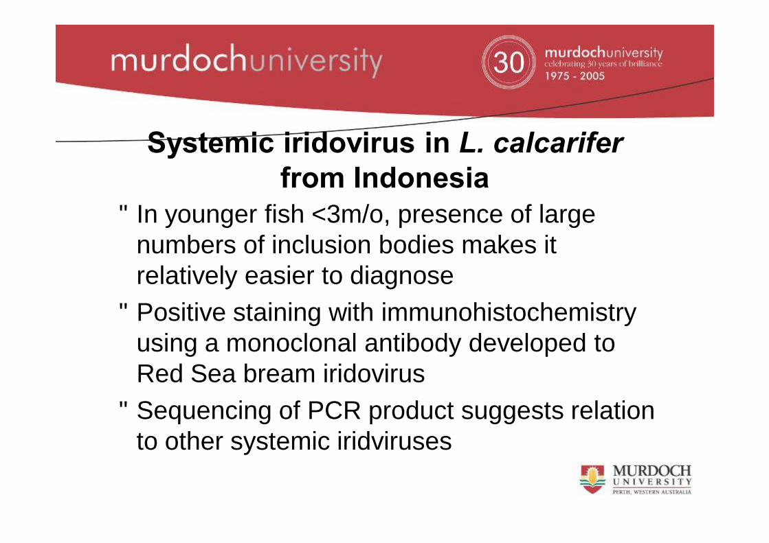
Systemic iridovirus in L. calcarifer from Indonesia
•Managed to obtain samples from an established farm in Indonesia•5 tonnes production of mainly 3kg fish every week•Earlier, farm had depended on PCR & clinical signs to diagnose disease •Histology examination showed iridovirus as a background disease on farm, more widespread than initially thought
Systemic iridovirus in L. calcarifer from Indonesia
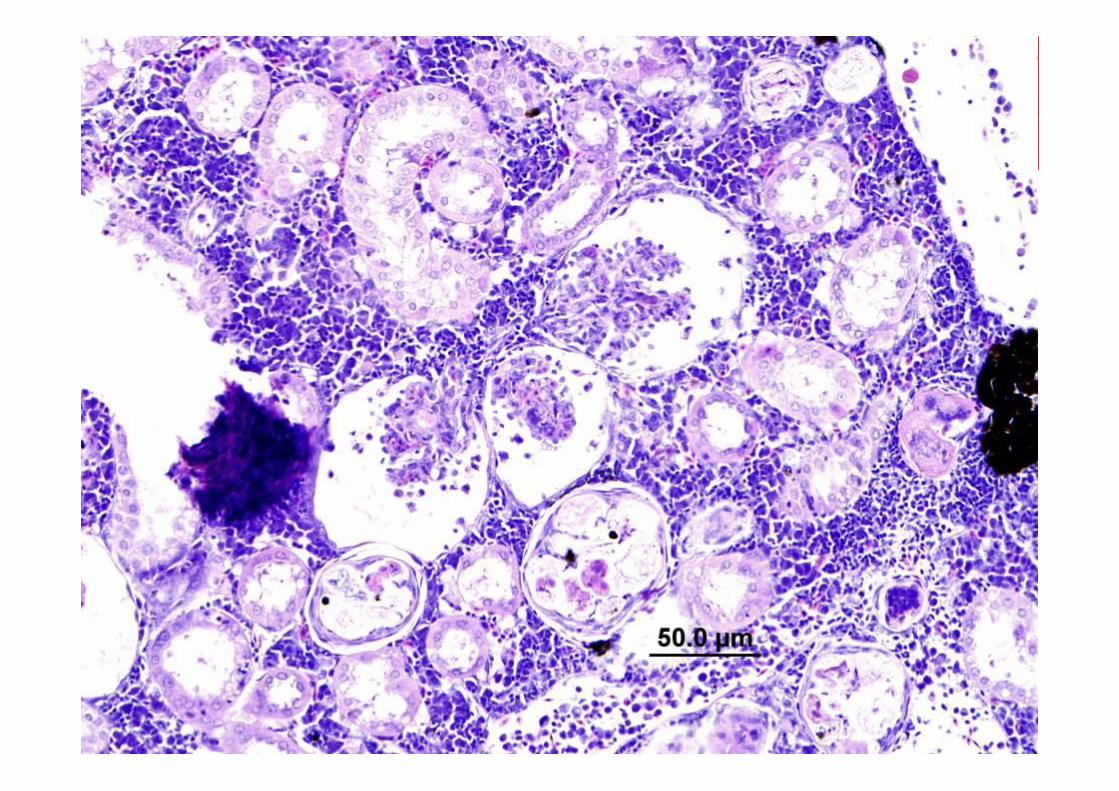
• In younger fish <3m/o, presence of large numbers of inclusion bodies makes it relatively easier to diagnose
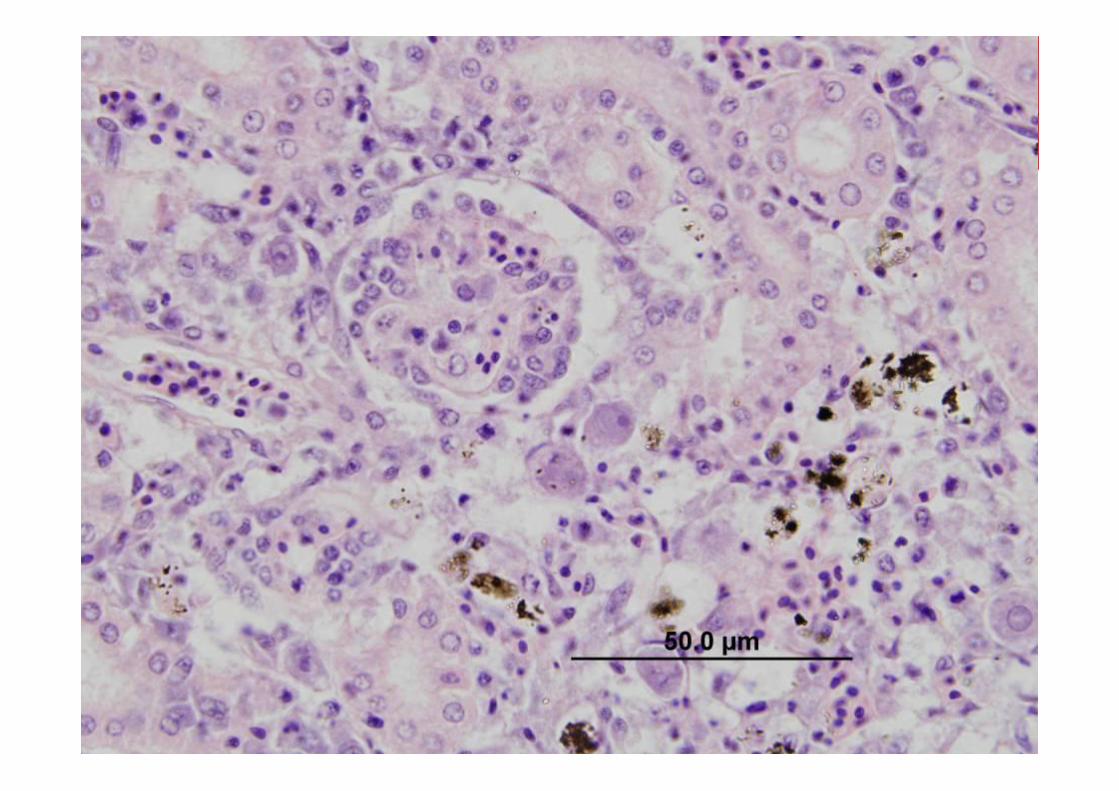
• Positive staining with immunohistochemistry using a monoclonal antibody developed to Red Sea bream iridovirus
• Sequencing of PCR product suggests relation to other systemic iridviruses
Viral inclusions stained red
Learning points with immunohistochemistry on fish tissues
• Many reagents have limited shelf life• Especially labeled antibodies• Fish tissues different so that when adapting
techniques need to optimise• Antigen retrieval using microwave had to be
reduced to avoid making fish tissues drop off slides
Systemic iridovirus TEM
•Pathology, immunohistochemistry & PCR suggests a systemic iridovirus infection in fish from Indonesia•TEM could not definitively demonstrate virus•Time of sampling is critical in looking for pathogen. Too early, virus not formed. Too late, no more pathogen.•Re-examine samples that have just arrived this week
Further work• Use Red sea bream iridovirus monoclonal antibody
immunohistochemistry on scale drop cases• Use DNA probe developed from PCR product of L
calcarifer systemic iridovirus (Indonesia) on scale drop cases
• Virion size of systemic iridovirus different from scale-drop virus
• Try get scale drop samples for PCR and sequencing to compare with 125nm L. calcarifer iridovirus (Singapore)
•One of 16 Nurseries in Vietnam where fish samples were taken•Imported fingerlings are held in fibreglass or cement tanks until sold to grow-out farms
History & Clinical signs of L. calcarifersampled from Vietnam
• Low grade mortality• Lethargic• Tail rot, skin ulcers, scale loss• Loss of appetite
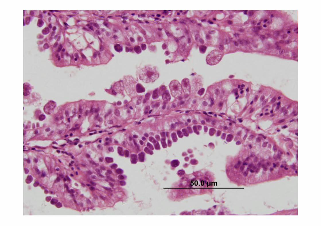
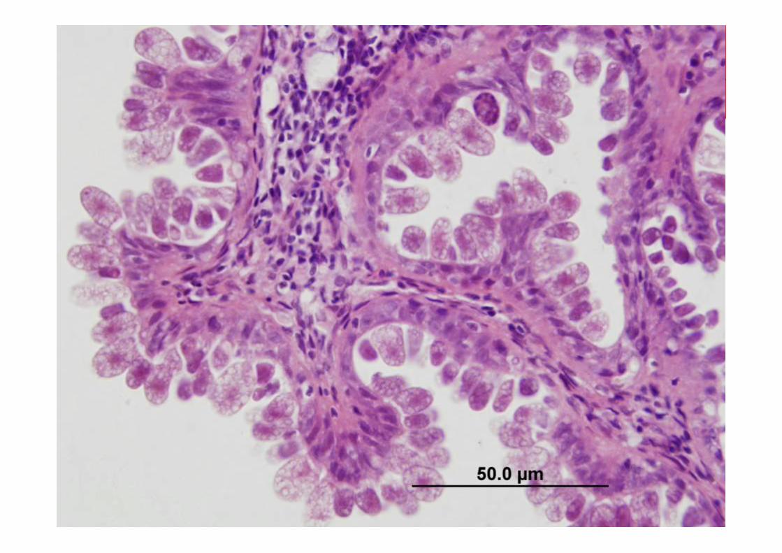
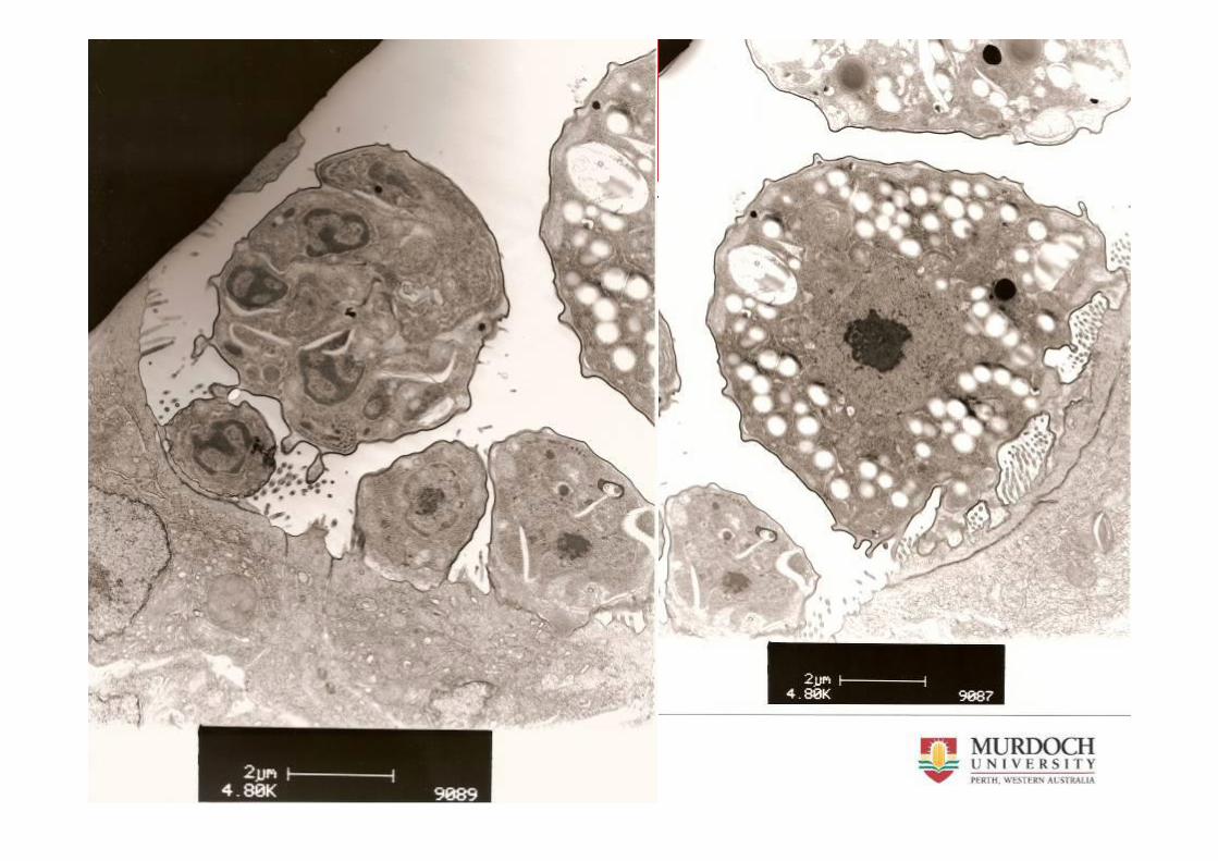
Histologic & TEM observations
•Intestinal protozoa infection often heavy & associated with degeneration/necrosis of gut•TEM images resemble cryptosporidium•PCR shows DNA sequence close to othercryptosporidium
Learning points of PCR on fixed tissues
• Fish tissues need to be decalcified with an acid prior to sectioning to make histology slides
• Poor success with PCR using acid treated decalcified tissues
• Cryptosporidia case was detected by PCR in spare formalin fixed tissues over a year old but not in wax block tissues
Further research on Crytopsoridia
• Developing of in-situ hybridization DNA probes using PCR products
• Immunohistochemistry using commercially available antibodies
Learning points on research in fish
• Information available from research in better known animal species often useful
• Materials generated from PCR can be useful to develop DNA probes to detect same or similar pathogens
• Published materials show successful outcomes only and do not show the pain taken to achieve it!
Acknowledgements
Supervisors Philip Nicholls & Brian JonesJun Kurita for monoclonal to red sea bream iridovirusHugh Ferguson & Miyazaki for help in pathologyMichael Slaven & Gerard Spoelstra from Histology LabPeter Fallon from Electron microscopy unitMark Bennett & Tim Hyndman for advice & help with molecular workThuy Ngo for the fish samples from VietnamDiana Chee for fish samples from SingaporeAlain for fish samples from IndonesiaAileen & Andrew Thompson for help with morphological identification of
cryptosporidiaUna Ryan & RongChang for 16S PCR on cryptosporidia cases