Diseases of Lates calcarifer by S Gibson-Kueh Page i Diseases of Asian seabass (or barramundi), Lates calcarifer Bloch Susan GIBSON-KUEH, B.V.Sc, M.Sc (Aquatic Veterinary Studies) This thesis is presented for the degree of Doctor of Philosophy of Murdoch University, 2012. Supervisors: Associate Prof. Philip K. Nicholls Prof. (adjunct) Brian Jones
Transcript
Diseases of Lates calcarifer by S Gibson-Kueh Page i
Diseases of Asian seabass (or barramundi), Lates
calcarifer Bloch
Susan GIBSON-KUEH, B.V.Sc, M.Sc (Aquatic Veterinary Studies)
This thesis is presented for the degree of Doctor of Philosophy of
Murdoch University, 2012.
Supervisors: Associate Prof. Philip K. Nicholls
Prof. (adjunct) Brian Jones
Diseases of Lates calcarifer by S Gibson-Kueh Page ii
Declaration
I declare that this thesis is an account of my research and contains work which
has not been previously submitted for a degree at any tertiary education
institution. Contributions by co-authors have been duly acknowledged.
Susan Gibson-Kueh
Diseases of Lates calcarifer by S Gibson-Kueh Page iii
Acknowledgements
Andrew Thompson, Andrea Valigurova & Miloslav Jirku for expert advice on
apicomplexans,
Una Ryan & RongChang Yang for molecular and phylogenetic analysis on the
Eimeria and Cryptosporidium from L. calcarifer,
Thuy, Alain, Diana Chee, Neil Wendover & John Jardine for some of the L.
calcarifer materials included in this study,
Aileen Elliot, Peter Fallon, Michael Slaven, Gerard Spoelstra, Wai-Yee Lee &
Micky Leong for technical laboratory support,
Jing Chen, Yahui Wang, Sophie Tay from the Animal & Plant Health Laboratories for
Red Sea bream iridovirus PCR and viral isolation on the scale drop cases,
Merck Aquatic Animal Health Laboratory in Singapore for the Red Sea bream
iridovirus PCR on L. calcarifer tissues from Farm A,
Mark Bennett & Masa Wayan for good advice on in situ hybridization,
Linda McInnes for providing the dioxygenin labeled Trypanosoma irwini DNA
probes
Jun Kurita for the Red Sea bream iridovirus monoclonal antibodies,
Jimmy Turnbull for introducing the mantra ‘the presence of a pathogen does not
equal the presence of a disease’
Mary Ng, who let me into the world of electron microscopy, what a great tool!
Hugh Ferguson, my teacher since Stirling, for providing expert opinion on the
pathology associated with scale drop syndrome…it was a great adventure
learning from you, and
Alan Lymbery for supporting all aquaculture related endeavours at Murdoch
University.
My supervisors, Phil & Brian for being patient with me.
To my husband Kelvin, and my daughters Leia, Sara, Rebecca & Clare, for all their
support and love.
Diseases of Lates calcarifer by S Gibson-Kueh Page iv
Preface
Chapter 1 serves as a brief introduction to husbandry practices and diseases
previously reported in cultured Lates calcarifer. It also includes a section on the
interactions between the host, environment and pathogens which need to be
considered in the investigation and managing of disease outbreaks. Chapters 2 to
4 are based on published papers while Chapter 5 is a manuscript intended for
publication in a scientific journal. There has been a need to adapt the chapters
based on published papers to integrate them into a thesis. Chapter 6 discusses
management strategies in relation to specific diseases in L. calcarifer at the
htachery, nursery and growout levels. Citations style is in keeping with that used
in Journal of Fish Diseases. Citations with more than 2 authors are quoted in full
when it appears in text within each chapter for the first time, and thereafter only
as first author & et al.
Diseases of Lates calcarifer by S Gibson-Kueh Page v
Abstract
Other than the study by Griffiths (2009) on gill diseases, there has been no
comprehensive study and report on the major diseases of Asian seabass (or
barramundi) Lates calcarifer Bloch. It is a food fish species of growing
importance in Asia and Australia. This study investigates some of the major
diseases encountered in the various stages of the culture of L. calcarifer, at the
histopathological, ultrastructural and molecular levels. Culture practices can
have significant impacts on fish health. Disease outbreaks are influenced by
factors involving the host, environment and pathogen. Current knowledge on
diseases of L. calcarifer, and these factors which may influence disease outbreaks
are discussed in Chapter 1.
This is the first report of an intestinal Eimeria infection in L. calcarifer.
The Eimeria infection was associated with severe pathology and significant
mortality in the absence of other pathogens. It was detected in diseased L.
calcarifer in all five nurseries in Ca Mau, Vietnam. Although these were small
scale nurseries which stocked an average of 3000 to 5000 fish at any one time, a
mortality rate of up to 30% was reported and is the cause of significant economic
losses for these nurseries. Moderate to heavy Eimeria infestation were observed
in greater than 80% of diseased fish examined. This high rate of Eimeria
infestation is suspected to be linked to the low daily water exchange rates
practised in these nurseries. However, the examination of only diseased fish does
not allow the determination of prevalence. A systemic iridovirus infection was
concurrently observed in some of the fishes but was not consistently present
when compared to the Eimeria infection. Molecular analysis showed that the
Eimeria of L. calcarifer from Vietnam formed clades with the Eimeria detected in
Diseases of Lates calcarifer by S Gibson-Kueh Page vi
L. calcarifer cultured in Australia, but clustered separately from other known
Eimeria species. Although Cryptosporidium was detected in these L. calcarifer
tissues, it could not be demonstrated histologically or ultrastructurally,
suggesting a low grade infestation or perhaps an environmental contaminant in
fish tissues tested. In situ hybridization using labeled PCR products showed that
labeled DNA probes generated from 18S PCR products could not be used to
distinguish between closely related genera such as Cryptosporidium and Eimeria.
Future investigation to determine the origin, transmission and risk factors
associated with this Eimeria infestation in L. calcarifer are needed.
‘Scale drop syndrome’ is a novel disease first reported in L. calcarifer
in Penang, Malaysia in 1992. Cases with similar gross and clinical presentations
were observed in Singapore in 2002, 2006 and 2009. Affected fish have loose
scales, which dropped off easily when handled. The disease was initially
observed in 100-300g fish, and later in larger fish up to 5kg bodyweight.
Cumulative mortalities of 40 to 50% were reported by farms, posing significant
economic losses of larger more valuable fish. This investigation forms the first
pathological description of ‘scale drop syndrome’ (SDS) in L. calcarifer. To aid
recognition of new cases for study, a case definition was developed for ‘scale
drop syndrome’ in L. calcarifer as a systemic vasculitis associated with tissue
necrosis in all major organs including the skin, with apparent targeting of cells of
epithelial origin. Attempts to isolate or detect the causative agent(s) by cell
culture, PCR and immunohistochemistry have proven unsuccessful. Further
studies to elucidate the definitive aetiology, isolate the causal agent(s) and
reproduce the disease will help better understanding and control of SDS.
Diseases of Lates calcarifer by S Gibson-Kueh Page vii
Although systemic iridoviral disease has been previously reported in
many freshwater and marine fish species, this study forms the first report of this
disease in L. calcarifer. Systemic iridoviral disease was observed in 5 to 20g L.
calcarifer usually 2 to 3 weeks post-transfer into sea cages at two farms.
Inclusion bodies suggestive of a systemic iridovirus infection were observed in
clinically healthy L. calcarifer from the land-based nursery of one of these two
farm; the presence of an iridovirus infection was supported by positive PCR
results using Red Sea bream iridovirus (RSIV) primer 1. The presence of
inclusions was not accompanied by any tissue necrosis in these clinically healthy
fish. This finding suggested that the systemic iridovirus infection occurred before
stocking at sea, and did not originate from wild fish or older fish in adjacent sea
cages as initially suspected by this farm. Immunohistochemistry on tissues of
clinical cases of systemic iridovirus gave positive results using the Red Sea
bream iridovirus monoclonal antibody (RSIV M10), although intensity varied
between tissues, possibly related to varying exposure of different tissues to
fixation chemicals. Inclusion bodies in clinically healthy fish from the same farm
did not show positive reaction with RSIV M10. This may be due to a lack of
antigenic expression by the viral infected cells at this early stage of infection.
Viral nervous necrosis (VNN) is a serious disease of hatchery reared L.
calcarifer fry in this study. Mortalities of 50 to 100% were reported in 3wo fry.
VNN can be difficult to diagnose in older fry, where it can be associated with few
vacuolations or an absence of viral inclusions
‘Pot belly disease’ (PBD) was previously reported in L. calcarifer fry less
than 1g, in association with an intracellular coccobacillus infection and
mortalities of 80 to 100%. In this study, PBD was observed in 120g L. calcarifer
Diseases of Lates calcarifer by S Gibson-Kueh Page viii
at two sea cage farms, in association with significant granulomatous enteritis.
The extent of the granulomatous enteritis is likely to have an effect on affected
fish. It was observed concurrently with systemic iridoviral disease at one farm
and nocardiosis at another farm. Diagnosis by histopathology and the lack of
other confirmatory tests for PBD may result in underdiagnosis of this disease.
The epidemiology of PBD needs further study to establish origin and modes of
transmission, to facilitate better disease control.
Diseases associated with infections by ubiquitous bacteria such as Vibrio,
Tenacibaculum were commonly observed in L. calcarifer post-handling.
Tenacibaculosis and vibriosis often occurred concurrently with other diseases
such as streptococcosis, systemic iridviral disease or PBD. Streptococcosis can
affect fish up to 3kg bodyweight, resulting in significant mortalities greater than
40 to 50%. Like SDS, because streptococcosis can affect up to market size fish,
they can cause considerable economic losses. Although vaccines against
Streptococcosis are available, conflicting views are held on the efficacy of
Streptococcus vaccines by various research groups. Overall, the South-east Asian
L. calcarifer farms which practiced vaccination against Streptococcus iniae
reported a reduction of mortality, especially in fish greater than 1 to 1.5kg
bodyweight.
Nocardiosis has been reported as an emerging disease in marine food fish
species caused by acid fast filamentous branching bacterium. Although
nocardiosis was observed histopathologically in L. calcarifer at two sea cage
farms, the numbers of samples examined were small and no other tests were
attempted due to lack of suitable samples. More intensive and extensive study is
needed to determine the significance of nocardiosis in L. calcarifer. Chronic
Diseases of Lates calcarifer by S Gibson-Kueh Page ix
granulomatous enteritis was not uncommon in the cases submitted to the Fish
Health Laboratory in Perth. Although the peritonitis was associated with heavy
bacteria infection, it is unclear if these are secondary invaders. Schipps, Bosmans
& Humphreys (2009) reported that Vibrio harveyi and Photobacterium damsela
damsela vaccinations appeared to be not efficacious, suggesting that these
bacteria were not the primary cause of the disease.
It is well recognized that disease outbreaks in farmed fish are influenced
by the interaction between host, the environment and pathogens. While serious
diseases are often reported in association with specific aquatic pathogens, not
much is known about the risk factors which trigger fish disease outbreaks.
Disease outbreaks often occur after stressful events such as net transfers, recent
handling or poor water quality. In fact, diseases are often caused by ubiquitous
pathogens that are commonly present in the culture environment. Although
further research is necessary to gather more information to improve diagnosis
and management of specific diseases, general health management strategies can
be applied at the various stages in the culture of L. calcarifer to minimize disease
outbreaks. This is discussed for L. calcarifer in Chapter 6.
Observations of types of disease agents may be influenced by site
conditions or the types of tests or materials examined. For example, some
parasites may be more prevalent in certain sites where intermediate hosts
abound, or loosely attached ectoparasites may be lost unless wet mount
microscopic examinations of fresh tissues were carried out. The study of
emerging diseases such as scale drop syndrome (SDS) or pot belly disease (PBD)
in L. calcarifer has been hampered by lack of confirmatory diagnostic tools and
inadequate knowledge on critical epidemiological factors such as mode of
Diseases of Lates calcarifer by S Gibson-Kueh Page x
transmission or potential reservoirs. While ideally identification and isolation of
the causal agent will help fulfil Koch’s postulates, it may be possible to improve
the understanding of disease via cohabitation or infectivity trials using tissue
homogenates from diseased fish when pure isolates are not available. There is a
need to conduct research to not only establish a definitive aetiology, but also to
identify risk factors to facilitate successful disease control. The successful
management of disease in aquaculture does not lie in any one strategy but an
integrated management of all risks encountered during the culture cycle against
disease occurrence or incursions.
Diseases of Lates calcarifer by S Gibson-Kueh Page xi
Table of Contents
Diseases of Asian seabass (or barramundi), Lates calcarifer Bloch ............................... i
Declaration ......................................................................................................................................... ii
Acknowledgements ........................................................................................................................ iii
Preface ................................................................................................................................................ iv
Table of Contents ............................................................................................................................ xi
Legends of Figures ........................................................................................................................ xv
Lists of Tables ................................................................................................................................. xxi
List of abbreviations ................................................................................................................... xxii
Meronts, gamonts and oocysts of piscine Cryptosporidium with a size
range of 3 to 5m are much smaller than corresponding stages of Eimeria and
Goussia, in the size range of 5 to 20m. The presence of invaginating feeder
organelles at the attachment juncture of Cryptosporidium distinguishes it from
the other genera (Valigurova, Jirku, Koudela, Gelnar, Modry & Slapeta 2008). The
attachment organelles of epicytoplasmic Eimeria and Goussia vary
ultrastructurally from monopodial to multiple finger-like attachment organelles
(Paperna 1991; Benajiba et al. 1994; Alvarez-Pellitero et al. 1997; Lukes 1992;
Lukes & Stary 1992).
This is the first report of an intestinal Eimeria infection in juveniles of L.
calcarifer, at the histopathological and ultrastructural levels. The Eimeria
infection was often associated with severe pathology even in the absence of
other significant pathogens, and is therefore a significant disease of L. calcarifer
in small scale nurseries in Ca Mau, Vietnam.
2.2 Materials and methods
2.2.1 Background information on samples examined
The emerging L. calcarifer industry in Vietnam depends on the grow-out of
juvenile fish from small scale nurseries, from which the samples in this study
were taken. Diseased juvenile L. calcarifer 2.5 to 7cm in body length were
Diseases of L. calcarifer Intestinal Eimeria infection
Diseases of Lates calcarifer by S Gibson-Kueh Page 23
sampled from a total of five nurseries in Ca Mau, Vietnam. It is estimated that
these nurseries stock 3000 to 5000 fish at any one time. These fish samples were
collected by field officers from the Minh Hai Sub-Institute for Fisheries Research,
Ca Mau, Vietnam in Jan to Mar 2008, Mar and Dec 2009, and Nov to Dec 2010, as
part of their active disease surveillance programs. Fixed tissue samples were
sent to Murdoch University, Australia for this study. A total of 181 fish were
processed for examination by histopathology and 10 fish for transmission
electron microscopy. Alcohol fixed oocysts obtained from discharged waste
water from culture tanks were also examined.
2.2.2 Light microscopy (LM)
Tissues were fixed in 10% phosphate buffered formalin for at least 24 h,
dehydrated in an ethanol-xylene series and embedded in paraffin blocks.
Formalin fixed bony tissues were decalcified in 5% nitric acid overnight prior to
dehydration and embedded in paraffin blocks. 5µm tissue sections were
dewaxed in xylene, rehydrated in an ethanol series and stained by haematoxylin
& eosin (H&E) or Giemsa stain.
2.2.3 Transmission electron microscopy (TEM)
Tissues were fixed in 5% glutaraldehyde in phosphate-buffered saline (PBS) at
4oC overnight, washed in several changes of PBS and post-fixed in Dalton’s
chrome osmic acid for1 h at 4oC. Fixed tissues were dehydrated through a graded
ethanol series to propylene oxide followed by immersion in a 60:40 solution of
propylene oxide/epoxy resin for 1 h, pure epoxy resin on a rotator overnight and
baked in an oven at 60oC for 24 h. Ultra-thin sections were stained with uranyl
acetate and lead citrate for viewing on a Philips CM100 Bio TEM.
Diseases of L. calcarifer Intestinal Eimeria infection
Diseases of Lates calcarifer by S Gibson-Kueh Page 24
2.3. Results
2.3.1 Field observations made on L. calcarifer nurseries sampled in this
study
The L. calcarifer nurseries in Ca Mau, Vietnam were mainly small scale with less
than five ½- to 1-tonne tanks. These nurseries obtained their fry from hatcheries
in Vung Tau or Khanh Hoa Province in Vietnam, or as imported fry from
Thailand. Fiber glass or cement tanks were mainly used as holding facilities with
static or closed water recirculation systems. Partial daily water exchange rates
less than 20-30% were practised. In earthen ponds which were less commonly
used, the fry were kept in nets suspended in the water column. Salinity of rearing
water ranged from 15 to 25 parts per thousand (ppt). Stocking density varied
from 280 to 350 fish/m3 water. Fish were fed commercial feed pellets
supplemented with coarsely chopped trash fish. The trash fish fed consisted of
wild fish caught from the sea.
Nurseries stocked 1 to 3 cm L. calcarifer fry obtained from hatcheries, and
grown to 7 to 9 cm body length fish to sell to grow-out farmers. Nursery reared
2.5 to 7.0 cm body length L. calcarifer juveniles were reported to suffer low grade
clinical disease soon after stocking, with a cumulative mortality of up to 30% of
stocked fish. Clinical signs and lesions included fish hanging at water surface,
inappetance, lethargy, darkened bodies, tail rot and scale loss.
2.3.2 Histopathology
An Eimeria infection was observed in greater than 60% of diseased L. calcarifer
sampled from each of the five nurseries, often as early as the first week post
stocking. The fish sampled in this study were kept in cement or fiber glass tanks,
or ponds, and in salinities of 15 or 25 ppt. Fish were found to be infected with
Diseases of L. calcarifer Intestinal Eimeria infection
Diseases of Lates calcarifer by S Gibson-Kueh Page 25
Eimeria in both tanks and ponds, and irrespective of salinities of rearing water.
Cytoplasmic inclusion bodies suggestive of systemic iridoviral infection were
observed histopathologically in approximately 20% of diseased L. calcarifer with
a concurrent Eimeria infestation. Low grade to heavy gill trichodinid infestation
was sometimes observed but not associated with any significant pathological
changes.
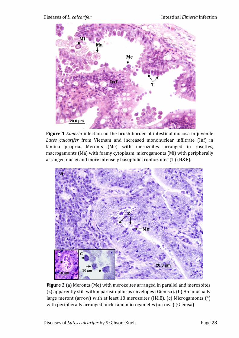
The primary infection site of the L. calcarifer Eimeria was the small
intestine. Both merogony and gamogony were epicytoplasmic and occurred
simultaneously (Fig. 1). Infection levels varied from light to heavy, often with
obliteration of the microvillous brush border. Meronts were much smaller than
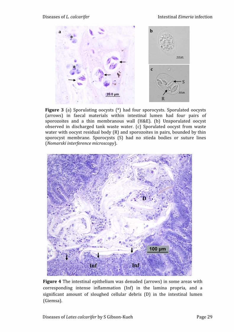
gamonts, and had merozoites arranged in rosettes or in parallel (Figs 1 & 2a).
Intracytoplasmic meronts or unusually large meronts with at least 18 merozoites
were occasionally observed (Fig. 2b). Macrogamonts with foamy cytoplasm due
to the presence of amylopectin granules often outnumbered microgamonts.
Microgamonts were smaller than macrogamonts and had peripherally arranged
nuclei (Figs 1 & 2c). Mature microgamonts had numerous flagellated
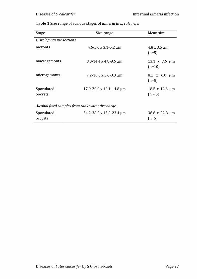
microgametes (Fig. 2c). Meronts measured 4.8 x 3.5 m (n=5), macrogamonts
13.1 x 7.6 m (n=10) and microgamonts 8.1 x 6.0 m (n=5). Table 1 shows a
summary of the size range of meronts, gamonts and oocysts.
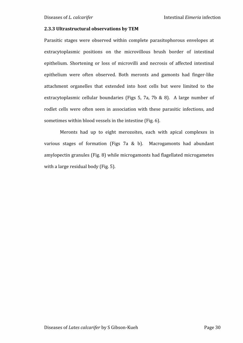
Sporulated oocysts were very rarely observed in histological tissue
sections, in fact in only 1 out of 181 fish examined, and measured 18.5 x 12.3 µm
(n = 5). These oocysts in faecal materials within the intestinal lumen had four
pairs of sporozoites held loosely within a thin membranous oocyst wall (Fig. 3a).
Both unsporulated and sporulated oocysts were readily observed in faeces
collected from tank bottom and in waste water from rearing tanks by wet mount
Diseases of L. calcarifer Intestinal Eimeria infection
Diseases of Lates calcarifer by S Gibson-Kueh Page 26
microscopic examinations (Figs 3b & c). Nomarski interference microscopy on
alcohol fixed sporulated oocysts showed the absence of Stieda bodies and suture
lines. Alcohol fixed sporulated oocysts measured 36.6 x 22.8 µm (n=5). A residual
body was present in oocysts, and each pair of sporozoites was held together by a
thin sporocyst membrane (Fig. 3c).
Squamous to cuboidal intestinal epithelium and low grade to severe
mononuclear inflammatory infiltrates in the lamina propria were frequently
observed in association with the Eimeria infection. The inflammation was
sometimes extended into the mucosal epithelium. There were often focal to
extensive areas of intestinal mucosal degeneration, necrosis and sloughed
necrotic cells in intestinal lumen (Fig. 4). Extra-intestinal parasite stages were
not commonly observed, except for two macrogamonts in renal tubules from 1
fish. Other abnormalities observed included dermatitis in caudal peduncle, renal
glomerular degeneration, moderately reactive spleens with white pulps depleted
of leucocytes, and reduced levels of hepatic glycogen stores.
Diseases of L. calcarifer Intestinal Eimeria infection
Diseases of Lates calcarifer by S Gibson-Kueh Page 27
Table 1 Size range of various stages of Eimeria in L. calcarifer
Stage Size range Mean size
Histology tissue sections
meronts 4.6-5.6 x 3.1-5.2 m 4.8 x 3.5 m
(n=5)
macrogamonts 8.0-14.4 x 4.8-9.6 m 13.1 x 7.6 m
(n=10)
microgamonts 7.2-10.0 x 5.6-8.3 m 8.1 x 6.0 m
(n=5)
Sporulated
oocysts
17.9-20.0 x 12.1-14.8 µm 18.5 x 12.3 µm
(n = 5)
Alcohol fixed samples from tank water discharge
Sporulated
occysts
34.2-38.2 x 15.8-23.4 µm 36.6 x 22.8 µm
(n=5)
Diseases of L. calcarifer Intestinal Eimeria infection
Diseases of Lates calcarifer by S Gibson-Kueh Page 28
Figure 1 Eimeria infection on the brush border of intestinal mucosa in juvenile
Lates calcarifer from Vietnam and increased mononuclear infiltrate (Inf) in
lamina propria. Meronts (Me) with merozoites arranged in rosettes,
macrogamonts (Ma) with foamy cytoplasm, microgamonts (Mi) with peripherally
arranged nuclei and more intensely basophilic trophozoites (T) (H&E).
Mi
Me
Ma
T Inf
a
Me
z
Figure 2 (a) Meronts (Me) with merozoites arranged in parallel and merozoites
(z) apparently still within parasitophorus envelopes (Giemsa). (b) An unusually
large meront (arrow) with at least 18 merozoites (H&E). (c) Microgamonts (*)
with peripherally arranged nuclei and microgametes (arrows) (Giemsa)
b
10 m
c
10 m
*
Diseases of L. calcarifer Intestinal Eimeria infection
Diseases of Lates calcarifer by S Gibson-Kueh Page 29
Figure 3 (a) Sporulating oocysts (*) had four sporocysts. Sporulated oocysts (arrows) in faecal materials within intestinal lumen had four pairs of sporozoites and a thin membranous wall (H&E). (b) Unsporulated oocyst observed in discharged tank waste water. (c) Sporulated oocyst from waste water with oocyst residual body (R) and sporozoites in pairs, bounded by thin sporocyst membrane. Sporocysts (S) had no stieda bodies or suture lines (Nomarski interference microscopy).
*
* a
b
c
R
S
Figure 4 The intestinal epithelium was denuded (arrows) in some areas with
corresponding intense inflammation (Inf) in the lamina propria, and a
significant amount of sloughed cellular debris (D) in the intestinal lumen
(Giemsa).
D
Inf Inf
Diseases of L. calcarifer Intestinal Eimeria infection
Diseases of Lates calcarifer by S Gibson-Kueh Page 30
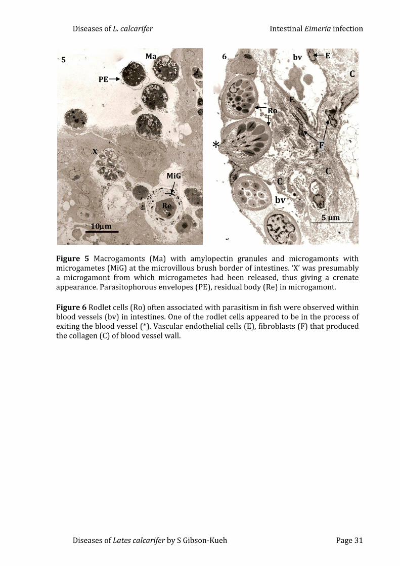
2.3.3 Ultrastructural observations by TEM
Parasitic stages were observed within complete parasitophorous envelopes at
extracytoplasmic positions on the microvillous brush border of intestinal
epithelium. Shortening or loss of microvilli and necrosis of affected intestinal
epithelium were often observed. Both meronts and gamonts had finger-like
attachment organelles that extended into host cells but were limited to the
extracytoplasmic cellular boundaries (Figs 5, 7a, 7b & 8). A large number of
rodlet cells were often seen in association with these parasitic infections, and
sometimes within blood vessels in the intestine (Fig. 6).
Meronts had up to eight merozoites, each with apical complexes in
various stages of formation (Figs 7a & b). Macrogamonts had abundant
amylopectin granules (Fig. 8) while microgamonts had flagellated microgametes
with a large residual body (Fig. 5).
Diseases of L. calcarifer Intestinal Eimeria infection
Diseases of Lates calcarifer by S Gibson-Kueh Page 31
Figure 5 Macrogamonts (Ma) with amylopectin granules and microgamonts with microgametes (MiG) at the microvillous brush border of intestines. ‘X’ was presumably a microgamont from which microgametes had been released, thus giving a crenate appearance. Parasitophorous envelopes (PE), residual body (Re) in microgamont.
Figure 6 Rodlet cells (Ro) often associated with parasitism in fish were observed within blood vessels (bv) in intestines. One of the rodlet cells appeared to be in the process of exiting the blood vessel (*). Vascular endothelial cells (E), fibroblasts (F) that produced the collagen (C) of blood vessel wall.
5 Ma
X
MiG
PE
10m
Re
6
*
Ro
E
C C
E
F
bv
bv
C
5 µm
Diseases of L. calcarifer Intestinal Eimeria infection
Diseases of Lates calcarifer by S Gibson-Kueh Page 32
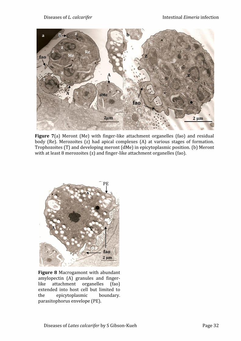
Figure 7(a) Meront (Me) with finger-like attachment organelles (fao) and residual body (Re). Merozoites (z) had apical complexes (A) at various stages of formation. Trophozoites (T) and developing meront (dMe) in epicytoplasmic position. (b) Meront with at least 8 merozoites (z) and finger-like attachment organelles (fao).
A
A
Re
T
fao Me
z
z
z
z
T dMe
2m
A a
2 µm
z
z
fao
b
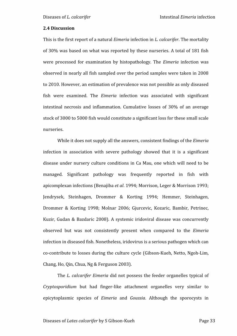
Figure 8 Macrogamont with abundant amylopectin (A) granules and finger-like attachment organelles (fao) extended into host cell but limited to the epicytoplasmic boundary. parasitophorus envelope (PE).
fao
A
A
2 µm
PE
Diseases of L. calcarifer Intestinal Eimeria infection
Diseases of Lates calcarifer by S Gibson-Kueh Page 33
2.4 Discussion
This is the first report of a natural Eimeria infection in L. calcarifer. The mortality
of 30% was based on what was reported by these nurseries. A total of 181 fish
were processed for examination by histopathology. The Eimeria infection was
observed in nearly all fish sampled over the period samples were taken in 2008
to 2010. However, an estimation of prevalence was not possible as only diseased
fish were examined. The Eimeria infection was associated with significant
intestinal necrosis and inflammation. Cumulative losses of 30% of an average
stock of 3000 to 5000 fish would constitute a significant loss for these small scale
nurseries.
While it does not supply all the answers, consistent findings of the Eimeria
infection in association with severe pathology showed that it is a significant
disease under nursery culture conditions in Ca Mau, one which will need to be
managed. Significant pathology was frequently reported in fish with
serum albumin in TBS 0.05% Tween 20) was added to tissue sections for 10
minutes before tapping off. Alkaline phosphatase (AP) conjugated anti-
dioxygenin (anti-DIG) (Roche, Germany) antibody diluted 1:600 in blocking
solution was added to each tissue section for a 60-minute incubation. The AP
conjugated anti-DIG was rinsed off followed by a 5-minute bath in TBS 0.05%
Tween 20. Liquid Permanent Red (Dako, USA) with 300 µg/ml levamisole to
block endogenous alkaline phosphatase was added to each slide for 20 minutes,
and rinsed off with tap water. Tissue sections were counterstained with
haematoxylin for 30 seconds and rinsed in tap water followed by a few short
dips in Scott’s solution. Slides were flicked dry prior to cover-slipping with
Faramount Mounting Media (Dako, USA) for visualization on an Olympus BX51
fluorescent microscope using the U-MWIBA2 filter.
3.3 Results
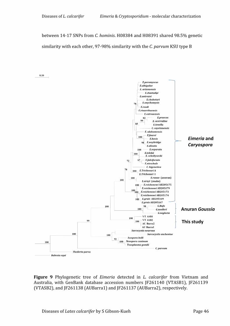
3.3.1 Sequence and phylogenetic analysis of Eimeria
Partial 18S rRNA gene sequences were obtained from two alcohol fixed
Vietnamese L. calcarifer tissue samples (VTASB1 and VTASB2) and two from
Australian L. calcarifer wax block tissue samples (AUBara1 and AUBara2). The
Diseases of L. calcarifer Eimeria & Cryptosporidium - molecular characterization
Diseases of Lates calcarifer by S Gibson-Kueh Page 45
partial 18S gene sequences VTASB2, AUBara1 and AUBara2 measured 1475bp,
while VTBSB1 measured 1468bp. Neighbour-joining, parsimony and ML analysis
of the 18S rRNA partial sequences from these four samples and a range of
Eimeria, Goussia and other species obtained from GenBank produced similar
results and showed that these isolates grouped separately from known Eimeria
and Goussia species (Fig. 9 - NJ tree shown). VTASB1 & 2 and AUBarra1 & 2
shared 99.93% - 98.1% similarity to each other and formed a distinct clade, but
shared only 92.8- 88.7% similarity with all other species.
The unique partial 18S rRNA gene sequences of the Vietnamese L.
calcarifer and Australian L. calcarifer Eimeria genotypes have been deposited in
the GenBank database under accession numbers JF261140 (VTASB1), JF261139
(VTASB2), JF261138 (AUBarra1) and JF261137 (AUBarra2), respectively.
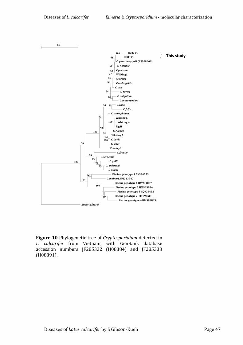
3.3.2 Sequence and phylogenetic analysis of Cryptosporidium
Partial 18S rRNA gene sequences were obtained from two formalin fixed
Vietnamese L. calcarifer tissue samples (H08384 and H08391). These sequences
obtained were 518 and 519bp, respectively. Neighbour-joining, parsimony and
ML analysis of the 18S rRNA sequences from these two samples and a range of
Cryptosporidium species and genotypes obtained from GenBank produced
similar results and showed that these DNA sequences grouped most closely with
C. parvum and C. hominis (Fig. 10 - NJ tree shown). H08384 and H08391 had 3
single nucleotide polymorphisms (SNPs) from each other, and between 7 and 12
SNPs from C. parvum KSU isolate type B (GenBank accession no. AF308600).
H08384 and H08391 had between 11-17 SNPs from C. parvum (GQ121019) and
Diseases of L. calcarifer Eimeria & Cryptosporidium - molecular characterization
Diseases of Lates calcarifer by S Gibson-Kueh Page 46
between 14-17 SNPs from C. hominis. H08384 and H08391 shared 98.5% genetic
similarity with each other, 97-98% similarity with the C. parvum KSU type B
Figure 9 Phylogenetic tree of Eimeria detected in L. calcarifer from Vietnam and Australia, with GenBank database accession numbers JF261140 (VTASB1), JF261139 (VTASB2), and JF261138 (AUBarra1) and JF261137 (AUBarra2), respectively.
Diseases of L. calcarifer Eimeria & Cryptosporidium - molecular characterization
Diseases of Lates calcarifer by S Gibson-Kueh Page 47
Figure 10 Phylogenetic tree of Cryptosporidium detected in L. calcarifer from Vietnam, with GenBank database accession numbers JF285332 (H08384) and JF285333 (H08391).
This study
0.1
Eimeria faurei
C. baileyi
C. saurophilum
C. fayeri
C. suis
C.meleagridis
C. wrairi
C. fragile
Piscine genotype 6 HM991857
Piscine genotype 5 HM989834
C. ryanae
C. serpentis
C. hominis
Pig II
Piscine genotype 3 GQ925452
Whiting 7
C. galli
C. parvum type B (AF308600)
H08384
H08391
C.parvum
Whiting1
C. ubiquitum
C. macropodum
C. canis
C. felis
Whiting 5
Whiting 4
C. bovis
C. xiaoi
C. andersoni
C. muris
Piscine genotype 1 AY524773
C. molnari_HM243547
Piscine genotype 2 FJ769050
Piscine genotype 4 HM989833
100
70
100
82
96
54
66
59
61
82
77
50
75
100
91
75
100
84
79
61
100
62
63
91
100
93
82
59
Diseases of L. calcarifer Eimeria & Cryptosporidium - molecular characterization
Diseases of Lates calcarifer by S Gibson-Kueh Page 48
isolate, and 96-97.5% similarity with C. parvum and C. hominis. Unfortunately all
attempts to amplify at the actin locus were unsuccessful.
The unique partial 18S rRNA sequences of the Cryptosporidium genotypes
detected in Vietnamese L. calcarifer have been deposited in the GenBank
database under accession numbers JF285332 (H08384) and JF285333
(H08391).
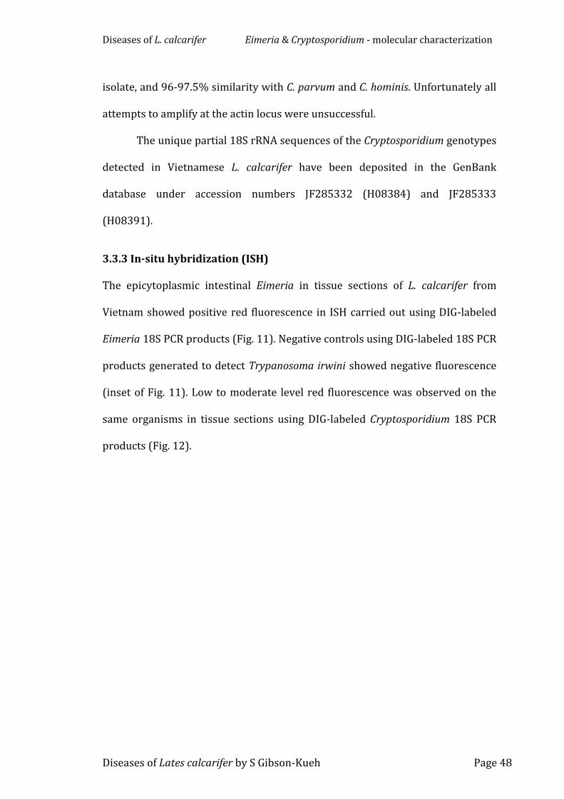
3.3.3 In-situ hybridization (ISH)
The epicytoplasmic intestinal Eimeria in tissue sections of L. calcarifer from
Vietnam showed positive red fluorescence in ISH carried out using DIG-labeled
products generated to detect Trypanosoma irwini showed negative fluorescence
(inset of Fig. 11). Low to moderate level red fluorescence was observed on the
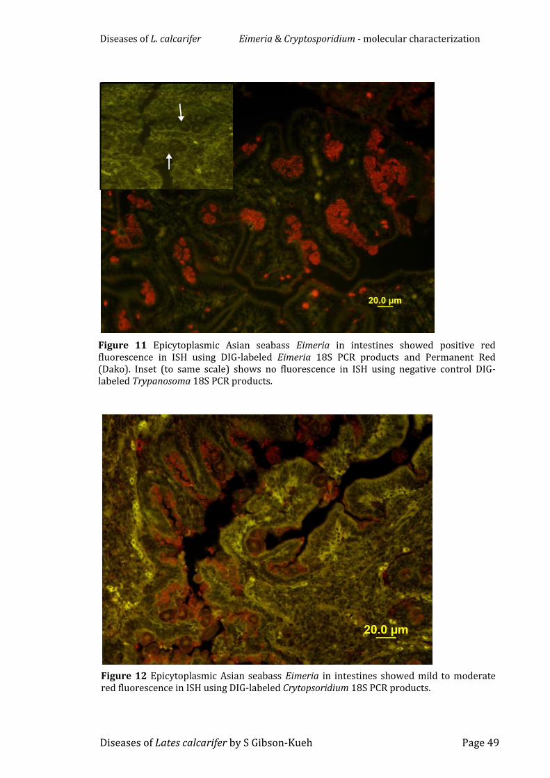
same organisms in tissue sections using DIG-labeled Cryptosporidium 18S PCR
products (Fig. 12).
Diseases of L. calcarifer Eimeria & Cryptosporidium - molecular characterization
Diseases of Lates calcarifer by S Gibson-Kueh Page 49
Figure 11 Epicytoplasmic Asian seabass Eimeria in intestines showed positive red fluorescence in ISH using DIG-labeled Eimeria 18S PCR products and Permanent Red (Dako). Inset (to same scale) shows no fluorescence in ISH using negative control DIG-labeled Trypanosoma 18S PCR products.
Figure 12 Epicytoplasmic Asian seabass Eimeria in intestines showed mild to moderate red fluorescence in ISH using DIG-labeled Crytopsoridium 18S PCR products.
Diseases of L. calcarifer Eimeria & Cryptosporidium - molecular characterization
Diseases of Lates calcarifer by S Gibson-Kueh Page 50
3.4 Discussion
Vietnamese and Australian L. calcarifer are the same fish species, but are from
distinct geographically separated populations. Analysis of Eimeria from both
Vietnamese and Australian L. calcarifer revealed that they are unique and form a
clade basal to the rest of Eimeriidae, which is clearly distinct from previously
described clades including anuran Eimeria and Goussia (Jirku, Jirku, Obornik,
Diseases of Lates calcarifer by S Gibson-Kueh Page 58
1995). L. calcarifer tissues from farm A in Indonesia (Chapter 5) diagnosed
histopathologically with a systemic iridoviral disease was found to produce
positive fluorescence in immunohistochemistry using the monoclonal antibody
RSIV M10, and served as positive control in testing L. calcarifer tissues with SDS.
4.2.5 PCR and tissue culture
Polymerase chain reaction (PCR) using RSIV primer set 1 which targets a large
range of iridoviruses (Kurita, Nakajima, Hirono & Aoki 1998), and viral isolation
on ATCC gruntfin, Haemulon sciurus Shaw (GF) and Asian seabass, Lates
calcarifer Bloch (SB) cells (Chong, Ngoh & Ng 1987) were carried out by the
Aquatic Animal Health Laboratory, AgriFood & Veterinary Authority of
Singapore.
4.3 Results
4.3.1 History, clinical & gross observations
‘Scale drop syndrome’ (SDS) was observed in 100-300g fish more than 3 to 4
months post-stocking in cages off the coast of Singapore. Fish were almost
always described as eating well with no signs of disease until onset of clinical
‘scale drop syndrome’. The disease appeared to progress within a few index
cages and then ‘spread’ onto surrounding cages stocked with L. calcarifer. The
disease did not affect other fish species stocked in the same farm. Based on
reports by farms, daily mortality of up to 1-2% was observed in affected cages
over a period of more than 3-4 weeks, with average cumulative losses of 40-50%
of stocked fish. Severely affected fish stopped schooling, and occasionally
showed abnormal neurological behaviour characterized by spiral swimming.
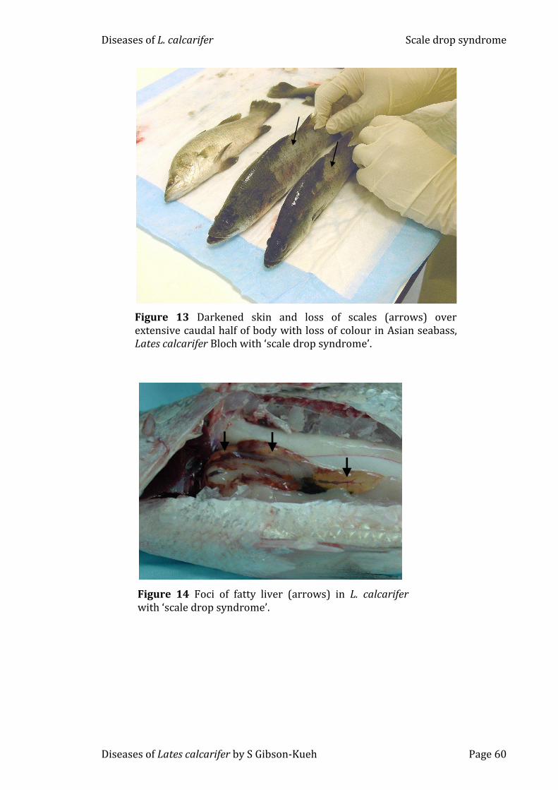
Gross observations included darkened bodies, scale loss over extensive areas
Diseases of L. calcarifer Scale drop syndrome
Diseases of Lates calcarifer by S Gibson-Kueh Page 59
with loss of skin colour (Fig. 13), tail/fin erosion, pallor of gills, focal to extensive
areas of hepatic lipidosis (Fig. 14), petechial to ecchymotic haemorrhage in the
liver, kidney and spleen, splenomegaly or atrophied shrunken spleen, and
renomegaly. Although low to moderate levels of parasites such as trichodinids,
monogeneans and myxosporeans were observed by wet mount microscopic
examinations in gills of some fish, they were not consistently present in affected
fish and therefore were considered opportunistic or incidental infestations.
Diseases of L. calcarifer Scale drop syndrome
Diseases of Lates calcarifer by S Gibson-Kueh Page 60
Figure 13 Darkened skin and loss of scales (arrows) over extensive caudal half of body with loss of colour in Asian seabass, Lates calcarifer Bloch with ‘scale drop syndrome’.
Figure 14 Foci of fatty liver (arrows) in L. calcarifer with ‘scale drop syndrome’.
Diseases of L. calcarifer Scale drop syndrome
Diseases of Lates calcarifer by S Gibson-Kueh Page 61
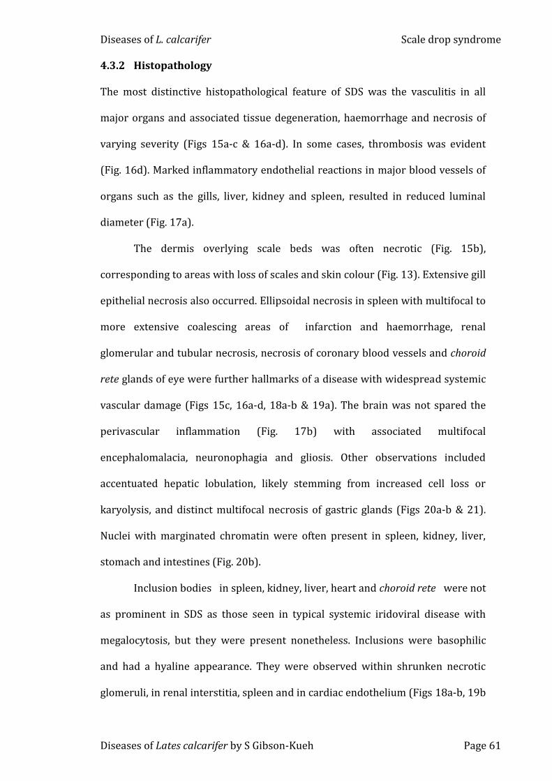
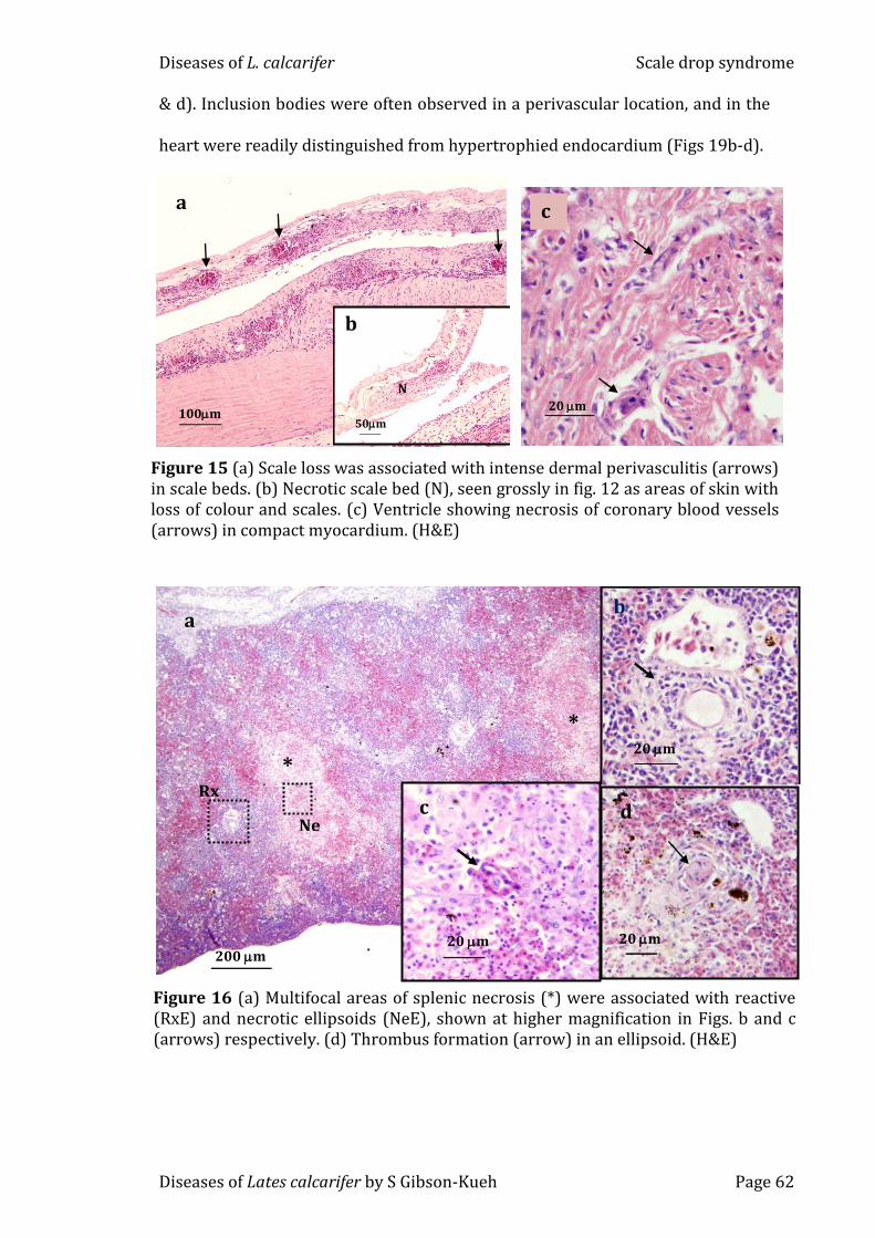
4.3.2 Histopathology
The most distinctive histopathological feature of SDS was the vasculitis in all
major organs and associated tissue degeneration, haemorrhage and necrosis of
varying severity (Figs 15a-c & 16a-d). In some cases, thrombosis was evident
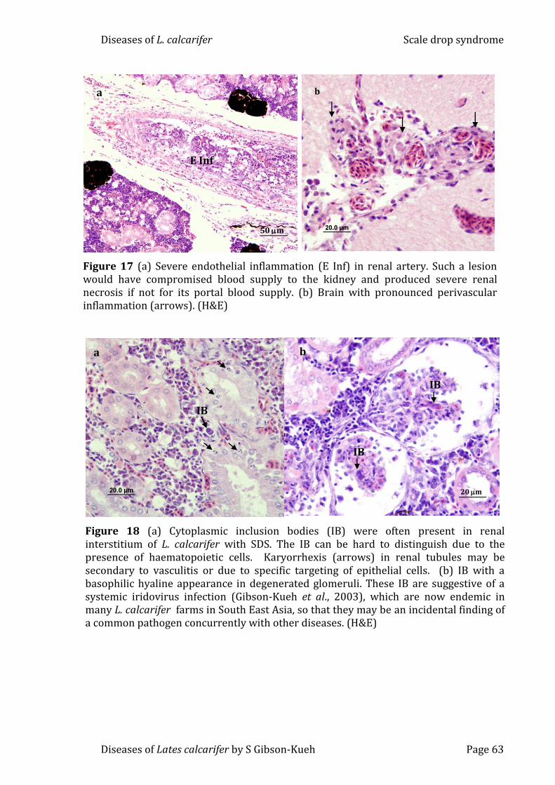
(Fig. 16d). Marked inflammatory endothelial reactions in major blood vessels of
organs such as the gills, liver, kidney and spleen, resulted in reduced luminal
diameter (Fig. 17a).
The dermis overlying scale beds was often necrotic (Fig. 15b),
corresponding to areas with loss of scales and skin colour (Fig. 13). Extensive gill
epithelial necrosis also occurred. Ellipsoidal necrosis in spleen with multifocal to
more extensive coalescing areas of infarction and haemorrhage, renal
glomerular and tubular necrosis, necrosis of coronary blood vessels and choroid
rete glands of eye were further hallmarks of a disease with widespread systemic
vascular damage (Figs 15c, 16a-d, 18a-b & 19a). The brain was not spared the
perivascular inflammation (Fig. 17b) with associated multifocal
encephalomalacia, neuronophagia and gliosis. Other observations included
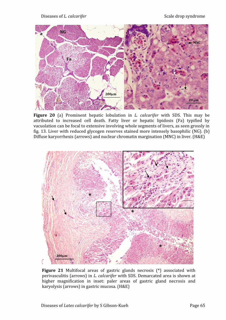
accentuated hepatic lobulation, likely stemming from increased cell loss or
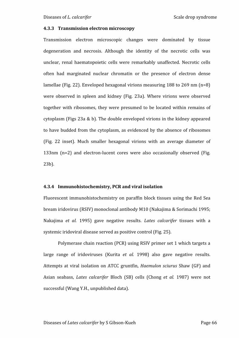
karyolysis, and distinct multifocal necrosis of gastric glands (Figs 20a-b & 21).
Nuclei with marginated chromatin were often present in spleen, kidney, liver,
stomach and intestines (Fig. 20b).
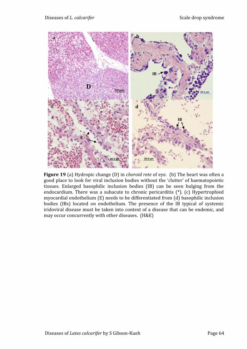
Inclusion bodies in spleen, kidney, liver, heart and choroid rete were not
as prominent in SDS as those seen in typical systemic iridoviral disease with
megalocytosis, but they were present nonetheless. Inclusions were basophilic
and had a hyaline appearance. They were observed within shrunken necrotic
glomeruli, in renal interstitia, spleen and in cardiac endothelium (Figs 18a-b, 19b
Diseases of L. calcarifer Scale drop syndrome
Diseases of Lates calcarifer by S Gibson-Kueh Page 62
& d). Inclusion bodies were often observed in a perivascular location, and in the
heart were readily distinguished from hypertrophied endocardium (Figs 19b-d).
100m
a
Figure 15 (a) Scale loss was associated with intense dermal perivasculitis (arrows) in scale beds. (b) Necrotic scale bed (N), seen grossly in fig. 12 as areas of skin with loss of colour and scales. (c) Ventricle showing necrosis of coronary blood vessels (arrows) in compact myocardium. (H&E)
N
50m
b
20 m
c
Figure 16 (a) Multifocal areas of splenic necrosis (*) were associated with reactive (RxE) and necrotic ellipsoids (NeE), shown at higher magnification in Figs. b and c (arrows) respectively. (d) Thrombus formation (arrow) in an ellipsoid. (H&E)
200 m
a
Rx
*
Ne
*
d
20 m
b
20 m
c
20 m
Diseases of L. calcarifer Scale drop syndrome
Diseases of Lates calcarifer by S Gibson-Kueh Page 63
Figure 18 (a) Cytoplasmic inclusion bodies (IB) were often present in renal interstitium of L. calcarifer with SDS. The IB can be hard to distinguish due to the presence of haematopoietic cells. Karyorrhexis (arrows) in renal tubules may be secondary to vasculitis or due to specific targeting of epithelial cells. (b) IB with a basophilic hyaline appearance in degenerated glomeruli. These IB are suggestive of a systemic iridovirus infection (Gibson-Kueh et al., 2003), which are now endemic in many L. calcarifer farms in South East Asia, so that they may be an incidental finding of a common pathogen concurrently with other diseases. (H&E)
b
20 m
IB
IB
a
IB
Figure 17 (a) Severe endothelial inflammation (E Inf) in renal artery. Such a lesion would have compromised blood supply to the kidney and produced severe renal necrosis if not for its portal blood supply. (b) Brain with pronounced perivascular inflammation (arrows). (H&E)
50 m
E Inf
a
b
Diseases of L. calcarifer Scale drop syndrome
Diseases of Lates calcarifer by S Gibson-Kueh Page 64
Figure 19 (a) Hydropic change (D) in choroid rete of eye. (b) The heart was often a good place to look for viral inclusion bodies without the ‘clutter’ of haematopoietic tissues. Enlarged basophilic inclusion bodies (IB) can be seen bulging from the endocardium. There was a subacute to chronic pericarditis (*). (c) Hypertrophied myocardial endothelium (E) needs to be differentiated from (d) basophilic inclusion bodies (IBs) located on endothelium. The presence of the IB typical of systemic iridoviral disease must be taken into context of a disease that can be endemic, and may occur concurrently with other diseases. (H&E)
* IB
b
IB
s
d
50 m D
a
c
E
Diseases of L. calcarifer Scale drop syndrome
Diseases of Lates calcarifer by S Gibson-Kueh Page 65
Figure 20 (a) Prominent hepatic lobulation in L. calcarifer with SDS. This may be attributed to increased cell death. Fatty liver or hepatic lipidosis (Fa) typified by vacuolation can be focal to extensive involving whole segments of livers, as seen grossly in fig. 13. Liver with reduced glycogen reserves stained more intensely basophilic (NG). (b) Diffuse karyorrhexis (arrows) and nuclear chromatin margination (MNC) in liver. (H&E)
200m
NG
Fa
a
b
20 m
MNC
20m
*
*
200m
*
Figure 21 Multifocal areas of gastric glands necrosis (*) associated with perivasculitis (arrows) in L. calcarifer with SDS. Demarcated area is shown at higher magnification in inset: paler areas of gastric gland necrosis and karyolysis (arrows) in gastric mucosa. (H&E)
Diseases of L. calcarifer Scale drop syndrome
Diseases of Lates calcarifer by S Gibson-Kueh Page 66
4.3.3 Transmission electron microscopy
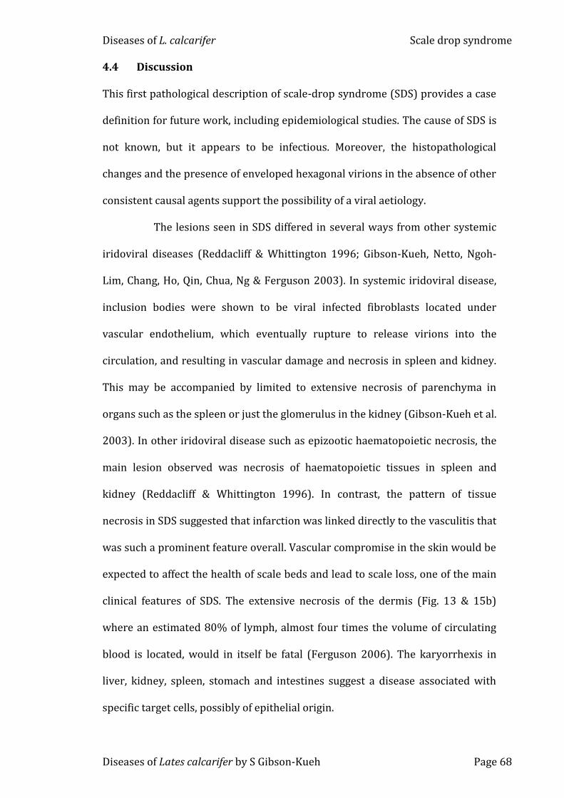
Transmission electron microscopic changes were dominated by tissue
degeneration and necrosis. Although the identity of the necrotic cells was
unclear, renal haematopoietic cells were remarkably unaffected. Necrotic cells
often had marginated nuclear chromatin or the presence of electron dense
Nakajima et al. 1995) gave negative results. Lates calcarifer tissues with a
systemic iridoviral disease served as positive control (Fig. 25).
Polymerase chain reaction (PCR) using RSIV primer set 1 which targets a
large range of iridoviruses (Kurita et al. 1998) also gave negative results.
Attempts at viral isolation on ATCC gruntfin, Haemulon sciurus Shaw (GF) and
Asian seabass, Lates calcarifer Bloch (SB) cells (Chong et al. 1987) were not
successful (Wang Y.H., unpublished data).
Diseases of L. calcarifer Scale drop syndrome
Diseases of Lates calcarifer by S Gibson-Kueh Page 67
Figure 22 L. calcarifer with SDS have remarkably normal renal haematopoietic cells (Hc). Necrotic cells have marginated chromatin (MNC) or presence of very electron dense lamellae (L) in nucleus. Red blood cells (rbc) in blood vessel. Demarcated area is shown at higher magnification in inset: double enveloped hexagonal virions with ribosomes absent, presumably as virions have budded out from cytoplasm of cell.
5 m
rbc
MNC
MNC
Hc
Hc
L
200nm
Figure 23 (a)Enveloped hexagonal virions in spleen in remains of cytoplasmic ribosomes (r), with a size range of 188 to 269 nm (n=8). (b) Smaller enveloped hexagonal virions measuring 133nm (n=2) in cytoplasmic remnants in kidney. These virions have electron-lucent nucleocapsids.
100 nm
b
200 nm
r
r
a
Diseases of L. calcarifer Scale drop syndrome
Diseases of Lates calcarifer by S Gibson-Kueh Page 68
4.4 Discussion
This first pathological description of scale-drop syndrome (SDS) provides a case
definition for future work, including epidemiological studies. The cause of SDS is
not known, but it appears to be infectious. Moreover, the histopathological
changes and the presence of enveloped hexagonal virions in the absence of other
consistent causal agents support the possibility of a viral aetiology.
The lesions seen in SDS differed in several ways from other systemic
Although vibriosis and tenacibaculosis can cause serious mortality in
cultured L. calcarifer, they are generally considered as opportunistic diseases in
stressed fish (Ruangpan 1988; Azad, Thirunavukkarasu, Kailasam & Rajan 2004;
Avendano-Herrera et al. 2006; Labrie et al. 2007; Humphrey et al. 2010).
Nocardiosis is an emerging disease in cultured marine finfish. Nocardia
can be recognized histologically as gram positive, acid fast with Fite Faraco stain,
filamentous and often branching bacteria in association with systemic
granulomatous necrotic lesions. Culture techniques and PCR have been
established for the detection of Nocardia (Kudo, Hatai & Seino 1988; Labrie, Ng,
Tan, Komar, Ho & Grisez 2008).
This is a first report of systemic iridoviral disease and nocardiosis in L.
calcarifer. Pot belly disease was found to affect older L. calcarifer than previously
reported. Viral and bacterial diseases reported in this study can cause significant
mortality in cultured L. calcarifer.
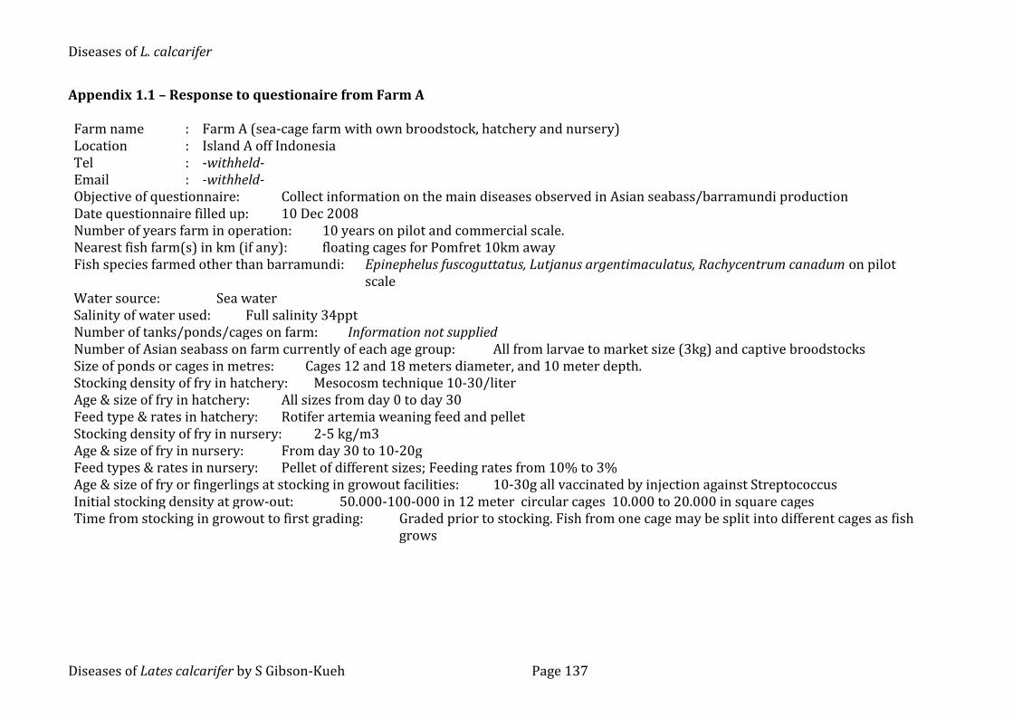
5.2 Materials and methods
5.2.1 Background
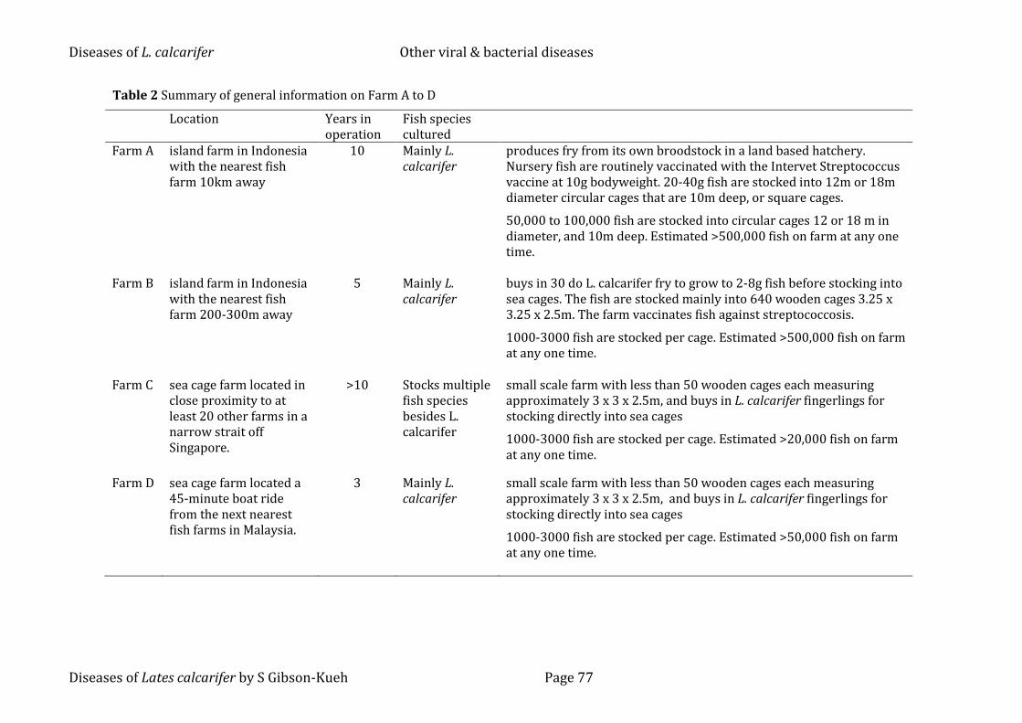
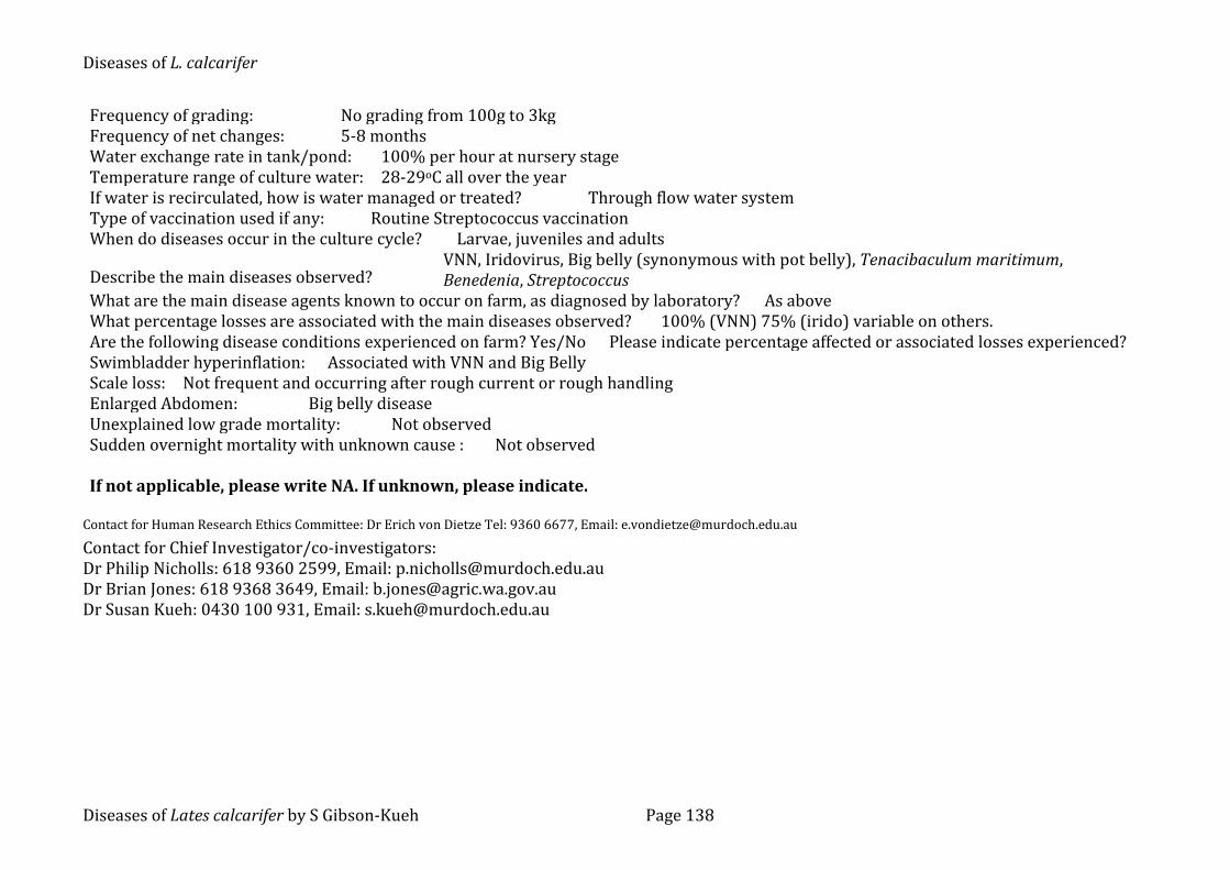
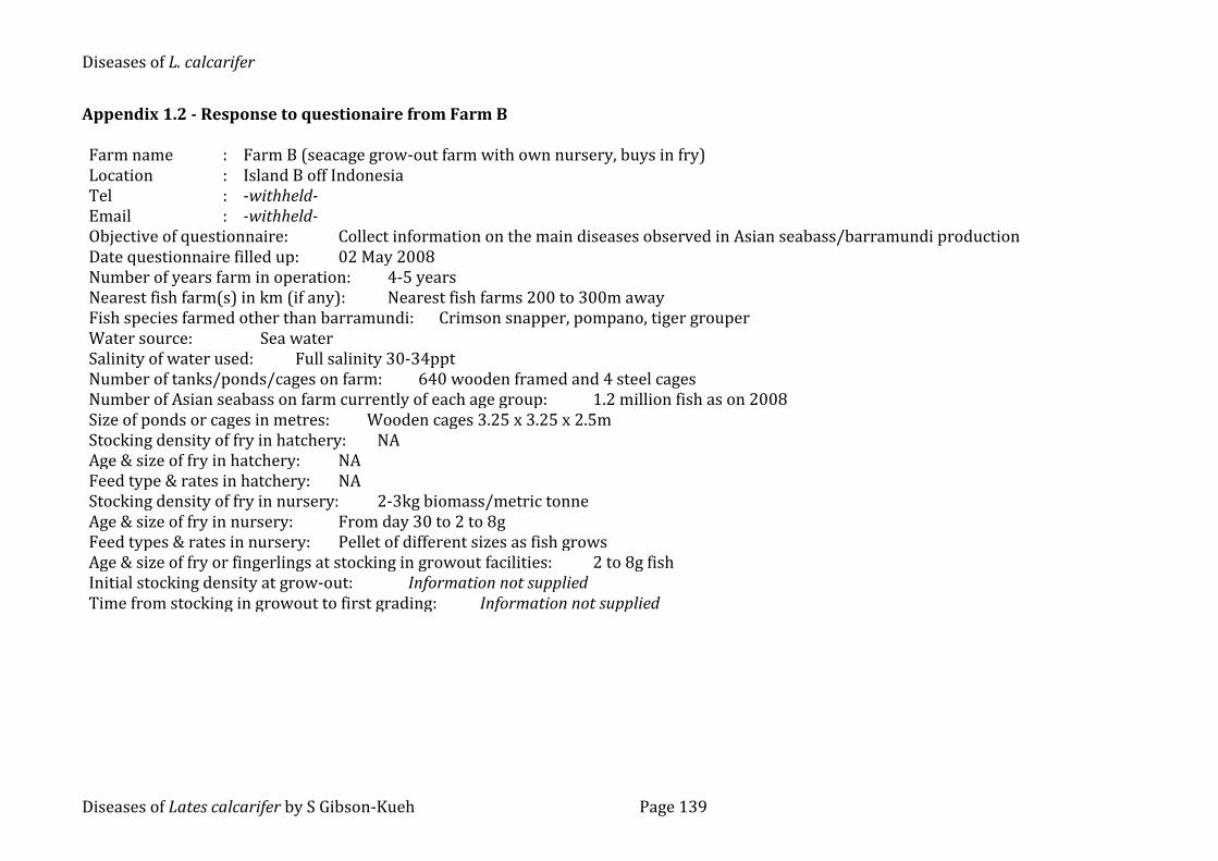
Table 2 is a summary of the general information on Farms A to D including
location, proximity to other farms, general husbandry practices and number of
Diseases of L. calcarifer Other viral & bacterial diseases
Diseases of Lates calcarifer by S Gibson-Kueh Page 75
years each farm has been in operation. Appendix 1.1 is a questionaire completed
by Farm A and appendix 1.2 by Farm B, on husbandry practices.
Materials examined included fixed L. calcarifer tissues sent to Murdoch
University, Perth by Farm A in Indonesia between 2007 and 2010. The range of
diseases encountered in Farm A is well documented. Farm A has a history of
systemic iridoviral disease in 10-20g L. calcarifer fingerlings two to three weeks
post transfer into sea cages. As the farm was comtemplating using an iridovirus
vaccine, the purpose of this study was to determine if the systemic iridovirus
infection occurred before transfer to sea cages. Lates calcarifer tissues were
examined by histopathology at Murdoch University and corresponding samples
sent to Merck Aquatic Animal Health Laboratory in Singapore for testing by PCR.
All samples examined at Murdoch University were either formalin fixed or
alcohol fixed L. calcarifer tissues, and presumptive diagnoses are based mainly
on histopathological evidence. PCR, immunohistochemistry and in-situ
hybridization was carried out only on selected L. calcarifer tissues from Farm A
with histopathological evidence of a systemic iridoviral disease.
Histology slides from L. calcarifer cases submitted to Aquatic Animal
Health Laboratory (AAHL), Agri-Food & Veterinary Authority of Singapore in
Singapore from 1993 to 2006, and Fish Health Laboratory, WA Department of
Fisheries from 2003 to 2008 were also examined. Reference to specific bacteria
species are based on results available in case records from routine culture or
PCR.
Diseases of L. calcarifer Other viral & bacterial diseases
Diseases of Lates calcarifer by S Gibson-Kueh Page 76
5.2.2 Light microscopy (LM)
Tissues were processed for light microscopy as outlined in 2.2.2. Tissue sections
were stained by haematoxylin & eosin (H&E) and Fite Faraco, a modified Ziehl
Neelsen stain.
5.2.3 Transmission electron microscopy (TEM)
Tissues were processed for electron microscopy as outlined in 2.2.3. Ultra-thin
sections were stained with uranyl acetate and lead citrate for viewing on a
Philips CM100 Bio TEM.
Diseases of L. calcarifer Other viral & bacterial diseases
Diseases of Lates calcarifer by S Gibson-Kueh Page 77
Table 2 Summary of general information on Farm A to D
Location Years in operation
Fish species cultured
Farm A island farm in Indonesia with the nearest fish farm 10km away
10 Mainly L. calcarifer
produces fry from its own broodstock in a land based hatchery. Nursery fish are routinely vaccinated with the Intervet Streptococcus vaccine at 10g bodyweight. 20-40g fish are stocked into 12m or 18m diameter circular cages that are 10m deep, or square cages.
50,000 to 100,000 fish are stocked into circular cages 12 or 18 m in diameter, and 10m deep. Estimated >500,000 fish on farm at any one time.
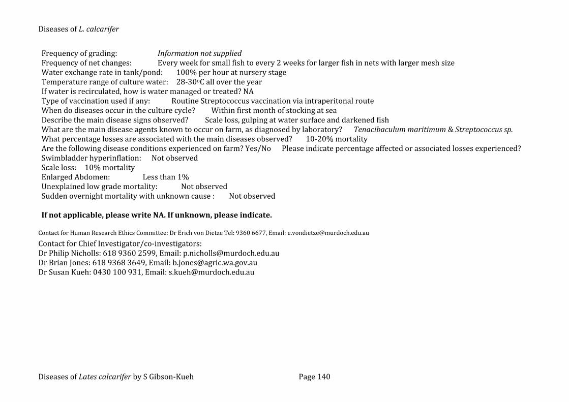
Farm B island farm in Indonesia with the nearest fish farm 200-300m away
5 Mainly L. calcarifer
buys in 30 do L. calcarifer fry to grow to 2-8g fish before stocking into sea cages. The fish are stocked mainly into 640 wooden cages 3.25 x 3.25 x 2.5m. The farm vaccinates fish against streptococcosis.
1000-3000 fish are stocked per cage. Estimated >500,000 fish on farm at any one time.
Farm C sea cage farm located in close proximity to at least 20 other farms in a narrow strait off Singapore.
>10 Stocks multiple fish species besides L. calcarifer
small scale farm with less than 50 wooden cages each measuring approximately 3 x 3 x 2.5m, and buys in L. calcarifer fingerlings for stocking directly into sea cages
1000-3000 fish are stocked per cage. Estimated >20,000 fish on farm at any one time.
Farm D sea cage farm located a 45-minute boat ride from the next nearest fish farms in Malaysia.
3 Mainly L. calcarifer
small scale farm with less than 50 wooden cages each measuring approximately 3 x 3 x 2.5m, and buys in L. calcarifer fingerlings for stocking directly into sea cages
1000-3000 fish are stocked per cage. Estimated >50,000 fish on farm at any one time.
Diseases of L. calcarifer Other viral & bacterial diseases
Diseases of Lates calcarifer by S Gibson-Kueh Page 78
5.2.4 Immunohistochemistry using RSIV M10
Formalin fixed paraffin embedded (FFPE) L. calcarifer tissue sections of selected
cases from Farm A with histopathological signs of a systemic iridoviral disease
(Gibson-Kueh et al. 2003) were tested by immunohistochemistry (IHC) using a
monoclonal antibody against the Red Sea bream iridovirus (RSIV M10)
(Nakajima & Sorimachi, 1995). The RSIV M10 was kindly supplied by Dr Jun
Kurita, National Research Institute of Aquaculture, Fisheries Research Agency,
Mie, Japan. The immunohistochemistry method after Adams and de Mateo
(1994) with modifications as described below was used. Negative control is
carried out with substitution of the RSIV M10 with blocking solution.
FFPE 5µm tissue sections on silanised slides were deparaffinized in two
changes of xylene and rehydrated through an ethanol series to tap water.
Antigen retrieval was performed by placing rehydrated tissue sections in Tris
EDTA buffer (pH 9) bath in a domestic microwave (Kambrook Model KER-
686LE) and subjected to ‘reheat’ for 4 minutes and ‘low heat’ function for 4
minutes, followed by cooling in running tap water.
A blocking step was carried out by adding 0.1% bovine serum albumin in
Tris buffered saline (TBS) (pH 7) with 0.05% Tween 20 directly to tissue
sections for a 10-minute incubation. After the blocking solution was tapped off,
the primary antibody RSIV M10 diluted 1/10 in blocking solution was added to
each tissue section for a 30-minute incubation. FFPE tissues incubated with
blocking solution instead of RSIV M10 served as the negative control. The
primary antibody was rinsed off, followed by addition of TBS (pH7) with 0.05%
Tween 20 to tissue sections for a 5-minute incubation.
Diseases of L. calcarifer Other viral & bacterial diseases
Diseases of Lates calcarifer by S Gibson-Kueh Page 79
Polyclonal rabbit anti-mouse immunoglobulin conjugated with alkaline
phosphatase (Dako, US) diluted 1:50 in blocking solution was added to tissue
sections for a 30-minute incubation, followed by rinsing in TBS (pH7) with
0.05% Tween 20 added to each slide for 5-minute incubation. Liquid permanent
red (Dako, US) with levamisole (300μg/ml) to block endogenous alkaline
phosphatase activity was added to tissue sections for a 20- minute incubation,
and rinsed off with tap water. Tissue sections were counterstained with
haematoxylin for 1 minute, rinsed in tap water and flicked dry prior to cover-
slipping with Farmount mounting media (Dako, US).
5.2.5 Polymerase chain reaction & in situ hybridization
Red Sea bream iridovirus (RSIV) PCR as published by Kurita et al. (1998) on L.
calcarifer tissues from Farm A was carried out by the Merck Aquatic Animal
Health Laboratory in Singapore. RSIV PCR was carried out at Murdoch University
to generate dioxigenin labeled DNA probes from PCR products for in situ
hybridization (ISH) on formalin fixed paraffin embedded (FFPE) tissues. DNA was
extracted from alcohol fixed L. calcarifer tissues from Farm A with histopathological
evidence of systemic iridoviral disease, using a QIAamp DNA FFPE Tissue kit
(Qiagen, Germany), according to the manufacturer’s instructions.
Briefly, PCR reaction mixes made up using TopTaq Master Mix (Qiagen, US)
according to the manufacturer’s instructions, containing 0.2 M of RSIV primer set 1
and 1 l of extracted DNA were incubated in an Eppendorf Mastercycler under PCR
conditions as described by Kurita et al., 1998. After amplification, 10 l of PCR
products from each reaction tube was analyzed in a 1 % agarose gel in 0.5x TBE
buffer with SybrSafe, and visualized under UV illumination.
Diseases of L. calcarifer Other viral & bacterial diseases
Diseases of Lates calcarifer by S Gibson-Kueh Page 80
RSIV PCR products obtained from fish tissues from Farm A were purified
using QIAquick PCR purification spin columns (Qiagen, Germany) and labeled with
dioxigenin (DIG) using DIG-nick translation mix (Roche, Germany), according to
the manufacturer’s instructions. The DIG-nick translation kit results in the
production of a mixture of DIG labeled DNA probes between 200 to 500bp in size
from purified PCR products. In situ hybridization was carried out using these DIG-
labeled DNA probes on FFPE L. calcarifer tissues with overt clinical and
histopathological evidence of a systemic iridoviral disease from the same Farm A.
PCR products were also purified using QIAquick PCR purification spin columns
(Qiagen, Germany), and send to a commercial company for sequencing.
DIG labeled DNA probes made from 1 g of PCR products were used to
make up 1 ml of DNA probe mixture that contained 50% formamide, 10%
dextran sulphate and 2x Saline Sodium Citrate (SSC) buffer. The DNA probe
mixture was stored at 4oC until required. A negative control DIG labeled DNA
probe generated from PCR products to detect a papilloma virus of bandicoot was
kindly provided by Mark Bennett, School of Veterinary and Biomedical Sciences,
Murdoch University (Bennett, Woolford, O’Hara, Warren & Nicholls 2008).
The ISH steps are carried out as described in Section 3.2.4 for the
detection of Eimeria in L. calcarifer tissues except for the use of DIG labeled RSIV
PCR product as the DNA probe.
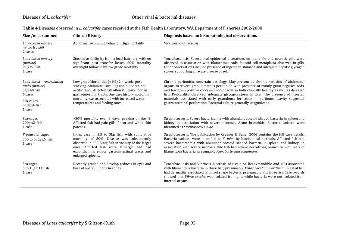
5.3 Diseases observed in L. calcarifer tissues examined
Table 3 is a summary of the diseases observed in L. calcarifer from Farms A to D,
and cases submitted to the Aquatic Animal Health Laboratory from 1993 to
2003. Table 4 is a summary of the diseases observed in L. calcarifer cases
Diseases of L. calcarifer Other viral & bacterial diseases
Diseases of Lates calcarifer by S Gibson-Kueh Page 81
submitted to the Fish Health Laboratory, WA Department of Fisheries from 2002
to 2008. Viral followed by bacterial diseases of L. calcarifer will be described.
5.3.1 Systemic iridoviral disease
Systemic iridoviral disease was observed histopathologically in 5 to 10g L.
calcarifer 2 to 3 weeks after transfer into sea cages at Farm A and C (Table 3).
Affected fish showed darkened bodies and no other clinical signs. Mortality in
batches of affected fish was reported as 95% in 5 to 6g fish and 75% in larger
10g fish in sea-cages in Farm A. Farm A has an estimated greater than 500,000
fish in sea cages at any one time, and at least 100,000 to 200,000 fish will fall into
the age group affected by systemic iridoviral disease. The most consistent gross
abnormalities were pale gills and splenomegaly. Systemic iridovirus infection
was diagnosed histopathologically in clinically healthy nursery 0.2-1.2 g
L.calcarifer from Farm A (Table 3). This was supported by positive PCR results
carried out by the Merck Aquatic Animal Health Laboratory in Singapore using
the RSIV primer 1 (unpublished data).
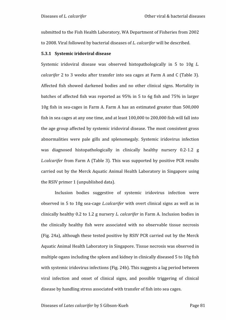
Inclusion bodies suggestive of systemic iridovirus infection were
observed in 5 to 10g sea-cage L.calcarifer with overt clinical signs as well as in
clinically healthy 0.2 to 1.2 g nursery L. calcarifer in Farm A. Inclusion bodies in
the clinically healthy fish were associated with no observable tissue necrosis
(Fig. 24a), although these tested positive by RSIV PCR carried out by the Merck
Aquatic Animal Health Laboratory in Singapore. Tissue necrosis was observed in
multiple ogans including the spleen and kidney in clinically diseased 5 to 10g fish
with systemic iridovirus infections (Fig. 24b). This suggests a lag period between
viral infection and onset of clinical signs, and possible triggering of clinical
disease by handling stress associated with transfer of fish into sea cages.
Diseases of L. calcarifer Other viral & bacterial diseases
Diseases of Lates calcarifer by S Gibson-Kueh Page 82
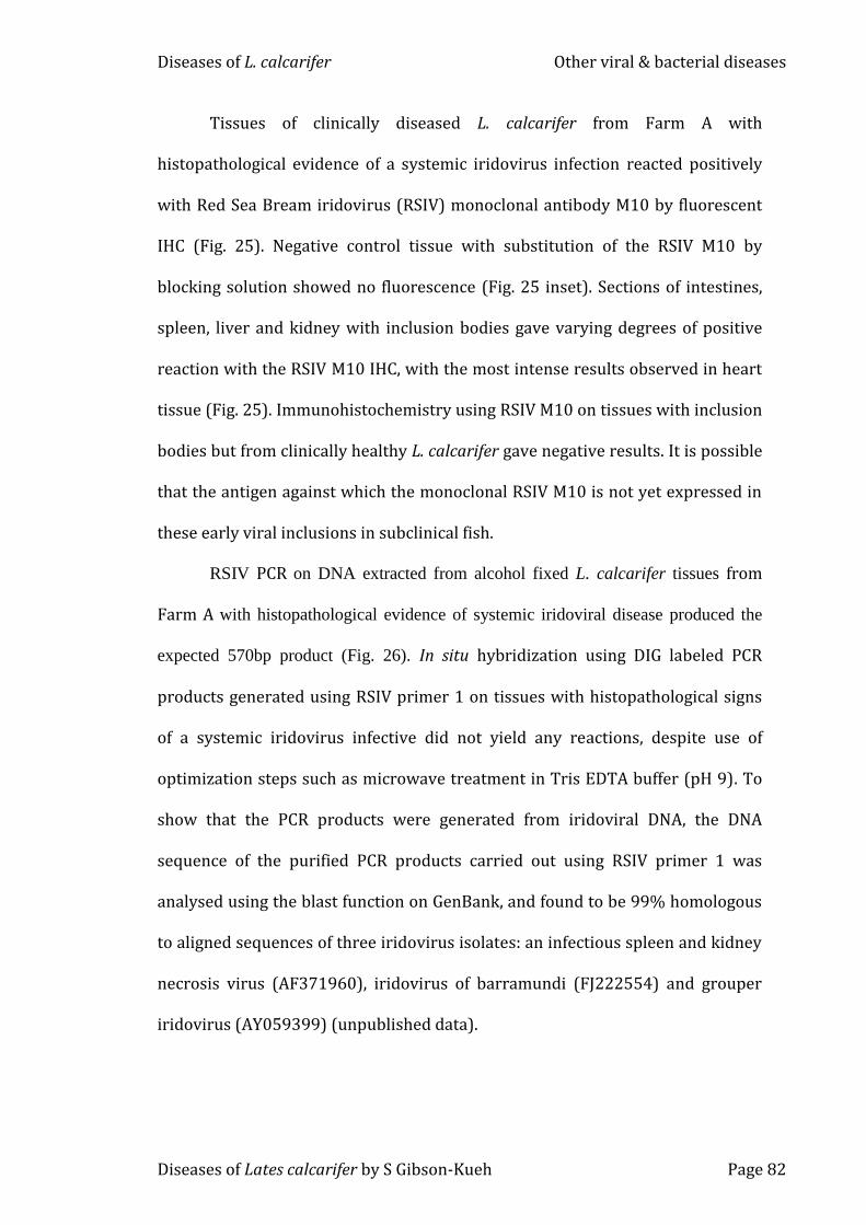
Tissues of clinically diseased L. calcarifer from Farm A with
histopathological evidence of a systemic iridovirus infection reacted positively
with Red Sea Bream iridovirus (RSIV) monoclonal antibody M10 by fluorescent
IHC (Fig. 25). Negative control tissue with substitution of the RSIV M10 by
blocking solution showed no fluorescence (Fig. 25 inset). Sections of intestines,
spleen, liver and kidney with inclusion bodies gave varying degrees of positive
reaction with the RSIV M10 IHC, with the most intense results observed in heart
tissue (Fig. 25). Immunohistochemistry using RSIV M10 on tissues with inclusion
bodies but from clinically healthy L. calcarifer gave negative results. It is possible
that the antigen against which the monoclonal RSIV M10 is not yet expressed in
these early viral inclusions in subclinical fish.
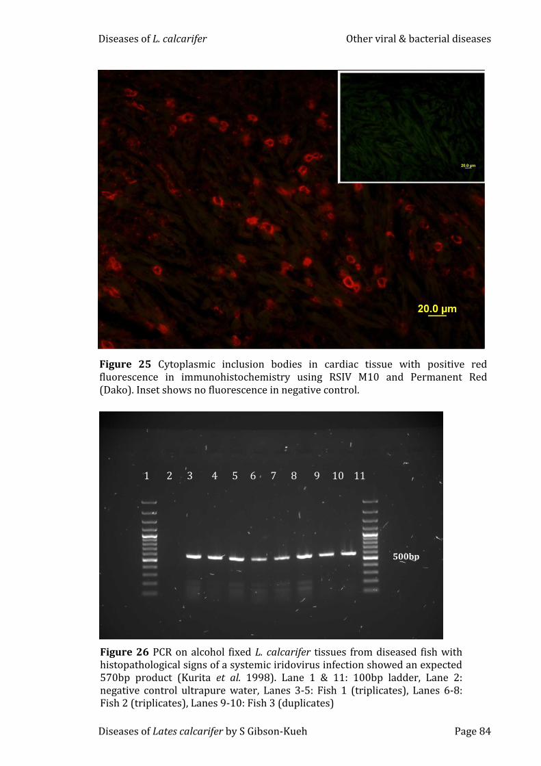
RSIV PCR on DNA extracted from alcohol fixed L. calcarifer tissues from
Farm A with histopathological evidence of systemic iridoviral disease produced the
expected 570bp product (Fig. 26). In situ hybridization using DIG labeled PCR
products generated using RSIV primer 1 on tissues with histopathological signs
of a systemic iridovirus infective did not yield any reactions, despite use of
optimization steps such as microwave treatment in Tris EDTA buffer (pH 9). To
show that the PCR products were generated from iridoviral DNA, the DNA
sequence of the purified PCR products carried out using RSIV primer 1 was
analysed using the blast function on GenBank, and found to be 99% homologous
to aligned sequences of three iridovirus isolates: an infectious spleen and kidney
necrosis virus (AF371960), iridovirus of barramundi (FJ222554) and grouper
iridovirus (AY059399) (unpublished data).
Diseases of L. calcarifer Other viral & bacterial diseases
Diseases of Lates calcarifer by S Gibson-Kueh Page 83
PCR using RSIV primer set 1 (Kurita et al. 1998) on alcohol fixed spleen
tissues of L. calcarifer from Farm A with overt clinical and histopathological signs
of systemic iridoviral disease produced the expected 570bp product (Fig. 26).
Attempts to perform in situ hybridizations (ISH) using DIG-labeled RSIV PCR
products on FFPE L. calcarifer tissues from fish with clinical and
histopathological signs of systemic iridoviral disease from Farm A gave negative
results.
Figure 24 Systemic iridovirus infections in (a) clinically healthy 0.2g L. calcarifer, and (b) clinically diseased 4g L. calcarifer. Tissue degeneration with hydropic changes and cell deaths (D) were more evident in the clinically diseased 4g fish in b. Inclusion bodies (IB) could be observed in both fish.
IB IB
20m
a
IB
IB D
D
20m
b
Diseases of L. calcarifer Other viral & bacterial diseases
Diseases of Lates calcarifer by S Gibson-Kueh Page 84
Figure 26 PCR on alcohol fixed L. calcarifer tissues from diseased fish with histopathological signs of a systemic iridovirus infection showed an expected 570bp product (Kurita et al. 1998). Lane 1 & 11: 100bp ladder, Lane 2: negative control ultrapure water, Lanes 3-5: Fish 1 (triplicates), Lanes 6-8: Fish 2 (triplicates), Lanes 9-10: Fish 3 (duplicates)
1 2 3 4 5 6 7 8 9 10 11
500bp
Figure 25 Cytoplasmic inclusion bodies in cardiac tissue with positive red fluorescence in immunohistochemistry using RSIV M10 and Permanent Red (Dako). Inset shows no fluorescence in negative control.
Diseases of L. calcarifer Other viral & bacterial diseases
Diseases of Lates calcarifer by S Gibson-Kueh Page 85
5.3.2 Viral nervous necrosis (VNN)
Viral nervous necrosis (VNN) was diagnosed histopathologically in 2 to 3 week-
old L. calcarifer with mortalities of up to 100% at Farm A. Mortality was reported
as less than 50% in older fry with VNN at Farm A. Clinical signs observed
included inappetance and pale fish, with or without abnormal neurological signs
(Table 3).
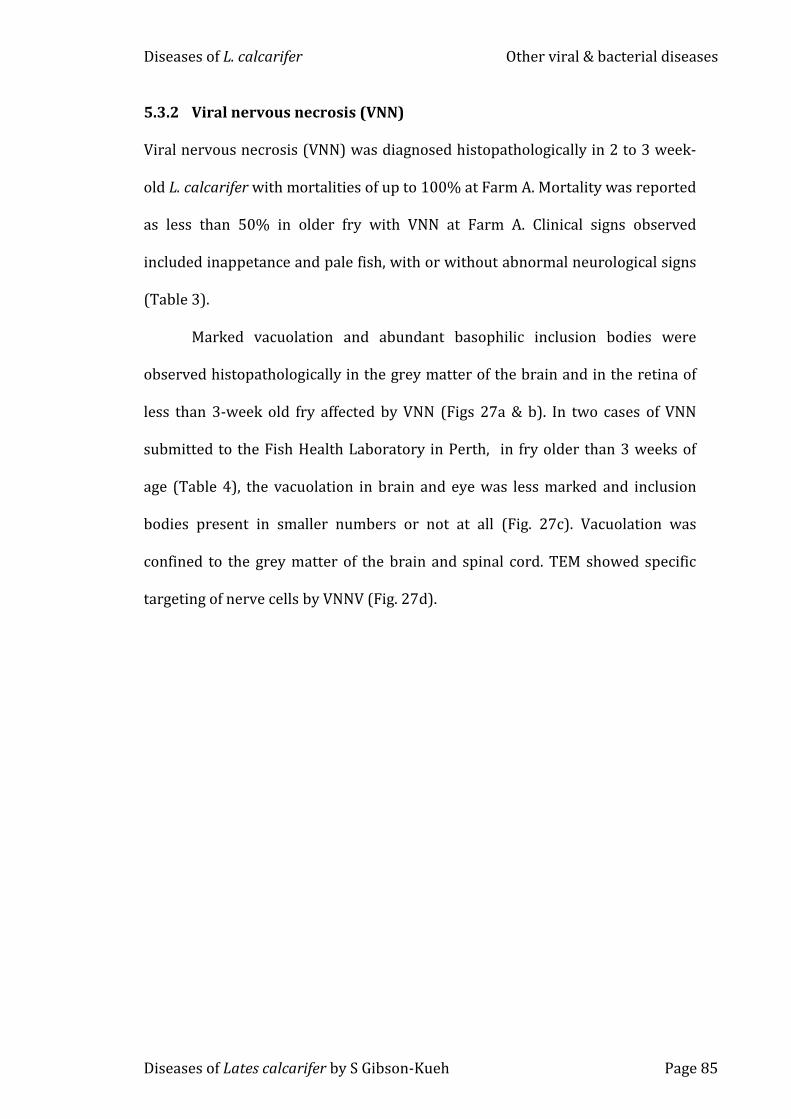
Marked vacuolation and abundant basophilic inclusion bodies were
observed histopathologically in the grey matter of the brain and in the retina of
less than 3-week old fry affected by VNN (Figs 27a & b). In two cases of VNN
submitted to the Fish Health Laboratory in Perth, in fry older than 3 weeks of
age (Table 4), the vacuolation in brain and eye was less marked and inclusion
bodies present in smaller numbers or not at all (Fig. 27c). Vacuolation was
confined to the grey matter of the brain and spinal cord. TEM showed specific
targeting of nerve cells by VNNV (Fig. 27d).
Diseases of L. calcarifer Other viral & bacterial diseases
Diseases of Lates calcarifer by S Gibson-Kueh Page 86
a
200m
Figure 27 (a, b & d) Viral encephalopathy and retinopathy or viral nervous necrosis (VNN) in 12 do L. calcarifer fry from Farm A (a) Severe vacuolation (arrows) in the brain and eye. (b) Vacuoles were often associated with basophilic inclusion bodies (arrows). (c) VNN in 22 do fry submitted to the Fish Health Laboratory in Perth, with mild vacuolation (arrows) and no inclusion bodies. (H&E) (d) Nerve cells with dendritic processes (arrows) and electron dense inclusions (IB) from VNNV infection (TEM). This explains the location of vacuolation in grey matter of the brain where the cell bodies of neurons are located.
b
50m
2m
IB
IB
IB
IB
d
100m
c
Diseases of L. calcarifer Other viral & bacterial diseases
Diseases of Lates calcarifer by S Gibson-Kueh Page 87
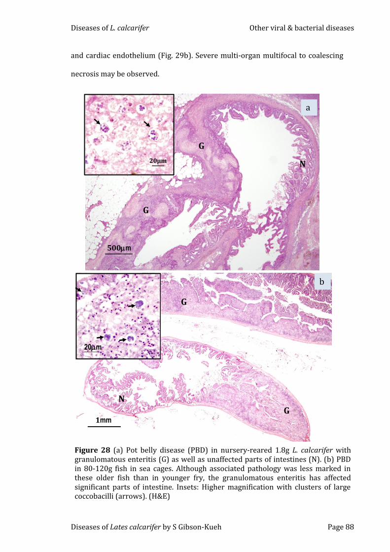
5.3.3 ‘Pot Belly Disease’ (PBD)
Mortality of 90 to 95% was reported in L. calcarifer fry at Farm A, in association
with ‘pot belly disease’ (PBD) (Table 3). PBD was also observed in older L.
calcarifer examined in this study: 1 to 5g L. calcarifer in the land-based nursery
of Farm A, and up to 120g L. calcarifer at two sea cage Farms B and D.
While the granulomatous enteritis tended to be less severe in older fish, it
affected significant portions of the intestine in affected fish (Figs 28a & b).
Clusters of large coccobacilli were observed in both groups of older fish (Figs 28a
& b insets). Remnants of peritonitis with and without the presence of coccobacilli
were observed in some fish. PBD often occurred concurrently with other
diseases such as systemic iridoviral disease in L. calcarifer in sea cages (Table 3).
The contribution of PBD to percentage mortality in grow-out cages in not known,
as it is often complicated by other concurrent diseases.
5.3.4 Streptococcosis
Streptococcosis was observed in 4.5 to 15g, 100 to 500g and up to 3kg L.
calcarifer with cumulative mortality greater than 40 to 50% (Tables 3 & 4). In
fish smaller than 15g, non-specific clinical signs such as darkened bodies,
lethargy and anorexia were observed. In 100 to 200g fish, additional clinical
signs such as exophthalmos, cloudy eyes and abnormal neurological signs were
observed (Table 3). Onset of mortality tended to be more acute, occurring over
several days in smaller fish while larger fish exhibited low grade mortality over
several weeks. Large numbers of gram positive, coccoid-shaped bacteria in pairs
or chains were present within tissues and blood vessels in all organs including
the brain (Fig. 29a) and intracellularly within phagocytic cells in spleen, kidney
Diseases of L. calcarifer Other viral & bacterial diseases
Diseases of Lates calcarifer by S Gibson-Kueh Page 88
and cardiac endothelium (Fig. 29b). Severe multi-organ multifocal to coalescing
necrosis may be observed.
Figure 28 (a) Pot belly disease (PBD) in nursery-reared 1.8g L. calcarifer with granulomatous enteritis (G) as well as unaffected parts of intestines (N). (b) PBD in 80-120g fish in sea cages. Although associated pathology was less marked in these older fish than in younger fry, the granulomatous enteritis has affected significant parts of intestine. Insets: Higher magnification with clusters of large coccobacilli (arrows). (H&E)
G
G
N
a
500m
20µm
N
G
G
b
1mm
20m
Diseases of L. calcarifer Other viral & bacterial diseases
Diseases of Lates calcarifer by S Gibson-Kueh Page 89
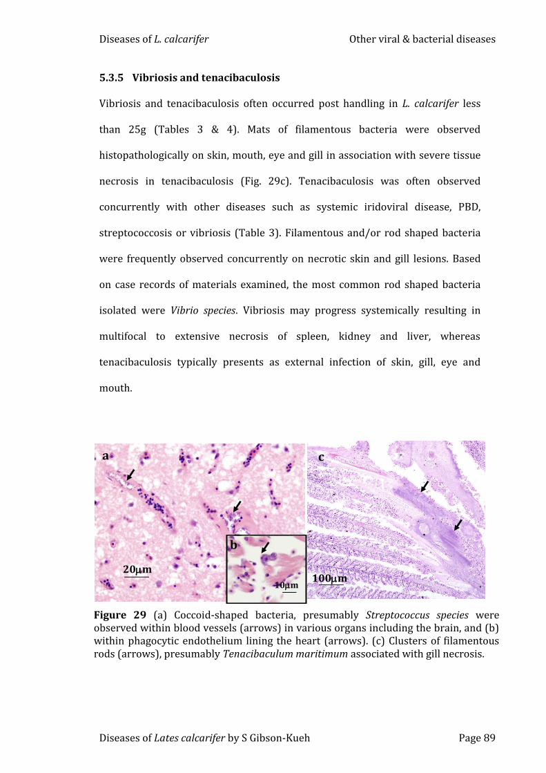
5.3.5 Vibriosis and tenacibaculosis
Vibriosis and tenacibaculosis often occurred post handling in L. calcarifer less
than 25g (Tables 3 & 4). Mats of filamentous bacteria were observed
histopathologically on skin, mouth, eye and gill in association with severe tissue
necrosis in tenacibaculosis (Fig. 29c). Tenacibaculosis was often observed
concurrently with other diseases such as systemic iridoviral disease, PBD,
streptococcosis or vibriosis (Table 3). Filamentous and/or rod shaped bacteria
were frequently observed concurrently on necrotic skin and gill lesions. Based
on case records of materials examined, the most common rod shaped bacteria
isolated were Vibrio species. Vibriosis may progress systemically resulting in
multifocal to extensive necrosis of spleen, kidney and liver, whereas
tenacibaculosis typically presents as external infection of skin, gill, eye and
mouth.
20m
a
10m
b
Figure 29 (a) Coccoid-shaped bacteria, presumably Streptococcus species were observed within blood vessels (arrows) in various organs including the brain, and (b) within phagocytic endothelium lining the heart (arrows). (c) Clusters of filamentous rods (arrows), presumably Tenacibaculum maritimum associated with gill necrosis.
100m
c
Diseases of L. calcarifer Other viral & bacterial diseases
Diseases of Lates calcarifer by S Gibson-Kueh Page 90
5.3.5 Nocardiosis
Nocardiosis was observed in 50 to 120g L. calcarifer at two sea-cage farms: Farm
B in Indonesia and Farm D in Malaysia (Table 3). Moderate to severe
granulomatous lesions were observed in multiple organs including the spleen,
with Fite Faraco, a modified Ziehl Neelsen stain, were observed in some of these
granulomatous lesions (Fig. 30 inset). Filamentous bacteria were present in
larger numbers in the choroid rete of the eye rather than other organs, in one
severely affected fish from Farm B (Fig. 30). In both these two cases of
nocardiosis in L. calcarifer in sea-cage farms, it occurred concurrently with PBD.
Both Farms B and D also culture pompano, Trachinotus blochii, in which
nocardiosis was frequently observed.
Figure 30 Severe granulomatous response (arrows) was observed in the choroid rete of the eye of this severely affected 50g L. calcarifer from Farm B. The branching filamentous rods stained positively with Fite Faraco (arrows), a modified Ziehl Neelsen stain (inset). Granulomatous lesions were also observed in the intestines, liver, gills, heart, kidney and spleen of this fish although with few to no filamentous bacteria (not shown). (H&E)
10m
Diseases of L. calcarifer Other viral & bacterial diseases
Diseases of Lates calcarifer by S Gibson-Kueh Page 91
5.3.6 Chronic peritonitis
Moderate to severe chronic peritonitis was observed in both clinically healthy as
well as diseased fish samples submitted to the Fish Health Laboratory in Perth
(Table 4). Low grade mortalities less than 1% occured 2-4 weeks post stocking.
Affected fish have abdominal swelling and blood stained ascitic fluid. According
to the case history, mortality was associated with increased water temperatures
and feeding rates.
Adequate glycogen stores in the liver and the presence of food in
gastrointestinal tracts were observed in clinically healthy fish with chronic
peritonitis. Severe granulomatous peritonitis was associated with the presence
of mainly gram negative rods, and the occasional gram positive cocci and
coccobacilli. Pericarditis may be observed. The presence of ingested materials
associated with early granuloma formation in the peritoneal cavity suggested
gastrointestinal perforation. Bacterial culture was generally insignificant. The
aetiology of chronic peritonitis is uncertain.
Diseases of L. calcarifer Other viral & bacterial diseases
Diseases of Lates calcarifer by S Gibson-Kueh Page 92
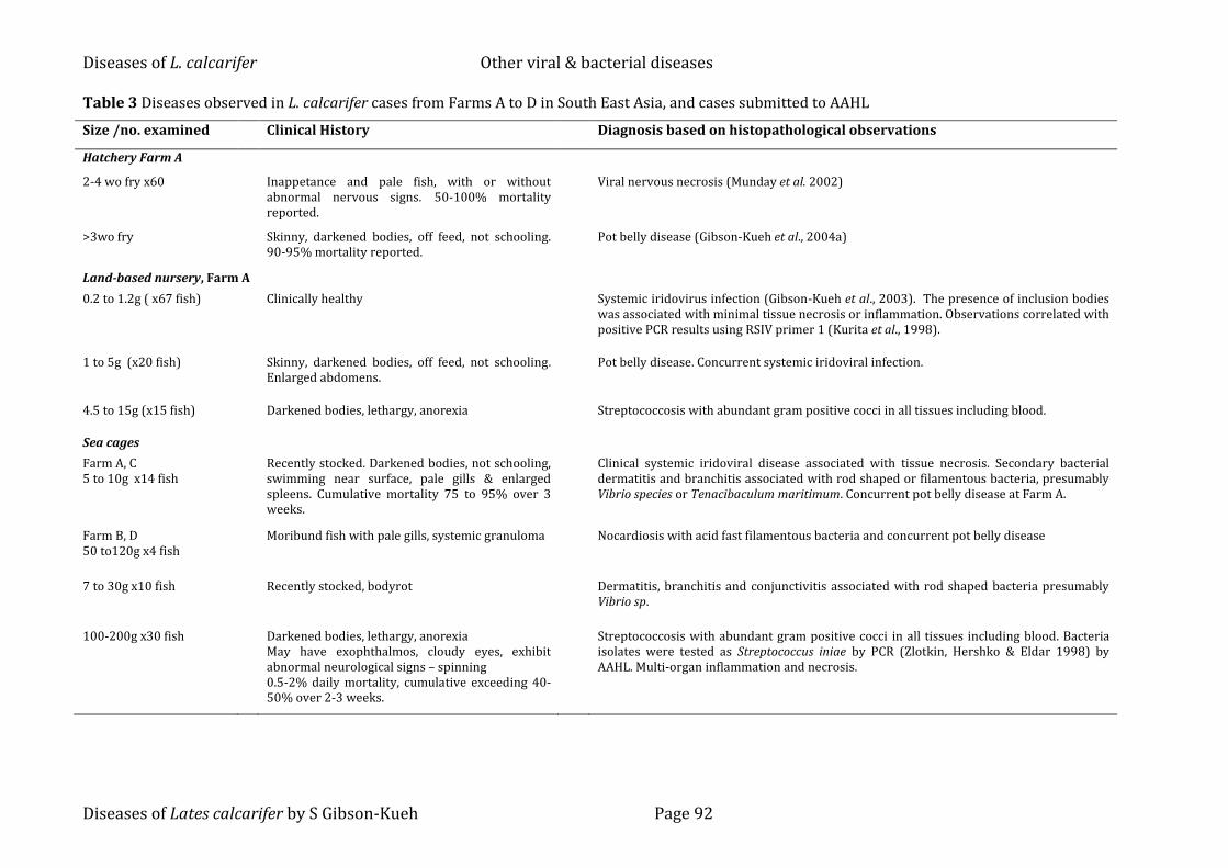
Table 3 Diseases observed in L. calcarifer cases from Farms A to D in South East Asia, and cases submitted to AAHL
Size /no. examined Clinical History Diagnosis based on histopathological observations
Hatchery Farm A
2-4 wo fry x60
Inappetance and pale fish, with or without abnormal nervous signs. 50-100% mortality reported.
Viral nervous necrosis (Munday et al. 2002)
>3wo fry Skinny, darkened bodies, off feed, not schooling. 90-95% mortality reported.
Pot belly disease (Gibson-Kueh et al., 2004a)
Land-based nursery, Farm A
0.2 to 1.2g ( x67 fish) Clinically healthy Systemic iridovirus infection (Gibson-Kueh et al., 2003). The presence of inclusion bodies was associated with minimal tissue necrosis or inflammation. Observations correlated with positive PCR results using RSIV primer 1 (Kurita et al., 1998).
1 to 5g (x20 fish) Skinny, darkened bodies, off feed, not schooling. Enlarged abdomens.
Pot belly disease. Concurrent systemic iridoviral infection.
4.5 to 15g (x15 fish) Darkened bodies, lethargy, anorexia Streptococcosis with abundant gram positive cocci in all tissues including blood.
Sea cages
Farm A, C 5 to 10g x14 fish
Recently stocked. Darkened bodies, not schooling, swimming near surface, pale gills & enlarged spleens. Cumulative mortality 75 to 95% over 3 weeks.
Clinical systemic iridoviral disease associated with tissue necrosis. Secondary bacterial dermatitis and branchitis associated with rod shaped or filamentous bacteria, presumably Vibrio species or Tenacibaculum maritimum. Concurrent pot belly disease at Farm A.
Farm B, D 50 to120g x4 fish
Moribund fish with pale gills, systemic granuloma Nocardiosis with acid fast filamentous bacteria and concurrent pot belly disease
7 to 30g x10 fish
Recently stocked, bodyrot Dermatitis, branchitis and conjunctivitis associated with rod shaped bacteria presumably Vibrio sp.
100-200g x30 fish
Darkened bodies, lethargy, anorexia May have exophthalmos, cloudy eyes, exhibit abnormal neurological signs – spinning 0.5-2% daily mortality, cumulative exceeding 40-50% over 2-3 weeks.
Streptococcosis with abundant gram positive cocci in all tissues including blood. Bacteria isolates were tested as Streptococcus iniae by PCR (Zlotkin, Hershko & Eldar 1998) by AAHL. Multi-organ inflammation and necrosis.
Diseases of L. calcarifer Other viral & bacterial diseases
Diseases of Lates calcarifer by S Gibson-Kueh Page 93
Table 4 Diseases observed in L. calcarifer cases received at the Fish Health Laboratory, WA Department of Fisheries 2002-2008
Size /no. examined Clinical History Diagnosis based on histopathological observations
Land-based nursery >3 wo fry x60 2 cases
Abnormal swimming behavior. High mortality Viral nervous necrosis
Land-based nursery (marine) 0.8g x7 fish 1 case
Stocked as 0.15g fry from a local hatchery, with no significant post transfer losses. 60% mortality overnight followed by low grade mortality.
Tenacibaculosis. Severe oral epidermal ulcerations on mandible and necrotic gills were observed in association with filamentous rods. Mucoid cell metaplasia observed in gills. Other observations include presence of ingesta in stomach and adequate hepatic glycogen stores, supporting an acute disease onset.
Land-based recirculation tanks (marine) 5g x 40 fish 6 cases
Sea-cages >10g x6 fish 1 case
Low grade Mortalities (<1%) 2-4 weeks post stocking. Abdominal swelling and blood stained ascitic fluid. Affected fish often still have food in gastrointestinal tracts. One case history noted that mortality was associated with increased water temperatures and feeding rates.
Chronic peritonitis, uncertain aetiology. May present as chronic serositis of abdominal organs to severe granulomatous peritonitis with presence of mainly gram negative rods, and few gram positive cocci and coccobacilli in both clinically healthy as well as diseased fish. Pericarditis observed. Adequate glycogen stores in liver. The presence of ingested materials associated with early granuloma formation in peritoneal cavity suggested gastrointestinal perforation. Bacterial culture generally insignificant.
Sea-cages 200g x2 fish 1 case
>50% mortality over 3 days, peaking on day 2. Affected fish had pale gills, finrot and white skin patches.
Streptococcosis. Severe bacteriaemia with abundant coccoid shaped bacteria in spleen and kidney in association with severe necrosis. Acute branchitis. Bacteria isolated were identified as Streptococcus iniae.
Freshwater cages
350 to 500g x6 fish 1 case
Index case in 2.5 to 3kg fish, with cumulative mortality of 50%. Disease was subsequently observed in 350-500g fish in vicinity of the larger ones. Affected fish were lethargic and had exophthalmia, empty gastrointestinal tracts and enlarged spleens.
Streptococcosis. The publication by Creeper & Buller 2006 contains the full case details. Bacteria isolated were identified as S. iniae by biochemical methods. Affected fish had severe bacteraemia with abundant coccoid shaped bacteria in spleen and kidney, in association with severe necrosis. One fish had severe necrotizing branchitis with mats of filamentous bacteria, presumably Flavobacterium columnare.
Sea cages 5 to 10g x 11 fish 1 case
Recently graded and develop redness in eyes and base of operculum the next day.
Tenacibaculosis and Vibriosis. Necrosis of tissue on head/mandible and gills associated with filamentous bacteria in three fish, presumably Tenacibaculum maritimum. Rest of fish had dermatitis associated with rod shape bacteria, presumably Vibrio species. Case records showed that Vibrio species was isolated from gills while bacteria were not isolated from internal organs.
Diseases of L. calcarifer Other viral & bacterial diseases
Diseases of Lates calcarifer by S Gibson-Kueh Page 94
5.4 Discussion
Although systemic iridoviral disease has been previously reported in many
marine fish species, this study forms the first report of this disease in L.
calcarifer. Our findings of systemic iridoviral infection in clinically healthy fish in
the land-based nursery of Farm A level suggested that the infection occurred
before stocking into sea cages, and did not originate from wild fish or older fish
in adjacent sea cages. The possibility of a systemic iridovirus infection in
clinically healthy fish reiterates the importance of routinely screening fish for
subclinical infections, and will impact any vaccination programs.
Attempts to perform in situ hybridizations (ISH) using DIG-labeled RSIV
PCR products on FFPE L. calcarifer tissues with histopathological signs of
systemic iridoviral disease from Farm A gave negative results. PCR products
were generated from and tested on tissues of fish with clinical systemic iridoviral
disease from farm A. It is suspected that ISH may have been affected by the nitric
acid decalcification process used on L. calcarifer tissues, which may result in
denatured DNA (Kucukodaci, Haholu, Sucullu & Baloglu 2012). Although
immunohistochemistry gave positive results using the RSIV M10, the intensity of
the antibody reaction varied between tissues such as intestines, liver, spleen and
kidney with abundant inclusion bodies, and was most intense in heart tissues.
This may be related to the prolonged exposure of different tissues in specimens
fixed as whole fish to histoprocessing chemicals and masking of antigen (Webster,
Miller, DuSold & Ramos-Vara 2010).
Viral nervous necrosis can be difficult to diagnose histopathologically in
cases where it is associated with few vacuolations and an absence of inclusion
bodies. In such cases, diagnosis should be verified by PCR (Chang et al. 1997).
Diseases of L. calcarifer Other viral & bacterial diseases
Diseases of Lates calcarifer by S Gibson-Kueh Page 95
In a previous publication of PBD, the causative agent was identified as a
large gram negative coccobacillus, tested positive by immunohistochemistry
using polyclonal antibodies to Edwardsiella ictaluri (Gibson-Kueh et al. 2004a).
Culture and PCR detection methods for the bacteria causative agent for pot belly
disease have been established by Global Aquatic Animal Health laboratory,
Merck Animal Health Singapore, although this has not been published and hence
is not available for use at diagnostic laboratories
Diseases of Lates calcarifer by S Gibson-Kueh Page 137
Appendix 1.1 – Response to questionaire from Farm A Farm name : Farm A (sea-cage farm with own broodstock, hatchery and nursery) Location : Island A off Indonesia Tel : -withheld- Email : -withheld- Objective of questionnaire: Collect information on the main diseases observed in Asian seabass/barramundi production Date questionnaire filled up: 10 Dec 2008 Number of years farm in operation: 10 years on pilot and commercial scale. Nearest fish farm(s) in km (if any): floating cages for Pomfret 10km away Fish species farmed other than barramundi: Epinephelus fuscoguttatus, Lutjanus argentimaculatus, Rachycentrum canadum on pilot
scale Water source: Sea water Salinity of water used: Full salinity 34ppt Number of tanks/ponds/cages on farm: Information not supplied Number of Asian seabass on farm currently of each age group: All from larvae to market size (3kg) and captive broodstocks Size of ponds or cages in metres: Cages 12 and 18 meters diameter, and 10 meter depth. Stocking density of fry in hatchery: Mesocosm technique 10-30/liter Age & size of fry in hatchery: All sizes from day 0 to day 30 Feed type & rates in hatchery: Rotifer artemia weaning feed and pellet Stocking density of fry in nursery: 2-5 kg/m3 Age & size of fry in nursery: From day 30 to 10-20g Feed types & rates in nursery: Pellet of different sizes; Feeding rates from 10% to 3% Age & size of fry or fingerlings at stocking in growout facilities: 10-30g all vaccinated by injection against Streptococcus Initial stocking density at grow-out: 50.000-100-000 in 12 meter circular cages 10.000 to 20.000 in square cages Time from stocking in growout to first grading: Graded prior to stocking. Fish from one cage may be split into different cages as fish
grows
Diseases of L. calcarifer
Diseases of Lates calcarifer by S Gibson-Kueh Page 138
Frequency of grading: No grading from 100g to 3kg Frequency of net changes: 5-8 months Water exchange rate in tank/pond: 100% per hour at nursery stage Temperature range of culture water: 28-29oC all over the year If water is recirculated, how is water managed or treated? Through flow water system Type of vaccination used if any: Routine Streptococcus vaccination When do diseases occur in the culture cycle? Larvae, juveniles and adults
Describe the main diseases observed? VNN, Iridovirus, Big belly (synonymous with pot belly), Tenacibaculum maritimum, Benedenia, Streptococcus
What are the main disease agents known to occur on farm, as diagnosed by laboratory? As above What percentage losses are associated with the main diseases observed? 100% (VNN) 75% (irido) variable on others. Are the following disease conditions experienced on farm? Yes/No Please indicate percentage affected or associated losses experienced? Swimbladder hyperinflation: Associated with VNN and Big Belly Scale loss: Not frequent and occurring after rough current or rough handling Enlarged Abdomen: Big belly disease Unexplained low grade mortality: Not observed Sudden overnight mortality with unknown cause : Not observed If not applicable, please write NA. If unknown, please indicate.
Contact for Human Research Ethics Committee: Dr Erich von Dietze Tel: 9360 6677, Email: [email protected]
Contact for Chief Investigator/co-investigators: Dr Philip Nicholls: 618 9360 2599, Email: [email protected] Dr Brian Jones: 618 9368 3649, Email: [email protected] Dr Susan Kueh: 0430 100 931, Email: [email protected]
Diseases of L. calcarifer
Diseases of Lates calcarifer by S Gibson-Kueh Page 139
Appendix 1.2 - Response to questionaire from Farm B
Farm name : Farm B (seacage grow-out farm with own nursery, buys in fry) Location : Island B off Indonesia Tel : -withheld- Email : -withheld- Objective of questionnaire: Collect information on the main diseases observed in Asian seabass/barramundi production Date questionnaire filled up: 02 May 2008 Number of years farm in operation: 4-5 years Nearest fish farm(s) in km (if any): Nearest fish farms 200 to 300m away Fish species farmed other than barramundi: Crimson snapper, pompano, tiger grouper Water source: Sea water Salinity of water used: Full salinity 30-34ppt Number of tanks/ponds/cages on farm: 640 wooden framed and 4 steel cages Number of Asian seabass on farm currently of each age group: 1.2 million fish as on 2008 Size of ponds or cages in metres: Wooden cages 3.25 x 3.25 x 2.5m Stocking density of fry in hatchery: NA Age & size of fry in hatchery: NA Feed type & rates in hatchery: NA Stocking density of fry in nursery: 2-3kg biomass/metric tonne Age & size of fry in nursery: From day 30 to 2 to 8g Feed types & rates in nursery: Pellet of different sizes as fish grows Age & size of fry or fingerlings at stocking in growout facilities: 2 to 8g fish Initial stocking density at grow-out: Information not supplied Time from stocking in growout to first grading: Information not supplied
Diseases of L. calcarifer
Diseases of Lates calcarifer by S Gibson-Kueh Page 140
Frequency of grading: Information not supplied Frequency of net changes: Every week for small fish to every 2 weeks for larger fish in nets with larger mesh size Water exchange rate in tank/pond: 100% per hour at nursery stage Temperature range of culture water: 28-30oC all over the year If water is recirculated, how is water managed or treated? NA Type of vaccination used if any: Routine Streptococcus vaccination via intraperitonal route When do diseases occur in the culture cycle? Within first month of stocking at sea Describe the main disease signs observed? Scale loss, gulping at water surface and darkened fish What are the main disease agents known to occur on farm, as diagnosed by laboratory? Tenacibaculum maritimum & Streptococcus sp. What percentage losses are associated with the main diseases observed? 10-20% mortality Are the following disease conditions experienced on farm? Yes/No Please indicate percentage affected or associated losses experienced? Swimbladder hyperinflation: Not observed Scale loss: 10% mortality Enlarged Abdomen: Less than 1% Unexplained low grade mortality: Not observed Sudden overnight mortality with unknown cause : Not observed If not applicable, please write NA. If unknown, please indicate.
Contact for Human Research Ethics Committee: Dr Erich von Dietze Tel: 9360 6677, Email: [email protected]
Contact for Chief Investigator/co-investigators: Dr Philip Nicholls: 618 9360 2599, Email: [email protected] Dr Brian Jones: 618 9368 3649, Email: [email protected] Dr Susan Kueh: 0430 100 931, Email: [email protected]