38
Diseases of the Eye Casey Conway Jeannie Stall , R.V.T. & ??? Credits : Clip Art graphics/Google images
| Date post: | 15-Dec-2015 |
| Category: |
Documents |
| Upload: | rita-helms |
| View: | 218 times |
| Download: | 2 times |
Diseases of the EyeCasey Conway
Jeannie Stall , R.V.T. & ??? Credits : Clip Art graphics/Google images
The Eye•CDCA has great picture of a cross section.
(hint)•Most highly developed of all senses•Most pets can live quality lives with a vision
loss•Proper diagnosis, quick treatment – Essential!•3 main categories of eye diseases:
▫Accessory structures▫The globe▫The retina and the neural pathways
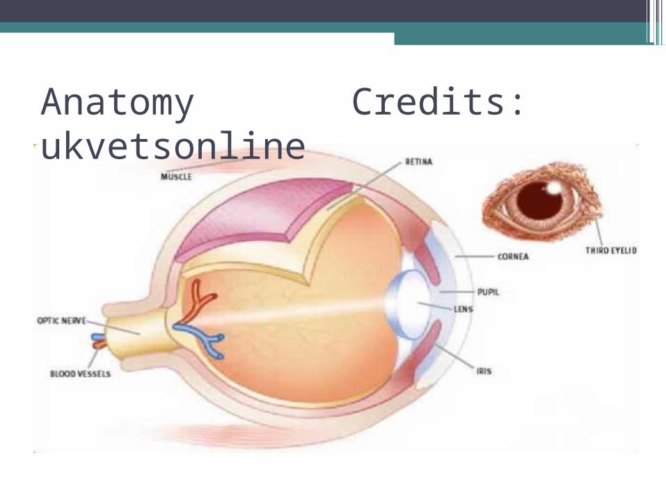
Anatomy Credits: ukvetsonline
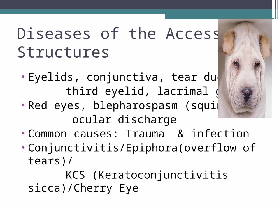
Diseases of the Accessory Structures
•Eyelids, conjunctiva, tear ducts, third eyelid, lacrimal glands•Red eyes, blepharospasm (squinting), ocular discharge•Common causes: Trauma & infection •Conjunctivitis/Epiphora(overflow of
tears)/ KCS (Keratoconjunctivitis
sicca)/Cherry Eye
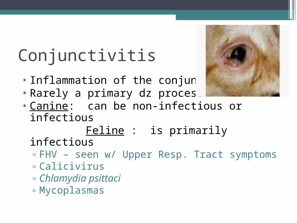
Conjunctivitis • Inflammation of the conjunctiva• Rarely a primary dz process • Canine: can be non-infectious or infectious Feline : is primarily infectious
▫FHV – seen w/ Upper Resp. Tract symptoms▫Calicivirus▫Chlamydia psittaci▫Mycoplasmas
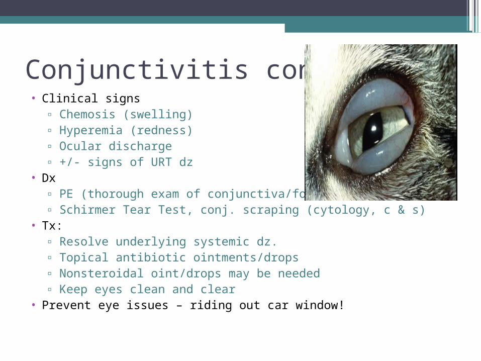
Conjunctivitis cont’d• Clinical signs
▫ Chemosis (swelling) ▫ Hyperemia (redness)▫ Ocular discharge▫ +/- signs of URT dz
• Dx ▫ PE (thorough exam of conjunctiva/foreign body) ▫ Schirmer Tear Test, conj. scraping (cytology, c & s)
• Tx: ▫ Resolve underlying systemic dz. ▫ Topical antibiotic ointments/drops▫ Nonsteroidal oint/drops may be needed ▫ Keep eyes clean and clear
• Prevent eye issues – riding out car window!
Epiphora • Overflow of tears
▫ Overproduction (pain or irritation) ▫ Faulty drainage: (lacrimal duct blocked by
swelling or trauma)
• Clinical Signs ▫ Watering of the eye▫ Wet facial hair▫ Secondary bacterial infection of facial skin▫ Discoloration of the facial hair
Epiphora cont’d……. • Dx:
▫ Eye exam▫ Fluorescein dye▫ Dacryocystorhinography ( Radiography of
nasolacrimal duct )• Tx:
Resolve primary cause of pain & irritation /Flush lacrimal ducts / Surgery / Topical abx/ Trim
hairs
Eyelid Diseases•At the base of each eyelash is a sebaceous gland Hordeolum: Meibomian gland abscess, usually caused by a staph. infection Chalazion: When inflammation involves meibomian glands & granulation occurs •Eyelid neoplasms : Older animals/ most are benign (cat usually malignant),
Squamous cell carcinoma most common tumor
Blepharitis: Swelling of the eyelids• Causes:
▫ Allergens ▫ Nutritional Deficiencies▫ Viral▫ Dermatitis from any cause
• Clinical signs ▫ Generalized swelling of lid ▫ Periocular pruritis (itchy eyes)▫ Periocular alopecia ( hairloss)▫ Rubbing of the eyes
Blepharitis Cont.• Dx:
▫ Eye exam▫ Skin scrape▫ Fungal culture▫ Bacterial cultures
• Tx: warm compresses▫ Express hordeolum(staph infection) ▫Surg. removal of chalazion (granulation tissue)▫ Topical abx or systemic abx ▫ Corticosteroids ( Prednisolone)▫ Antifungals ( Conofite or Tresaderm)
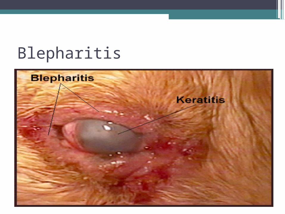
Blepharitis
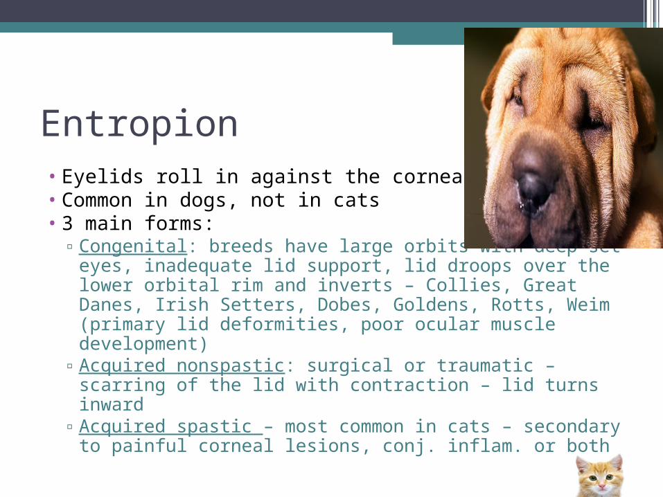
Entropion • Eyelids roll in against the cornea • Common in dogs, not in cats• 3 main forms:
▫Congenital: breeds have large orbits with deep-set eyes, inadequate lid support, lid droops over the lower orbital rim and inverts – Collies, Great Danes, Irish Setters, Dobes, Goldens, Rotts, Weim (primary lid deformities, poor ocular muscle development)
▫Acquired nonspastic: surgical or traumatic – scarring of the lid with contraction – lid turns inward
▫Acquired spastic – most common in cats – secondary to painful corneal lesions, conj. inflam. or both
Entropion
Entropion
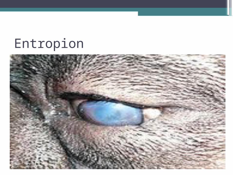
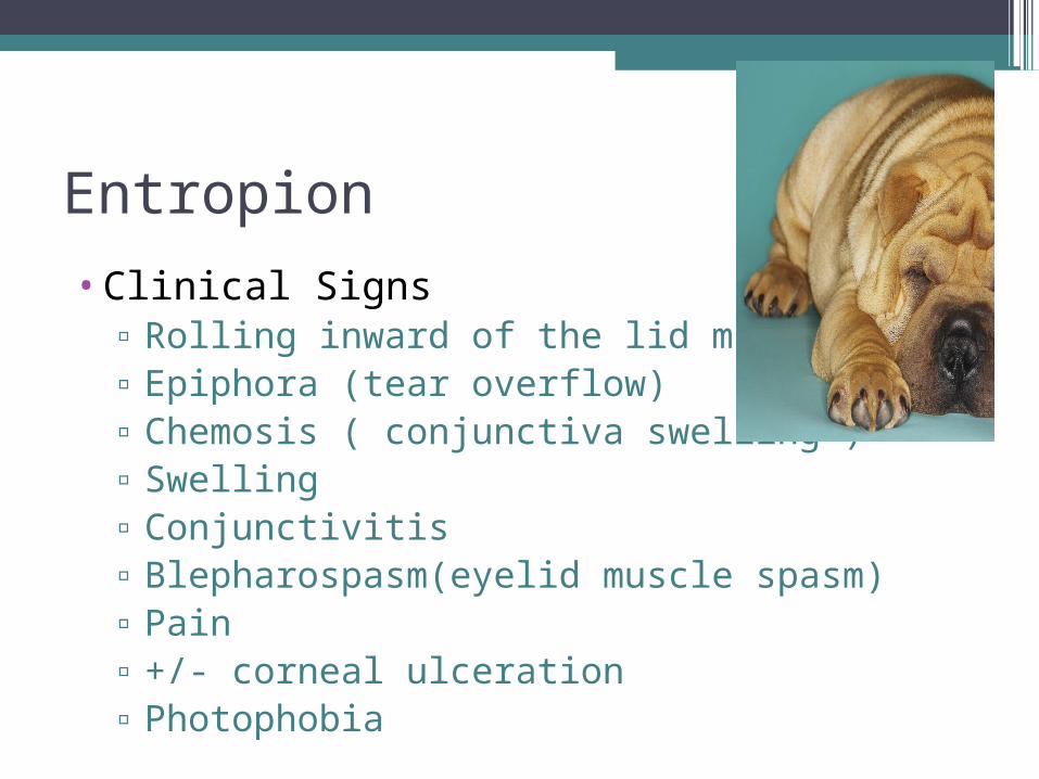
•Clinical Signs▫ Rolling inward of the lid margin(s)▫ Epiphora (tear overflow)▫ Chemosis ( conjunctiva swelling )▫ Swelling▫ Conjunctivitis▫ Blepharospasm(eyelid muscle spasm)▫ Pain▫ +/- corneal ulceration▫ Photophobia

Entropion Con’t……
•Dx: ▫Observe lids interaction with globe▫Eye exam while awake
•Tx: Surgical correction
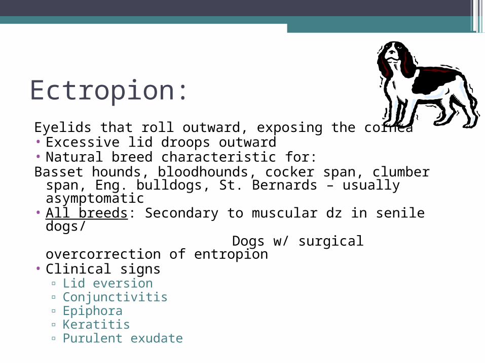
Ectropion:Eyelids that roll outward, exposing the cornea• Excessive lid droops outward• Natural breed characteristic for: Basset hounds, bloodhounds, cocker span, clumber
span, Eng. bulldogs, St. Bernards – usually asymptomatic
• All breeds: Secondary to muscular dz in senile dogs/ Dogs w/ surgical overcorrection of
entropion• Clinical signs
▫ Lid eversion▫ Conjunctivitis▫ Epiphora▫ Keratitis▫ Purulent exudate
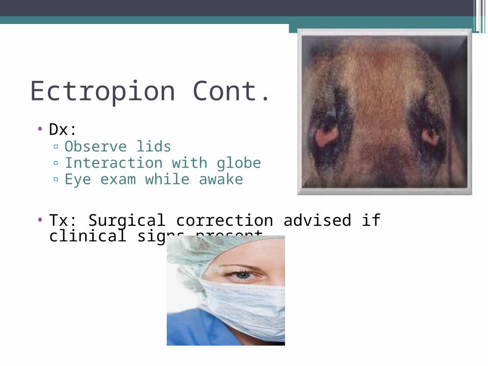
Ectropion Cont.• Dx:
▫ Observe lids▫ Interaction with globe▫ Eye exam while awake
• Tx: Surgical correction advised if clinical signs present
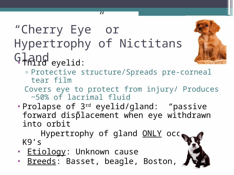
“Cherry Eye” orHypertrophy of Nictitans Gland•Third eyelid:
▫ Protective structure/Spreads pre-corneal tear film
Covers eye to protect from injury/ Produces ~50% of lacrimal fluid
•Prolapse of 3rd eyelid/gland: “passive forward displacement when eye withdrawn into orbit”
Hypertrophy of gland ONLY occurs in K9’s• Etiology: Unknown cause • Breeds: Basset, beagle, Boston, cocker
Cherry Eye
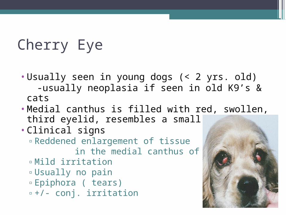
•Usually seen in young dogs (< 2 yrs. old) -usually neoplasia if seen in old K9’s & cats•Medial canthus is filled with red, swollen,
third eyelid, resembles a small cherry•Clinical signs
▫Reddened enlargement of tissue in the medial canthus of the eye▫Mild irritation▫Usually no pain▫Epiphora ( tears)▫+/- conj. irritation
Cherry Eye continued…..•Dx:
▫ Clinical signs▫ Predisposed breed▫ Rule out tumor (usually older dogs & cats)
•Tx: ▫ Surgical replacement of the gland, Tuck
sutured▫ Avoid excision – predisposes to KCS – only excise in cases of neoplasia
•W/O sx., corneal damage can occur & may affect
vision

Cherry Eye
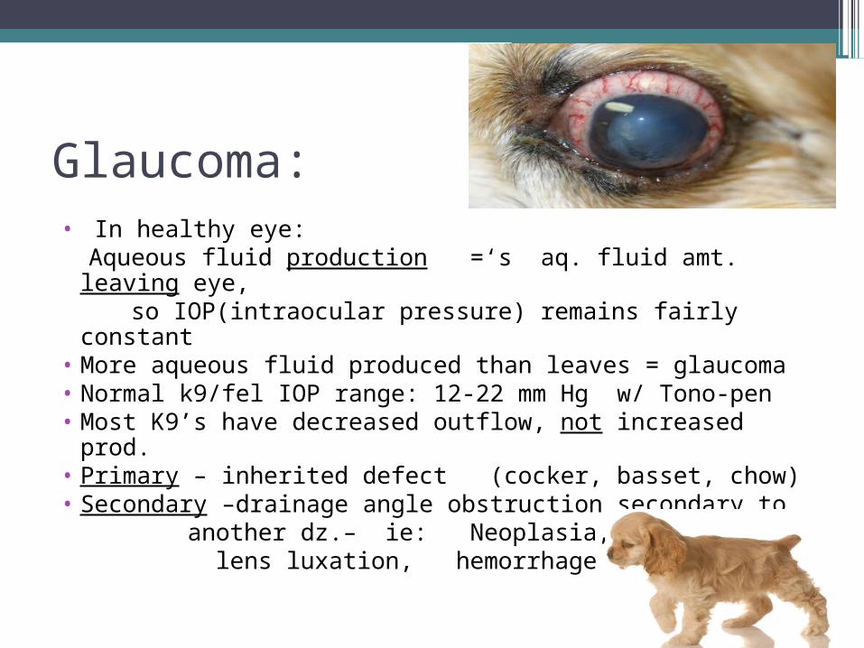
Glaucoma: • In healthy eye: Aqueous fluid production =‘s aq. fluid amt. leaving eye, so IOP(intraocular pressure) remains fairly constant• More aqueous fluid produced than leaves = glaucoma• Normal k9/fel IOP range: 12-22 mm Hg w/ Tono-pen • Most K9’s have decreased outflow, not increased prod. • Primary – inherited defect (cocker, basset, chow)• Secondary –drainage angle obstruction secondary to another dz.– ie: Neoplasia, uveitis, lens luxation, hemorrhage
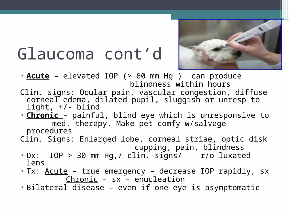
Glaucoma cont’d• Acute – elevated IOP (> 60 mm Hg ) can produce blindness within hours Clin. signs: Ocular pain, vascular congestion, diffuse
corneal edema, dilated pupil, sluggish or unresp to light, +/- blind
• Chronic – painful, blind eye which is unresponsive to med. therapy. Make pet comfy w/salvage proceduresClin. Signs: Enlarged lobe, corneal striae, optic disk cupping, pain, blindness • Dx: IOP > 30 mm Hg,/ clin. signs/ r/o luxated lens• Tx: Acute – true emergency – decrease IOP rapidly, sx Chronic – sx – enucleation• Bilateral disease – even if one eye is asymptomatic
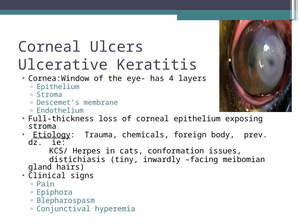
Corneal UlcersUlcerative Keratitis• Cornea:Window of the eye- has 4 layers
▫ Epithelium▫ Stroma▫ Descemet’s membrane▫ Endothelium
• Full-thickness loss of corneal epithelium exposing stroma• Etiology: Trauma, chemicals, foreign body, prev. dz.
ie: KCS/ Herpes in cats, conformation issues, distichiasis (tiny, inwardly –facing meibomian gland
hairs)• Clinical signs
▫ Pain▫ Epiphora▫ Blepharospasm▫ Conjunctival hyperemia
Corneal Ulcers
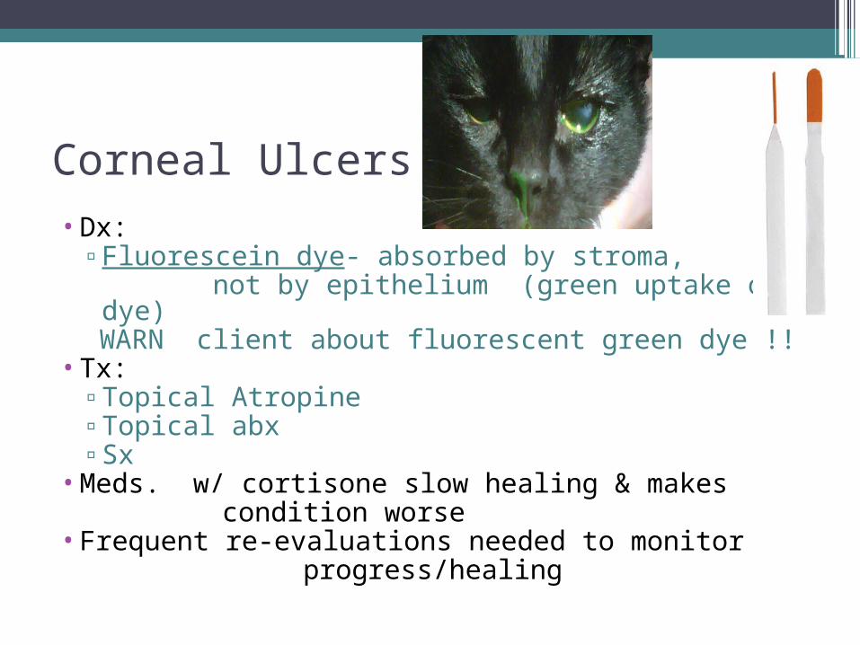
•Dx: ▫Fluorescein dye- absorbed by stroma, not by epithelium (green uptake of dye) WARN client about fluorescent green dye !!
•Tx: ▫Topical Atropine▫Topical abx▫Sx
•Meds. w/ cortisone slow healing & makes condition worse•Frequent re-evaluations needed to monitor progress/healing
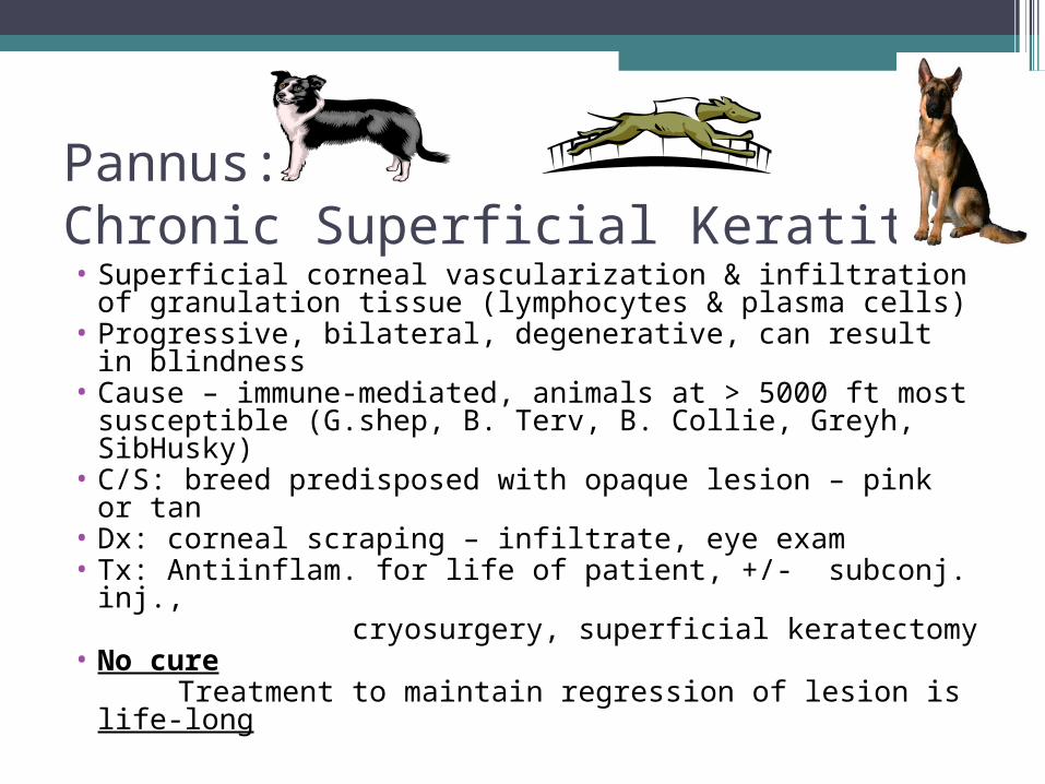
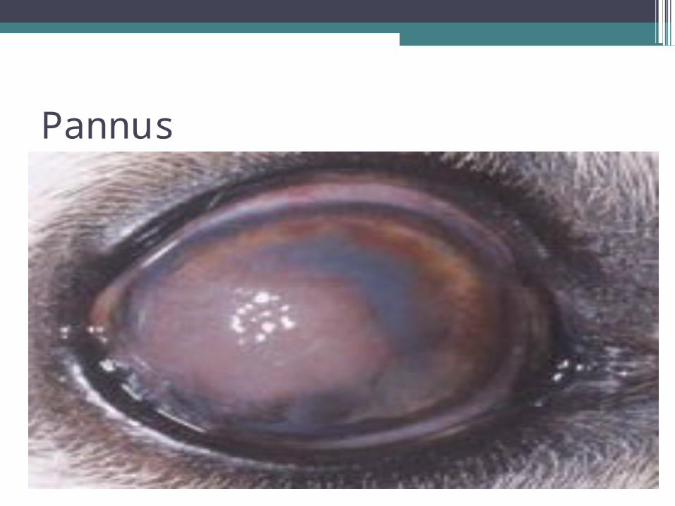
Pannus: Chronic Superficial Keratitis • Superficial corneal vascularization & infiltration of
granulation tissue (lymphocytes & plasma cells) • Progressive, bilateral, degenerative, can result in
blindness• Cause – immune-mediated, animals at > 5000 ft most
susceptible (G.shep, B. Terv, B. Collie, Greyh, SibHusky)
• C/S: breed predisposed with opaque lesion – pink or tan
• Dx: corneal scraping – infiltrate, eye exam• Tx: Antiinflam. for life of patient, +/- subconj. inj., cryosurgery, superficial keratectomy • No cure Treatment to maintain regression of lesion is life-
long
Pannus
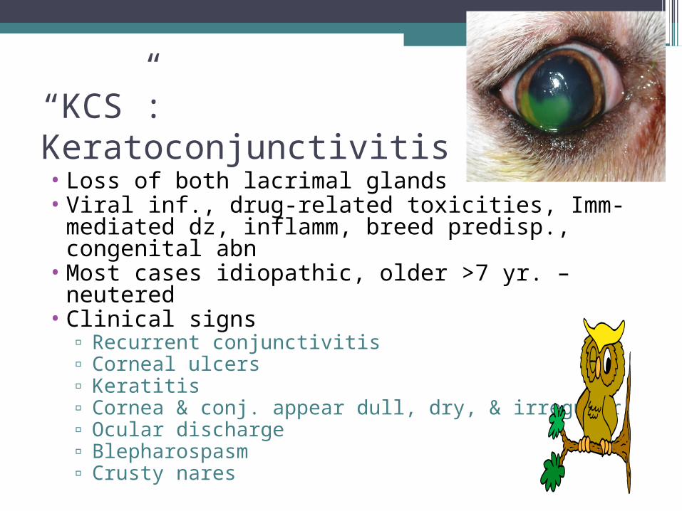
“KCS”:Keratoconjunctivitis Sicca• Loss of both lacrimal glands• Viral inf., drug-related toxicities, Imm-
mediated dz, inflamm, breed predisp., congenital abn
• Most cases idiopathic, older >7 yr. – neutered• Clinical signs
▫ Recurrent conjunctivitis▫ Corneal ulcers▫ Keratitis▫ Cornea & conj. appear dull, dry, & irregular▫ Ocular discharge▫ Blepharospasm▫ Crusty nares
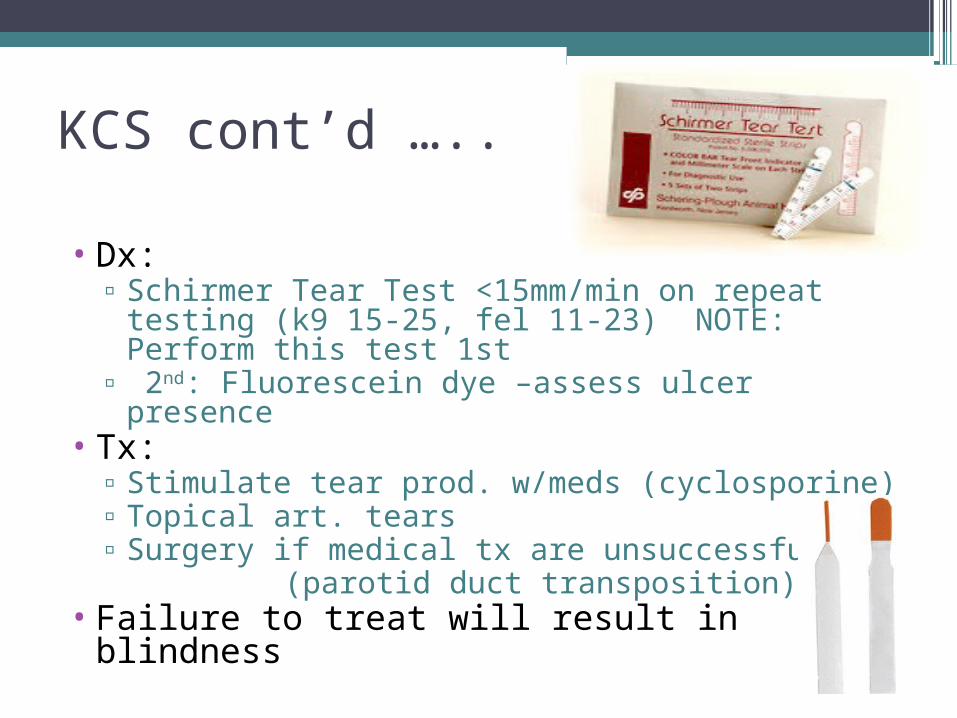
KCS cont’d …..
• Dx: ▫ Schirmer Tear Test <15mm/min on repeat
testing (k9 15-25, fel 11-23) NOTE: Perform this test 1st
▫ 2nd: Fluorescein dye –assess ulcer presence• Tx:
▫ Stimulate tear prod. w/meds (cyclosporine)▫ Topical art. tears▫ Surgery if medical tx are unsuccessful (parotid duct transposition)
• Failure to treat will result in blindness
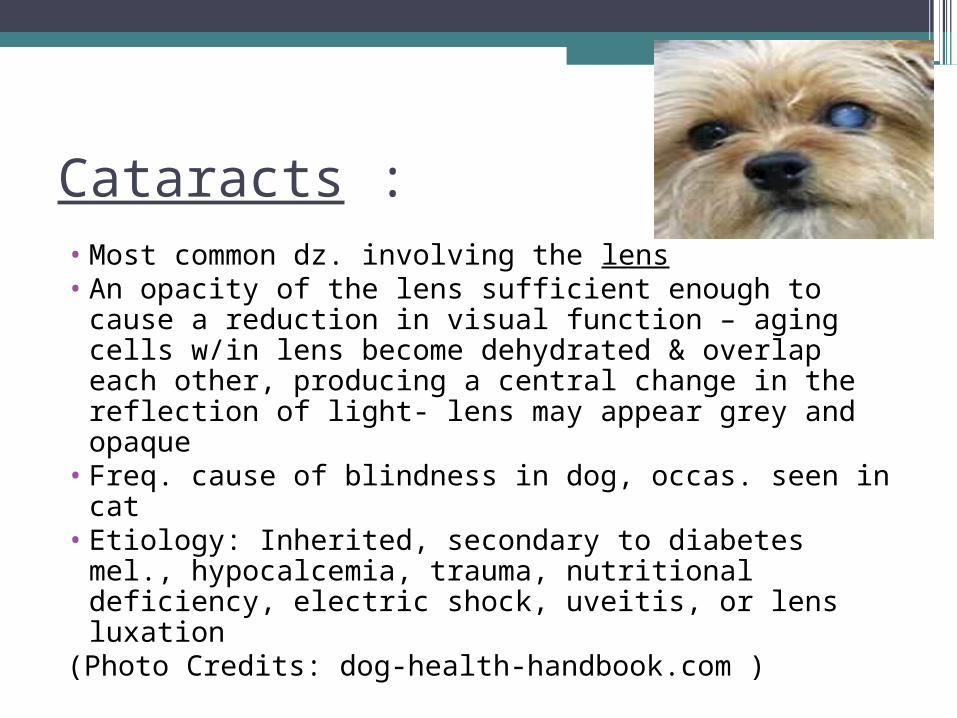
Cataracts :• Most common dz. involving the lens • An opacity of the lens sufficient enough to cause
a reduction in visual function – aging cells w/in lens become dehydrated & overlap each other, producing a central change in the reflection of light- lens may appear grey and opaque
• Freq. cause of blindness in dog, occas. seen in cat
• Etiology: Inherited, secondary to diabetes mel., hypocalcemia, trauma, nutritional deficiency, electric shock, uveitis, or lens luxation
(Photo Credits: dog-health-handbook.com )

Cataracts cont’d•Clinical signs
▫ Progressive loss of vision▫ Opaque pupillary opening▫ Signs related to systemic dz. (diabetes mellitus
or hypocalcemia) •Dx:
▫ Complete eye exam/Assess via obstacle course/Lack of menace /Failure to track visual responses
Photo Credits: petdig.com
Cataracts cont’d……..
•Pupillary light response is usually normal•Tx:
▫Sx removal▫Tx of any other dz
Anterior Uveitis • Inflammation of the uvea (iris, ciliary body,
choriod) • Trauma, extension of local infx, foreign body,
neoplasm, thermal trauma, parasites, protozoa (bact, viral, mycotic dz – hematogenous spread)
• C/S: epiphora, photophobia, blepharospasm, +/- vision defects, corneal edema, chemosis, prolapsed 3rd eyelid, pain, change in iris color
• Dx: c/s, hx, labs, x-ray, ultrasound, tonometry• Tx: I.D. & elim. cause, control inflamm w/topical
steroids (w/o tx, vision will eventually be lost)
Progressive Retinal Atrophy•Group of hereditary retinal disorders seen in
many breeds of dogs (can occur in cats, but not as frequently) •Toy poodles, min poodles, goldens, Irish sett,
cockers, min sch, collies, samoyed, gordon sett, Norw. Elkhound
•C/S: defective night vision, slowly progressive loss of day vision, cataract formation•Dx: labs, eye exam of retina•No Tx. currently exists
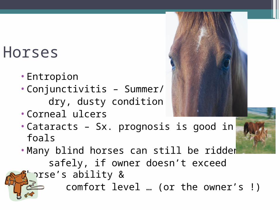
Horses
•Entropion•Conjunctivitis – Summer/ dry, dusty condition /flies•Corneal ulcers •Cataracts – Sx. prognosis is good in foals•Many blind horses can still be ridden safely, if owner doesn’t exceed horse’s
ability & comfort level … (or the owner’s !)
“Moon Blindness”Periodic Opthalmia, Recurrent Uveitis•Comes & goes/ exact cause undetermined•Many animals have high Lepto antibody titer•Signs: cloudy eye, blepharospasm, excessive tears•Dx: visualization of protein flare in fluid of
anterior chamber, affected eye may be smaller, corneal stain, Lepto titer
•Tx: topical corticosteroids, atropine, banamine or Subconjunctival long-acting steroid
injections
Sheep and Goats
•Entropion – most common ocular abnormality in neonatal lambs
•Infectious Conjunctivitis (Pinkeye) – responds well to medication – herd management
•Cataracts – most common lens abnormality in sheep and goats


