52
DISORDERS OF THE PARATHYROID GLANDS
| Date post: | 17-Dec-2015 |
| Category: |
Documents |
| Upload: | darren-raymond-hodges |
| View: | 226 times |
| Download: | 4 times |
DISORDERS OF THE PARATHYROID
GLANDS
Disorders of the Parathyroid Glands
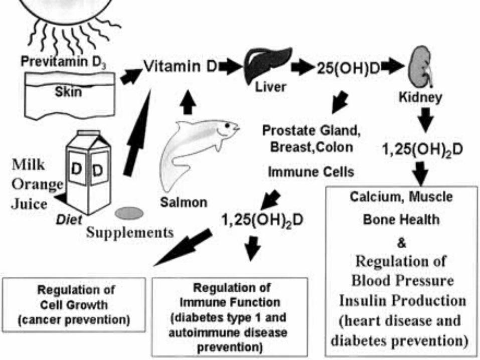
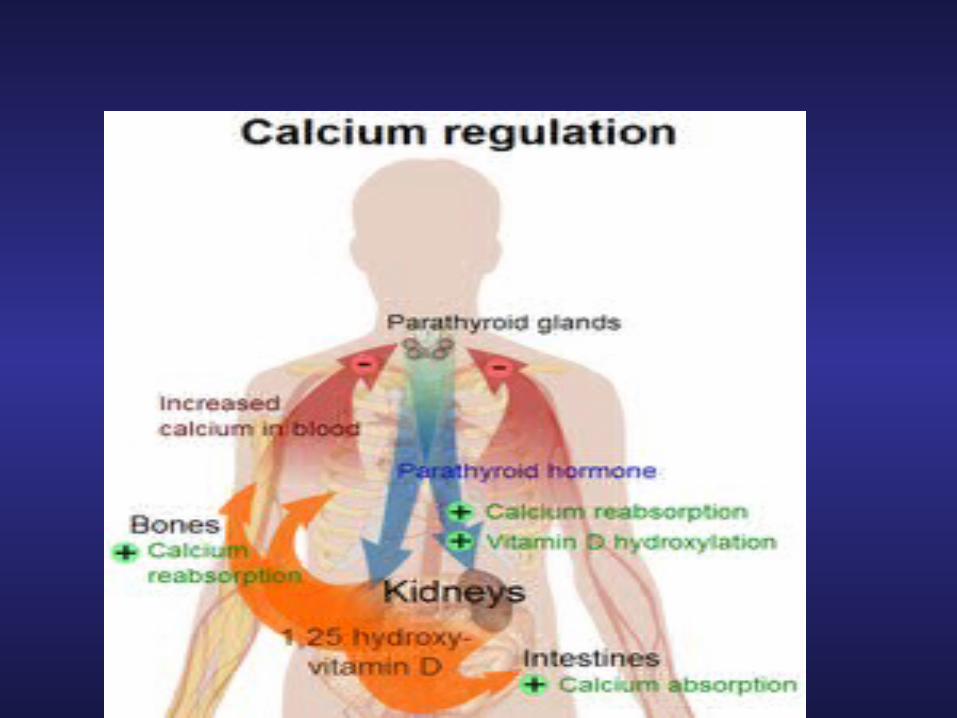
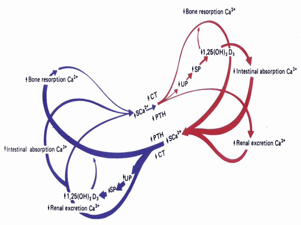
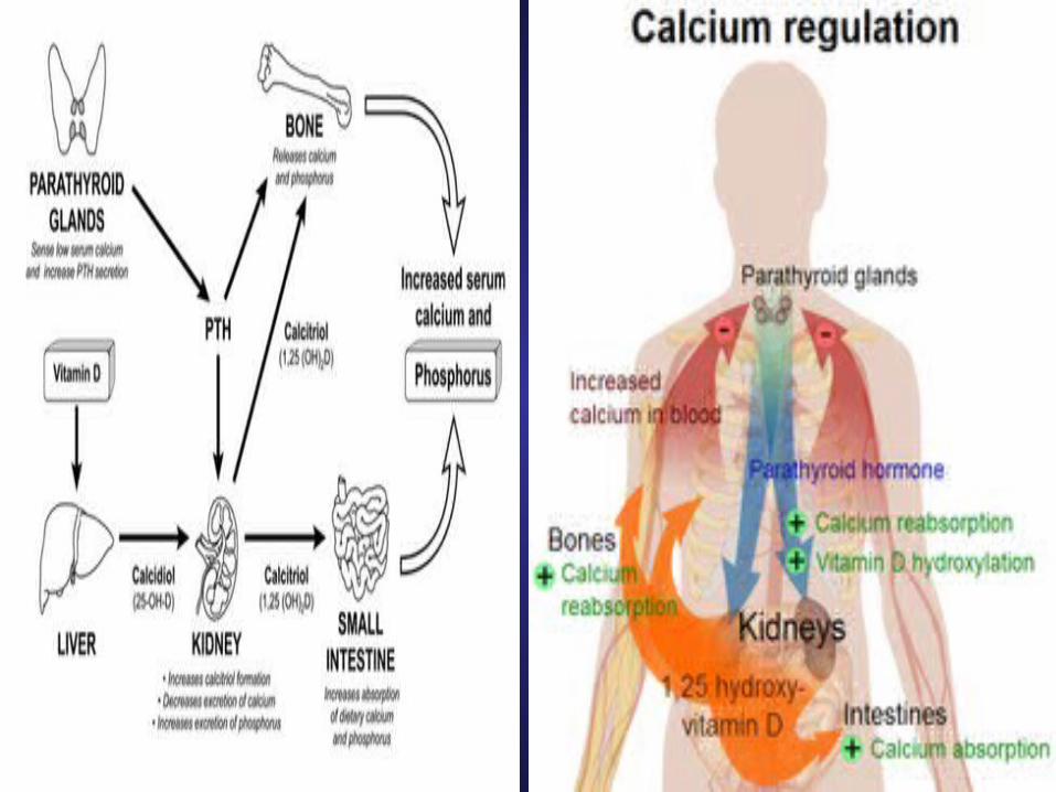
Maintenance of calcium, phosphate and magnesium homeostasis is under the influence of two polypeptide hormones; parathyroid hormone(PTH), and calcitonin (CT), as well as a sterol hormone, 1,25 dihydroxy cholecalciferol (1,25 (OH)2D3.
Disorders of the Parathyroid Glands
These hormones regulate the flow of minerals in and out of the extracellular fluid compartments through their actions on intestine, kidneys, and bones.
Disorders of the Parathyroid Glands
The PTH acts directly on the bones and kidneys and indirectly on the intestine through its effect on the synthesis of 1,25 (OH)2D3.
Its production is regulated by the concentration of serum ionized calcium. Lowering of the serum calcium levels will induce an increased rate of parathyroid hormone secretion
Disorders of the Parathyroid Glands
Calcitonin is released by the “C” cells (parafollicular cells in the thyroid gland) in response to small increases in plasma ionic calcium.
It acts on the kidney and bones to restore the level of calcium to just below a normal set point which in turn inhibits secretion of the hormone.
Disorders of the Parathyroid Glands
Calcitonin is therefore the physiological antagonist of PTH. The two hormones act in concert to maintain normal concentration of calcium ion in the extracellular fluid.
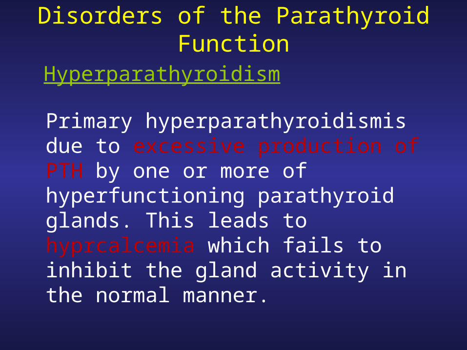
Disorders of the Parathyroid Function
Primary hyperparathyroidismis due to excessive production of PTH by one or more of hyperfunctioning parathyroid glands. This leads to hyprcalcemia which fails to inhibit the gland activity in the normal manner.
Hyperparathyroidism
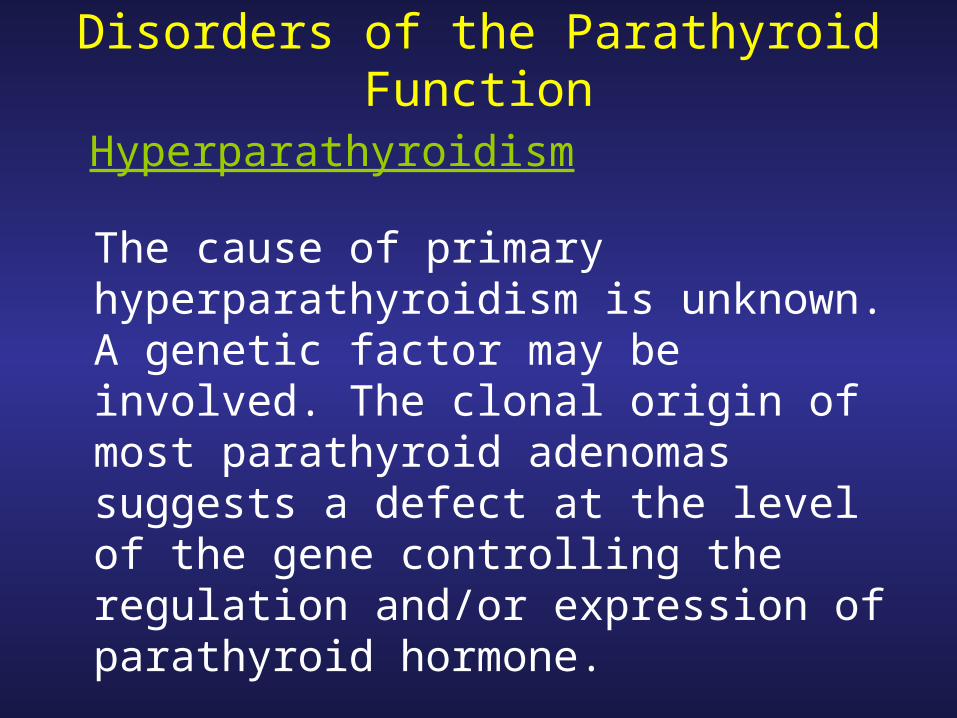
Disorders of the Parathyroid Function
The cause of primary hyperparathyroidism is unknown. A genetic factor may be involved. The clonal origin of most parathyroid adenomas suggests a defect at the level of the gene controlling the regulation and/or expression of parathyroid hormone.
Hyperparathyroidism

Disorders of the Parathyroid Function
The incidence of the disease increases dramatically after the age of 50 and it is 2-4 folds more common in women.
A single adenoma occurs in about 80% of patients with primary hyperparathyroidism.
Four glands hyprplasia account for 15-20% of cases.
A parathyroid carcinoma could be the etiology in a rare incidence of less then 1%.
Hyperparathyroidism
Disorders of the Parathyroid Function
The two major sites of potential complications are the bones and the kidneys.
The kidneys may have renal stones (nephrolithiasis) or diffuse deposition of calcium-phosphate complexes in the parachyma (nephrocalcinosis).
Now a days such complications are seen less commonly and around 20% of patients or less show such complications.
Clinical Features:
Disorders of the Parathyroid Function
In skeleton a condition called osteitis fibrosa cystica could occur with subperiosteal resorption of the distal phalanges, distal tappering of the clavicles, a “salt and pepper” appearance of the skull as well as bone cysts and brown tumors of the long bones.
Such overt bone disease even though typical of primary hyperparathyroidism is very rarely encountered.
Clinical Features:
Disorders of the Parathyroid Function
Now a days almost 90% of diagnosed cases in the developed countries are picked up by routine screening for calcium level using the new automated machines.
Clinical Features:
Disorders of the Parathyroid Function
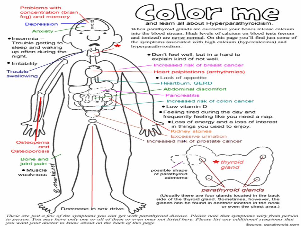
Other symptoms include muscle weakness, easy fatigability, peptic ulcer disease, pancreatitis, hypertension, gout and pseudogout as well as anemia and depression have been associated with primary hyperparathyroidism.
Clinical Features:
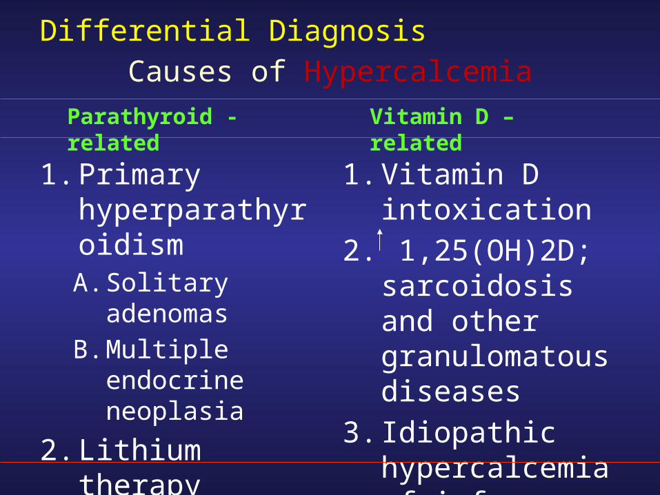
Differential Diagnosis
1. Primary hyperparathyroidismA. Solitary
adenomas
B. Multiple endocrine neoplasia
2. Lithium therapy
3. Familial hypocalciuric hypercalcemia
1. Vitamin D intoxication
2. 1,25(OH)2D; sarcoidosis and other granulomatous diseases
3. Idiopathic hypercalcemia of infancy
Causes of Hypercalcemia
Parathyroid - related Vitamin D – related
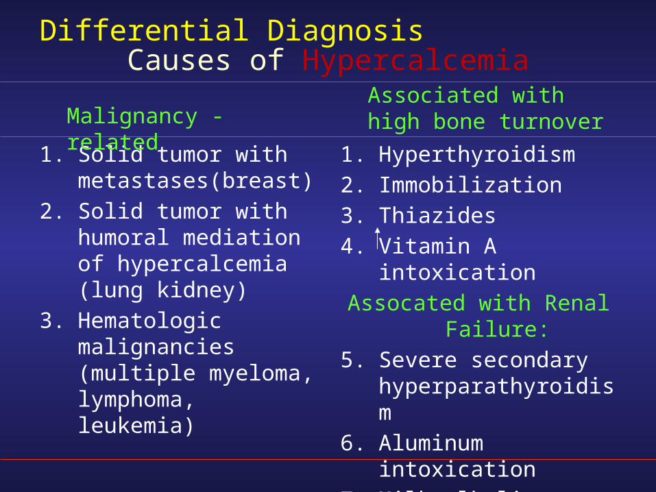
Differential Diagnosis
1. Solid tumor with metastases(breast)
2. Solid tumor with humoral mediation of hypercalcemia (lung kidney)
3. Hematologic malignancies (multiple myeloma, lymphoma, leukemia)
1. Hyperthyroidism
2. Immobilization
3. Thiazides
4. Vitamin A intoxication
Assocated with Renal Failure:
5. Severe secondary hyperparathyroidism
6. Aluminum intoxication
7. Milk alkali syndrome
Causes of Hypercalcemia
Malignancy - relatedAssociated with high bone turnover
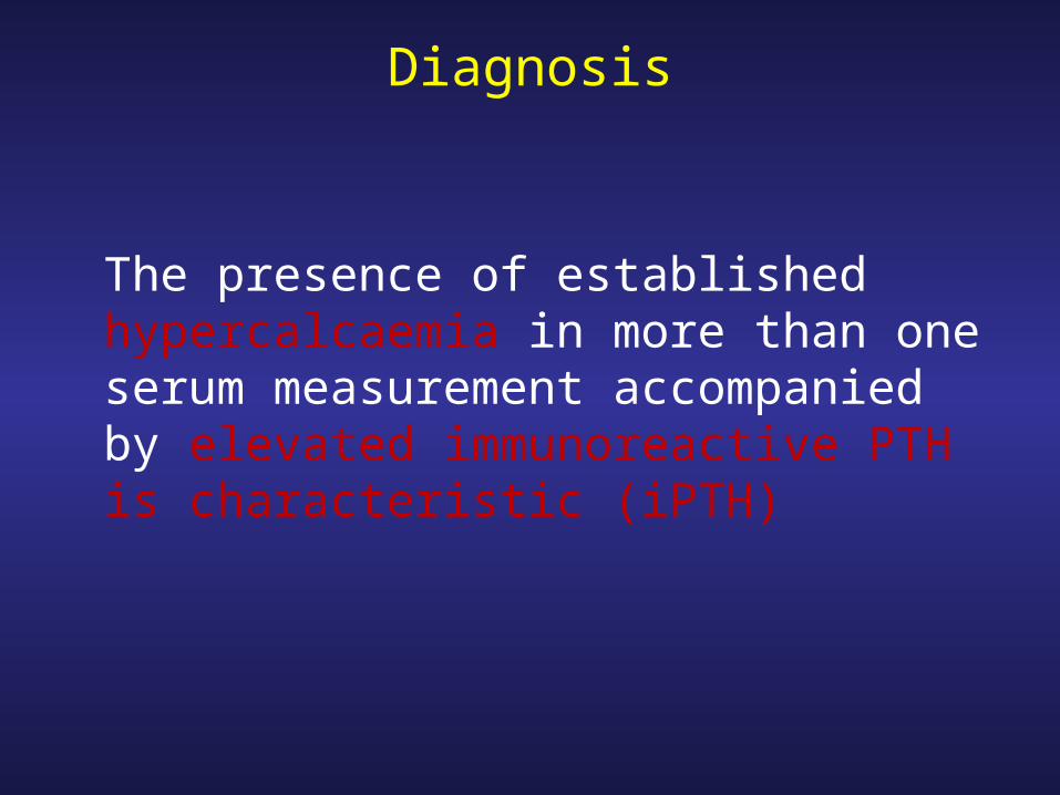
Diagnosis
The presence of established hypercalcaemia in more than one serum measurement accompanied by elevated immunoreactive PTH is characteristic (iPTH)

Diagnosis
Serum phosphate is usually low but may be normal. Hypercalcaemia is common and blood alkaline phosphatase (of bone origin) and the urinary hydroxyproline concentrations are commonly elevated when the bones are involved. Nephrogenous CAMP is elevated in about 80% of patients but the test is rarely used because of technical difficulties
Other Diagnostic tests
The heypercalcaemic of non-parathyroid origin e.g., vitamin D intoxication, sarcoidosis and lymphoproliferative syndromes generally respond to the administration of prednisolone in a dose of 40-60 mg daily for 10 days by a decrease in serum calcium level.
The Glucocortisoid suppression test:

Other Diagnostic tests
Plain X-ray of hands can be diagnostic showing subperiosteal bone resorption usually on the radial surfacy of the distal phalanx with distal phalangeal tufting as well as cysts formation and generalzed osteopenia.
Radiograph:
Other Diagnostic tests
• Ultrasonography• MRI• CT• Thallium 201 – Tehcnichum99m scan
(subtraction study)
Pre-operative localization of the abnormal parathyroid gland(s):
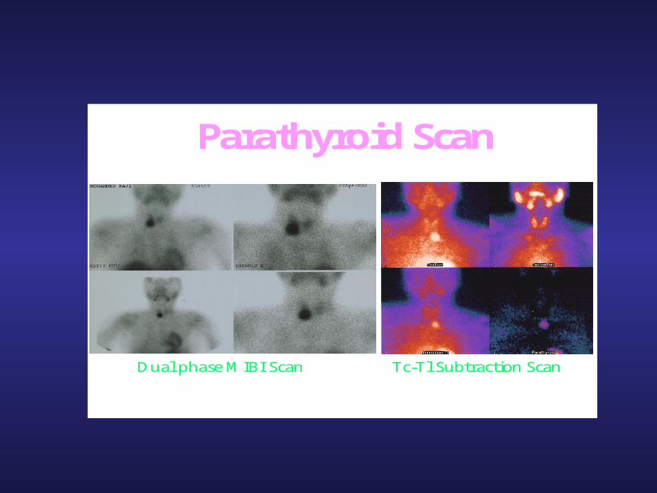
Parathyroid Scan
Dual phase MIBI Scan Tc-Tl Subtraction Scan
Treatment
A large proportion of patients have “biochemical” hyperparathyroidism but with prolonged follow up they progress to overt clinical presentation.
Resection of the parathyroid lesion is curative with recurrences observed mainly in the multiple glandular disease.
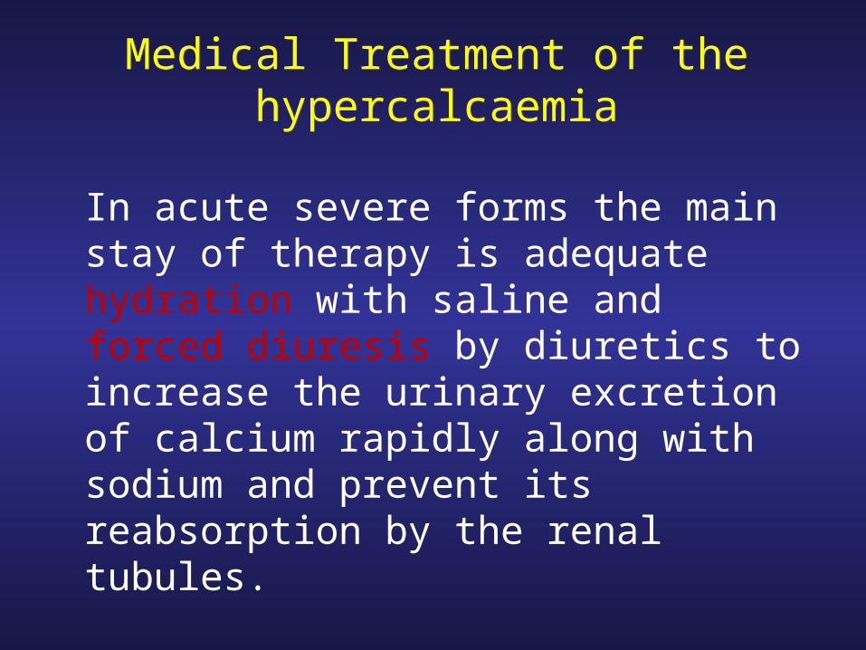
Medical Treatment of the hypercalcaemia
In acute severe forms the main stay of therapy is adequate hydration with saline and forced diuresis by diuretics to increase the urinary excretion of calcium rapidly along with sodium and prevent its reabsorption by the renal tubules.
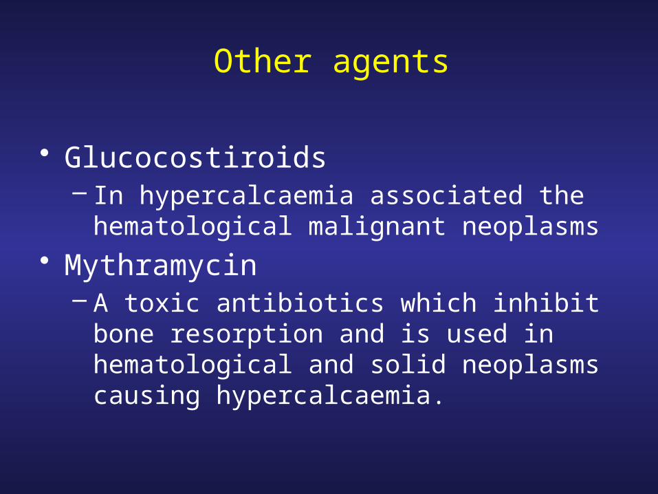
Other agents
• Glucocostiroids– In hypercalcaemia associated the
hematological malignant neoplasms• Mythramycin
– A toxic antibiotics which inhibit bone resorption and is used in hematological and solid neoplasms causing hypercalcaemia.
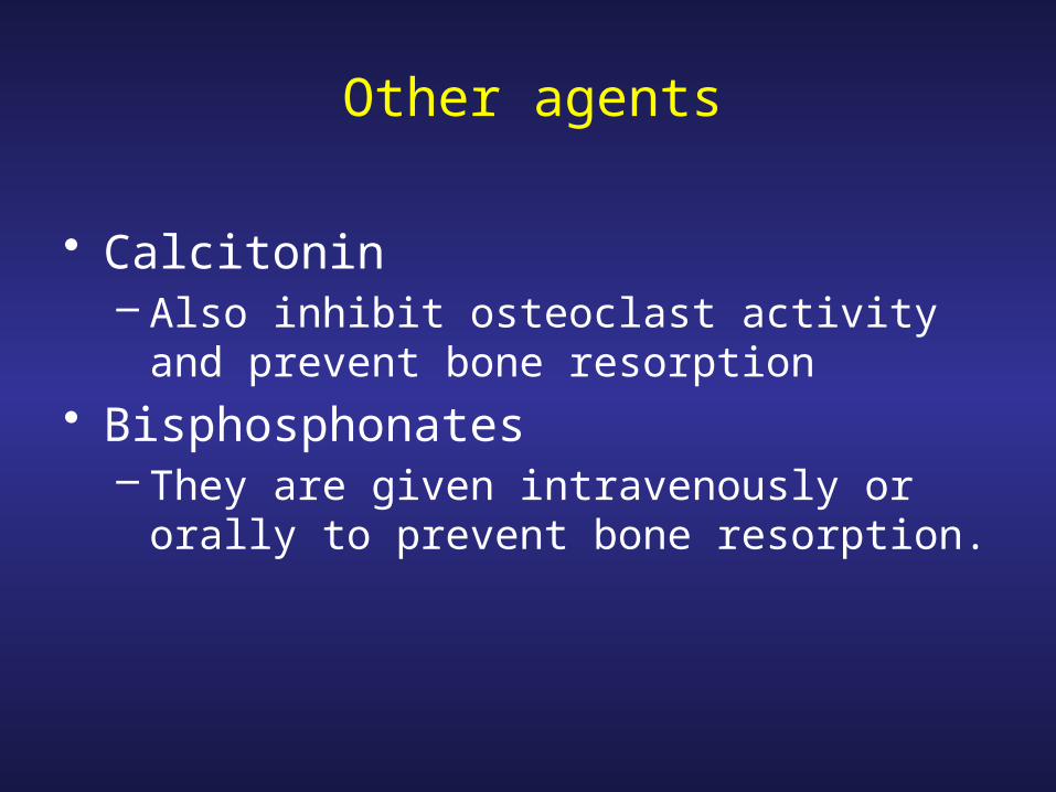
Other agents
• Calcitonin– Also inhibit osteoclast activity and prevent
bone resorption• Bisphosphonates
– They are given intravenously or orally to prevent bone resorption.
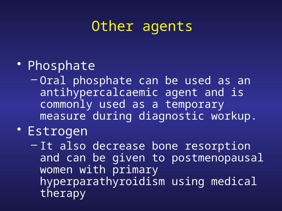
Other agents
• Phosphate– Oral phosphate can be used as an
antihypercalcaemic agent and is commonly used as a temporary measure during diagnostic workup.
• Estrogen– It also decrease bone resorption and can be
given to postmenopausal women with primary hyperparathyroidism using medical therapy
Surgery
• Surgical treatment should be considered in all cases with established diagnosis of primary hyperparthyroidism.
• During surgery the surgeon identifies all four parathyroid glands (using biopsy if necessary) followed by the removal of enlarged parathyroid or 3 ½ glands in multiple glandular disease.
Other Complications
• Deterioration of renal function• Metabolic disturbance e.g. hypomagnesia,
pancreatitis, gout or pseudogout
Secondary hyperparathyroidism
An increase in PTH secretion which is adaptive and unrelated to intrinsic disease of the parathyroid glands is called secondary hyperparathyroidism.
This is due to chronic stimulation of the parathyroid glands by a chronic decrease in the ionic calcium level in the blood

Major causes of chronic hypocalcemia other than hypoparathyroidism
• Dietary deficiency of vitamin D or calcium• Decreased intestinal absorption of vitamin
D or calcium due to primary small bowel disease, short bowel syndrome, and post-gastrectomy syndrome.
• Drugs that cause rickets or osteomalacia such as phenytoin, phenobarbital, cholestyramine, and laxative.
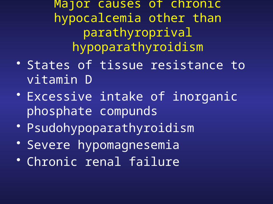
Major causes of chronic hypocalcemia other than parathyroprival hypoparathyroidism
• States of tissue resistance to vitamin D• Excessive intake of inorganic phosphate
compunds• Psudohypoparathyroidism• Severe hypomagnesemia• Chronic renal failure
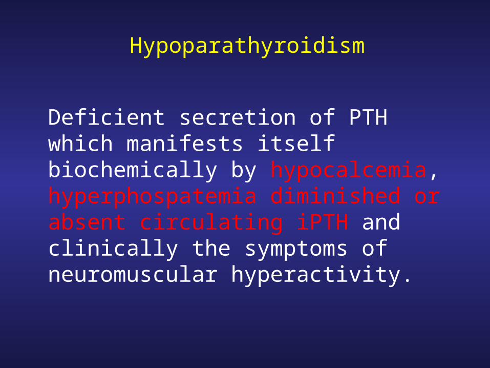
Hypoparathyroidism
Deficient secretion of PTH which manifests itself biochemically by hypocalcemia, hyperphospatemia diminished or absent circulating iPTH and clinically the symptoms of neuromuscular hyperactivity.
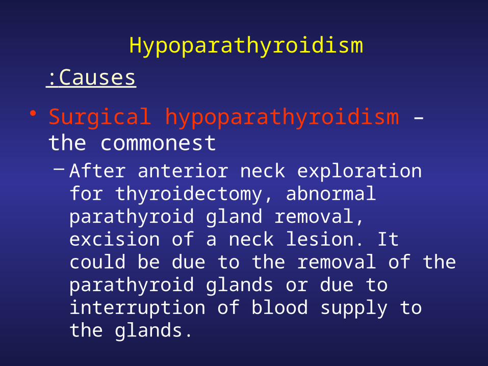
Hypoparathyroidism
• Surgical hypoparathyroidism – the commonest– After anterior neck exploration for
thyroidectomy, abnormal parathyroid gland removal, excision of a neck lesion. It could be due to the removal of the parathyroid glands or due to interruption of blood supply to the glands.
Causes:
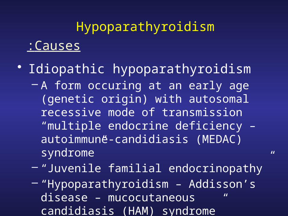
Hypoparathyroidism
• Idiopathic hypoparathyroidism– A form occuring at an early age (genetic
origin) with autosomal recessive mode of transmission “multiple endocrine deficiency –autoimmune-candidiasis (MEDAC) syndrome”
– “Juvenile familial endocrinopathy”– “Hypoparathyroidism – Addisson’s disease –
mucocutaneous candidiasis (HAM) syndrome”
Causes:
Hypoparathyroidism
• Idiopathic hypoparathyroidism– Circulating antibodies for the parathyroid
glands and the adrenals are frequently present.
– Other associated disease:• Pernicious anemia• Ovarian failure• Autoimmune thyroiditis• Diabetes mellitus
Causes:
Hypoparathyroidism
• Idiopathic hypoparathyroidism– The late onset form occurs sporadically
without circulating grandular autoantibodies.• Functional hypoparathyroidism
– In patients who has chronic hypomagesaemia of various causes.
– Magnesium is necessary for the PTH release from the glands and also for the peripheral action of the PTH.
Causes:
Hypoparathyroidism
A. Neuromuscular– The rate of decrease in serum calcium is the
major determinant for the development of neuromuscular complications.
– When nerves are exposed to low levels of calcium they show abnormal neuronal function which may include decrease threshold of excitation, repetitive response to a single stimulus and rarely continuous activity.
Clinical Features:
Hypoparathyroidism
A. Neuromuscular– Parathesia– Tetany– Hyperventilation– Adrenergic symptoms– Convulsion (More common in young people and it
can take the form of either generalized tetany followed by prolonged tonic spasms or the typical epileptiform seizures.
– Signs of latent tetany• Chvostek sign• Trousseau sign• Extrapyramidal signs (due to basal ganglia calcification)
Clinical Features:
Hypoparathyroidism
B. Other clinical manifestation1. Posterio lenticular cataract
2. Cardiac manifestation:
Prolonged QT interval in the ECG
Resistance to digitalis
Hypotension
Refractory heart failure with cardiomegally can occur.
Clinical Features:
Hypoparathyroidism
B. Other clinical manifestation3. Dental Manifestation
Abnormal enamel formation with delayed or absent dental eruption and defective dental root formation.
4. Malabsorption syndrome
Presumably secondary to decreased calcium level and may lead to steatorrhoea with long standing untreated disease.
Clinical Features:
Hypoparathyroidism
In the absence of renal failure the presence of hypocalcaemia with hyperphosphataemia is virtually diagnostic of hypoparathyroidism. Undetectable serum iPTH confirms the diagnosis or it can be detectable if the assay is very sensitive.
Diagnosis:
Hypoparathyroidism
The mainstay of treatment is a combination of oral calcium with pharmacological doses of vitamin D or its potent analogues.
Phosphate restriction in diet may also be useful with or without aluminum hydroxide gel to lower serum phosphate level.
Treatment:
Emergency Treatment for Hypocalcaemic
Calcium should be given parenterally till adequate serum calcium level is obtained and then vitamin D supplementation with oral calcium should be initiated.
Tetany:
Emergency Treatment for Hypocalcaemic
In patients with hyperparathyroidism and severe bone disease who undergo successful parathyroidectomy hypocalcaemia may be severe and parenteral calcium infusion with later supplementation with oral calcium and vitamin D.
Hungry bone syndrome:
Pseudohypoparathysoidism and Pseudopseudohypoparathyroidism
A rare familial disorders with target tissue resistance to PTH.
There is hypocalcaemia, hyperphosphataemia, with increased parathyroid gland function.
There is also a variety of congenital defects in the growth and development of skeleton including:
• Short statue• Short metacarpal and metatarsal bones
Pseudohypoparathysoidism and Pseudopseudohypoparathyroidism
In pseudopseudohypoparathyroidism they have the developmental defects without the biochemical abnormalitiesThe diagnosis is established when low serum calcium level with hyperphosphataemia is associated with increased serum iPTH as well as diminished nephrogenous CAMP and phosphature response to PTH administration