34
Disorders of the Tongue and Nails Stephanie Blackburn, DO PGY 4 Affiliated Dermatology Date: 3/29/2017

Disorders of the Tongue and Nails
Stephanie Blackburn, DO PGY 4
Affiliated Dermatology Date: 3/29/2017

Relevant Financial Relationship(s)
None
Off Label Usage
None
Disclosure
• Review disorders of the tongue and oral lesions
• Discuss diagnosis and potential treatment options for dermatologic disorders of the tongue and disorders of the oral cavity
• Expand differential diagnosis in regards to tongue/oral lesions
• Review board relevant nail disorders
Learning Objectives

Introduction
• Diagnosis and treatment of dermatologic lesions of the oral cavity and tongue is challenging
• In a study from 2001, almost all (84%) hospital doctors in general and geriatric medicine felt that it was important to examine the patient’s mouth, however less than one-fifth (19%) routinely performed such examinations [1]

Fissured Tongue
• Congenital disorder with enlarged tongue and plicate superficial or deep grooves
• Seen in Melkersson-Rosenthal syndrome(facial paralysis/lip edema/scrotal tongue) and many patients with Down syndrome
• Occurs with geographic tongue in 50% of patients and both are commonly seen in psoriasis [2]
• No treatment is necessary, however recommending mouthwash to keep the fissures clean is important http://diseasespictures.com/fissured-tongue/
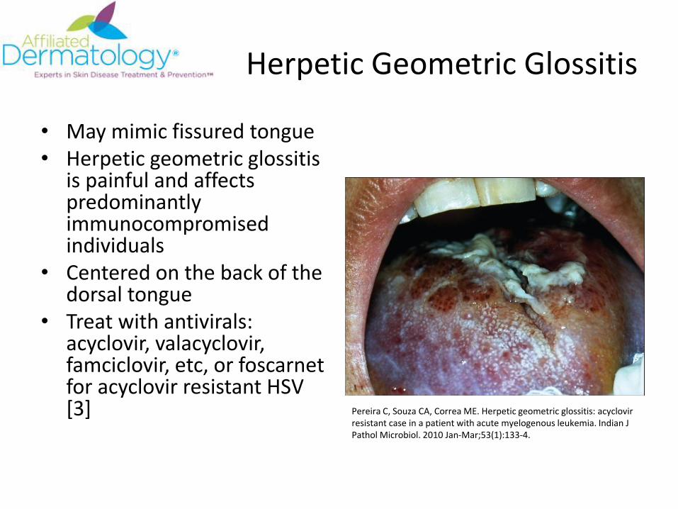
Herpetic Geometric Glossitis
• May mimic fissured tongue• Herpetic geometric glossitis
is painful and affects predominantly immunocompromised individuals
• Centered on the back of the dorsal tongue
• Treat with antivirals: acyclovir, valacyclovir, famciclovir, etc, or foscarnetfor acyclovir resistant HSV [3] Pereira C, Souza CA, Correa ME. Herpetic geometric glossitis: acyclovir
resistant case in a patient with acute myelogenous leukemia. Indian J Pathol Microbiol. 2010 Jan-Mar;53(1):133-4.
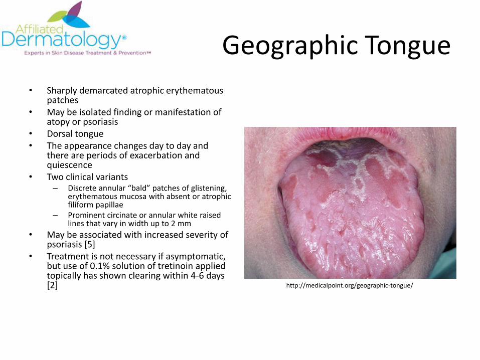
Geographic Tongue
• Sharply demarcated atrophic erythematous patches
• May be isolated finding or manifestation of atopy or psoriasis
• Dorsal tongue• The appearance changes day to day and
there are periods of exacerbation and quiescence
• Two clinical variants– Discrete annular “bald” patches of glistening,
erythematous mucosa with absent or atrophic filiform papillae
– Prominent circinate or annular white raised lines that vary in width up to 2 mm
• May be associated with increased severity of psoriasis [5]
• Treatment is not necessary if asymptomatic, but use of 0.1% solution of tretinoin applied topically has shown clearing within 4-6 days [2] http://medicalpoint.org/geographic-tongue/

Annulus Migrans
• Geographic tongue associated with psoriasis and/or reactive arthritis
James W, Elston D, Berger T, Andrews G. Andrews’ Diseases of the skin. [London]: Saunders/Elsevier, ©2001. 11th
edition

Black Hairy Tongue
• On the dorsum of the tongue anterior to the circumvallate papillae
• The “hair” is due to benign hyperplasia of the filiform papillae
• Associated with smoking, use of oral antibiotics, psychotropic drugs, and Candida
• Differentiated from oral hairy leukoplakia due to clinical location. Hairy leukoplakia is on the lateral tongue
• Treatment is exfoliation of the tongue with toothbrush alone or with 1-2% hydrogen peroxide. May use urea, tretinoin or papain (meat tenderizer) [2,7]
• Discontinue predisposing factors (smoking) and increase oral hygiene
http://diseasespictures.com/black-hairy-tongue/

Atrophic Glossitis
• Bald tongue/smooth tongue
• Painful
• Results from atrophy of the filiform and fungiform papillae
• Moeller/Hunter glossitis-B12 deficiency
• Iron deficiency, pellagra, malabsorption syndrome, anorexia nervosa, alcoholism
• Treat underlying cause
http://www.hxbenefit.com/glossitis.html
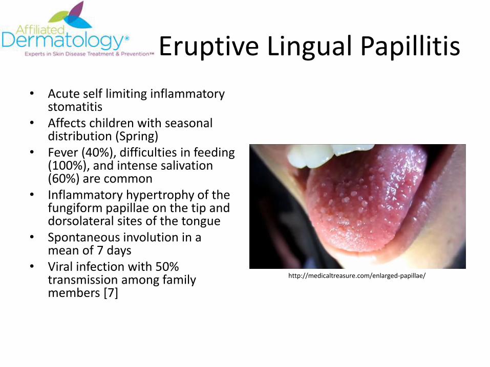
Eruptive Lingual Papillitis
• Acute self limiting inflammatory stomatitis
• Affects children with seasonal distribution (Spring)
• Fever (40%), difficulties in feeding (100%), and intense salivation (60%) are common
• Inflammatory hypertrophy of the fungiform papillae on the tip and dorsolateral sites of the tongue
• Spontaneous involution in a mean of 7 days
• Viral infection with 50% transmission among family members [7]
http://medicaltreasure.com/enlarged-papillae/

Median Rhomboid Glossitis
• Shiny oval or diamond-shaped elevation on the dorsum in the midline immediately in front of circumvallate papillae
• No change in size and no link to cancer
• May result from abnormal fusion of the posterior portion of the tongue, but it is nearly always chronically infected with Candida
• Histologically there is chronic inflammation with fibrosis
• Eosinophilic ulcer of the oral mucosa may look similar
• Treat with oral antifungalshttp://www.intelligentdental.com/2010/04/26/how-diabetes-can-affect-your-oral-health-part-2-2/

Granular Cell Tumor
• 1/3 of reported cases of granular cell tumor occur on the tongue (1/3 skin, 1/3 internal organs) [2]
• About 2/3 of patients are black and 2/3 are women [2]
• Well circumscribed, solitary, firm nodule ranging from 5-30 mm
• Histologically distinct with sheets of large polygonal cells with abundant eosinophilic granular cytoplasm with central nucleus [12]
• Pustulo-ovoid bodies of Milian-discrete round eosinophilic giant lysosomal granules
• Overlying PEH [12]• S100+ • Complete excision is advisable due to
potential difficulties distinguishing between malignant granular cell tumor
http://www.rdhmag.com/articles/print/volume-33/issue-9/columns/tongue-granular-cell-tumor.html
Elston D, Ferringer T. Dermatopathology. Elsevier, ©2014. 2nd Edition

Granular Cell Tumor
Elston D, Ferringer T. Dermatopathology. Elsevier, ©2014. 2nd Edition
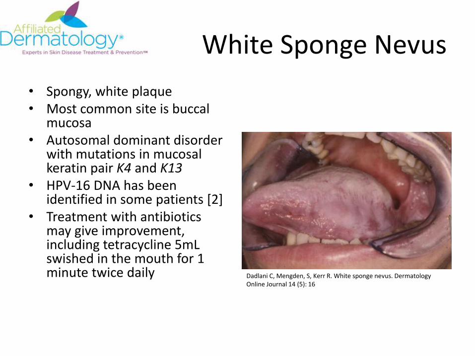
White Sponge Nevus
• Spongy, white plaque • Most common site is buccal
mucosa• Autosomal dominant disorder
with mutations in mucosal keratin pair K4 and K13
• HPV-16 DNA has been identified in some patients [2]
• Treatment with antibiotics may give improvement, including tetracycline 5mL swished in the mouth for 1 minute twice daily Dadlani C, Mengden, S, Kerr R. White sponge nevus. Dermatology
Online Journal 14 (5): 16

Leukoplakia
• Presents as whitish thickening of the epithelium of the mucous membranes
• White pellicle is adherent to underlying mucosa, attempts to remove result in bleeding
• Benign form is usually in response to irritation
• If progresses to carcinoma, follows a 1 to 20 year lag time, unless patient is immunosuppressed
• Associated with tobacco, alcohol and poorly fitting dentures
• Treatment: surgery or destruction, fulguration, excision, cryosurgery, CO2 laser ablation
http://diseasesforum.com/wp-content/uploads/2013/07/Leukoplakia-2.jpg

Oral Hairy Leukoplakia
• Distinctive condition strongly associated with HIV/immunosuppression
• HHV4/Epstein-Barr virus
• In immunosuppressed patients there is continuous shedding of EBV virus in oral secretions
• If noted, a workup for immunosuppression is recommended
James W, Elston D, Berger T, Andrews G. Andrews’ Diseases of the skin. [London]: Saunders/Elsevier, ©2001. 11th edition
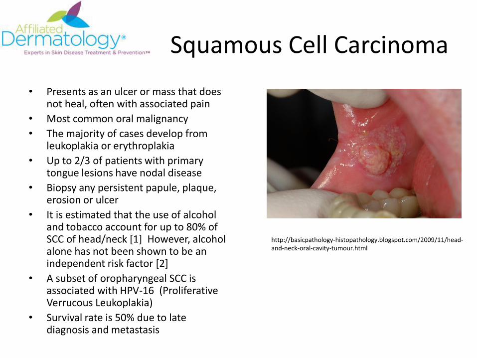
Squamous Cell Carcinoma
• Presents as an ulcer or mass that does not heal, often with associated pain
• Most common oral malignancy
• The majority of cases develop from leukoplakia or erythroplakia
• Up to 2/3 of patients with primary tongue lesions have nodal disease
• Biopsy any persistent papule, plaque, erosion or ulcer
• It is estimated that the use of alcohol and tobacco account for up to 80% of SCC of head/neck [1] However, alcohol alone has not been shown to be an independent risk factor [2]
• A subset of oropharyngeal SCC is associated with HPV-16 (Proliferative Verrucous Leukoplakia)
• Survival rate is 50% due to late diagnosis and metastasis
http://basicpathology-histopathology.blogspot.com/2009/11/head-and-neck-oral-cavity-tumour.html
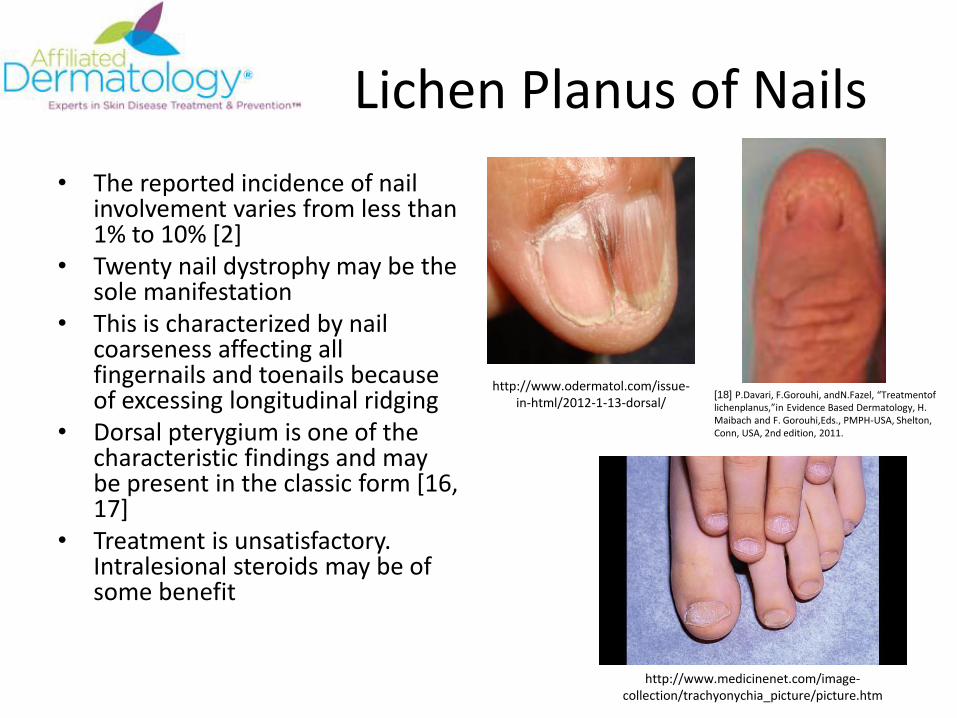
Lichen Planus of Nails
• The reported incidence of nail involvement varies from less than 1% to 10% [2]
• Twenty nail dystrophy may be the sole manifestation
• This is characterized by nail coarseness affecting all fingernails and toenails because of excessing longitudinal ridging
• Dorsal pterygium is one of the characteristic findings and may be present in the classic form [16, 17]
• Treatment is unsatisfactory. Intralesional steroids may be of some benefit
http://www.odermatol.com/issue-in-html/2012-1-13-dorsal/
http://www.medicinenet.com/image-collection/trachyonychia_picture/picture.htm
[18] P.Davari, F.Gorouhi, andN.Fazel, “Treatmentoflichenplanus,”in Evidence Based Dermatology, H. Maibach and F. Gorouhi,Eds., PMPH-USA, Shelton, Conn, USA, 2nd edition, 2011.
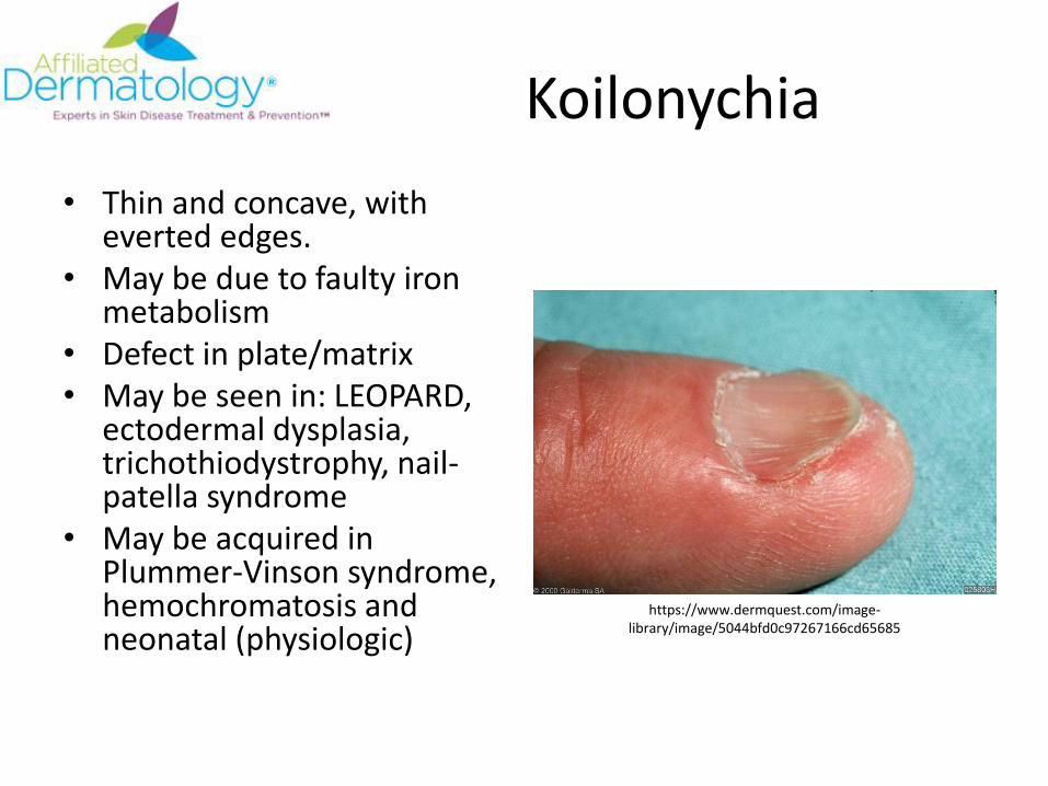
Koilonychia
• Thin and concave, with everted edges.
• May be due to faulty iron metabolism
• Defect in plate/matrix• May be seen in: LEOPARD,
ectodermal dysplasia, trichothiodystrophy, nail-patella syndrome
• May be acquired in Plummer-Vinson syndrome, hemochromatosis and neonatal (physiologic)
https://www.dermquest.com/image-library/image/5044bfd0c97267166cd65685
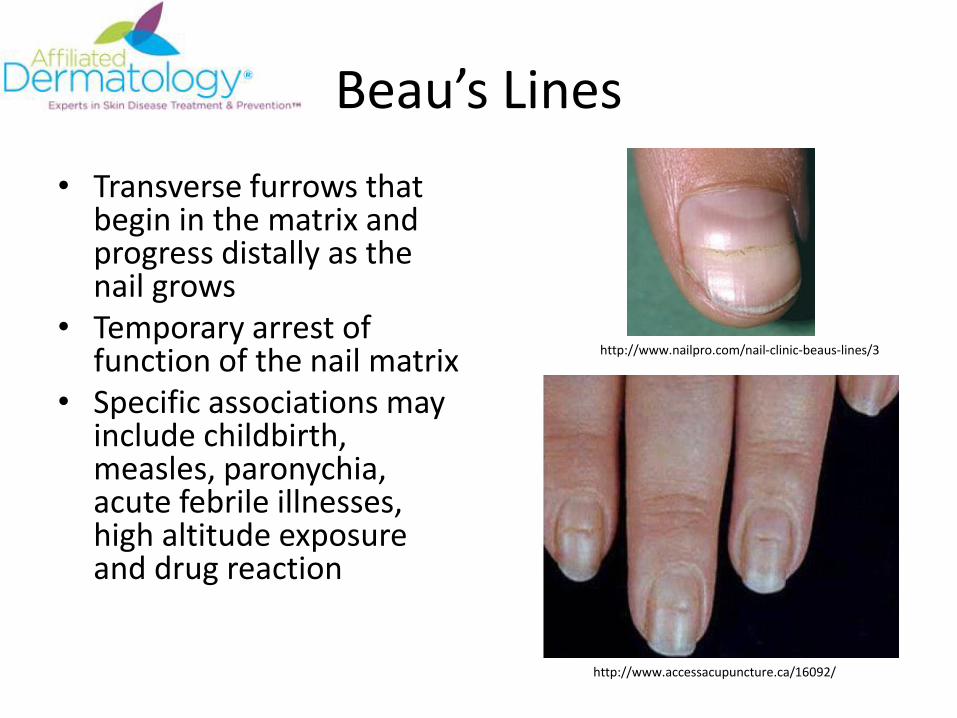
Beau’s Lines
• Transverse furrows that begin in the matrix and progress distally as the nail grows
• Temporary arrest of function of the nail matrix
• Specific associations may include childbirth, measles, paronychia, acute febrile illnesses, high altitude exposure and drug reaction
http://www.accessacupuncture.ca/16092/
http://www.nailpro.com/nail-clinic-beaus-lines/3
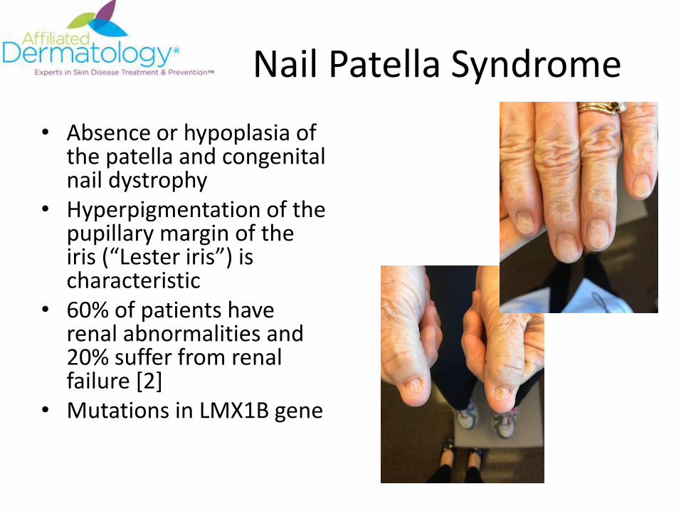
Nail Patella Syndrome
• Absence or hypoplasia of the patella and congenital nail dystrophy
• Hyperpigmentation of the pupillary margin of the iris (“Lester iris”) is characteristic
• 60% of patients have renal abnormalities and 20% suffer from renal failure [2]
• Mutations in LMX1B gene

Darier’s Disease
• V-shaped distal nicking
• Alternating red and white longitudinal bands with subungual hyperkeratosis
• AD inheritance
• Mutation in ATP2A2 gene encoding SERCA2, calcium ATPase
http://dermatologyoasis.net/nails-in-dariers-disease/
http://creativecommons.org/licenses/by-nc-nd/3.0/nz/
http://www.dermnetnz.org/topics/darier-disease/

Pachyonychia CongenitaType 1
• AD
• Defect in K6a, K16
• Focal PPK
• Benign oral leukokeratosis
• Nail dystrophy with significant subungual hyperkeratosis
http://drugline.org/medic/term/pachyonychia-congenita-type-1/

Pachyonychia CongenitaType II
• AD
• Defect in K6b, K17
• Nail dystrophy
• Steatocystomas
• Eruptive vellus hair cyst
• Natal teeth
• Pili torti
http://www.huidziekten.nl/zakboek/dermatosen/ptxt/pachyonychia-congenita.htm
http://www.huidziekten.nl/zakboek/dermatosen/ptxt/pachyonychia-congenita.htm

Half and Half Nails
• Proximal ½ with white zone
• Distal ½ with red/brown zone
• Due to chronic renal disease and nail bed edema
https://www.dermquest.com/image-library/image/5044bfd0c97267166cd6569f

Meuhrcke’s bands
• Transverse white bands parallel to lunula
• Disappear with squeezing of nail
• Due to hypoalbuminemia, nephrotic syndrome, liver disease, malnutrition and chemotherapy
http://imgarcade.com/1/muehrckes-lines-causes/

Terry’s nails
• Proximal 2/3 white nail color
• Distal 1/3 brown-pink band
• Cirrhosis, hypoalbuminemia, diabetes, cardiac disease https://www.dermquest.com/image-library/image/5044bfd0c97267166cd650ba
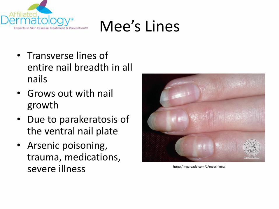
Mee’s Lines
• Transverse lines of entire nail breadth in all nails
• Grows out with nail growth
• Due to parakeratosis of the ventral nail plate
• Arsenic poisoning, trauma, medications, severe illness http://imgarcade.com/1/mees-lines/

Tumors Affecting the Nail
• Myxoid Cyst:– Smooth, soft nodule
most commonly adjacent to the DIP joint
– May cause longitudinal grooving in the nail plate
– Contains clear yellow viscous fluid
• Glomus Tumor:– Small reddish-blue
tender subungual tumor
http://www.suggest-keywords.com/Z2xvbXVzICB0dW1vcg/
https://www.dermquest.com/image-library/image/5044bfd0c97267166cd63334

Tumors Affecting the Nail
• Acquired Digital Fibrokeratoma:– Firm excrescence on the
finger or toe– Pathology: collagen with
no prominent nerves
• Accessory digit:– Firm excrescence on the
finger or toe, most commonly at proximal portion of 5th digit
– Pathology: Collagen with prominent nerve fascicles
https://ozmedgirl.wordpress.com/support-pages/
http://doctorv.ca/cosmetic-services/lump-and-bump-removal/acquired-digital-fibrokeratoma/

Tumors Affecting the Nail
• Bowen’s disease:
– Hyperkeratotic plaques often with spread under nail plate
• Wart:
– Well defined hyperkeratotic plaques around nail plate
http://www.eatonhand.com/img/img00046.htm
http://www.dermatalk.com/threads/3587-Dry-Skin-around-Nails
Resources1. Morgan R, Tsang J, Harrington N, Fook L. Survery of hospital doctors’ attitudes and knowledge of oral conditions in older patients.
Postgrad Med J 2001; 77;392.
2. James W, Elston D, Berger T, Andrews G. Andrews’ Diseases of the skin. [London]: Saunders/Elsevier, ©2001. 11th edition
3. Pereira C, Souza CA, Correa ME. Herpetic geometric glossitis: acyclovir resistant case in a patient with acute myelogenous leukemia. Indian J Pathol Microbiol. 2010 Jan-Mar;53(1):133-4.
4. http://medicalpoint.org/geographic-tongue/
5. Zargari O. The prevalence and significance of fissured tongue and geographical tongue in psoriatic patients.Clin Exp Dermatol. 2006 Mar;31(2):192-5.
6. http://medicalpoint.org/geographic-tongue/
7. Langtry JA, Carr MM, Steele MC, Ive FA. Topical tretinoin: a new treatment for black hairy tongue (lingua villosa nigra). Clin Exp Dermatol. 1992 May;17(3):163-4.
8. http://www.hxbenefit.com/glossitis.html
9. Whitaker SB, Krupa JJ 3rd, Singh BB. Transient lingual papillitis. Oral Surg Oral Med Oral Pathol Oral Radiol Endod. 1996 Oct;82(4):441-5.
10. http://www.intelligentdental.com/2010/04/26/how-diabetes-can-affect-your-oral-health-part-2-2/
11. Burkhart N. Tongue: granular cell tumor. http://www.rdhmag.com/articles/print/volume-33/issue-9/columns/tongue-granular-cell-tumor.html
12. Elston D, Ferringer T. Dermatopathology. Elsevier, ©2014. 2nd Edition
13. http://diseasesforum.com/wp-content/uploads/2013/07/Leukoplakia-2.jpg
14. Dadlani C, Mengden, S, Kerr R. White sponge nevus. Dermatology Online Journal 14 (5): 16
15. Jain, Sima. Dermatology. Springer, 2012.
16. A. Tosti, B. M. Piraccini, S. Cambiaghi, and M. Jorizzo, “Nail Lichen Planus in children: clinical features, response to treatment, and long-term follow-up,” Archives of Dermatology, vol.137, no. 8, pp. 1027–1032, 2001.
17. E. N. Nnoruka, “Lichen Planus in African children: a study of 13 patients,” Pediatric Dermatology, vol. 24, no. 5, pp. 495–498,2007.
18. P.Davari, F.Gorouhi, andN.Fazel, “Treatmentof lichenplanus,”in Evidence Based Dermatology, H. Maibach and F. Gorouhi,Eds., PMPH-USA, Shelton, Conn, USA, 2nd edition, 2011.
Thank You
• Affiliated Dermatology– Kevin Miller DO
– Sarah Belden DO
– Jason Barr DO (Program Director)
– Stephanie Blackburn DO
– Dylon Howard DO
– Dustin Mullens DO


