Page 1
COMPARISON OF ULTRASOUND GUIDED TRANSVERSUS
ABDOMINIS PLANE BLOCK AND CAUDAL EPIDURAL
BLOCK FOR PAIN RELIEF IN CHILDREN UNDERGOING
UNILATERAL INGUINAL HERNIOTOMY
Dissertation submitted to
The Tamil Nadu Dr.M.G.R. Medical University,
Chennai 600032
with fulfilment of regulations for the award of Degree
M.D.ANAESTHESIOLOGY
BRANCH – X
DEPARTMENT OF ANAESTHESIOLOGY
K.A.P.V. GOVT. MEDICAL COLLEGE,
TRICHY.
APRIL 2016
Page 2
CERTIFICATE
This is to certify that this dissertation titled “COMPARISON OF
ULTRASOUND GUIDED TRANSVERSUS ABDOMINIS PLANE
BLOCK AND CAUDAL EPIDURAL BLOCK FOR PAIN RELIEF IN
CHILDREN UNDERGOING UNILATERAL INGUINAL
HERNIOTOMY” is a bonafide work of Dr.R.AYSHVARYA, Post Graduate
in M.D. Anaesthesiology, Department of Anaesthesiology, K.A.P.V.
Government Medical College, Trichy and has been prepared by her under our
guidance. This has been submitted in partial fulfilment of regulations of The
Tamil Nadu Dr. M.G.R. Medical University, Chennai -32 for the award of
M.D. Degree in Anaesthesiology.
Prof. Dr.M.Suresh M.D, D.A., Prof. Dr. N. Jothi MD, DA.
Associate Professor Professor and Head of Department,
Department of Anaesthesiology, Department of Anaesthesiology,
K.A.P.V.Govt Medical College, K.A.P.V.Govt Medical College,
Trichy Trichy.
Prof. Dr. M.K.Muralidharan, M.S.,M.Ch.,
Dean,
K.A.P.V.Government Medical College,
Trichy.
Page 3
DECLARATION
I Dr. R.AYSHVARYA, solemnly declare that this dissertation titled,
“COMPARISON OF ULTRASOUND GUIDED TRANSVERSUS
ABDOMINIS PLANE BLOCK AND CAUDAL EPIDURAL BLOCK
FOR PAIN RELIEF IN CHILDREN UNDERGOING UNILATERAL
INGUINAL HERNIOTOMY” is a bonafide work done by me at K.A.P.V.
Government Medical College, during the period, 2013-2016 under the
guidance and supervision of Head Of the Department, Department of
Anaesthesiology, Prof. Dr. N.Jothi, M.D, D.A. The dissertation is submitted
to The Tamil Nadu Dr. M.G.R. Medical University, towards the partial
fulfilment of requirement for the award of M.D. Degree in Anaesthesiology
Branch X.
Place: Trichy
Date: Dr. R.AYSHVARYA
Page 4
ACKNOWLEDGEMENT
First of all, I thank the DEAN of K.A.P.V. Govt. Medical College,
Trichy, Prof.Dr.M.K.Muralidharan M.S.,Mch for permitting me to conduct
this study in Department of Anaesthesiology of K.A.P.V. Government
Medical College, Trichy. My sincere thanks to the Head of the Department,
Dr.N.Jothi M.D.,D.A for her continuous support and guidance during this
study.
My heart felt gratitude to Prof.Dr.R.Selvakumar, M.D., D.A., D.N.B
and Prof. Dr.M.Suresh M.D, D.A., Prof. Dr.Sivakumar M.D.,
Prof.Dr.P.Elango M.D. for their immense knowledge and valuable comments
which helped me in my research work.
My deepest gratitude to my guide and mentor, Dr.L.R.Ganessan, for
his patience and motivation. I was fortunate to work under his technical
guidance, and his valuable inputs helped me at every stage of my research
work.
I am greatly indebted to all my Assistant professors who have been an
indispensable part of my research work and I thank them for their
encouragement, support and guidance.
I would also like to thank Dr.Bhaskar, M.S., Mch, Prof. of Dept. of
Paediatric Surgery for his support.
I would be failing in my duty if I forget to mention the help rendered by
my fellow postgraduates in conducting this study.
Page 5
I would also like to thank Dr.Veerakumar, M.D. for helping me in the
statistical work.
Last but not the least, I would like to thank my parents
CA.A.Navaneetha Ramalingam, Dr.T.S.Ramalingam PhD and my husband,
Dr.A.Balasubramanian M.D. for all their love and support, which helped me in
completing this study.
Page 9
TABLE OF CONTENTS
S. No: CONTENTS PAGE NO.
1. INTRODUCTION 1
2. AIM OF THE STUDY 4
3. TRANSVERSUS ABDOMINIS PLANE BLOCK 5
4. CAUDAL ANAESTHESIA 14
5. PHARMACOLOGY OF BUPIVACAINE 20
6. LARYNGEAL MASK AIRWAY 25
7. REVIEW OF LITERATURE 29
8. MATERIALS AND METHODS 38
9. OBSERVATIONS AND RESULTS 48
10. DISCUSSION 67
11. SUMMARY 77
12. CONCLUSION 79
13. BIBLIOGRAPHY 80
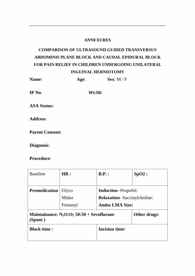
14. ANNEXURES 89
Page 10
LIST OF FIGURES
S.NO: TITLE PAGE
NO.
1. TRANSVERSUS ABDOMINIS PLANE BLOCK 6
2. ANATOMY OF ANTERO-LATERAL ABDOMINAL
WALL AND LUMBAR TRIANGLE OF PETIT 7
3. PATHWAY OF THORACIC NERVES IN THE
ABDOMINAL WALL 8
4. TYPICAL DISTRIBUTION OF NERVES IN
TRANSVERSUS ABDOMINIS PLANE 9
5. POSTERIOR APPROACH IN USG-GUIDED TAP
BLOCK 12
6. ANATOMY OF CAUDAL SPACE 15
7 ULTRASOUND VIEW OF THE THREE MUSCLE
LAYERS IN THE ABDOMINAL WALL 41
8
HYPOECHOIC SHADOW SEEN IN THE
TRANSVERSUS ABDOMINIS PLANE AFTER DRUG
DEPOSITION
41
9 CAUDAL BLOCK TECHNIQUE 42
Page 11
LIST OF TABLES
S.NO: TITLE PAGE
NO.
1. DEMOGRAPHIC VARIABLES 48
2. INTRA-OPERATIVE VARIATIONS IN HEART RATE 53
3. INTRA-OPERATIVE VARIATIONS IN MEAN
ARTERIAL PRESSURE 55
4. INTRAOPERATIVE ANALGESIC REQUIREMENTS 57
5. DURATION OF POSTOPERATIVE ANALGESIA 59
6. POST OPERATIVE PAIN SCORES 60
7. REQUIREMENTS OF RESCUE ANALGESIA IN THE
FIRST 12HRS OF POST-OPERATIVE PERIOD 62
8. NUMBER OF DOSES OF RESCUE ANALGESIA
REQUIRED 63
9. INCIDENCE OF SIDEEFFECTS 65
10. TIME TO FIRST URINE VOIDING IN THE POST-
OPERATIVE PERIOD 65
Page 12
LIST OF CHARTS
S.NO: TITLE OF THE CHART PAGE
NO.
1. AGE DISTRIBUTION 49
2. SEX DISTRIBUTION 50
3. WEIGHT DISTRIBUTION 51
4. HEIGHT DISTRIBUTION 52
5. HEART RATE VARIABILITY DURING INTRA-
OPERATIVE PERIOD 54
6. MEAN ARTERIAL PRESSURE VARIABILITY DURING
INTRA-OPERATIVE PERIOD 56
7.
INTRA-OPERATIVE REQUIREMENT OF
SUPPLEMENTATION BY FENTANYL GROUP T:
USG-GUIDED TAP BLOCK
58
8.
INTRA-OPERATIVE REQUIREMENT OF
SUPPLEMENTATION OF FENTANYL IN GROUP C:
CAUDAL BLOCK 58
9. DURATION OF POST-OPERATIVE ANALGESIA 59
10. FLACC PAIN SCORE (MEDIAN) IN THE POST-
OPERATIVE PERIOD (0-12HRS) 61
11. CUMULATIVE DOSE OF RESCUE ANALGESIA
REQUIRED IN FIRST 12 POST-OPERATIVE HOURS 62
12. NUMBER OF DOSES RESCUE ANALGESIA IN THE
FIRST 12hrs OF POST-OPERATIVE PERIOD 64
13. TIME TO FIRST URINE VOIDING IN THE POST-
OPERATIVE PERIOD 66
Page 14
INTRODUCTION
Perioperative pain in paediatric population is undertreated in a
substantial percentage, due to myths that children do not feel pain. It is also due
to the developmental and cognitive differences in children that pose difficulty
in assessment of their pain.1
In reality, children tend to have more physical and emotional reactions
to pain than adults. They require adequate pain relief to prevent acute and long
term adverse effects.
In order to provide optimal perioperative pain relief for children, local
anaesthetics should be a part of the initial pain management plan which is
accomplished by choosing a regional anaesthetic technique such as neuraxial
blockade, peripheral nerve blockade or local infiltration of the wound along
with General anaesthesia or sedation.2
Among the regional techniques, Caudal block is the oldest and most
commonly used regional technique of anaesthesia.3
It has been the most preferred technique for lower abdominal & lower
limb surgeries for infants & children but is associated with side effects such as
motor blockade in lower limbs and retention of urine.
Page 15
The main disadvantage of a single-shot Caudal block, is that it can give
only a short duration of post-op pain relief, requiring supplementationof other
analgesics.4
There is a recent trend towards regional nerve blocks, under ultrasound
guidance, as they provide better safety and are associated with lower incidence
of adverse effects compared to neuraxial blocks.
Transverse Abdominis Plane block (TAP) block, is an abdominal field
block, which provides myocutaneous analgesia, by depositing local anaesthetic
drug in the plane between the two muscles, namely Internal Oblique and
Transversus Abdominis.5 This fascial plane is a potential space where the
anterior rami of the thoracolumbar nerves (T6-L1) traverse and can be
effectively blocked before they supply the anterior abdominal wall muscles and
the skin. The plane can be reached after two pop offs felt while piercing the
fascial extensions of external and internal oblique, with the help of a needle
perpendicular to the skin while entering through the lumbar triangle of petit. It
has been shown that TAP block is easy and safe to perform under ultrasound
guidance.
It has been studied to be effective in reducing the post-operative pain
scores and morphine consumption in adult patients undergoing
appendicectomies, infra-umbilical surgeries, and caesarean sections.6 There are
few recent studies describing the efficacy of TAP block in paediatric
population. But there is not much information on how far it is superior to the
most preferred caudal block in paediatric surgeries.
Page 16
This study was conducted to compare the efficacy of the Ultrasound-
guided TAP block with the Caudal epidural block for intra-operative and post-
operative pain relief.
Since Inguinal hernia repair is one of the most frequently performed
paediatric surgical procedure7, this study was conducted in children undergoing
inguinal herniotomy.
Page 18
AIM OF THE STUDY
To evaluate the efficacy of the Ultrasound-guided Transversus
Abdominis Plane Block in comparison with the Caudal Epidural
Block, for pain relief in paediatric inguinal hernia repair surgeries.
Objectives of the study:
To compare the requirement of analgesia in the intra-operative
period with USG guided TAP block and Caudal block.
To Compare the Duration of Postoperative analgesia in Transversus
Abdominis Plane Block and Caudal Epidural Block.
To Compare the Quality of pain relief & Requirement of Rescue
Analgesia for the first 12hrs postoperatively
Incidence of any side effects
Page 19
TRANSVERSUS
ABDOMINIS PLANE
BLOCK
Page 20
REGIONAL ANAESTHESIA IN PAEDIATRICS
When regional technique was combined with general anaesthesia, it
results in reduced concentrations of potent inhaled agents used and reduction or
nil requirement of opioid during intra-operative period. This results in quick
recovery times and less nausea and vomiting post-operatively. It also
suppresses the neuroendocrine responses associated with general anaesthesia.
Regional anaesthesia as the sole technique for inguinal hernia repair has been
shown to decrease the incidence of postoperative apnea in former preterm
infants.
TRANSVERSUS ABDOMINIS PLANE BLOCK
HISTORY OF TAP BLOCK:
The TAP block is a novel regional anaesthetic technique. It provides
pain relief to the parietal peritoneum, skin and muscles of the anterior
abdominal wall.
TAP block was first described by Rafi in 2001.8 Rafi called it a refined
abdominal field infiltration. He utilised the lumbar triangle of Petit as the
anatomical landmark for his block and entered it to reach the TAP via single
pop off felt. The triangle is formed by the Lattisimus dorsi posteriorly,External
oblique medially and Iliac crest inferiorly.
McDonnell et al. presented the preliminary work on TAP blocks. He
studied it in cadavers and healthy volunteers, in the year 2004. Although the
technique was referred to as RAFI (Refined Abdominal Field Infiltration), by
Page 21
FIGURE 1: TRANSVERSUS ABDOMINIS PLANE BLOCK
(N, needle; ST, subcutaneous tissue; EO, external oblique muscle; IO,
internal oblique; TA, transversus abdominis; LD latissumis dorsi; QL,
quadratus lumborum, IL- Longissimus, Iliocostalis, PM-Psoas major,
MM- Multifidus muscle )
Page 22
the time Rafi had published his study in 2007, describing the sensory loss from
xiphoid area to pubic symphysis, McDonnell et al had already established the
term TAP block. He had demonstrated its use in open retropubic
prostatectomy.
In the same year, 2007, Hebbard et al described the use of Ultrasound
for administration of TAP block by real time imaging of the muscle layers and
the needle path, for better accuracy of the block.9
Hebbard, then described the subcostal approach in TAP block to target
the upper abdominal nerves, in the year 2008.10
TAP blocks continue to be studied for analgesia in various abdominal
surgeries.
ANATOMY OF TRANSVERSUS ABDOMINIS PLANE
There are 4 paired muscles in the abdominal wall, Rectus Abdominis
anteriorly and 3 musclesin the lateral abdominal wall namelyExternal oblique,
Internal Oblique and Transversus Abdominisfromsuperficial to deep.These
three muscles are fleshy only in the lateral abdominal region and become
aponeurotic medially.
Transversus Abdominis Block is performed in the lateral part of the
abdominal wall, between the TA an IO muscles. (Figure 1)
External Oblique Muscle
The External oblique is the most superficial muscle, originates from the
anterior angles of the lower eight ribs, the fibres take an oblique course
Page 23
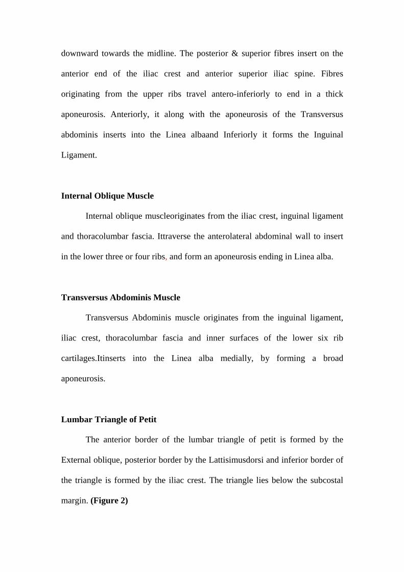
FIGURE 2: ANATOMY OF ANTERO-LATERAL ABDOMINAL WALL
ANDLUMBAR TRIANGLE OF PETIT
Page 24
downward towards the midline. The posterior & superior fibres insert on the
anterior end of the iliac crest and anterior superior iliac spine. Fibres
originating from the upper ribs travel antero-inferiorly to end in a thick
aponeurosis. Anteriorly, it along with the aponeurosis of the Transversus
abdominis inserts into the Linea albaand Inferiorly it forms the Inguinal
Ligament.
Internal Oblique Muscle
Internal oblique muscleoriginates from the iliac crest, inguinal ligament
and thoracolumbar fascia. Ittraverse the anterolateral abdominal wall to insert
in the lower three or four ribs, and form an aponeurosis ending in Linea alba.
Transversus Abdominis Muscle
Transversus Abdominis muscle originates from the inguinal ligament,
iliac crest, thoracolumbar fascia and inner surfaces of the lower six rib
cartilages.Itinserts into the Linea alba medially, by forming a broad
aponeurosis.
Lumbar Triangle of Petit
The anterior border of the lumbar triangle of petit is formed by the
External oblique, posterior border by the Lattisimusdorsi and inferior border of
the triangle is formed by the iliac crest. The triangle lies below the subcostal
margin. (Figure 2)
Page 25
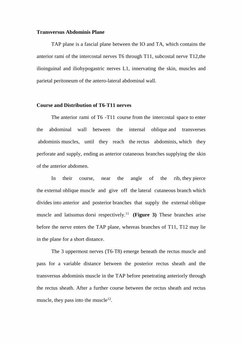
FIGURE 3: PATHWAY OF THORACIC NERVES IN THE
ABDOMINAL WALL
Page 26
Transversus Abdominis Plane
TAP plane is a fascial plane between the IO and TA, which contains the
anterior rami of the intercostal nerves T6 through T11, subcostal nerve T12,the
ilioinguinal and iliohypogastric nerves L1, innervating the skin, muscles and
parietal peritoneum of the antero-lateral abdominal wall.
Course and Distribution of T6-T11 nerves
The anterior rami of T6 -T11 course from the intercostal space to enter
the abdominal wall between the internal oblique and transverses
abdominis muscles, until they reach the rectus abdominis, which they
perforate and supply, ending as anterior cutaneous branches supplying the skin
of the anterior abdomen.
In their course, near the angle of the rib, they pierce
the external oblique muscle and give off the lateral cutaneous branch which
divides into anterior and posterior branches that supply the external oblique
muscle and latissmus dorsi respectively.11 (Figure 3) These branches arise
before the nerve enters the TAP plane, whereas branches of T11, T12 may lie
in the plane for a short distance.
The 3 uppermost nerves (T6-T8) emerge beneath the rectus muscle and
pass for a variable distance between the posterior rectus sheath and the
transversus abdominis muscle in the TAP before penetrating anteriorly through
the rectus sheath. After a further course between the rectus sheath and rectus
muscle, they pass into the muscle12.
Page 27
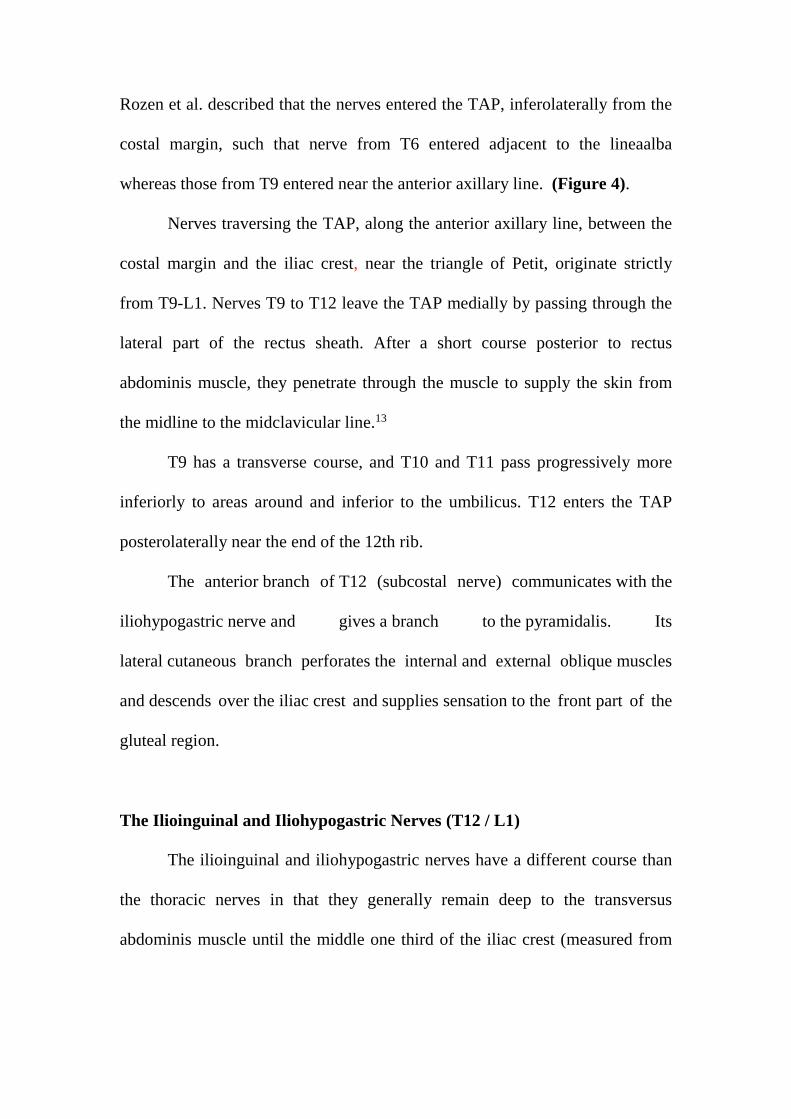
FIGURE 4: TYPICAL DISTRIBUTION OF NERVES IN
TRANSVERSUS ABDOMINIS PLANE
Page 28
Rozen et al. described that the nerves entered the TAP, inferolaterally from the
costal margin, such that nerve from T6 entered adjacent to the lineaalba
whereas those from T9 entered near the anterior axillary line. (Figure 4).
Nerves traversing the TAP, along the anterior axillary line, between the
costal margin and the iliac crest, near the triangle of Petit, originate strictly
from T9-L1. Nerves T9 to T12 leave the TAP medially by passing through the
lateral part of the rectus sheath. After a short course posterior to rectus
abdominis muscle, they penetrate through the muscle to supply the skin from
the midline to the midclavicular line.13
T9 has a transverse course, and T10 and T11 pass progressively more
inferiorly to areas around and inferior to the umbilicus. T12 enters the TAP
posterolaterally near the end of the 12th rib.
The anterior branch of T12 (subcostal nerve) communicates with the
iliohypogastric nerve and gives a branch to the pyramidalis. Its
lateral cutaneous branch perforates the internal and external oblique muscles
and descends over the iliac crest and supplies sensation to the front part of the
gluteal region.
The Ilioinguinal and Iliohypogastric Nerves (T12 / L1)
The ilioinguinal and iliohypogastric nerves have a different course than
the thoracic nerves in that they generally remain deep to the transversus
abdominis muscle until the middle one third of the iliac crest (measured from
Page 29
anterior superior iliac spine to posterior superior iliac spine); anterior to this,
they are usually found in the TAP.
The Iliohypogastric nerve divides into an anterior cutaneous branch,
supplying the skin over the hypogastrium, and a lateral cutaneous branch
supplying skin over the gluteal region. The Ilioinguinal nerve travels within the
inguinal canal and supplies sensation to the skin of the upper thigh, base of
penis and scrotum.
INDICATIONS
TAP block has been used as an adjunct for postoperative pain control in
abdominal, gynecologic and urologic surgery involving the T6 to L1
distribution. This block is indicated for postoperative analgesia for lower
abdominal surgeries such as appendicectomy, hernia repair, caesarean section,
abdominal hysterectomy and prostatectomy. Efficacy in laparoscopic surgeries
and renal transplantation have been demonstrated.14 Bilateral blocks can
be given for midline incisions or laparoscopic surgery.
CONTRAINDICATIONS
Infection at the site of injection, patient refusal or inability to cooperate,
and allergy to local anaesthetics.
COMPLICATIONS
Potential complications are intraperitoneal injection, owel
haematoma,transientfemoral nerve palsyand hepatic injury. Femoral nerve
Page 30
palsy can occur as the fascia iliaca is continuous with transversalis fascia.
Possibility of Local anaesthetic toxicity if large volumes are used.
TECHNIQUE
Various approaches for TAP Block:
Anatomical Landmark-Based Approaches
Ultrasound-Guided Approaches
Posterior Approach
Subcostal Approach
Oblique SubcostalApproach
Anatomical Landmark Based Technique:
Patientis placed in the supine position. A finger is rolled from the
anterior superior iliac spine along the top of the iliac crest until it dipped
inward into the lumbar triangle of petit. Further posterior movement, made the
fingertip, slip over the lateral border of latissimus dorsi, near its attachment to
iliac crest.15
A blunt tipped 24 gauge needle is inserted anterior to the fingertip, to
meet the external lip of the iliac crest and adjusted to advance further
perpendicular to the skin, until a distinct “pop” is felt, while piercing the
internal oblique, is the method described by Rafi.
Page 31
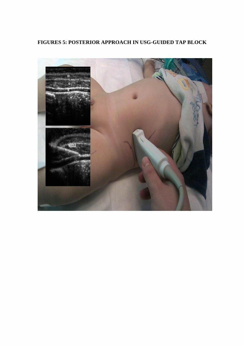
FIGURES 5: POSTERIOR APPROACH IN USG-GUIDED TAP BLOCK
Page 32
Whereas O’Donnell et al, describe “two pop”, entering cephalad to the iliac
crest, where the two pops are due to the passage of the needle through the
fascial extensions of External and Internal oblique.16
Ultrasound-Guided Approaches
Posterior Approach
In obese patients, and those with anatomical variations, the blind
anatomical landmark guided procedure may not be accurate in finding the TA
plane. Real time ultrasound imaging helps in locating the plane precisely. In
TAP block, performed under Ultrasound guidance, the probe is placed over the
anterolateral abdomen transversely to visualize the three muscles namely,
External, Internal oblique and Transversus Abdominis distinctly. The probe is
moved laterally to lie transversely across the midaxillary line. The Transversus
Abdominis Plane is visualised and a block needle is inserted in plane to the
probe, from antero-medial to postero-lateral direction and drug is deposited in
TA plane, which is seen as a hypoechoic shadow between the two muscles
(Internal Oblique and Transversus Abdominis). Figure 5.
Ultrasound-guided Subcostal Approach
The subcostal TAP block involves injection of local anesthetic into the
TAP lateral to the lineasemilunaris immediately inferior and parallel to the
costal margin, and is suitable for Periumbilical surgeries. In this approach, the
ultrasound probe is placed near the xiphoid process, parallel to the subcostal
Page 33
margin. TA is the most hypoechoic muscle, lying below the Rectus muscle.
After identification of the neurofascial plane between the TA muscle and
Rectus Abdominis, the block needle is inserted anteriorly, in plane to the probe.
Oblique Subcostal Approach
For supra umbilical procedures, Hebbard developed the oblique
subcostal approach. In this approach, the needle is inserted near the xiphoid and
directed inferolaterally along the costal margin.
LOCAL ANAESTHETIC DRUGS AND DOSAGE
Rafi described 20ml of Local anaesthetic for TAP block on each side for
adults.17 Long acting local anaesthetics such as 0.25% Bupivacaine, 0.25%
Levobupivacaine and 0.2-0.375% Ropivacaine at 0.3-0.6ml/kg, are generally
used with total dose not exceeding the toxic dose.
Page 34
CAUDAL
ANAESTHESIA
Page 35
CAUDAL ANAESTHESIA
Caudal anaesthesia is a technique of epidural blockade, performed via
the sacral hiatus, first reported to be used in children by Campbell in 1933.
Caudal anaesthesia is basically a single shot technique, but continuous
infusions as well as repeat doses can be given using placement of an indwelling
catheter.
Anatomy of Caudal space
Sacrum is a triangular bone formed by the fusion of the five sacral
vertebrae, located between the lumbar vertebrae and the coccyx. It is wedge-
shaped and presents markedly concave anterior and convex posterior surfaces.
The anterior surface bears four transverse lines which terminate on each side in
the four anterior sacral foramina, lateral to which is the fused lateral mass.
The anterior primary rami of the upper four sacral spinal nerves, as they
emerge from the anterior foramina, produce distinct neural grooves on the
lateral mass. The posterior surface of the sacrum is made up of the fused
vertebral arches which form the roof of the sacral canal. It presents a median
crest of fused spines, represented by small spinous tubercles. On either side of
this crest are the fused laminae which bear laterally an articular crest composed
of fused articular facets, each represented by a small tubercle.
Lateral to the articular tubercles are the four posterior sacral foramina,
which lie directly opposite their corresponding anterior foramina and which are
closed laterally by the posterior aspect of the lateral mass. The posterior rami of
Page 36
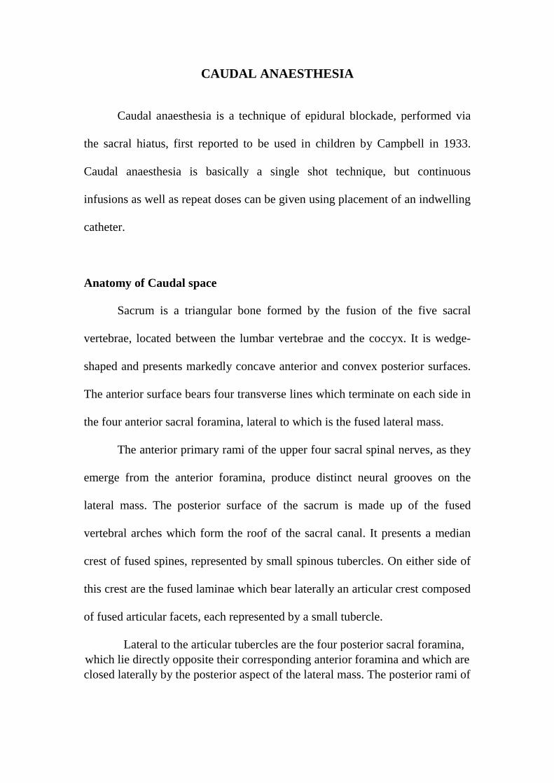
FIGURE 6: ANATOMY OF CAUDAL SPACE
Page 37
the sacral nerves emerge from the posterior sacral foramina. The posterior
sacral foramina are continuous with the epidural space in the sacral canal.
The caudal epidural space can be accessed by the defect formed due to
the failure of fusion of the fifth sacral vertebral arch, (sometimes fourth) which
is called as the Sacral hiatus.
It is covered by the sacrococcygeal membrane, which is about 1-3mm in
thickness, is a continuation of the ligamentum flavum. Sacral cornua are the
downward prolongation of the inferior articular processes of the fifth sacral
vertebrae, which form an important landmark, in identifying the hiatus. Sacral
hiatus is identified an indentation in the midline, that is found immediately
caudal to the sacral cornua. When the sacral cornua are difficult to identify due
to anatomical variations, the area of sacral hiatus may be identified as the apex
of an equilateral triangle, whose base is formed by a line joining the two
posterior superior iliac spines. (Figure 6)
The sacral hiatus is located more cephalad and the dura may end more
caudal, in comparison to adults, increasing the risk of inadvertent dural
puncture. The less densely packed epidural fat in infants and children,
facilitates cephalad spread of the local anaesthetics Caudal space is difficult to
reach in children older than 6-7yrs, as the space becomes more angulated and
there is reduced spread of local anaesthetic due to the increased density of the
epidural fat.
Page 38
INDICATIONS:
Caudal anaesthesia can be used for various lower abdominal surgeries,
below the level of the umbilicus, such as inguinal herniorrhaphy, orchidopexy,
circumcision, hypospadias repair, and for distal orthopaedic procedures. It can
be used for even upper abdominal, thoracic surgeries in selected patients, under
special circumstances. It can be combined with general anaesthesia for post-
operative pain relief or can be used as the sole anaesthetic technique.
CONTRAINDICATIONS:
Major malformations of the sacrum (myelomeningocele, open spina
bifida), meningitis and intracranial hypertension.
TECHNIQUE:
Patient Position:
Technique can be performed with the child in the lateral decubitus
position, with hips flexed, with the dependent leg less flexed than the non-
dependent leg. Patient can also be placed in semiprone or prone position with a
rolled towel under the pelvis, or with the legs flexed as in “frog” position.
Landmark Identification:
The two sacral cornua which limit the V-shaped sacral hiatus, are
located by palpation along the spinal process line at the level of the
sacrococcygeal joint.
Page 39
Needle Insertion:
After sterile preparation, the hiatus is identified with the index or middle
finger of the non-dominant hand and a short beveled, 22 or 23 gauge needle is
inserted in the midline, at an angle of 45-60 degrees to the plane of the skin.
The needle is advanced until the sacrococcygeal membrane is pierced, which is
felt as a distinct pop. The angle of the needle is then decreased to 20-30 degrees
and the needle is advanced 2-4mm into the caudal space.
The successful needle placement can be confirmed using the “whoosh”
test, where an assistant keeps a stethoscope over the midline lumbar spine for a
characteristic whoosh sound on injection of 2-3ml of air via the caudal needle.
Ultrasound imaging can also be used to confirm the successful needle
placement, by the displacement of the dura mater, during injection of saline
into the caudal space. After careful aspiration, confirming the absence of CSF
or blood, the local anaesthetic is injected.
Factors affecting the dose and spread of local anaesthetic in caudal space:
Factors which are known and can be controlled are: Age, weight, height,
dose (both volume and concentration of the drug), speed of injection, and
patient position
Page 40
Factors which are unknown or which cannot be controlled are:
Size of the caudal space
Size of the sacral canal and its patency
Presence of any bony distortions or septa in the canal
Nature of the fatty tissues in the epidural space
Neural tissue and dural cuff permeability to the local anaesthetic
Page 41
Local anaesthetic Drugs:
Bupivacaine is the most commonly used local anaesthetic in children.
Local anaesthetic concentration and volume are important factors in
determining the density and sensory level of blockade. Mostly, paediatric
patients receive light general anaesthesia along with the regional technique, and
hence it is enough if the regional technique is able to provide good intra-
operative and post-operative analgesia. Therefore low doses of local
anaesthetics are preferred to prevent any drug toxicity.
The volume of local anaesthetic necessary is calculated based on the
patient’s weight, given by using the Armitage formula,
0.5ml/kg blocks all the sacral dermatomes,
1ml/kg blocks all the sacral and lumbar dermatomes,
1.25ml/kg blocks till mid-thoracic level.
Hernia repair requires blockade up to T10 dermatome level, but
1.25ml/kg is usually not used for the fear of the excessive cephalic spread.
Hence 1ml/kg of 0.25% bupivacaine was used in our study.
COMPLICATIONS:
Subcutaneous injection, intraosseous injection, intravascular injection,
subarachnoid injection
Page 42
PHARMACOLOGY
OF BUPIVACAINE
Page 43
PHARMACOLOGY OF BUPIVACAINE
Bupivacaine was the first local anaesthetic that combined the properties
of an acceptable onset, long duration of action, profound conduction blockade,
and significant separation of sensory anaesthesia and motor blockade.
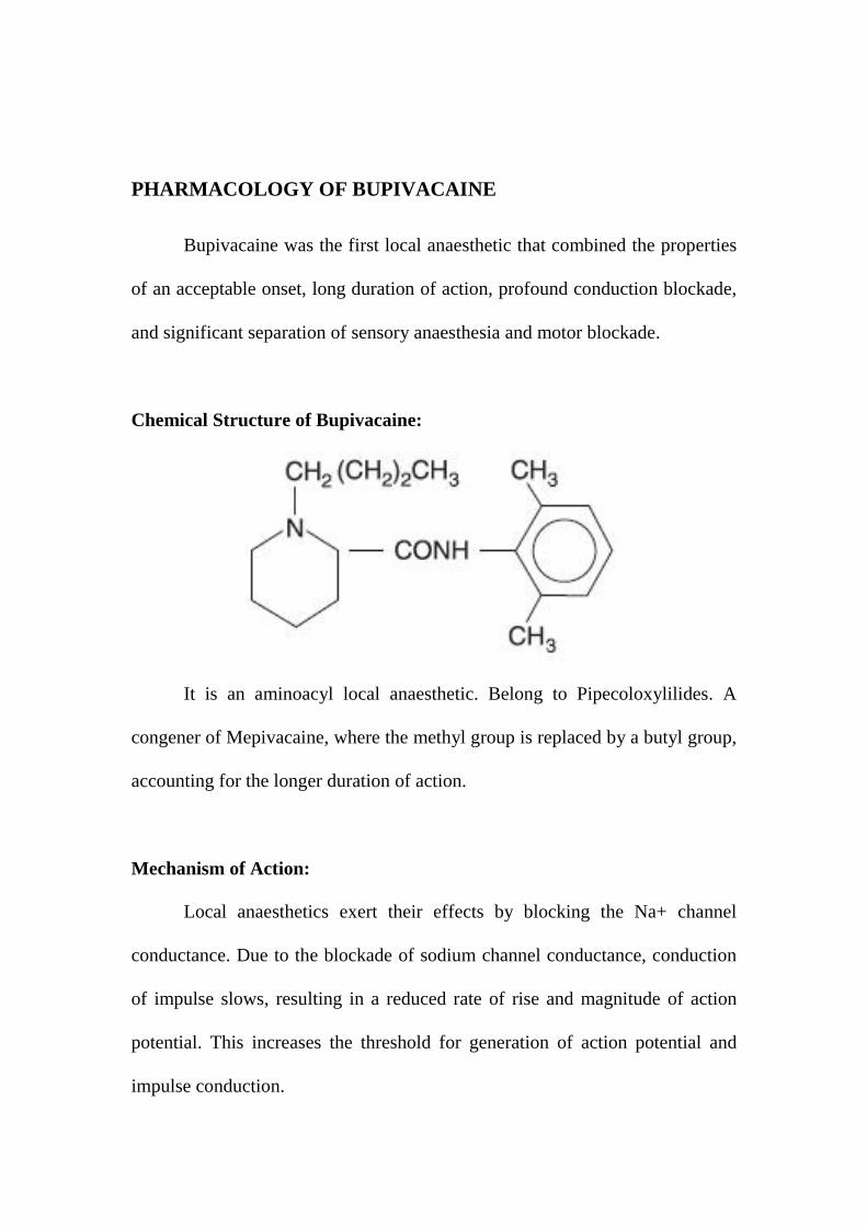
Chemical Structure of Bupivacaine:
It is an aminoacyl local anaesthetic. Belong to Pipecoloxylilides. A
congener of Mepivacaine, where the methyl group is replaced by a butyl group,
accounting for the longer duration of action.
Mechanism of Action:
Local anaesthetics exert their effects by blocking the Na+ channel
conductance. Due to the blockade of sodium channel conductance, conduction
of impulse slows, resulting in a reduced rate of rise and magnitude of action
potential. This increases the threshold for generation of action potential and
impulse conduction.
Page 44
Onset of Action:
Depends on lipid solubility, pKa. Bupivacaine has a lipid solubility of
28 and a pKa of 8.1. Less potent, less lipid soluble agents tend to have a faster
onset. Bupivacaine is highly lipid soluble and has a slower onset of action.
pKa is the pH at which the unionised form exists in equilibrium with
ionised form. The greater the fraction of unionised form, the faster the
permeation into the nerve cell membranes and faster the onset of action. At
physiological pH, 17% of the molecule exist in unionised fraction, resulting in
slower onset of action.
USES:
This agent is used for various regional anesthetic procedures, including
infiltration, peripheral nerve blocks, and epidural and spinal anesthesia. It is
used in epidural labour analgesia, where only sensory blockade is required.
Bupivacaine causes differential blockade of sensory and motor and hence is the
most preferred drug in labour analgesia
DURATION OF ACTION:
Since bupivacaine has a high lipid solubility and is 95% protein bound,
it has a longer duration of action. The average duration of surgical anaesthesia
with bupivacaine varies from approximately 3 to 10 hours. Its longest duration
Page 45
of action occurs with major peripheral nerve blocks such as brachial plexus
blockade
Pharmacokinetics:
Absorption:
Since bupivacaine is more lipid soluble and highly protein bound, its
absorption into systemic circulation is slow. It also depends on the site of
injection.
Absorption of bupivacaine is high in caudal block.
Distribution:
Has a volume of distribution of 73L
Elimination and Metabolism:
Has a clearance of 0.47L/min. Bupivacaine undergoes aromatic
hydroxylation, N-dealkylation, amide hydrolysis, and conjugation.
Dosage in Paediatric age group:
2 mg/kg of plain bupivacaine in concentration of 0.125-0.25%.
Pharmacokinetic studies of a single dose of racemic bupivacaine (2.5
mg/kg) injected in the caudal space have demonstrated differences between
infants and children. Infants have a greater volume of distribution (3.9 L/kg
versus 2.7 L/kg), an increased elimination half-life (7.7 versus 4.6 hours), and
Page 46
decreased clearance (7.1 versus 10.0 mL/kg per min) compared with older
children
Systemic toxicity
Systemic toxicity is due to an excess plasma concentration of the drug.
Plasma concentrations of local anesthetics are determined by the rate of drug
entrance into the systemic circulation, relative to their redistribution to inactive
tissue sites and clearance by metabolism.
Accidental direct intravascular injection of local anesthetic solutions
during the performance of peripheral nerve block anesthesia or epidural
anesthesia is the most common mechanism for the production of excess plasma
concentrations of local anesthetics. Less often, excess plasma concentrations of
local anesthetics result from the absorption of the local anesthetic from the
injection site.
Systemic toxicity manifests as CNS toxicity and CVS toxicity
depending on the concentration in plasma.
Acid–base status can alter the CNS activity of local anesthetic agents.
The convulsive threshold of various local anesthetics is inversely related to the
arterial CO2 tension. An increase in PaCO2, decreases the convulsive threshold
of local anaesthetics.
A decrease in arterial pH also decreases the convulsant threshold of
these agents. Hypercarbia increases cerebral blood flow, which results in a
greater uptake of local anesthetic by the brain. Hypercarbia and/or acidosis also
Page 47
decrease the plasma protein binding of local anesthetic agents, which will
increase the proportion of free drug available for diffusion into the brain.
Bupivacaine has a direct effect on both cardiac muscle and vascular smooth
muscle. These agents alter the heart’s electrical and mechanical activity. The S
forms of bupivacaine are less cardiotoxic than the R form.
Bupivacaine shows selective cardiac toxicity on toxic plasma
concentrations. It causes hypotension, cardiac dysrhythmias, and
atrioventricular heart block.
Bupivacaine depresses Vmax(depresses the rapid phase of depolarization)
considerably more than lidocaine. During diastole, highly lipid soluble
bupivacaine dissociates from sodium ion channels at a slow rate when
compared with lidocaine, thus accounting for the drug's persistent depressant
effect on Vmax and subsequent cardiac toxicity. At normal heart rates, diastolic
time is sufficiently long for lidocaine dissociation, but bupivacaine block
intensifies and depresses electrical conduction, causing reentrant-type
ventricular dysrhythmias.
Page 48
LARYNGEAL MASK
AIRWAY
Page 49
LARYNGEAL MASK AIRWAY
Laryngeal mask airway (LMA) is a supraglottic airway device
developed by British Anesthesiologist Dr. Archie Brain. It has been in use
since 1988.
The LMA is a good airway device in many settings, including the
operating room, the emergency department, and out-of-hospital care, because it
is easy to use and quick to place, even for the inexperienced provider18.
It is a device which occupies the space shared by alimentary and
reparatory tract. When properly inserted, it doesn’t stimulate the respiratory
tract.
INDICATIONS
Its use in operating room for short elective procedures, when
endotracheal intubation is not necessary.19
Difficult airway
Cardiac arrest
Conduit for intubation
Pre-hospital airway management
CONTRAINDICATIONS
Absolute contraindications
Limited mouth opening
Complete upper airway obstruction
Page 50
Relative contraindications (in the elective setting)
Increased risk of aspiration
Prolonged bag-valve-mask ventilation, Morbid obesity, Second or third
trimester pregnancy, Patients who have not fasted before ventilation, Upper
gastrointestinal bleed
Suspected or known abnormalities in supraglottic anatomy
Need for high airway pressures (except Proseal)
LMA USE IN PAEDIATRICS
LMA is increasingly being used in paediatrics as it is less invasive
compared to endotracheal intubation. LMA was found to provide a better and
more secure airway than the face mask without direct tracheal intervention. It is
easy to use and can be used in place of the face mask20
LMA has several advantages over endotracheal tube (ETT):
Rapidity & Ease of insertion in securing the airway
Laryngoscopy and muscle relaxants are not required
Changes in haemodynamic parameters are less with LMA use than
with endotracheal intubation.21
Less stimulating than ETT, hence advantageous in situations of
reactive airway
Lesser incidence of sore throat
Page 51
LMA is advantageous over Facemask
An airtight seal is easier to obtain with an LMA
Episodes of desaturation are lesser with LMA
Work of breathing is less
Useful in cases of difficult mask ventilation
Frees hands of the Anaesthesiologist
Limitations and complications of LMA use in children
Laryngospasm and airway obstruction, when it is inserted/removed
in a lighter plane.
Airway obstruction can also occur due to malpositioning
Risk of aspiration as LMA does not form a tight seal around the
larynx.
Younger and smaller the child, the risk of complications are higher.22
Most problems were documented with the use of 1 size LMA, which reduced
with increasing experience.23
Timing of Removal of LMA
Timing of removal of the LMA in children is controversial. Both awake
and deep removal of LMA have been advocated.
Awake removal ensures return of protective reflexes.But it also
attends to the problems of airway reactivity. The LMA should not be
Page 52
removed in light plane as this may cause coughing and
laryngospasm.24
Removing the LMA when the child is deep avoids the risk of
laryngospasm. The LMA can be removed at a plane that would allow
an endotracheal tube to be removed.
Ambu Laryngeal Mask
The Ambu Laryngeal Mask is a disposable device that has a cuff that is
tapered at the tube. The airway tube is larger and more rigid than that of the
LMA and is precurved. It has no aperture bars. The inflation line is attached to
the airway tube. It has a reinforced tip. It is available in seven sizes
Size
Patient
Weight
(kg)
Maximum Cuff
Inflation Volume (mL)
1 Up to 5 4
1.5 5 to 10 7
2 10 to 20 10
2.5 20 to 30 14
3 30 to 50 20
4 50 to 70 30
5 70–100 40
6 >100 50
Page 53
REVIEW OF
LITERATURE
Page 54
REVIEW OF LITERATURE
A systematic search of the literature identified some randomized clinical
trials investigating the effect of TAP block on post-operative pain. The surgical
procedures included large bowel resection with a midline abdominal incision,
caesarean delivery via the Pfannenstiel incision, abdominal hysterectomy via a
transverse lower abdominal wall incision, open appendectomy and
laparoscopic cholecystectomy. Overall, most studies have demonstrated
clinically significant reductions of post-operative opioid requirements and pain
scores.25
Petersen et al. reviewed 7 randomized, double-blinded, clinical trials
of both landmark-based (n = 3) and ultrasound-guided (n = 4) TAP
blocks for managing postoperative pain after abdominal surgery with
incisions below the level of the umbilicus.26
All 7 studies compared pain-related outcomes with TAP blocks as
part of a multimodal postoperative analgesic regimen. Morphine PCA
± acetaminophen ±nonsteroidal anti-inflammatory drugs was most
commonly used to complement TAP blocks. In one study, intrathecal
morphine was also part of the analgesic regimen.
A meta-analysis of these 7 studies (180 cases and 184 controls)
demonstrated an average reduction in 24-hour morphine consumption
of 22mg in favour of TAP block patients compared with standard
management.
Page 55
Furthermore, TAP blocks were associated with reduced early
postoperative visual analog scores (VAS) both at rest and during
mobilization in 4 of the 7 studies (1 study did not record VAS
scores).
Postoperative sedation, as well as postoperative nausea and vomiting
(PONV) was marginally reduced in patients with TAP blocks.27
Another meta-analysis by Charlton et al., which reviewed 236
participants from 5 studies (including landmark- and ultrasound-
guided TAP blocks), demonstrated a significant reduction in 24-hour
morphine requirements in TAP block patients compared to controls.28
A significant difference in postoperative sedation, nausea and
vomiting was not appreciated between TAP-block and non-TAP
block patients in this paper.
Bharti et al. randomized 40 patients undergoing colorectal surgery to
standard treatment (diclofenac and intravenous morphine) and
bilateral intraoperative TAP block with either 0.25% bupivacaine (n
= 20) or saline (n = 20).29
The bupivacaine group had a significant reduction in 24-hour
morphine requirements (6.45 ± 3.26mg versus 17.55 ± 5.78mg;
P<0.0001) as well as a significant reduction in early postoperative
pain scores both at rest and with coughing.
Page 56
Furthermore, early postoperative sedation scores were significantly
lower in the bupivacaine group, and patient satisfaction was higher
(6.8 ± 1.1mg versus 3.5 ± 1.5mg; P<0.001).
There was no significant difference between groups in the incidence
of PONV. Patients in the control group experienced significantly
more severe PONV, requiring pharmacological intervention.
Hivelin et al. studied the effect of TAP blocks for postoperative
analgesia in patients with abdominal deep inferior epigastric
perforator flaps for breast reconstruction.30
The TAP block group (n = 15) required significantly less morphine
(median and interquartile range: 28mg (27mg– 38mg) versus 42mg
(36mg–46mg); P = 0.0057) than controls (n = 15) in the first 24 hours
after surgery.
Early postoperative numerical pain scale scores were also
significantly lower in the TAP block group compared to the non-
TAP-block patients.
However, no difference was observed between groups for
postoperative sedation, PONV and 48-hour satisfaction with pain
management.
In another study, Baaj et al. randomized 40 women to receive either
local anaesthetic (n = 20) or saline (n = 20) for TAP blocks in
addition to a plain bupivacaine spinal block for elective caesarean
section.31
Page 57
A significant reduction in 24-hour morphine requirement was
observed in the local anaesthetic TAP block group versus controls
(26mg ± 5mg versus 63mg ± 5mg; P<0.05).
McDonnell et al. and Belavy showed decreased 24-hour morphine
consumption following C-section in patients who received TAP
blocks in addition to plain local anaesthetic spinal blocks when
compared to patients with just local anaesthetic spinal blocks.3233
Reports which do not demonstrate an analgesic benefit to TAP blocks
when compared to standard therapy.
Griffiths et al. randomized 65 patients undergoing surgery for
presumed gynecologic malignancy to standard treatment (parecoxib,
acetaminophen, and morphine) plus ultrasound-guided TAP block
with either Ropivacaine (n = 32) or saline (n = 33).34
No significant difference was found in the two groups for 24-hour
morphine consumption, VAS scores at rest, VAS scores with
coughing, patient satisfactionor incidence of nausea and pruritis.
The authors speculated that the negative study may have been due to
a combination of factors including a high incidence of obesity in the
study population leading to potentially more technical failures, a wide
age rangeand the fact that 18 of the 65 patients had incisions that
extended above the umbilicus.
Page 58
The authors also hypothesized that the study population had a larger
variation in “surgical insult” that is, some cases involved more organ
manipulation and dissection resulting in more visceral pain, for which
TAP blocks would be less effective than those for parietal/incisional
pain.
McMorrow et al. randomised 80 patients to 4 equal groups (n = 20 in
each arm) and reported that they found no overall analgesic advantage
to TAP blocks and no incremental benefit of adding TAP blocks when
patients receive intrathecal morphine35.
They also reported similar overall patient satisfaction among groups
despite more frequent pruritis in patients who received intrathecal
morphine.
Studies comparing TAP blocks to epidural analgesia.
Kadam and Moran had conducted a retrospective matched case-
control study comparing continuous TAP block catheters (posterior
and subcostal approaches; n = 15) to thoracic epidural analgesia (n =
15).36
Except for assessments in the post anaesthesia care unit, there was no
appreciable difference in pain scores between the two groups over a
3-day follow-up period. While patient satisfaction was similar
between groups, the TAP block group required a significantly higher
amount of breakthrough fentanyl over the study period. Therapeutic
Page 59
failure rate was higher in the epidural group (patchy block in 4
patients) versus the TAP catheter group (unilateral block in 2
patients). Hypotension was reported in 2 patients from the epidural
group.
Clinical trials which had studied use of TAP blocks in pediatric population
Tanaka M et al investigated 64 pediatric patients of age 5-12 years
undergoing TAP and non-TAP block for receiving bone graft from
the ilium to the alveolar cleft, and concluded that TAP block was
effective due to reduced requirement of postop analgesics.
Sandermanertal studied the effect of adding TAP blocks to local
anaesthetic infiltration on morphine consumption and postoperative
pain in 93 children undergoing laparoscopic appendicectomy of age
7-13yearsand found that TAP blocks increased anaesthesia
administration time by 14 min on average but offered no clinically
important benefit over local anaesthetic port-site infiltration.37
Fredrickson MJ et al compared ilio-inguinal block to TAP block in
children undergoing inguinal surgery and showed that pain was more
frequent and increased ibuprofen use in TAP block, whereas
recovery room pain, morphine consumption and post discharge
ibuprofen use, comfort and satisfaction scores were similar between
groups38
Page 60
Review of studies which compared TAP block with Caudal block
Dalia et al studied 40 patients belonging to age group 6 months to
6years undergoing surgical open pyeloplasty, with 20 under caudal
group ( with 1.25ml/kg of 0.25% bupivacaine as a single shot) and
20 under TAP block ( under ultrasound guidance, with 0.3ml/kg).
The study revealed that ultrasound-guided TAP block provided
significantly prolonged postoperative analgesia and reduced the
postoperative analgesic requirements as compared with caudal block
in pediatric patients undergoing open pyeloplasty39
Wafaa Mohammed Aslam et al studied 60 children of age group 2-
7years undergoing lower abdominal surgery, by allocating them
under three groups, namely USG guided TAP block, USG guided
Caudal block and control block (20 in each group).
The study revealed that only 3/20 in TAP block group required
rescueanalgesia whereas all patients in Caudal block group and
control group required rescue analgesia. It also showed that there
was significant difference in pain score after 6hrs postoperative
between USG guided TAP block and USG guided Caudal block.
Review of Safety of TAP block
In a study conducted by Long, Justin B et al, on Safety analysis of
TAP block, 1994 cases undergoing TAP block were included in the
study and it was found that the upper incidence of overall
Page 61
complications associated with the TAP block in children was 0.3%.
It said that the complications were minor and did not require any
intervention.40
Reviews of journals which compared the efficacy of Caudal block with
other regional techniques such as wound infiltration, inguinal field block,
paravertebral block
In 2 studies which reviewed Caudal block with local infiltration,
both interventions were performed after surgery.41 There were 4
other studies that performed caudal preoperatively and infiltration
postoperatively.42 Except for Lafferty and colleagues (only
orchidopexy), all studies included hernia surgeries only. All used
bupivacaine in a concentration of 0.25% for Caudal Block and
0.25%–0.5% for Local infiltration. The volume ranged from 0.7 to
1.0mLkg−1 (Caudal block) and from 0.2 to 0.7mLkg−1 (Local
infiltration). Only Conroy and colleagues used epinephrine along
with bupivacaine.43
Variations of the infiltration techniques involved infiltration of the
wound site through the skin and infiltration of fascia or aponeurosis
before closure. No study used image guidance.
Abdellatif compared Ultrasound-guided Inguinal Nerve block (INB)
with blind Caudal Block in children having inguinal hernia
surgeries.44 Average pain scores and use of rescue medications
Page 62
were not found to be significantly different. Use of rescue analgesia:
early period: 5/25(CB) and 7/25(INB); late period: 9/25 (CB) and
8/23 (INB)
Tug and colleagues used a single shot lumbar Paravertebral block
(PVB) to compare with Caudal block (CB) for inguinal surgeries.45
70 patients were evaluated,with 35 in each group. Six patients had a
failed block (two in PVB and four in CB) and 12/35 patients in CB
and 4/35 patients in PVB needed rescue analgesia during the early
period with a relative ratio of 3.0. They also observed 2 cases of
motor block in CB compared to 0 in PVB.
Page 63
MATERIALS
AND
METHODS
Page 64
MATERIALS AND METHODS
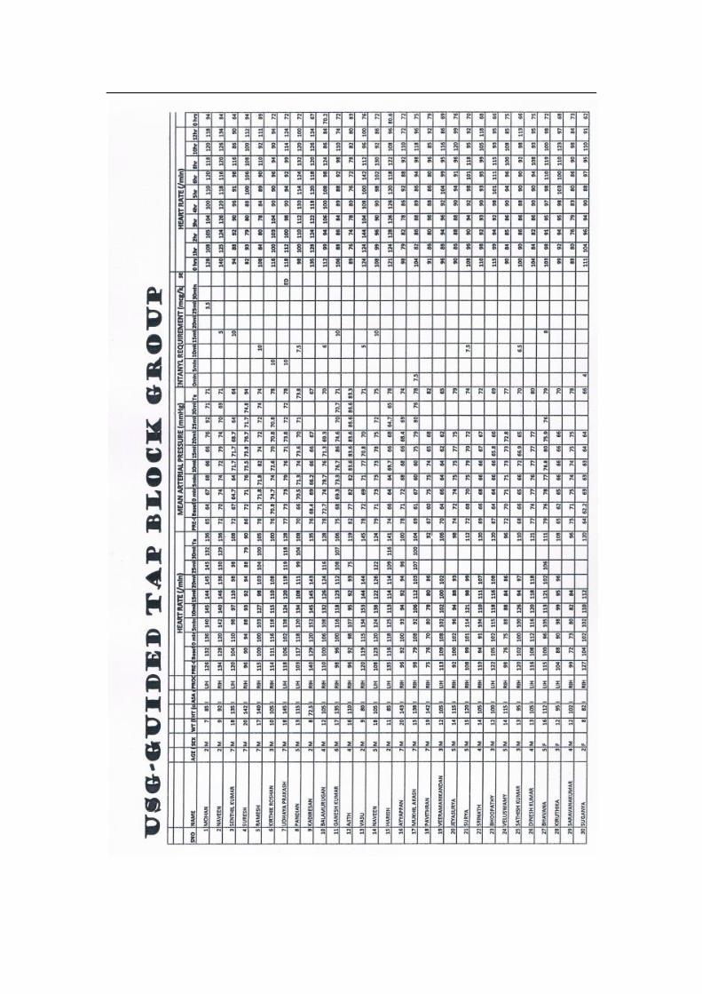
A Randomised Control Trial was conducted in 60 children, undergoing
unilateral inguinal herniotomy in Mahatma Gandhi Memorial Government
Hospital, attached to K.A.P.V. Government Medical College, Trichy.
The Institutional Ethical Committee Approval was obtained. Sixty
children satisfying the inclusion criteria were included in the study, after
obtaining informed consent from the parents/guardian of the patients. They
were randomly allocated into two groups, Group T and Group C, with 30
patients in each, using computer allocated random numbers.
Group T (n=30) Receiving USG-guided TAP Block with 0.5ml/kg of
0.25% bupivacaine.
Group C (n= 30) Receiving Caudal Epidural Block with 1ml/kg of
0.25% bupivacaine
INCLUSION CRITERIA:
Children of age group 1-7 yrs, weighing 5-20kg, of ASA status I – II
To undergo Unilateral Inguinal Herniotomy, were included in the
study.
EXCLUSION CRITERIA:
Infants less than 2yrs of age and more than 7yrs of age
Infants less than 5 kg and children more than 20kg
Children undergoing Bilateral Inguinal Herniotomy
Page 65
Children with known allergy to the drugs used in the study
Local infection at the site of the block
Children belonging to ASA status III, IV
Children with contraindications for caudal anaesthesia such as major
sacral malformations, those with meningitis, with raised intracranial
hypertension.
Parent refusal for consent
EXCLUSION OF PATIENTS AFTER SELECTION FOR THE STUDY:
Cases in which the block had failed, were excluded from the study.
METHODOLOGY
All children were fasted 8hrs for milk and solids and 3 hrs for clear
liquids.
PROCEDURE
PREMEDICATION:
All the children in both the groups were sedated with oral midazolam
syrup, 0.5mg/kg 30 minutes before the surgery and were shifted to the
operating room and an intravenous access was secured using a 22gauge IV
cannula. Baseline vital signs were recorded following application of standard
monitoring (ECG, HR, NIBP, SpO2).
Page 66
Children in both the groups were premedicated with glycopyrrolate
8mcg/kg given intravenously 5 min before induction.
GENERAL ANAESTHESIA:
Jackson Rees modification of Ayre’s T-piece was used for General
anaesthesia. Fentanyl 1mcg/kg was given intravenously, during preoxygenation
with 100% oxygen for 3 mins with an appropriate size face mask. Anaesthesia
was induced with propofol 2mg/kg and muscle paralysis was achieved using
succinylcholine 1.5mg/kg. An Ambu LMA of appropriate size was inserted and
the child was allowed to breathe spontaneously. Anaesthesia was maintained
with 50 %N2O: 50% O2 and 1.5-2% Sevoflurane titrated accordingto BP.
The children in Group T received Ultrasound guided Transversus
Abdominis Plane block with 0.5ml/kg of 0.25% isobaric bupivacaine after
insertion of the LMA Children in Group C received caudal epidural block with
1ml/kg of 0.25% isobaric bupivacaine after the insertion of LMA
Group T: Ultrasound-guided Transversus Abdominis Plane Block:
After insertion of the LMA, with the child in the supine position, skin
disinfection was done using povidone-iodine solution, and a high frequency (6-
13Hz) linear probe, connected to Sonoray Ultrasound machine was used to
scan the anteriorabdominal wall.
Sterility was maintained by covering the edge of the probe with a sterile
transducer sheath and applying a sterile gel over the area to be scanned.
Page 67
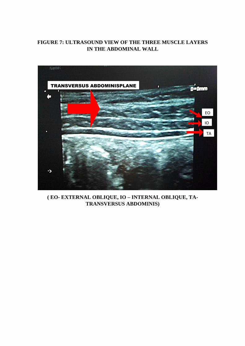
FIGURE 7: ULTRASOUND VIEW OF THE THREE MUSCLE LAYERS
IN THE ABDOMINAL WALL
( EO- EXTERNAL OBLIQUE, IO – INTERNAL OBLIQUE, TA-
TRANSVERSUS ABDOMINIS)
TRANSVERSUS ABDOMINISPLANE
EO
IO
TA
Page 68
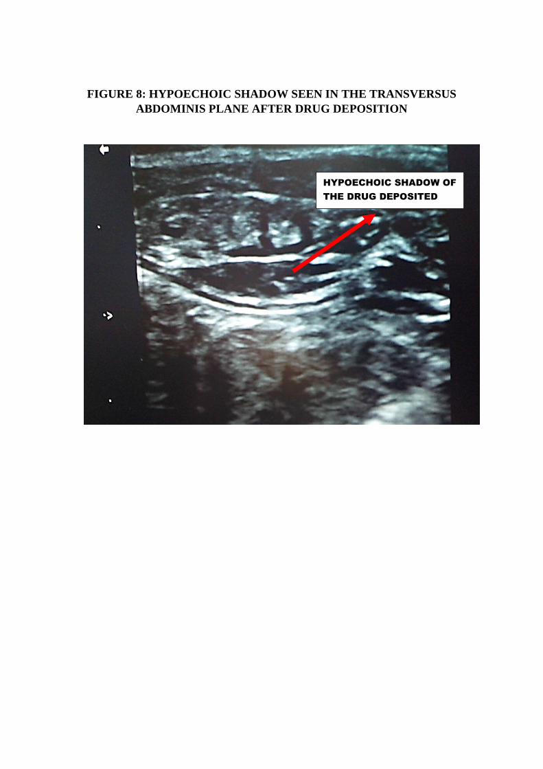
FIGURE 8: HYPOECHOIC SHADOW SEEN IN THE TRANSVERSUS
ABDOMINIS PLANE AFTER DRUG DEPOSITION
HYPOECHOIC SHADOW OF
THE DRUG DEPOSITED
Page 69
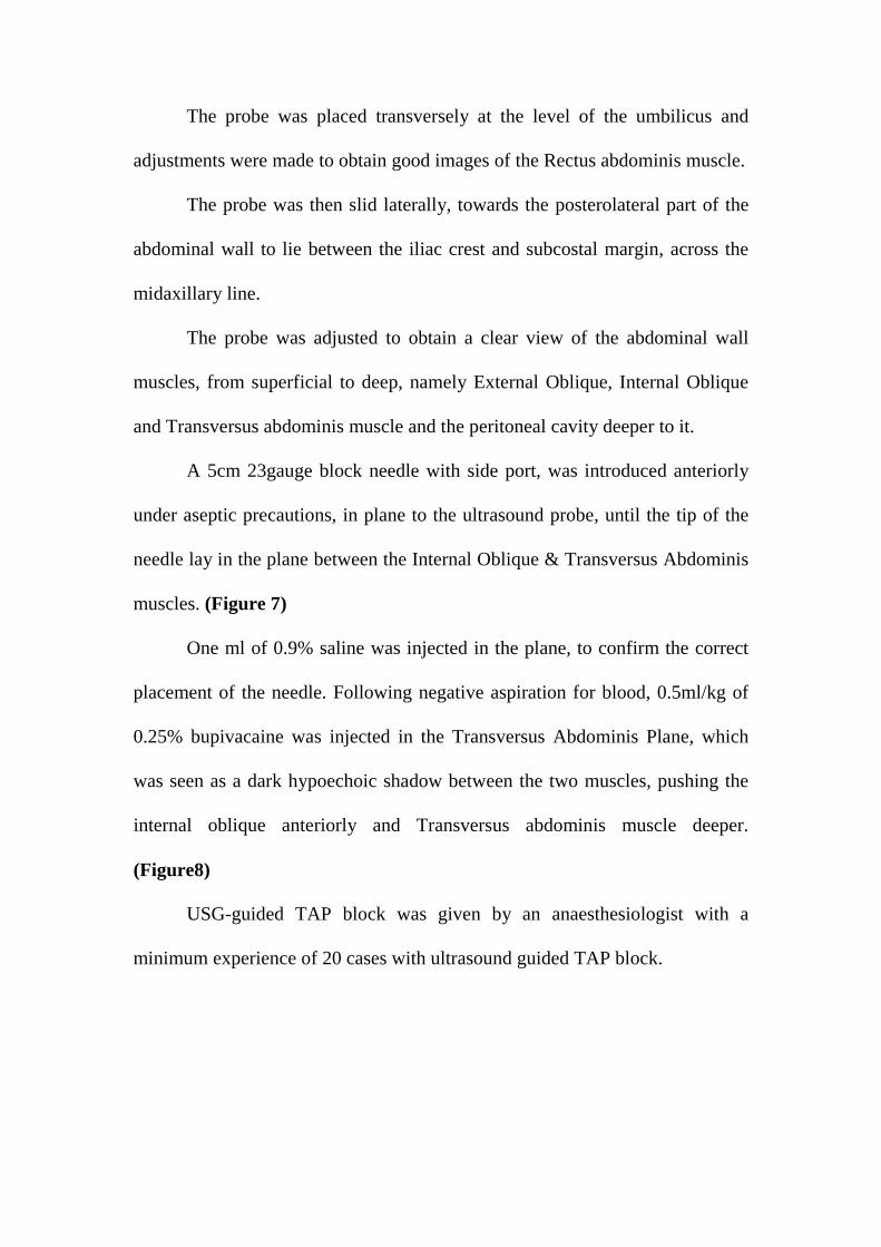
The probe was placed transversely at the level of the umbilicus and
adjustments were made to obtain good images of the Rectus abdominis muscle.
The probe was then slid laterally, towards the posterolateral part of the
abdominal wall to lie between the iliac crest and subcostal margin, across the
midaxillary line.
The probe was adjusted to obtain a clear view of the abdominal wall
muscles, from superficial to deep, namely External Oblique, Internal Oblique
and Transversus abdominis muscle and the peritoneal cavity deeper to it.
A 5cm 23gauge block needle with side port, was introduced anteriorly
under aseptic precautions, in plane to the ultrasound probe, until the tip of the
needle lay in the plane between the Internal Oblique & Transversus Abdominis
muscles. (Figure 7)
One ml of 0.9% saline was injected in the plane, to confirm the correct
placement of the needle. Following negative aspiration for blood, 0.5ml/kg of
0.25% bupivacaine was injected in the Transversus Abdominis Plane, which
was seen as a dark hypoechoic shadow between the two muscles, pushing the
internal oblique anteriorly and Transversus abdominis muscle deeper.
(Figure8)
USG-guided TAP block was given by an anaesthesiologist with a
minimum experience of 20 cases with ultrasound guided TAP block.
Page 70
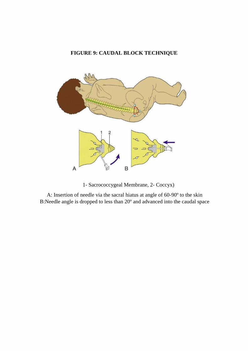
FIGURE 9: CAUDAL BLOCK TECHNIQUE
1- Sacrococcygeal Membrane, 2- Coccyx)
A: Insertion of needle via the sacral hiatus at angle of 60-90º to the skin
B:Needle angle is dropped to less than 20º and advanced into the caudal space
Page 71
Group C: Caudal Epidural Block:
Patients were placed in left lateral position with knees drawn up to
chest. After skin preparation with betadine solution, the sacral hiatus was
identified by palpating the sacral cornua with the index finger of the non-
dominant hand.
A 23gauge needle was inserted at 45-60 degrees to the skin over the
sacral hiatus. After piercing the sacrococcygeal membrane which was felt as a
distinct pop, the needle angle was dropped to 20 to 40 degrees from the skin
and the needle was advanced about 2-4mm into the caudal space. (Figure 9)
The position of the needle in the caudal space was confirmed by the
“whoosh test” by injecting saline.
After careful negative aspiration for blood or CSF, 1ml/kg of 0.25%
bupivacaine was injected into the caudal space.
Caudal block was given by an experienced anaesthetist with a minimum
experience of 2 years in anaesthesiology and has performed more than 30
Caudal blocks.
AFTER ADMINISTRATION OF THE BLOCK
The surgical procedure was started 15mins after the administration of
the block, Caudal or USG-guided TAP block, according to the group to which
they belonged to HR, NIBP and SpO2 were recorded every 5mins from the
beginning of the surgical procedure until the removal of the LMA, by a blinded
investigator, who was not aware of the block given.
Page 72
When there was more than 20% increase in heart rate or mean arterial
pressuredespite administration of 1 MAC of sevoflurane intraoperatively, the
patient was supplemented with fentanyl at a dose of 1mcg/kg.
Sevoflurane and Nitrous oxide gas mixture were stopped at the end of
the procedure and the children were given 100% oxygen and LMA was
removed after they regained consciousness andwere shifted to the recovery
room.
Any side effects or adverse events which occurred during the block
procedure, intra-operative period and after LMA removal were recorded. All
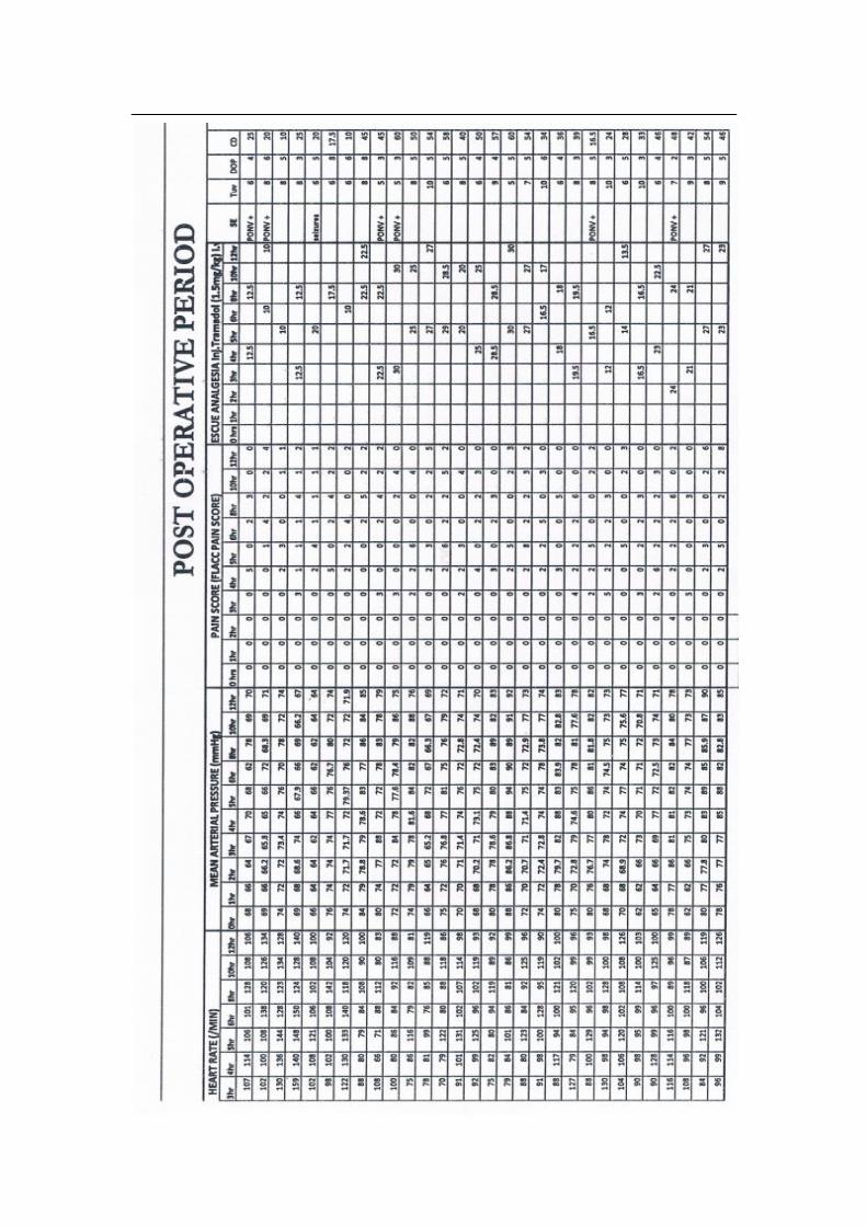
children were assessed for pain using FLACC behavioural pain assessment
score (Facial expression, Crying, Legs, Activity state and Consolability) and
their vitals were monitored during the immediate post-operative period in the
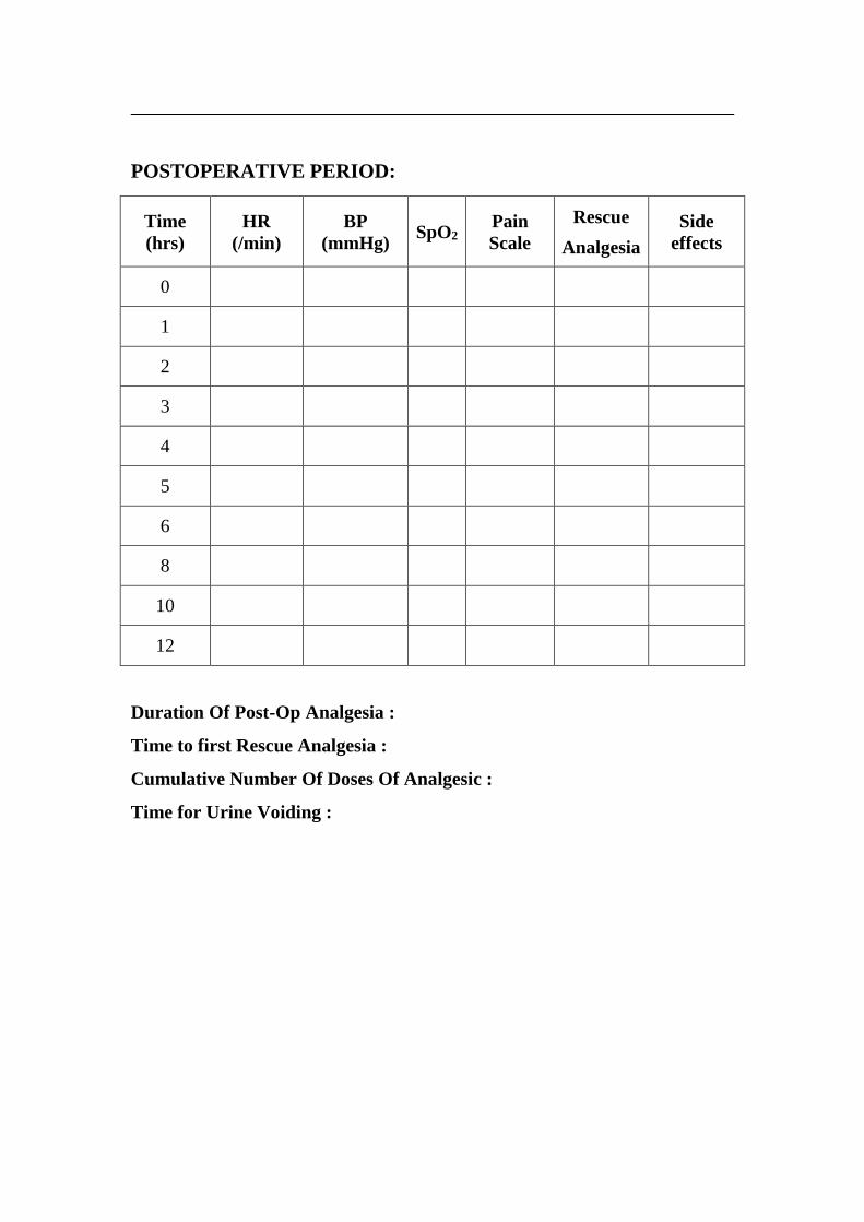
recovery room, then every hour for the first 6hrsand every two hourly for the
next 6hrs,after surgery.
PAIN SCORE
Various composite observational pain measurement tools have been
developed to aid in assessment of pain in paediatric population. FLACC score
is one such score to assess pain in children belonging to age group 2 months to
7 years. It takes into account the Facial expression, Crying, Legs, Activity state
and Consolability.46
Page 73
In awake patients:
Patients are observed for 1-5mins. Their legs and body uncovered.
Patient should be repositioned and activity should be observed. Consoling
conversations should be initiated if required.
In patients who are asleep:
Patients are observed for 1-5mins. Their legs and body uncovered.
Patient should be repositioned if possible and body should be assessed for
tenseness and tone.
Page 74
FLACC BEHAVIOURAL PAIN ASSESSMENT SCORE
Criteria Score 0 Score 1 Score 2
Face
No particular
expression or
smile
Occasional grimace or
frown, withdrawn,
uninterested
Frequent to
constant quivering
chin, clenched jaw
Legs Normal position
or relaxed Uneasy, restless, tense
Kicking ,or legs
drawn up
Activity
Lying quietly,
normal position,
moves easily
Squirming, shifting,
back and forth, tense
Arched, rigid or
jerking
Cry No cry
(awake or asleep)
Moans or whimpers;
occasional complaint
Crying steadily,
screams or sobs,
frequent
complaints
Consolability Content, relaxed
Reassured by occasional
touching, hugging or
being talked to,
distractible
Difficult to
console or comfort
Interpreting the Behavioural Score
Each category is scored on the 0–2 scale, which results in a total score of 0–10.
Score of 0 = Relaxed and comfortable
Score 1–3 = Mild discomfort
Score 4–6 = Moderate pain
Score 7–10 = Severe discomfort or pain or both
RESCUE ANALGESIA:
When the FLACC pain score >3, the children were given 1.5mg/kg of
tramadol intravenously as rescue analgesia
Page 75
MEASURED PARAMETERS
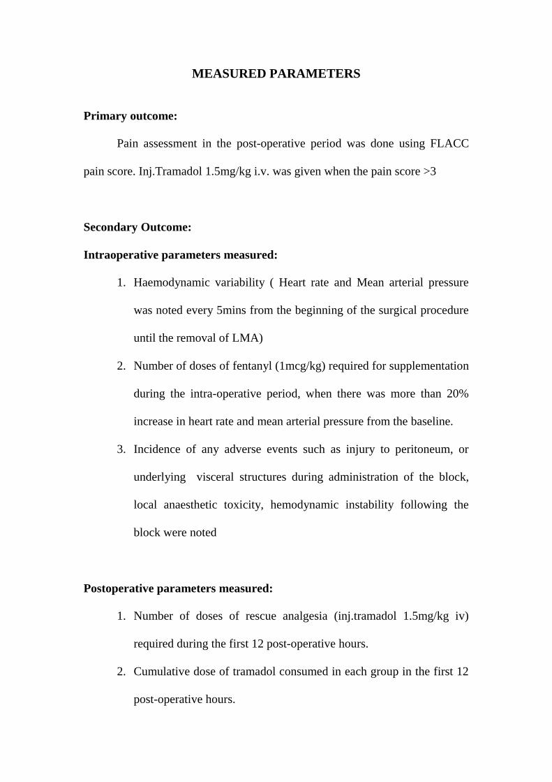
Primary outcome:
Pain assessment in the post-operative period was done using FLACC
pain score. Inj.Tramadol 1.5mg/kg i.v. was given when the pain score >3
Secondary Outcome:
Intraoperative parameters measured:
1. Haemodynamic variability ( Heart rate and Mean arterial pressure
was noted every 5mins from the beginning of the surgical procedure
until the removal of LMA)
2. Number of doses of fentanyl (1mcg/kg) required for supplementation
during the intra-operative period, when there was more than 20%
increase in heart rate and mean arterial pressure from the baseline.
3. Incidence of any adverse events such as injury to peritoneum, or
underlying visceral structures during administration of the block,
local anaesthetic toxicity, hemodynamic instability following the
block were noted
Postoperative parameters measured:
1. Number of doses of rescue analgesia (inj.tramadol 1.5mg/kg iv)
required during the first 12 post-operative hours.
2. Cumulative dose of tramadol consumed in each group in the first 12
post-operative hours.
Page 76
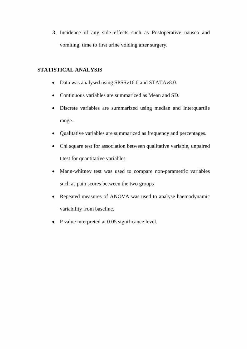
3. Incidence of any side effects such as Postoperative nausea and
vomiting, time to first urine voiding after surgery.
STATISTICAL ANALYSIS
Data was analysed using SPSSv16.0 and STATAv8.0.
Continuous variables are summarized as Mean and SD.
Discrete variables are summarized using median and Interquartile
range.
Qualitative variables are summarized as frequency and percentages.
Chi square test for association between qualitative variable, unpaired
t test for quantitative variables.
Mann-whitney test was used to compare non-parametric variables
such as pain scores between the two groups
Repeated measures of ANOVA was used to analyse haemodynamic
variability from baseline.
P value interpreted at 0.05 significance level.
Page 77
OBSERVATIONS
AND
RESULTS
Page 78
OBSERVATIONS AND RESULTS
Sixty patients were involved in the study and were randomly allocated
into two groups, Group T (n=30) and Group C (n=30)
TABLE 1: DEMOGRAPHIC VARIABLES
S.No Characteristic USG guided
TAP block
Caudal
Epidural block P value
1. Age 4.40± 1.831 4.37± 1.650 0.941
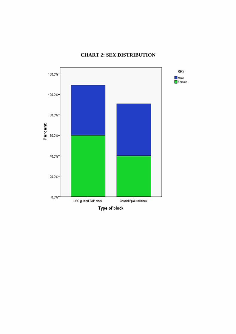
2. Sex- Male 27(90) 28(93.3) 0.640
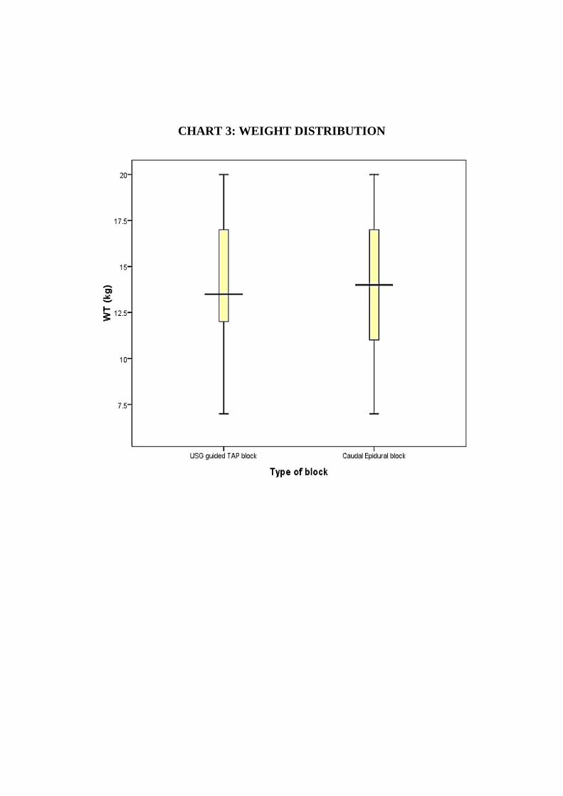
3. Weight 13.73± 3.667 13.40±4.090 0.741
4. Height 110.83± 21.050 104.72± 20.365 0.257
Demographic Variable such as Age, Sex, Weight and Height were
comparable in both the groups
Page 79
CHART 1: AGE DISTRIBUTION (MEAN + SD)
Page 80
CHART 2: SEX DISTRIBUTION
Page 81
CHART 3: WEIGHT DISTRIBUTION
Page 82
CHART 4: HEIGHT DISTRIBUTION
50
60
70
80
90
100
110
120
MEA
N H
EIG
HT
(cm
)
USG TAP Block Caudal Block
Page 83
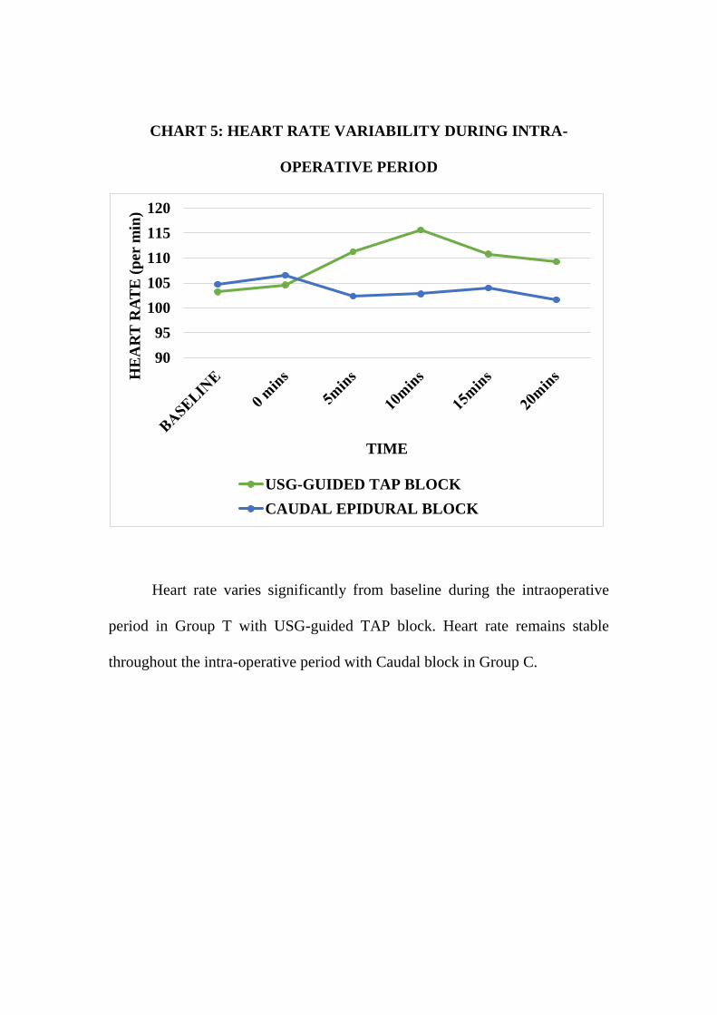
TABLE 2: INTRA-OPERATIVE VARIATIONS IN HEART RATE
TIME CHARACTERISTIC
USG-GUIDED
TAP BLOCK
(MEAN+SD)
CAUDAL
EPIDURAL
BLOCK
(MEAN + SD)
T0
BASELINE
(After Block
Administration)
103.25
+15.096
104.75
+16.712
T1 0 mins
(SKIN INCISION)
104.57
+13.908
106.57
+16.630
T2 5mins 111.29
+18.208
102.36
+15.956
T3 10mins 115.64
+19.425
102.82
+15.635
T4 15mins 110.82
+18.886
104.04
+13.898
T5 20mins 109.29
+17.049
101.64
+12.870
F VALUE 11.086 1.550
P VALUE <0.001 0.208
Page 84
CHART 5: HEART RATE VARIABILITY DURING INTRA-
OPERATIVE PERIOD
Heart rate varies significantly from baseline during the intraoperative
period in Group T with USG-guided TAP block. Heart rate remains stable
throughout the intra-operative period with Caudal block in Group C.
90
95
100
105
110
115
120
HE
AR
T R
AT
E (
per
min
)
TIME
USG-GUIDED TAP BLOCK
CAUDAL EPIDURAL BLOCK
Page 85
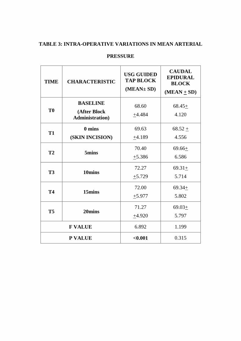
TABLE 3: INTRA-OPERATIVE VARIATIONS IN MEAN ARTERIAL
PRESSURE
TIME CHARACTERISTIC
USG GUIDED
TAP BLOCK
(MEAN± SD)
CAUDAL
EPIDURAL
BLOCK
(MEAN + SD)
T0
BASELINE
(After Block
Administration)
68.60
+4.484
68.45+
4.120
T1 0 mins
(SKIN INCISION)
69.63
+4.189
68.52 +
4.556
T2 5mins 70.40
+5.386
69.66+
6.586
T3 10mins 72.27
+5.729
69.31+
5.714
T4 15mins 72.00
+5.977
69.34+
5.802
T5 20mins 71.27
+4.920
69.03+
5.797
F VALUE 6.892 1.199
P VALUE <0.001 0.315
Page 86
CHART 6: MEAN ARTERIAL PRESSURE VARIABILITY DURING
INTRA-OPERATIVE PERIOD
Change in mean arterial pressure from baseline is significant during the
intraoperative period in Group T with USG-guided TAP block, whereas change
in mean arterial pressure is not significant with Caudal block in Group C
66
67
68
69
70
71
72
73
MA
P (
mm
Hg)
USG-GUIDED TAP BLOCK
CAUDAL EPIDURAL BLOCK
Page 87
TABLE 4: INTRAOPERATIVE ANALGESIC REQUIREMENTS
S.No Characteristic
USG guided
TAP block
N (%)
Caudal
Epidural
block
N (%)
P value
1. Intra operative
requirement of Fentanyl 16 (53.3) 1(3) <0.0001
Around 53.3% of patients belonging to Group T required intra-operative
fentanyl supplementation, whereas only 3% in Group C, required intra-
operative fentanyl supplementation. The difference was found to be statistically
significant.
Page 88
CHART 7: INTRA-OPERATIVE REQUIREMENT OF
SUPPLEMENTATION OF FENTANYL IN GROUP T: USG-GUIDED
TAP BLOCK
CHART 8: INTRA-OPERATIVE REQUIREMENT OF
SUPPLEMENTATION OF FENTANYL IN GROUP C: CAUDAL
BLOCK
Opioid
required
53%
Opioid Not
required
47%
USG guided TAP block
Opioid required
Opioid Not required
Opioid
required
10%
Opioid Not
required
90%
Caudal Epidural block
Opioid required
Opioid Not required
Page 89
TABLE 5: DURATION OF POSTOPERATIVE ANALGESIA
S.No Characteristic USG guided
TAP block
Caudal Epidural
block P value
1
Duration of Post op
analgesia
(hrs)
8.60± 1.840
4.57± 1.406 <0.0001
CHART 9: DURATION OF POST-OPERATIVE ANALGESIA
Post-operative analgesia was defined as the duration of analgesia from
the immediate post-op period to the time at which the first rescue analgesic was
required.
Duration of Post-operative analgesia was longer in Group T than Group
C. TAP block provided postop analgesia for 8.60hrs on average whereas caudal
block provided a post-op analgesia of duration 4.57hrs on average. The
difference was found to be statistically significant.
0 2 4 6 8 10
TIME (hrs)
TEC
HN
IQU
E
DURATION OF POST-OP ANALGESIA
CAUDAL BLOCK USG TAP BLOCK
Page 90
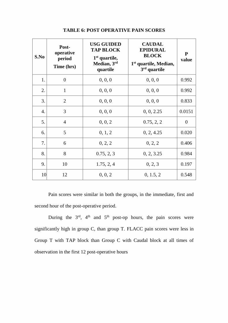
TABLE 6: POST OPERATIVE PAIN SCORES
S.No
Post-
operative
period
Time (hrs)
USG GUIDED
TAP BLOCK
1st quartile,
Median, 3rd
quartile
CAUDAL
EPIDURAL
BLOCK
1st quartile, Median,
3rd quartile
P
value
1. 0 0, 0, 0 0, 0, 0 0.992
2. 1 0, 0, 0 0, 0, 0 0.992
3. 2 0, 0, 0 0, 0, 0 0.833
4. 3 0, 0, 0 0, 0, 2.25 0.0151
5. 4 0, 0, 2 0.75, 2, 2 0
6. 5 0, 1, 2 0, 2, 4.25 0.020
7. 6 0, 2, 2 0, 2, 2 0.406
8. 8 0.75, 2, 3 0, 2, 3.25 0.984
9. 10 1.75, 2, 4 0, 2, 3 0.197
10. 12 0, 0, 2 0, 1.5, 2 0.548
Pain scores were similar in both the groups, in the immediate, first and
second hour of the post-operative period.
During the 3rd, 4th and 5th post-op hours, the pain scores were
significantly high in group C, than group T. FLACC pain scores were less in
Group T with TAP block than Group C with Caudal block at all times of
observation in the first 12 post-operative hours
Page 91
CHART 10: FLACC PAIN SCORE (MEDIAN) IN THE POST-
OPERATIVE PERIOD (0-12HRS)
0
0.5
1
1.5
2
2.5
3
3.5
4
4.5
0 1 2 3 4 5 6 8 10 12
PAIN
SC
OR
E
POST-OP HOURS
PAIN SCORES IN THE 12HR POST-OP PERIOD
USG TAP Block Caudal Block
Page 92
TABLE 7: REQUIREMENTS OF RESCUE ANALGESIA
S.No Characteristic USG guided TAP
block
Caudal
Epidural
block
P value
1 Cumulative dose of
rescue analgesia (mg)
23.40± 11.322
38.23± 15.434 <0.0001
2
No. of doses of rescue
analgesia
1.1000±.40258
1.8333±
.37905 <0.0001
CHART 11: CUMULATIVE DOSE OF RESCUE ANALGESIA
REQUIRED IN THE FIRST 12HRS OF POST-OPERATIVE PERIOD
The cumulative doses of tramadol required to rescue the patient from
post-operative pain was significantly less in Group T than in Group C.
23.4
38.23
0
5
10
15
20
25
30
35
40
45
CU
MU
LA
TIV
E D
OS
E O
F T
RA
MA
DO
L
(mg
)
TECHNIQUE USED
CUMULATIVE DOSE OF RESCUE ANALGESIA
USG TAP BLOCK CAUDAL BLOCK
Page 93
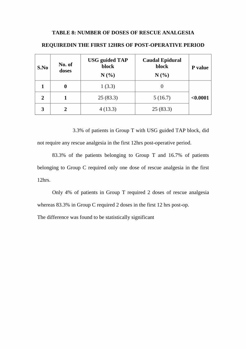
TABLE 8: NUMBER OF DOSES OF RESCUE ANALGESIA
REQUIREDIN THE FIRST 12HRS OF POST-OPERATIVE PERIOD
S.No No. of
doses
USG guided TAP
block
N (%)
Caudal Epidural
block
N (%)
P value
1 0 1 (3.3) 0
<0.0001 2 1 25 (83.3) 5 (16.7)
3 2 4 (13.3) 25 (83.3)
3.3% of patients in Group T with USG guided TAP block, did
not require any rescue analgesia in the first 12hrs post-operative period.
83.3% of the patients belonging to Group T and 16.7% of patients
belonging to Group C required only one dose of rescue analgesia in the first
12hrs.
Only 4% of patients in Group T required 2 doses of rescue analgesia
whereas 83.3% in Group C required 2 doses in the first 12 hrs post-op.
The difference was found to be statistically significant
Page 94
CHART 12: NUMBER OF DOSES RESCUE ANALGESIA IN THE
FIRST 12hrs OF POST-OPERATIVE PERIOD
0
5
10
15
20
25
30
No Dose 1 Dose 2 DosesNU
MB
ER
OF
PA
TIE
NT
S
NUMBER OF DOSES
NUMBER OF DOSES OF RESCUE ANALGESIA REQUIRED IN POST
OPERATIVE PERIOD
USG GUIDED TAP BLOCK
CAUDAL EPIDURAL BLOCK
Page 95
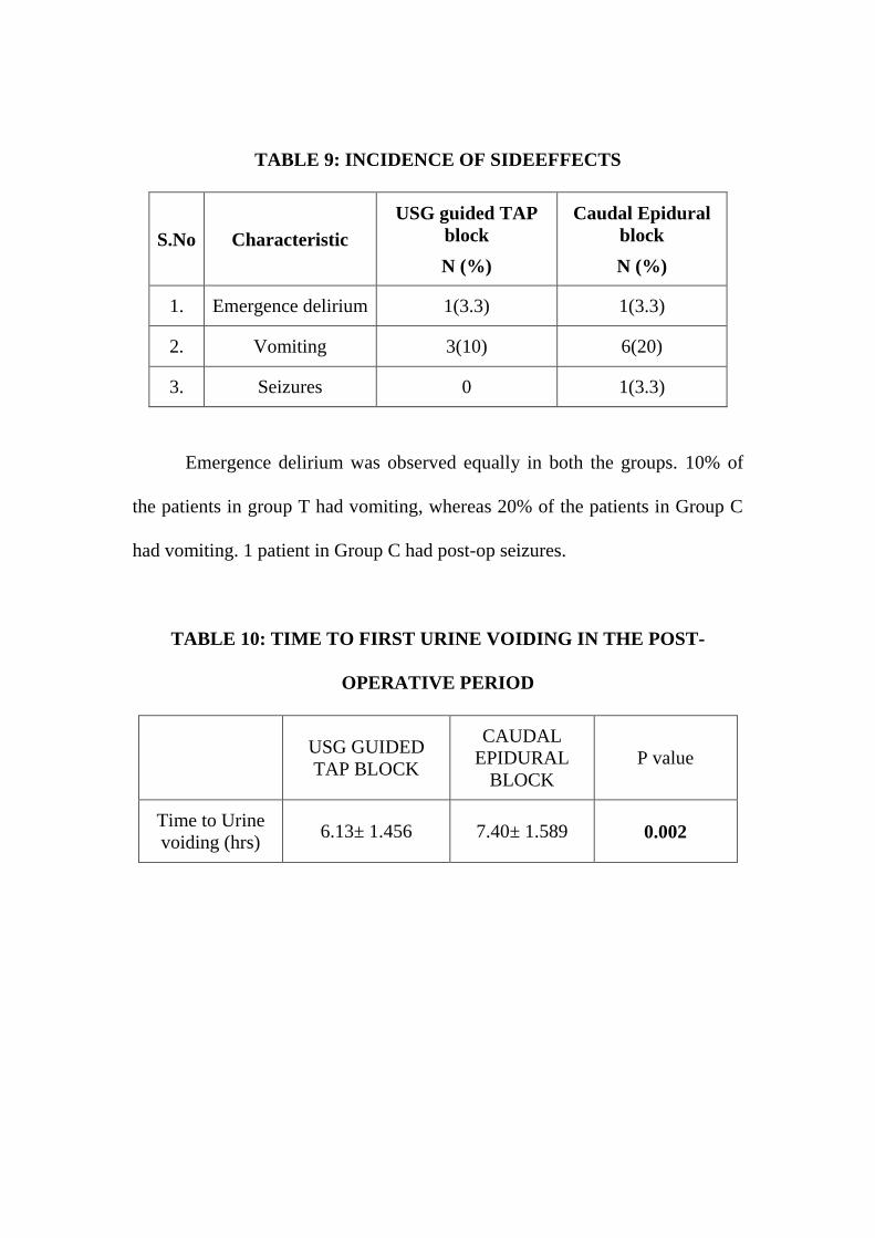
TABLE 9: INCIDENCE OF SIDEEFFECTS
S.No Characteristic
USG guided TAP
block
N (%)
Caudal Epidural
block
N (%)
1. Emergence delirium 1(3.3) 1(3.3)
2. Vomiting 3(10) 6(20)
3. Seizures 0 1(3.3)
Emergence delirium was observed equally in both the groups. 10% of
the patients in group T had vomiting, whereas 20% of the patients in Group C
had vomiting. 1 patient in Group C had post-op seizures.
TABLE 10: TIME TO FIRST URINE VOIDING IN THE POST-
OPERATIVE PERIOD
USG GUIDED
TAP BLOCK
CAUDAL
EPIDURAL
BLOCK
P value
Time to Urine
voiding (hrs) 6.13± 1.456 7.40± 1.589 0.002
Page 96
CHART 13: TIME TO FIRST URINE VOIDING IN THE POST-
OPERATIVE PERIOD
Time to Urine voiding was longer in Group C than in Group T and the
difference was found to be statistically significant.
0
1
2
3
4
5
6
7
8
TIM
E (h
rs)
CAUDAL EPIDURAL BLOCK USG GUIDED TAP BLOCK
Page 98
DISCUSSION
Optimal treatment of perioperative pain is usually multimodal. Even in
procedures which are done under regional anaesthesia, a general anaesthesia or
sedation is usually given for the child to cooperate for the regional technique.
This is because it is both unethical and dangerous to perform a regional
technique in an agitated, moving child.
In our study, we used the technique of general anaesthesia via ambu
LMA, which was inserted after obtunding the reflexes using propofol 2mg/kg
and fentanyl 1mcg/kg and succinylcholine 1.5mg/kg to facilitate insertion.
Anaesthesia was maintained with 0.75-1 MAC sevoflurane with nitrous-oxide
and oxygen (50:50) gas mixture.
INTRA-OPERATIVE ANALGESIA:
Analgesia during the procedure is provided by a regional technique
either Caudal or TAP block, according to the group, which cannot be assessed
directly.
Adequacy of the regional block in supplementing the general
anaesthesia can be assessed only indirectly using the changes in haemodynamic
parameters and requirement of supplementation by analgesics like opioid.
Since pain is associated with stress response resulting in increase in heart rate
and blood pressure, significant increase in these haemodynamic parameters
would imply an inadequate analgesia by the regional technique. The
cardiovascular responses were used as a surrogate for adequacy of analgesia. In
Page 99
our study, we assigned thatdespite the administration of 1 MAC of sevoflurane,
if there was a 20% increase in heart rate and mean arterial pressure from the
baseline, it was due to inadequate analgesia requiring supplementation. In such
cases, we supplemented them with 1mcg/kg of fentanyl intravenously.
The heart rate and mean arterial pressure variables remained constant
throughout the procedure in Group C (with caudal block) whereas the heart rate
and MAP were significantly high from the baseline, during the 10-15mins
period after the beginning of surgical procedure in Group T (with TAP block)
This is because Caudal block is a neuraxial blockade which offers
complete blockade of sensory, motor and autonomic innervation up to the level
of blockade. Hence there is complete analgesia in Caudal block, whereas TAP
block anaesthetises only the nerves supplying the parietal peritoneum, skin and
muscles of anterior abdominal wall. Hence cord traction and visceral peritoneal
handling can result in stress response, causing rise in heart rate and mean
arterial pressure in Group T (TAP block).
Results in our study show that 53% of the patients in Group T with TAP
block require supplementation with fentanyl, in contrast to Group C, where
only 10% required supplemental analgesia during the intra-operative period.
Among the initial research studies with TAP block, a study by
Fredrickson47 on TAP block for inguinal herniotomy in 8 paediatric patients,
demonstrated that 5 patients of the 8 did not require any intra-operative
supplementation of fentanyl. The rest of the 3 patients required intraoperative
Page 100
supplementation of fentanyl (<0.5 mcg/kg fentanyl), which he attributed to the
pain felt during spermatic cord manipulation.
In our study 16/30in TAP block group required fentanyl
supplementation, unlike the study by Fredricksonwhere only 3/8 required
fentanyl supplementation. This discrepancy between the studies may be due to
the small sample size used in his study.
Ray et al48studied Caudal block with bupivacaine and ropivacaine
administered pre-operatively in paediatric patients undergoing urogenital
procedures. He demonstrated no change in haemodynamic parameters during
the intra-operative period and no supplementation was required in both the
groups. Whereas 3% in Group C in our study, required supplementation of
fentanyl.
Since the onset time was not observed in either of the block techniques
in our study, the delayed onset of Caudal block might be the responsible for the
requirement of fentanyl supplementation in one patient in Group C, at the time
of skin incision.
DURATION OF POST-OPERATIVE ANALGESIA:
The duration of post-operative analgesia with TAP block was found to
be significantly longer thanCaudal block.
Studies with Caudal block have demonstrated a post-op analgesia of 4-
6hrs. In our study, the mean duration of post-op analgesia with caudal block
was 4.6hrs (274min).
Page 101
Ivani et al49conducted a double blind study with forty patients belonging
to the age group 1-9 years. They were posted for elective minor surgery under
caudal anaesthesia. The children were randomly divided into two groups,
twenty in each. One group received 0.25% bupivacaine 2mg/kg and the other
received 0.2% ropivacaine 2mg/kg. Hehad demonstrated a post-operative
analgesia duration of 253min following single shot caudal with 0.25%
bupivacaine which is consistent with the results of our study.
In our study, the time to first rescue analgesia in TAP block group was
516min. The duration of post-operative pain relief was longer in TAP group
when compared to caudal group. This may be explained by the following
reasons.
Owing to the high vascularity of the caudal space, the absorption of
local anaesthetic into systemic circulation is more in Caudal block, resulting in
faster clearance of the local anaesthetic. Transversus Abdominis Plane is a
relatively avascular fascial plane.
The local anaesthetic drug volume deposited in the caudal space has to
spread over a larger area to achieve the level of blockade whereas the drug
volume injected in TAP spreads in a narrow fascial plane between two
muscles.
The post-operative pain felt in a superficial surgery like inguinal hernia
repair, is mainly due to pain sensations from the skin, muscles and parietal
peritoneum, which is effectively blocked by the TAP block, making it effective
in providing prolonged post-operative analgesia.
Page 102
Dalia M50compared USG-guided TAP block with Caudal block for post-
operative pain relief in open pyeloplasty. He included 39 patients belonging to
age group 6months to 6years randomly allocated into two groups. In his study,
he demonstrated that the patients with TAP block had a significantly longer
time to first rescue analgesia, 602min in contrast to 280min in caudal block;
which is comparable to our study (TAP 516min vs Caudal 253min)
In a study conducted by Sahin L et al51, he evaluated the analgesic
efficacy of ultrasound-guided TAP block in comparison with wound
infiltration, during the first 24 h after surgery in children of age 2-8years
undergoing inguinal hernia repair. He showed that TAP block provided
prolonged duration of post-op analgesia (17hrs vs. 4.7hrs) in comparison to
wound infiltration. The analgesia duration was significantly longer (17hrs)
when compared to (8.6hrs) in our group. This might be because, we had not
observed the pain scores in the patients after 12 postoperative hours, and they
had used levobupivacaine, which provides a longer duration of sensory
blockade than bupivacaine.
Tobias52 studied efficacy of TAP block for lower abdominal surgery in
10 paediatric patients belonging to age group 10months to 8years and reported
that 80% had good post-operative analgesia with the duration of analgesia
lasting for 7 to 11 hours, which is consistent with the results (average of 8.6hrs)
of our study.
Paleti Sophia53 studied the efficacy of TAP block in 50 children of age
7-13years undergoing lower abdominal surgeries. The children were randomly
Page 103
allocated into two groups. After general anaesthesia, 25 children received TAP
block and rest of the 25 received systemic analgesia. PaletiSophia showed that
the mean time to first request of analgesia was 8.5 to 9 hours in TAP block,
similar to the results (8.6hrs) of our study.
PAIN SCORES
The pain scores in both the groups were comparable in the immediate
post-op, upto 2hrs. This shows that both caudal and TAP block are equally
effective in providing pain relief in the immediate post-op. Pain scores were
significantly higher in 3-5 hours in Caudal group requiring administration of
rescue analgesia.
In our study, pain scores in Group T with TAP block were lower than
that with Group C with Caudal block,at 3, 4 and 5 hrs post-operatively.FLACC
scores were lower in TAP block group when compared to caudal block group,
at all timepoints of observation upto 12 post-operative hours.
Dalia M, who compared USG-guided TAP block with Caudal block for
post-operative pain relief in open pyeloplasty, observed that FLACC pain
scores were significantly less in TAP block than Caudal block at
2,4,6,8,10,12,24 hrs post-operatively, consistent with the results of our study.
In a study conducted by Neha Kanojia, who compared USG guided TAP
block with Caudal block for lower abdominal surgeries, employed 60 children
of age 1-12years.
Page 104
They were randomly allocated into two groups, 30 in each. Neha
demonstrated lower mean VAS scores in both the groups for first 3-4 hours
postoperatively, similar to our study. After 3-4 hours, there was a rise in the
number of patients with mean VAS score >3 in Caudal group compared to
group TAP, as in our study.
Carney et al studied 40 children undergoing open appendicectomy, with
twenty in each group, where one group was given TAP block and the other was
given placebo.54He observed that TAP block significantly reduced VAS pain
scores at rest and on movement at all times, similar to our study.
Sahin L et al showed lower CHEOPS pain scores in children receiving
ultrasound guided TAP block compared to wound infiltration at all points of
their assessment for the first 24 post-operative hours, as in our study.
McDonnell et al. reported that TAP block given to 32 adults undergoing
large bowel resection reduced the VAS pain scores in the post-anaesthesia care
unit (1 ± 1.4 vs. 6.6 ± 2.8) and at all postoperative observation time points,
including at 24 h (1.7 ± 1.7 vs. 3.1 ± 1.5).
MEAN OPIOID CONSUMPTION
Cumulative doses of tramadol as rescue analgesic were significantly
more in Caudal block, with a mean tramadol consumption of 38.23mg
compared to 23.40mg in TAP block group. Since the time to first rescue
analgesic was longer in TAP block, children in Group T required lesser number
of doses of rescue analgesia.
Page 105
Carney et al who studied TAP block vs placebo in open
appendicectomy, showed that TAP block reduced mean morphine requirements
in the first 48hrs postoperatively, (10.3mg vs 22.3mg) and he found it superior
to placebo in being part of a multimodal analgesia regimen in the first 48hrs
post-operatively.
In a study conducted by Niraj G, studying the efficacy of USG guided
TAP block in 50 adult patients undergoing open appendicectomy. He randomly
allocated them into two groups, where 26 patients received TAP block and 24
patients received standard care with general anaesthesia.He demonstrated that
TAP block significantly reduced the VAS scores and postoperative morphine
consumption in the first 24 hr (28mg in TAP block group vs 50mg). 55
INCIDENCE OF SIDE EFFECTS:
Emergence delirium was observed in both the groups during the
immediate post-operative period in 10% of the patients. Sevoflurane which was
used in the general anaesthesia may be responsible for the emergence delirium
in both the groups.
Volatile anaesthetics and opioids (fentanyl) used in General anaesthesia
can cause PONV. Incidence of PONV was higher in Caudal group than in TAP
block group, probably because of the increased requirements of tramadol in
Caudal group. No complications or adverse events related to TAP block was
noted in our study.
Page 106
There are rare instances such as Liver trauma with regional anaesthesia
block needle while performing landmark guided TAP block, reported by
Farooq M and Carey M56. No such complications occurred in our study, as the
procedure was done under ultrasound guidance.
There was an instance of seizure episode in the postoperative period,
3hours after the surgical procedure in Caudal block group. Since the seizure
was associated with feverand the fact that local anaesthetic toxicity usually
manifests immediately after block, the seizure episode might not have been
due to the block.
In our study, the time to first urine voiding, in caudal group (7.4hrs+1.6)
was significantly longer than TAP block group (6.13hrs ± 1.5).
Fischer QA studied the postoperative voiding interval and duration of
analgesia following Caudal analgesia and Ilioinguinal-iliohypogastric (IIN/IIH)
nerve block. Study was done in 82 children of age 6months to 10years
undergoing herniorrhaphy and orchidopexy under general anaesthesia. He
allocated them into 3 groups, one group to receive caudal block 0.75ml/kg of
bupivacaine, second group - 0.75ml bupivacaine with epinephrine, and the
third group to receive IIN/IIH nerve block with epinephrine. The Mean time to
micturition in Caudal group with plain bupivacaine was 202min, in second
caudal group with bupivacaine with epinephrine was 262min and in IIN/IIH
group was 196min. He demonstrated that the time to postoperative voiding is
variable in children and not prolonged by caudal analgesia.
Page 107
In studies which compared caudal to non-caudal procedures, many of
them showed that caudal block was associated with longer time to urine
voiding in the post-operative period. In a study by Markham et al, the incidence
of urinary retention was 12/26 in caudal group and 5/26 in nerve block group.
Though our study showed significant difference in time to urine voiding
between the two groups, there were no cases of urinary retention which
required intervention in both the groups.
Page 109
SUMMARY
This study titled “COMPARISON OF ULTRASOUND GUIDED
TRANSVERSUS ABDOMINIS PLANE BLOCK AND CAUDAL
EPIDURAL BLOCK FOR PAIN RELIEF IN CHILDREN UNDERGOING
UNILATERAL INGUINAL HERNIOTOMY” was conducted at Mahatma
Gandhi Memorial Government Hospital, Trichy, during the period of
November 2013 to April 2015.
Sixty children belonging to age group 1-7 yrs, weighing 5-20kg,
undergoing unilateral inguinal herniotomy, belonging to ASA I and II status
were selected. Children undergoing bilateral inguinal herniotomy, children with
known allergy to bupivacaine, children with altered sacral, caudal anatomy,
children with local infection at the site of the block and children with renal or
hepatic insufficiency, belonging to ASA III, IV were excluded from the study.
Our study sample were divided into two groups of 30 each. After
institution of general anaesthesia, Group T received Ultrasound-guided TAP
block with 0.5ml/kg of 0.25% bupivacaine and Group C received Caudal
epidural block with 1ml/kg of 0.25% bupivacaine
Intra-operative haemodynamic parameters such Heart rate and mean
arterial pressure; requirement of fentanyl and incidence of any adverse events
were noted. FLACC pain scores were observed at 1, 2, 3, 4, 5, 6, 8, 10 and 12
hrs in the post-operative period. Any side effects such as post-operative nausea
and vomiting, seizures and urinary retention were also noted.
Page 110
In this prospective randomised control trial, the following results were
noted:
1. Both groups were comparable in age, sex, weight and height.
2. Group C had no significant variations in haemodynamics and did not
require any fentanyl supplementation. Caudal block provided good
analgesia during the intra-operative period.
3. TAP block is not adequate to provide complete intra-operative
analgesiain inguinal herniotomies, requiring supplementation by
other analgesics.
4. Group T showed prolonged duration of post-operative analgesia,
compared to Group C.
5. FLACC scores were similar in both the groups up to 2 hrs post-
operatively, after which Caudal group showed significant increase in
pain scores. Pain scores in TAP block were lower throughout the
post-operative period when compared to caudal block
6. Mean opioid consumption was lower in TAP block group.
7. Increased incidence of PONV and increased time to urine voiding
was noted in Caudal block group.
Page 112
CONCLUSION
Caudal epidural block provided better intra-operative analgesia than
USG guided TAP block for inguinal hernia repair.
Ultrasound-guided TAP block provided prolonged post-operative pain
relief than single shot Caudal epidural block and reduced the mean opioid
consumption in the first 12 post-operative hours after inguinal herniotomy in
children of age 1-7years.
Page 114
BIBLIOGRAPHY
1Miller R. Miller's Anesthesia. 7th ed. Philadelphia, Pa: Elsevier,
Churchill Livingstone; p2521.
2Motoyama E, Davis P. Smith's anesthesia for infants and children.
Philadelphia: Mosby Elsevier; 2006.
3Miller R. Miller's Anesthesia. 7th ed. Philadelphia, Pa: Elsevier,
Churchill Livingstone; p2532-2533.
4Lloyd-Thomas AR. Pain management in paediatric patients. Br J
Anaesth 1990; 64:85–104
5Galante D, Caruselli M, Dones F, et al. Ultrasound transversus
abdominis plane (TAP) block in pediatric patients: Not only a regional
anesthesia technique for adults. Anaesth Pain & Intensive Care 2012;
16(2): 201-204
6 El- Dawlatly AA, Turkistani A, Kettner SC, et al. Ultrasound-
guided transversus abdominis plane block: description of a new technique
and comparison with conventionalsystemic analgesia during
laparoscopic cholecystectomy . Br J Anaesth 2009; 102:763–767
7Kapur P, Caty MG, Glick PL. Pediatric hernias and hydroceles.
PediatrClin North Am. 1998 Aug;45(4):773-89. Review.
8 Rafi AN. Abdominal field block: a new approach via the lumbar
triangle. Anaesthesia. 2001;56(10):1024-1026
Page 115
9 P Hebbard, Y Fujiwara, Y Shibata, C Royse. Ultrasound Guided Transversus
Abdominis Plane Block. Anaesthesia & Intensive Care; 2007; 35(4): 616-7
10Hebbard P. Subcostal transversus abdominis plane block under ultrasound
guidance. AnesthAnalg. 2008;106(2):674-675
11 Davies FGR, Stibbe EP. The anatomy of the intercostal nerves. J Anat.
1931;66:323Y333.
12Borley N. Anterior abdominal wall, posterior abdominal wall and
retroperitoneum. In: Standring S. Gray’s Anatomy. 40th ed. New York, NY:
Churchill Livingstone Elsevier; 2008
13Rozen WM, Tran TM, Ashton MW, Barrington MJ, Ivanusic JJ, Taylor GI.
Refining the course of the thoracolumbar nerves: a new understanding of the
innervation of the anterior abdominal wall. Clin Anat. 2008;21:325Y333
14 El-Dawlatly A, Turkistani A, Kettner S, et al. Ultrasound-guided
transversus abdominis plane block: description of a new technique and
comparison with conventional systemic analgesia during laparoscopic
cholecystectomy. Br J Anaesth 2009 102(6): 763-767
15 Young, Mark J., Andrew W. Gorlin, Vicki E. Modest, and Sadeq A.
Quraishi. 2012. Clinical implications of the transversus abdominis plane block
in adults. Anesthesiology Research and Practice 2012:731645.
16 J. G. McDonnell, B. O’Donnell, G. Curley, A. Heffernan, C. Power, and J.
G. Laffey, “The analgesic efficacy of transversus abdominis plane block after
abdominal surgery: a prospective randomized controlled trial,” Anesthesia and
Analgesia, vol. 104, no. 1, pp. 193–197, 2007
Page 116
17 R. J. Kearns and S. J. Young, “Transversus abdominis plane blocks; a
national survey of techniques used by UK obstetric anaesthetists,” International
Journal of Obstetric Anesthesia, vol. 20, no. 1, pp. 103–104, 2011
18Berry AM, Brimacombe JR, Verghese C. The laryngeal mask airway in
emergency medicine, neonatal resuscitation, and intensive care
medicine. IntAnesthesiolClin. 1998 Spring. 36(2):91-109.
19Miller RD. Miller's Anesthesia. 6th ed. Philadelphia, Pa: Elsevier Churchill
Livingstone; 2005. 1625-28
20Miyasaka K, Suzuki Y, Kondo Y, Sakai H, Nakagawa S, Asahara S. The use
of the laryngeal mask airway in pediatric anesthesia. J Anesth. 1991
Apr;5(2):160-5.PubMed PMID: 15278650.
21Jamil SN, Alam M, Usmani H, Khan MM. A Study of the Use of Laryngeal
Mask Airway (LMA) in Children and its Comparison with Endotracheal
Intubation.Indian Journal of Anaesthesia. 2009;53(2):174-178.
22 Lopez-Gil M, Brimacombe J, Alvarez M. Safety and efficacy of the
laryngeal mask airway: A prospective survey of 1400
children. Anaesthesia. 1996;51:969–72.
23Sanders JC, Olumu PN, Furman JR. Detection, frequency and prediction of
problems in the use of the proseal laryngeal mask airway in
children. PediatrAnesth. 2008;18:1183–9
24 Laffon M, Plaud B, Dubousset AM, Ben R. Haj’hmida removal of laryngeal
mask airway: Airway complications in Children anesthetized versus
awake. PediatrAnesth. 1994;4:35–7
Page 117
25 Petersen PL, Mathiesen O, Torup H, Dahl JB. The transversus abdominis
plane
block: a valuable option for postoperative analgesia? A topical review.
ActaAnaesthesiol Scand. 2010 May;54(5):529-35. doi: 10.1111/j.1399-
6576.2010.02215.x.Epub 2010 Feb 17. Review. PubMed PMID: 20175754
26 G. Niraj, A. Kelkar, I. Jeyapalan et al., “Comparison of analgesic efficacy of
subcostal transversus abdominis plane blocks with epidural analgesia following
upper abdominal surgery,” Anaesthesia, vol. 66, no. 6, pp. 465–471, 2011.
27 M. R.S. Siddiqui, M. S. Sajid, D. R. Uncles, L. Cheek, and M. K. Baig, “A
meta-analysis on the clinical effectiveness of transversus abdominis plane
block,” Journal of Clinical Anesthesia, vol. 23, no. 1, pp. 7–14, 2011
28 S. Charlton, A. M. Cyna, P. Middleton, and J. D. Griffiths, “Perioperative
transversus abdominis plane (TAP) blocks for analgesia after abdominal
surgery,” Cochrane Database of Systematic Reviews, vol. 8, Article ID
CD007705, 2010
29 N. Bharti, P. Kumar, I. Bala, and V. Gupta, “The efficacy of a novel
approach to transversus abdominis plane block for postoperative analgesia after
colorectal surgery,” Anesthesia and Analgesia, vol. 112, no. 6, pp. 1504–1508,
2011
30 M. Hivelin, A. Wyniecki, B. Plaud, J. Marty, and L. Lantieri, “Ultrasound-
guided bilateral transversus abdominis plane block for postoperative analgesia
after breast reconstruction by DIEP flap,” Plastic and Reconstructive Surgery,
vol. 128, no. 1, pp. 44–55, 2011
Page 118
31 J. M. Baaj, R. A. Alsatli, H. A. Majaj, Z. A. Babay, and A. K. Thallaj,
“Efficacy of ultrasound-guided transversus abdominis plane (TAP) block for
post-cesarean section delivery analgesia—a double-blind, placebo-controlled,
randomized study,” Middle East Journal of Anesthesiology, vol. 20, no. 6, pp.
821–826, 2010
32 J. G. McDonnell, G. Curley, J. Carney et al., “The analgesic efficacy of
transversus abdominis plane block after cesarean delivery: a randomized
controlled trial,” Anesthesia and Analgesia, vol. 106, no. 1, pp. 186–191, 2008
33 D. Belavy, P. J. Cowlishaw, M. Howes, and F. Phillips, “Ultrasound-guided
transversus abdominis plane block for analgesia after Caesarean delivery,
”British Journal of Anaesthesia, vol. 103, no. 5, pp. 726–730, 2009
34 J. D. Griffiths, J. V. Middle, F. A. Barron, S. J. Grant, P. A. Popham, and
C.F.Royse, “Transversus abdominis plane block does not provide additional
benefit to multimodal analgesia in gynaecological cancer surgery, ”Anesthesia
and Analgesia, vol. 111, no. 3, pp. 797–801, 2010
35 R. C.N. McMorrow, R. J. Ni Mhuircheartaigh, K. A. Ahmed et al.,
“Comparison of transversus abdominis plane block vs spinal morphine for pain
relief after Caesarean section, ”British Journal of Anaesthesia, vol. 106, no. 5,
pp. 706–712, 2011
36 V.R. Kadam and J. L. Moran, “Epidural infusions versus transversus
abdominis plane(TAP) block infusions :retrospective study,” Journal of
Anesthesia, vol. 25, no. 5, pp. 786–787, 2011.
37Sandeman DJ, Bennett M, Dilley AV, Perczuk A, Lim S, Kelly KJ.
Page 119
Ultrasound-guided transversus abdominis plane blocks for laparoscopic
appendicectomy in children: a prospective randomized trial. Br J Anaesth.
2011Jun;106(6):882-6. doi: 10.1093/bja/aer069. Epub 2011 Apr 18. PubMed
PMID: 21504934.
38 Fredrickson MJ, Paine C, Hamill J. Improved analgesia with the ilioinguinal
block compared to the transversus abdominis plane block after pediatric
inguinal surgery: a prospective randomized trial. PaediatrAnaesth. 2010
Nov;20(11):1022-7. doi: 10.1111/j.1460-9592.2010.03432.x. PubMed PMID:
20964768.
39El Fawy DM, El Gendy HA. Ultrasound-guided transversus abdominis plane
blockversus caudal block for postoperative pain relief in infants and children
undergoing surgical pyeloplasty. Ain-Shams J Anaesthesiol 2014;7:177-81
40 Long J, Birmingham P, De Oliveira G, Schaldenbrand K, Suresh S.
Transversus Abdominis Plane Block in Children. Anesthesia & Analgesia.
2014;119(2):395-399.
41 A. Machotta, A. Risse, S. Bercker, R. Streich, and D. Pappert, “Comparison
between instillation of bupivacaine versus caudal analgesia for postoperative
analgesia following inguinal herniotomy in children,” Paediatric Anaesthesia,
vol. 13, no. 5, pp. 397–402,2003
42P.M.Lafferty, N.H.Gordon, and T.J.Winning, “A comparison of postoperative
pain relief techniques in orchidopexy, ”Annals of the Royal College of
Surgeons of England, vol.72, no.1, pp.7–8, 1990
Page 120
43J.M.Conroy, H.B.Othersen Jr., B.H.Dorman, J.D.Gottesman, C. T. Wallace,
and N. H. Brahen, “A comparison of wound instillation and caudal block for
analgesia following pediatric inguinal herniorrhaphy,”Journal of Pediatric
Surgery, vol.28, no. 4, pp.565–567,1993.
44 A. Abdellatif, “Ultrasound-guided ilioinguinal/iliohypogastric nerve blocks
versus caudal block for postoperative analgesia in children undergoing
unilateral groin surgery, ”Saudi Journal of Anaesthesia,vol.6,no.4, pp.367–372,
2012
45 R. Tug, D. Ozcengiz, and Y. G¨une¸s, “Single level paravertebral versus
caudal block in paediatric inguinal surgery, ”Anaesthesia and Intensive Care,
vol.39, no.5, pp.909–913, 2011.
46 Merkel, S. I., Voepel-Lewis, T., Shayevitz, J. R., &Malviya, S. (1997). The
FLACC: A behavioral scale for scoring postoperative pain in young children.
Pediatric Nursing, 23(3), 293–297.
47M. Fredrickson, P. Seal, J. Houghton, Early experience with the transversus
abdominis plane block in children, PediatrAnesth, 18 (2008), pp. 891–892
48M. Ray, S.K. Mondal, A. Biswa, Caudal analgesia in paediatric patients:
comparison between bupivacaine and ropivacaine, Indian J Anaesth, 47 (4)
(2003), pp. 275–278
49Ivani G, Mereto N, Lampugnani E, De Negri P, Torre M, Mattioli G, Jasonni
V, Lonnqvist PA : Ropivacaine in paediatric surgery : Preliminary results.
PaediatrAnaesth 1998; 8: 127-29.
Page 121
50El Fawy DM, El Gendy HA. Ultrasound-guided transversus abdominis plane
block versus caudal block for postoperative pain relief in infants and children
undergoing surgical pyeloplasty. Ain-Shams JAnaesthesiol2014;7:177-81
51Sahin L, Sahin M, Gul R, Saricicek V, Isikay N. Ultrasound-guided
transversusabdominis plane block in children: a randomised comparison with
wound infiltration. Eur J Anaesthesiol. 2013 Jul;30(7):409-14. doi:
10.1097/EJA.0b013e32835d2fcb.
52 Tobias JD. Preliminary experience with transversus abdominis plane block
for postoperative pain relief in infants and children. Saudi J Anaesth 2009;3:2-6
53Paleti Sophia, Pothula Krishna Prasad, Samanthula Kiran Kumar, B.
Sowbhagya Lakshmi. To Evaluate the Analgesic Efficacy of Ipsilateral
Transversus Abdominis Plane Block for Lower Abdominal Surgeries in
Children: A Prospective Randomised Controlled Study. Journal of Evolution of
Medical and Dental Sciences 2014; Vol. 3, Issue 28, July 14; Page: 7682-7691,
DOI: 10.14260/jemds/2014/2956
54J. Carney, O. Finnerty, J. Rauf, et al.Ipsilateral transversus abdominis plane
block provides effective analgesia after appendectomy in children: a
randomized controlled trial, AnaesthAnalg, 111 (2010), pp. 998–1003.
55Niraj G, Searle A, Mathews M, Misra V, Baban M, Kiani S, Wong M.
Analgesic efficacy of ultrasound-guided transversus abdominis plane block in
patients undergoing open appendicectomy. Br J Anaesth. 2009 Oct;103(4):601-
5. doi:10.1093/bja/aep175.
Page 122
56Farooq M, Carey M . A case of liver trauma with a blunt regional anesthesia
needle while performing transversus abdominis plane block. RegAnesth Pain
Med 2008;33:274
Page 124
ANNEXURES
COMPARISON OF ULTRASOUND GUIDED TRANSVERSUS
ABDOMINIS PLANE BLOCK AND CAUDAL EPIDURAL BLOCK
FOR PAIN RELIEF IN CHILDREN UNDERGOING UNILATERAL
INGUINAL HERNIOTOMY
Name: Age: Sex: M / F
IP No. Wt:Ht:
ASA Status:
Address:
Parent Consent:
Diagnosis:
Procedure:
Baseline HR : B.P. : SpO2 :
Premedication Glyco
Midaz
Fentanyl
Induction -Propofol:
Relaxation- Succinylcholine:
Ambu LMA Size:
Maintainance: N2O:O2 50:50 + Sevoflurane
(Spont )
Other drugs:
Block time : Incision time:
Page 125
INTRA-OPERATIVE PERIOD:
Time
(mins)
HR
(/min)
BP
(mmHg) SpO2
Fentanyl
Requirement
Sevoflurane
%
Side
effects
0
5
10
15
20
25
30
35
40
45
50
55
60
Page 126
POSTOPERATIVE PERIOD:
Time
(hrs)
HR
(/min)
BP
(mmHg) SpO2
Pain
Scale
Rescue
Analgesia
Side
effects
0
1
2
3
4
5
6
8
10
12
Duration Of Post-Op Analgesia :
Time to first Rescue Analgesia :
Cumulative Number Of Doses Of Analgesic :
Time for Urine Voiding :
Page 130
ABBREVIATIONS
ASA- American society of Anaesthesiologist
CB - Caudal block
EO- External Oblique
ETT – Endotracheal Tube
FLACC - Facial expression, Crying, Legs, Activity state and Consolability
HR- Heart rate
IIN/IIH - Ilioinguinal-iliohypogastric
INV - Inguinal Nerve block
IO- Internal Oblique
IV - Intravenous
LMA - Laryngeal Mask Airway
MAP- Mean arterial pressure
NIBP – Non-Invasive Blood Pressure
PCA – Patient Controlled Analgesia
PONV – Post-Operative Nausea and Vomiting
PVB - Paravertebral block
RAFI - Refined Abdominal Field Infiltration
TA- Transversus Abdominis
TAP- Transversus Abdominis Plane
USG- Ultrasound
Page 131
VAS – Visual Analog Scale
Page 136
Key to Master chart:
Wt – Weight
Ht – Height
Te – Time of LMA Removal
SE- Side effects
ED – Emergence Delirium
PONV – Post-operative Nausea and Vomiting
DOP – Duration of post-operative analgesia
CD – Cumulative doses of post-operative analgesia
Tuv- Time for Urine Voiding