Dissociable networks for the expectancy and perception of emotional stimuli in the human brain Felix Bermpohl, a, * Alvaro Pascual-Leone, a Amir Amedi, a Lotfi B. Merabet, a Felipe Fregni, a Nadine Gaab, b,1 David Alsop, c Gottfried Schlaug, b and Georg Northoff a,2 a Center for Non-Invasive Brain Stimulation, Department of Neurology, Beth Israel Deaconess Medical Center, Harvard Medical School, Boston, MA 02132, USA b Laboratory for Functional Neuroimaging, Department of Neurology, Beth Israel Deaconess Medical Center, Harvard Medical School, Boston, MA 02132, USA c Center for Advanced Imaging, Department of Radiology, Beth Israel Deaconess Medical Center, Harvard Medical School, Boston, MA 02132, USA Received 5 November 2004; revised 21 July 2005; accepted 24 September 2005 Available online 7 November 2005 William James posited that comparable brain regions were implicated in the anticipation and perception of a stimulus; however, dissociable networks (at least in part) may also underlie these processes. Recent functional neuroimaging studies have addressed this issue by comparing brain systems associated with the expectancy and perception of visual, tactile, nociceptive, and reward stimuli. In the present fMRI study, we addressed this issue in the domain of pictorial emotional stimuli (IAPS). Our paradigm involved the experimental conditions emotional expect- ancy, neutral expectancy, emotional picture perception, and neutral picture perception. Specifically, the emotional expectancy cue was uncertain in that it did not provide additional information regarding the positive or negative valence of the subsequent picture. Neutral expectancy and neutral picture perception served as control conditions, allowing the identification of expectancy and perception effects specific for emotion processing. To avoid contamination of the perception conditions by the preceding expectancy periods, 50% of the pictorial stimuli were presented without preceding expectancy cues. We found that the emotional expectancy cue specifically produced activation in the supracallosal anterior cingulate, cingulate motor area, and parieto- occipital sulcus. These regions were not significantly activated by emotional picture perception which recruited a different neuronal network, including the amygdala, insula, medial and lateral prefrontal cortex, cerebellum, and occipitotemporal areas. This dissociation may reflect a distinction between anticipatory and perceptive components of emotional stimulus processing. D 2005 Elsevier Inc. All rights reserved. Introduction Immediate identification of motivationally relevant informa- tion and its translation into prompt action is critical for survival (Darwin, 1872). The expectancy (anticipation) of future events allows one to optimize the speed and accuracy of these processes (Ingvar, 1985). Expectancy may be regarded as preceding attention to an upcoming stimulus which is predicted by a contextual cue. Previously acquired knowledge in combination with current environmental information provides the basis for the generation of expectancy (Pavlov and Anrep, 1927). Expectancy can be observed in a variety of domains, including vision, somatosensation, reward, and emotion. Emo- tional expectancy concerns the anticipation of emotionally salient events. It prepares for focused affective and cognitive information processing and for early motor and autonomic reactions. Functional neuroimaging has been used to study the neuronal correlates of various aspects in emotion processing (Phan et al., 2002). However, investigations directed at identifying brain regions associated with the expectancy of pictorial emotional stimuli have only recently begun (Ueda et al., 2003; Simmons et al., 2004). In contrast, expectancy-related processes have been investigated extensively in other domains. These include vision (Kastner et al., 1999; Shulman et al., 1999; Hopfinger et al., 2000), olfaction (Gottfried et al., 2002), touch sensation (Carlsson et al., 2000), viscerosensation (Phillips et al., 2003b), taste reward (O’Doherty et al., 2002), monetary reward (Breiter et al., 2001; Knutson et al., 2001; Kahn et al., 2002; Kirsch et al., 2003; Knutson et al., 2003; Tanaka et al., 2004), and pain (Reiman et al., 1989; Ploghaus et al., 2003; Singer et al., 2004). Common to expectancy studies in all domains is the question of the relationship between expectancy- and percep- tion-related activities in the human cortex. Two different 1053-8119/$ - see front matter D 2005 Elsevier Inc. All rights reserved. doi:10.1016/j.neuroimage.2005.09.040 * Corresponding author. Present address: Department of Psychiatry and Psychotherapy, Charite ´ Medical School, University Medicine Berlin, Schumannstr. 20/21, D-10117 Berlin, Germany. Fax: +49 30 517905. E-mail address: [email protected](F. Bermpohl). 1 Present address: Dept. of Brain and Cognitive Sciences, Massachusetts Institute of Technology, USA. 2 Present address: Dept. of Psychiatry, University of Magdeburg, Germany. Available online on ScienceDirect (www.sciencedirect.com). www.elsevier.com/locate/ynimg NeuroImage 30 (2006) 588 – 600
Transcript
www.elsevier.com/locate/ynimg
NeuroImage 30 (2006) 588 – 600
Dissociable networks for the expectancy and perception
of emotional stimuli in the human brain
Felix Bermpohl,a,* Alvaro Pascual-Leone,a Amir Amedi,a Lotfi B. Merabet,a Felipe Fregni,a
Nadine Gaab,b,1 David Alsop,c Gottfried Schlaug,b and Georg Northoff a,2
aCenter for Non-Invasive Brain Stimulation, Department of Neurology, Beth Israel Deaconess Medical Center,
Harvard Medical School, Boston, MA 02132, USAbLaboratory for Functional Neuroimaging, Department of Neurology, Beth Israel Deaconess Medical Center,
Harvard Medical School, Boston, MA 02132, USAcCenter for Advanced Imaging, Department of Radiology, Beth Israel Deaconess Medical Center, Harvard Medical School, Boston, MA 02132, USA
Received 5 November 2004; revised 21 July 2005; accepted 24 September 2005
Available online 7 November 2005
William James posited that comparable brain regions were implicated in
the anticipation and perception of a stimulus; however, dissociable
networks (at least in part) may also underlie these processes. Recent
functional neuroimaging studies have addressed this issue by comparing
brain systems associated with the expectancy and perception of visual,
tactile, nociceptive, and reward stimuli. In the present fMRI study, we
addressed this issue in the domain of pictorial emotional stimuli (IAPS).
Our paradigm involved the experimental conditions emotional expect-
ancy, neutral expectancy, emotional picture perception, and neutral
picture perception. Specifically, the emotional expectancy cue was
uncertain in that it did not provide additional information regarding
the positive or negative valence of the subsequent picture. Neutral
expectancy and neutral picture perception served as control conditions,
allowing the identification of expectancy and perception effects specific
for emotion processing. To avoid contamination of the perception
conditions by the preceding expectancy periods, 50% of the pictorial
stimuli were presented without preceding expectancy cues. We found
that the emotional expectancy cue specifically produced activation in the
supracallosal anterior cingulate, cingulate motor area, and parieto-
occipital sulcus. These regions were not significantly activated by
emotional picture perception which recruited a different neuronal
network, including the amygdala, insula, medial and lateral prefrontal
cortex, cerebellum, and occipitotemporal areas. This dissociation may
reflect a distinction between anticipatory and perceptive components of
emotional stimulus processing.
D 2005 Elsevier Inc. All rights reserved.
1053-8119/$ - see front matter D 2005 Elsevier Inc. All rights reserved.
doi:10.1016/j.neuroimage.2005.09.040
* Corresponding author. Present address: Department of Psychiatry and
Psychotherapy, Charite Medical School, University Medicine Berlin,
tional perception, and neutral perception) comprised of 64 trials
presented over 8 runs. The conditions were pseudorandomized and
counterbalanced within and across runs. The non-pictorial stimuli
presented during these conditions (upright and horizontal arrows)
were of equal shape, size, color, and luminance and were centered
on a black background. The duration of both expectancy period
and picture presentation was 5 s each. The relatively long duration
of picture presentation was chosen to match the durations of
expectancy and perception conditions. Furthermore, it was
ascertained during behavioral pilot testing that several of the more
complex pictures required longer processing times in order to be
fully comprehended and appreciated (and thus induce the
respective emotional responses). Similar durations were previously
used in other studies (e.g., Schaefer et al., 2002).
Prior to the experiment, subjects were familiarized with the
paradigm and completed a test run with 20 trials. The subjects were
instructed to promptly press a button whenever they saw a
photograph. This button press allowed the monitoring of the
attentiveness of the subjects. The button response did not require a
specific judgment because such cognitive demand could have
interfered with emotional stimulus processing (Taylor et al., 2003).
Due to technical difficulties, reaction times were not recorded in
three subjects.
A day after the fMRI session, the paradigm was presented to the
subjects again. This time, each picture was followed by a task
period consisting of emotional valence and intensity rating as well
as a surprise recognition test. Valence and intensity ratings were
scored using a 9-point visual analogue scale, in which (1) meant
Fvery negative_ or Flow intensity,_ (5) meant Fneutral_ or Fmedium
intensity,_ and (9) meant Fvery positive_ or Fhigh intensity,_respectively. Although these post hoc ratings do not reflect the
actual performance during scanning, it would seem reasonable to
assume that subjects had similar experiences during the scanning
and post hoc session. The valence ratings given by our subjects
indicated that pictures classified as emotional and neutral in the
paradigm were experienced as such. The average valence rating
scores for the negative, neutral, and positive pictures employed
were 1.81 (T0.54, SD), 5.14 (T0.30), and 7.26 (T0.73), respectively.Post hoc intensity ratings showed mean scores of 5.99 (T0.96) and3.08 (T1.05) for emotional and neutral pictures, respectively. The
recognition task tested for recognition of pictures presented during
the fMRI session. We found mean hit rates of 0.74 (T0.00) and 0.63(T0.00) and mean false alarm rates of 0.08 (T0.02) and 0.06 (T0.01)for emotional and neutral pictures, respectively. These relatively
high recognition scores suggest that subjects had been attentive
during the picture perception throughout the fMRI session.
fMRI data acquisition
MR images were acquired on a 3 T GE VH/1 (Milwaukee, WI,
USA) whole-body scanner equipped with echo planar imaging
(EPI) capabilities using the standard head coil for radio-frequency
transmission and signal reception. A 3D T1-weighted structural
image (1 mm3 voxel size) was acquired for each subject for
anatomical reference. For functional imaging, a gradient-echo EPI
sequence was used with a repetition time (TR) of 3.016 s, an echo
time (TE) of 20 ms, and a matrix of 64 � 64. Using a midsagittal
scout image, a total of 36 contiguous axial slices were acquired
parallel to the bicommissural plane covering the entire brain in less
than 3 s (flip angle = 90-, FOV = 24 cm, 3 mm slices, skip 1 mm).
A total of 196 T2*-weighted functional images were acquired per
run. The first four acquisitions of each run were discarded due to
T1 saturation effects. BOLD images were reconstructed to yield
isotropic voxels, 4 mm on edge.
fMRI image analysis
Image processing and statistical analysis were performed using
SPM99 (Wellcome Department of Imaging Neuroscience, London,
UK). Each set of functional volumes was realigned to the first
volume (Friston et al., 1995a), spatially normalized (Friston et al.,
1995a) to a standard SPM99 template based upon the MNI
reference brain (Evans et al., 1993), and finally smoothed using an
8-mm FWHM Gaussian kernel. The effect of global differences in
scan intensity was removed by scaling each scan in proportion to
its global intensity. Low-frequency drifts were removed using a
temporal high-pass filter with a frequency of 1/200 Hz. High-
frequency drifts were removed applying a low-pass filter convolv-
ing our data with the hemodynamic response function (HRF). Prior
to statistical analysis, a whole-brain mask was created and was
explicitly specified based on each subject’s normalized inplane
anatomical image. This was done to ensure that statistics are
F. Bermpohl et al. / NeuroImage 30 (2006) 588–600 591
performed in all brain regions, including those where signals may
be low in some subjects due to susceptibility artifacts (K.
Fig. 4. Dissociation between networks activated during emotional expect-
ancy and emotional picture perception. The contrasts Femotional expect-
ancy > neutral expectancy_ (red) and Femotional perception > neutral
perception_ (green), superimposed on one glass-brain. Yellow color code
was used where contrasts appear overlapping in the respective projection
view. Together, the three projection views reveal that the two contrasts
involve distinct neuronal networks. P < 0.05 FDR-corrected.
F. Bermpohl et al. / NeuroImage 30 (2006) 588–600 593
lum, and occipitotemporal visual regions (P < 0.05 FDR-
corrected; Table 2). When the serial subtraction term was
exclusively masked with Fneutral expectancy > emotional expect-
ancy,_ we observed a similar pattern of activation, however, with
smaller clusters in the amygdala and absent effects in medial
prefrontal cortex and midbrain.
Conjunction and dissociation between expectancy and perception
of emotional pictures
While the above analyses served to identify differences
between expectancy and perception networks, the next step was
to determine a potential overlap between neuronal networks
involved in the expectancy and perception of emotional stimuli.
For this purpose, we carried out a conjunction analysis between
the two constituents of the above serial subtraction, i.e., the
contrasts Femotional expectancy > neutral expectancy_ and
Femotional perception > neutral perception._ The conjunction
analysis revealed no overlapping voxels at P < 0.05 FDR-
corrected. This dissociation of networks is illustrated in Fig. 4
which displays both contrasts with different color coding in one
glass-brain (P < 0.05 FDR-corrected). When the threshold was
exploratorily lowered to P < 0.001 uncorrected, the conjunction
analysis revealed common activation in the right pre-supplemen-
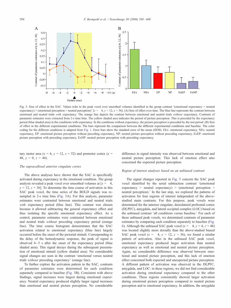
Fig. 5. Size of effect in the SAC. Values refer to the peak voxel over smoothed volumes identified in the group contrast F(emotional expectancy > neutral
expectancy) > (emotional perception > neutral perception)_ [x = �4, y = 12, z = 36]. (A) Size of effect over time. The blue line represents the contrast between
emotional and neutral trials with expectancy. The orange line depicts the contrast between emotional and neutral trials without expectancy. Contrasts of
parameter estimates were extracted from 2-s time bins. The yellow shaded area indicates the period of picture perception. This is preceded by the expectancy
period (blue shaded area) in the conditions with expectancy. In the conditions without expectancy, the picture perception is preceded by the rest period. (B) Size
of effect in the different experimental conditions. The bars represent the comparison between the different experimental conditions and baseline. The color
coding for the different conditions is adapted from Fig. 1. Error bars show the standard error of the mean (SEM). EEx: emotional expectancy, NEx: neutral
expectancy, EP: emotional picture perception without preceding expectancy, NP: neutral picture perception without preceding expectancy, ExEP: emotional
picture perception with preceding expectancy, ExNP: neutral picture perception with preceding expectancy.
F. Bermpohl et al. / NeuroImage 30 (2006) 588–600594
tary motor area (x = 4, y = 12, z = 52) and premotor cortex (x =
48, y = 0, z = 44).
The supracallosal anterior cingulate cortex
The above analyses have shown that the SAC is specifically
activated during expectancy in the emotional condition. The group
analysis revealed a peak voxel over smoothed volumes at [x = �4,y = 12, z = 36]. To determine the time course of activation in this
SAC peak voxel, the time series of the BOLD signals was re-
sampled in 2-s time bins (Fig. 5A). For this analysis, parameter
estimates were contrasted between emotional and neutral trials
with expectancy period (blue line). This contrast was chosen
because it allowed subtracting the general expectancy effect and
thus isolating the specific emotional expectancy effect. As a
control, parameter estimates were contrasted between emotional
and neutral trials without preceding expectancy period (orange
line). The time course histogram demonstrates that the SAC
activation related to emotional expectancy (blue line) largely
occurred before the onset of the pictorial stimuli. Corresponding to
the delay of the hemodynamic response, the peak of signal is
observed 4–5 s after the onset of the expectancy period (blue
shaded area). This signal decays during the subsequent presenta-
tion of emotional stimuli (yellow shaded area). No considerable
signal changes are seen in the contrast Femotional versus neutral
trials without preceding expectancy_ (orange line).
To further explore the activation pattern in the SAC, contrasts
of parameter estimates were determined for each condition
separately compared to baseline (Fig. 5B). Consistent with above
findings, signal increases were largest during emotional expect-
ancy. Neutral expectancy produced slightly larger signal increases
than emotional and neutral picture perception. No considerable
difference in signal intensity was observed between emotional and
neutral picture perception. This lack of emotion effect also
concerned the expected picture perception.
Region of interest analyses based on an unbiased contrast
The signal changes reported in Fig. 5 concern the SAC peak
voxel identified by the serial subtraction contrast F(emotional
neutral perception)._ In the last step, we explored the patterns of
activation for four regions of interest independent of the above-
studied main contrasts. For this purpose, peak voxels were
determined for the anterior cingulate, dorsolateral prefrontal cortex
(DLPFC), amygdala, and lateral occipital complex (LOC) based on
the unbiased contrast Fall conditions versus baseline._ For each of
these unbiased peak voxels, we determined contrasts of parameter
estimates by comparing each condition separately to baseline (Fig.
6). Although the unbiased SAC peak voxel (x = �4, y = 4, z = 48)
was located slightly more dorsally than the above-studied biased
SAC peak voxel (x = �4, y = 12, z = 36), we found a similar
pattern of activation. Also in the unbiased SAC peak voxel,
emotional expectancy produced larger activation than neutral
expectancy as well as emotional and neutral picture perception.
Again, no considerable difference was observed between emo-
tional and neutral picture perception, and this lack of emotion
effect concerned both expected and unexpected picture perception.
A different pattern of activation was observed in the DLPFC,
amygdala, and LOC: in these regions, we did not find considerable
activation during emotional expectancy compared to the other
conditions. These regions consistently showed larger activation
during emotional picture perception compared to neutral picture
perception and to emotional expectancy. In addition, the amygdala
Fig. 6. Size of effect in four regions of interest: anterior cingulate (A), dorsolateral prefrontal cortex (B), amygdala (C), and lateral occipital complex (D). The
bars represent the comparison between the different experimental conditions and baseline. Error bars show the standard error of the mean (SEM). The color
coding for the different conditions is adapted from Fig. 1. Peak voxels were determined for each region of interest, based on the unbiased contrast Fall
conditions versus baseline._ Right and left hemisphere showed comparable results (see Supplementary data). EEx: emotional expectancy, NEx: neutral
expectancy, EP: emotional picture perception without preceding expectancy, NP: neutral picture perception without preceding expectancy, ExEP: emotional
picture perception with preceding expectancy, ExNP: neutral picture perception with preceding expectancy.
F. Bermpohl et al. / NeuroImage 30 (2006) 588–600 595
showed larger activation during expected compared to non-
expected emotional picture perception. This modulation of
perception by expectancy was specific for the emotional condition
and was not present in the SAC, DLPFC, and LOC.
Discussion
The present fMRI study examined the neural correlates of the
expectancy of pictorial emotional stimuli in comparison to the
perception of these stimuli. Neutral expectancy and neutral picture
perception were used as control conditions in order to identify
brain regions activated during expectancy versus perception
specifically in the emotional condition. Our analyses revealed that
the supracallosal anterior cingulate cortex (SAC), cingulate motor
area (CMA), and parieto-occipital sulcus are specifically activated
during expectancy in the emotional condition (emotional expect-
ancy network). A different neuronal network was specifically
associated with emotional picture perception. This emotional
perception network involved a variety of brain regions previously
reported in neuroimaging studies of emotion perception (Phan et
al., 2002), including the amygdala, insula, medial and lateral
prefrontal cortex, cerebellum, and occipitotemporal areas. Using
conjunction analysis, we were not able to document a potential
overlap between these two networks. Taken together, our findings
suggest that separate networks are involved in the expectancy and
perception of pictorial emotional stimuli.
Dissociation between the expectancy and perception of emotional
stimuli
Our finding is in contrast to the hypothesis ventured by
William James (1892) that largely the same brain regions were
implicated in the anticipation and perception of a stimulus.
Carlsson et al. (2000) have previously observed activation of the
primary and secondary sensory cortex during both the expectancy
and perception of tactile stimuli, lending some support to James’
hypothesis. This anticipatory activation in sensory areas was
interpreted as the result of tonic top–down regulation of neuronal
activity. Our data suggest that such tonic pre-activation is less
pronounced or even absent in the domain of emotional picture
processing. Instead of anticipatory activation in the emotional
F. Bermpohl et al. / NeuroImage 30 (2006) 588–600596
perception network, we observed the involvement of a separate
network during emotional expectancy. A similar dissociation has
been observed in pain (Ploghaus et al., 1999) and reward
(Knutson et al., 2001; O’Doherty et al., 2002; Knutson et al.,
2003), although there are also indications for overlapping
networks in reward (Breiter et al., 2001). O’Doherty et al.
(2002) found activation in the ventral tegmental area, amygdala,
and striatum during the expectancy of taste reward, whereas the
insula and operculum were involved in reward consumption.
Such dissociation seems to reflect the distinction between
expectancy-related ‘‘wanting’’ and consumption-related ‘‘liking’’
in reward processing (Berridge, 1996). Similarly, our present
finding of dissociable patterns of activation observed during
different periods of our paradigm may reflect a distinction
between anticipatory and perceptive components of emotional
stimulus processing.
The expectancy of pictorial emotional stimuli has recently been
studied using fMRI (Ueda et al., 2003; Simmons et al., 2004).
These paradigms differed from ours in two aspects. First, they used
valence-selective (certain) emotional expectancy, while we
explored uncertain emotional expectancy. Second, they did not
include a condition of Femotional picture perception without
preceding expectancy_ so that a within-study comparison between
emotional expectancy and perception networks could not be
completed. Nonetheless, it appears that in these previous studies
positive and negative expectancy produced signal increases in
regions that are also activated during emotional stimulus percep-
tion in our study and elsewhere (Bush et al., 2000; Phan et al.,
2002). These regions include the amygdala, insula, medial and
lateral prefrontal cortex, cerebellum, and PAC. Thus, contrary to
our findings, their data suggest that there is a considerable overlap
between networks involved in emotional expectancy and percep-
tion. This discrepancy between study results might be related to
differences between certain and uncertain emotional expectancy.
This assumption is consistent with expectancy studies in other
domains. For instance, findings for certain and uncertain pain
expectancy are largely analogous to the results in emotional
expectancy. Specifically, certain pain expectancy involves the PAC
(Ploghaus et al., 1999, 2003), whereas uncertain pain expectancy is
associated with activation in the SAC including the CMA (Hsieh et
al., 1999; Porro et al., 2002; Jensen et al., 2003; Porro et al., 2003).
Similarly, in the reward domain, Critchley et al. (2001) found that
the expectancy of monetary reward produced larger activation in
the SAC when higher outcome uncertainty was present. It seems
that these findings are now extended to the domain of emotional
picture processing. While certain emotional expectancy has
previously been shown to produce activation in parts of the
emotional perception network including the PAC, amygdala,
insula, and lateral prefrontal cortex (Ueda et al., 2003; Simmons
et al., 2004), the present study demonstrates that uncertain
emotional expectancy involves brain regions (SAC, CMA,
parieto-occipital sulcus) dissociable from the emotional perception
network. However, it is acknowledged that the distinction between
certain and uncertain emotional expectancy remains speculative as
the within-study comparison between certain and uncertain expect-
ancy of emotional pictures was not carried out.
The baseline comparisons shown in Fig. 6 revealed three
different patterns of activation associated with our paradigm. First,
the SAC showed differential activation during expectancy in the
emotional condition (interaction between expectancy and emotion).
Larger signal increases were observed during emotional expect-
ancy compared to both neutral expectancy and emotional
perception. No difference was found between emotional and
neutral perception. Second, the DLPFC and LOC showed differ-
ential activation during picture perception in the emotional
condition (interaction between perception and emotion). Larger
signal increases were observed during emotional picture perception
compared to both neutral picture perception and emotional
expectancy; no difference was found between emotional and
neutral expectancy. Third, the amygdala showed differential
activation during emotional picture perception similar to the
DLPFC and LOC. In addition, this region showed a specific effect
of emotional expectancy on the period of picture perception.
Larger signal increases were observed during emotional picture
perception when it was preceded by emotional expectancy, while
expectancy had no effect on neutral picture perception. The period
of emotional expectancy itself was not associated with consid-
erable signal changes in this region. Taken together, these findings
illustrate that emotional expectancy and emotional picture percep-
tion produce activation in dissociable networks. In addition, these
findings suggest that different brain regions are involved in the
effect of emotional expectancy at distinct stages of emotional
picture presentation. The SAC showed this effect during the
expectancy period and the amygdala during the picture perception
period, while the DLPFC and LOC were not affected by emotional
expectancy.
In contrast to the present investigation, studies on aversive and
appetitive conditioning have observed amygdalar activation related
to conditioned stimuli (Buchel et al., 1998; LaBar et al., 1998;
Buchel et al., 1999; Parkinson et al., 2000; Gottfried et al., 2002). It
seems that this difference in findings is related to the difference in
valence specificity between expectancy cues. In the mentioned
conditioning studies, the cue was linked to either aversive or
appetitive stimuli, whereas, in our study, the emotional expectancy
cue is followed in equal proportions by both positive and negative
stimuli. It might be speculated that the valence ambiguity of our
expectancy cues might have prevented specific aversive or
appetitive conditioning processes and related activation of the
amygdala in our study.
A methodological challenge associated with expectancy studies
is to disentangle cue- from target-related BOLD signals (Rees et
al., 1997). Because of the temporal characteristics of the
hemodynamic response, the regressors for Fexpectancy_ may be
confounded by the subsequent picture periods. This confounder
could be reduced by the inclusion of unpaired (Buchel et al., 1998)
or misleading expectancy cues or the use of very irregular
expectancy intervals (Chawla et al., 1999). These measures were
not taken in our study for psychological reasons. Behavioral pilot
tests indicated that the emotional expectancy cue would have
become too Farbitrary_ and would not have sufficiently differed
from the rest condition. This tendency to Farbitrariness_ of the
emotional expectancy cue is related to two features of our
paradigm: (1) we used uncertain emotional expectancy cues which
by themselves introduce a considerable degree of uncertainty. (2)
The fixation cross was followed by emotional pictures in 25% of
the trials because our control condition consisted of pictures
without preceding expectancy. While these two features were
essential for our paradigm, we chose to omit unpaired, misleading,
and irregular cues in order to not further lower the predictive value
of the emotional expectancy cue. Given this situation, it must be
acknowledged that decorrelation of expectancy- and picture-related
BOLD responses can only be partially achieved in our study. This
F. Bermpohl et al. / NeuroImage 30 (2006) 588–600 597
raises the possibility that the SAC activation observed in the serial
subtraction contrast using Femotional > neutral expectancy_ as thefirst constituent could be related to the perception of expected
emotional pictures rather than the emotional expectancy period per
se. However, our results argue against this possibility. First, we
found dissociable networks for the expectancy and perception of
emotional stimuli. Rather than dissociable networks, one would
have anticipated overlapping networks as a result of insufficient
decorrelation. Second, the time course histogram (Fig. 5A)
demonstrates that the SAC activation induced by emotional
expectancy occurred before the onset of the subsequent picture
presentation. Third, baseline comparisons showed a trend towards
lower, rather than higher, signal intensities in the SAC during
expected emotional pictures compared to unexpected emotional
pictures and to expected neutral pictures (Figs. 5B and 6A). Taken
together, our findings indicate that the SAC activation attributed to
emotional expectancy was not critically confounded by the
subsequent picture period.
Emotional expectancy and the supracallosal anterior cingulate
cortex
In our paradigm, activation in the SAC (including CMA) was
observed during expectancy specifically in the emotional con-
dition. Based on lesion and functional neuroimaging studies, this
region is considered a multi-integrative structure that is implicated
in a variety of affective, cognitive, and motor processes related to
adaptive behavior (Devinsky et al., 1995; Paus, 2001). Our
findings contribute to this notion in that they highlight the
anticipatory aspect in these processes.
Although the SAC is considered the Fcognitive division_ of
the anterior cingulate (Devinsky et al., 1995; Bush et al., 2000),
several affective functions have also been proposed for this
region. These functions relate to the processing of emotional
attention (Lane et al., 2001; Vuilleumier et al., 2001), autonomic
arousal (Fredrikson et al., 1998; Critchley et al., 2003), reward
(Breiter et al., 1997; Bush et al., 2002), and pain (Rainville et al.,
1997; Becerra et al., 2001; Rolls et al., 2003; Singer et al.,
2004). Our data indicate that the processes mediated by the SAC
are independent of the presence of emotional stimuli. In our
study, mere expectancy of emotional pictures produced SAC
activation. Even more, this response was clearly larger than the
one related to the actual perception of emotional photographs.
The latter did not differ from neutral picture perception and
tended to produce smaller SAC activation than neutral expect-
ancy. Taken together, these findings highlight the anticipatory
character of SAC function. They suggest a role for the SAC in
preceding emotional attention (e.g., emotional expectancy) rather
than attentional processes requiring the actual presence of
emotional stimuli.
Activation in the SAC (as well as the CMA and parieto-
occipital sulcus) has previously been observed in paradigms used
to study anticipatory anxiety (Chua et al., 1999), anticipatory
arousal (Critchley et al., 2001), and the expectancy of reward
(Kirsch et al., 2003) and pain (Hsieh et al., 1999; Porro et al., 2002;
Jensen et al., 2003; Porro et al., 2003). Although these paradigms
were not explicitly designed to study the expectancy of emotional
stimuli, it seems plausible that they implicitly involved this aspect.
In addition, these paradigms involved processes specifically related
to reward and pain, which may interact with both the emotion and
the expectancy network. Using standardized and validated stimuli
from the IAPS, the present paradigm was designed to study
emotional expectancy independent of reward and pain. In contrast
to reward paradigms, subjects were aware that they could not
influence the outcome of the trial. In contrast to pain paradigms,
the nociceptive system was not activated, and the emotional
expectancy cue did not distinguish between aversive and pleasant
stimuli which might have prevented specific aversive conditioning
processes (see above). In view of the present results, one might
suggest that the SAC, CMA, and parieto-occipital sulcus are
involved in emotional expectancy independent of reward and pain.
Other brain regions activated during reward and pain expectancy
may be related to non-emotional aspects of these paradigms; these
regions include the ventral tegmental area, ventral striatum, and
orbitofrontal cortex in reward expectancy (Breiter et al., 2001;
Knutson et al., 2001; O’Doherty et al., 2002; Knutson et al., 2003)
and the primary somatosensory cortex, medial prefrontal cortex,
insula, and medial thalamus in pain expectancy (Ploghaus et al.,
1999; Porro et al., 2002, 2003).
Figs. 5B and 6A show that signal increases in the SAC not only
related to emotional expectancy, but also to neutral expectancy.
This finding suggests that the arrows presented in the expectancy
condition may also induce a nonspecific expectancy effect (atten-
tional capture) in the SAC. Since the signal increase is greater
during emotional compared to neutral expectancy, it might be
concluded that both nonspecific attentional capture and specific
emotional expectancy contribute to the activation observed during
emotional expectancy.
It is also important to note that the observed SAC activation
cannot simply be explained by a nonspecific arousal effect. A
general arousal effect would be hypothesized to produce activation
not only during emotional expectancy, but also during emotional
picture perception. According to the normative data of the IAPS
(Lang et al., 1999), the emotional pictures presented can be
considered high arousing stimuli and the neutral pictures low
arousing stimuli. Since the comparison Femotional picture percep-
tion > neutral picture perception_ did not produce differential SAC
activation in our experiment (Figs. 4–6), we conclude that the
SAC activation observed during emotional expectancy does not
simply reflect general arousal. However, we cannot exclude a
specific contribution of anticipatory arousal to the observed
activation. One could argue for a distinction between anticipatory
and general arousal and speculate that the SAC is specifically
involved in the former.
The cognitive roles previously proposed for the SAC are related
to Pavlovian conditioning (Buchel et al., 1998; LaBar et al., 1998)
and the representation of conflict (Carter et al., 2000) and
uncertainty (Critchley et al., 2001; Keri et al., 2004). The present
study focused on emotional expectancy, which naturally involves
elements of conditioning. Our paradigm, however, does not
represent conventional Pavlovian conditioning (Pavlov and Anrep,
1927) because our subjects were familiarized with the association
between the expectancy cues and subsequent pictorial stimuli prior
to the experiment. Moreover, in conventional conditioning, the cue
is linked to either aversive or appetitive stimuli. In our study, by
contrast, the emotional expectancy cue was followed in equal
proportions by positive and negative pictures. The emotional
expectancy cue thus involved uncertainty with regard to the
valence of the subsequent picture (positive or negative) which
might have resulted in a conflict between approach and with-
drawal. Our data therefore show that SAC involvement in
conditioning or, more generally, in expectancy does not presuppose
F. Bermpohl et al. / NeuroImage 30 (2006) 588–600598
cues unequivocally associated with either aversive or appetitive
stimuli.
Besides affective and cognitive processes, the SAC (especially
its most caudal part, the CMA) has been implicated in the
processing of motor response to behaviorally relevant stimuli.
Because of its dense connections to primary and secondary motor
regions, this region appears well suited to translate affective and
cognitive information into action (Paus, 2001). It has been
demonstrated that CMA activation does not reflect action perform-
ance per se but rather the anticipatory state in which one is ready to
select an action in response to a motivationally salient stimuli
(Woldorff et al., 1999). In our study, the CMA is activated during
emotional expectancy compared to both neutral expectancy and
emotional stimulus perception (Fig. 6). One might suggest that the
expectancy of emotional pictures also implicates a state of
preparedness for motor response (e.g., approach or withdrawal).
Besides the SAC and CMA, the parieto-occipital sulcus (which
includes mesial parts of BA 7 and BA 19, extending into BA 31) was
identified by the contrast F(emotional expectancy > neutral expect-
ancy) > (emotional perception > neutral perception)._ This finding isin accordance with previous studies showing activation in this
region during the expectancy of pain (Buchel et al., 1998; Porro et
al., 2003), tickling (Carlsson et al., 2000), monetary reward (Bjork et
al., 2004), and emotional photographs (Ueda et al., 2003). The
parieto-occipital sulcus can be considered the anterior part of the
dorsal visual pathway, which projects from early visual areas to the
posterior parietal cortex. Like the CMA, this dorsal stream is
associated with processes related to action (Goodale and Milner,
1992; Goodale and Westwood, 2004). Specifically, this stream
appears to mediate the required sensorimotor transformations for
visually guided action. In our study, as well as in the other mentioned
expectancy studies, the parieto-occipital sulcus is activated during
the expectancy period, which does not involve visually guided
action. However, we suggest that the expectancy of motivationally
relevant stimuli might implicate a state of preparedness for action.
This might produce anticipatory activation in the dorsal stream even
in the absence of action-related visual stimulation. Such expectancy-
related activation of specialized visual regions has extensively been
studied for basic visual features such as color (Chawla et al., 1999),
motion (Shulman et al., 1999), or spatial location (Kastner et al.,
1999; Hopfinger et al., 2000).
Taken together, our findings point out the anticipatory character
of SAC function. Based on the present results and previous studies,
it might be suggested that this multi-integrative region is involved
in emotional expectancy and its attendant state of preparedness for
motor and autonomic response in situations of emotional salience.
Conclusions
Building on previous studies of visual, tactile, pain, and reward
anticipation, we compared brain systems activated during the
expectancy and perception of pictorial emotional stimuli. During
the expectancy of emotional pictures, we observed activation in
the supracallosal anterior cingulate, cingulate motor area, and
parieto-occipital sulcus. This network of emotional expectancy
was dissociable from regions specifically activated during emo-
tional picture perception. We suggest that this dissociation reflects
a distinction between anticipatory and perceptive components of
emotional stimulus processing, as similarly proposed for pain and
reward.
Acknowledgments
This work was supported by a grant within the Postdoc-
Programme of the German Academic Exchange Service (DAAD,
D/02/46858) to F.B., a Heisenberg grant from the German
Research Foundation to G.N. (DFG, 304/4-1), a Human Frontier
Science Program award to A.A., grant K24 RR018875 from the
National Institutes of Health (NCRR) to A.P.-L., and the Harvard
Thorndike General Clinical Research Center (NCRR MO1
RR01032).
Appendix A. Supplementary data
Supplementary data associated with this article can be found in
the online version at doi:10.1016/j.neuroimage.2005.09.040.
References
Amedi, A., Jacobson, G., Hendler, T., Malach, R., Zohary, E., 2002.
Convergence of visual and tactile shape processing in the human