DISTAL RADIOULNAR JOINT INJURIES Presented by Dr Sunil poonia, PGT, Orthopaedics, SMCH Moderated by Dr S. K. Das, Assoc. Prof of Orthopaedics, SMCH Binu P Thomas, Raveendran Sreekanth Dr. Paul Brand Centre for Hand Surgery, CMC Hospital, Vellore, Tamil Nadu, India
Transcript
DISTAL RADIOULNAR JOINT INJURIES
Presented by Dr Sunil poonia, PGT, Orthopaedics, SMCH
Moderated by Dr S. K. Das, Assoc. Prof of Orthopaedics, SMCH
Binu P Thomas, Raveendran Sreekanth
Dr. Paul Brand Centre for Hand Surgery, CMC Hospital,
Important joint in pronosupination and load transmission
EVOLUTION OF DRUJ
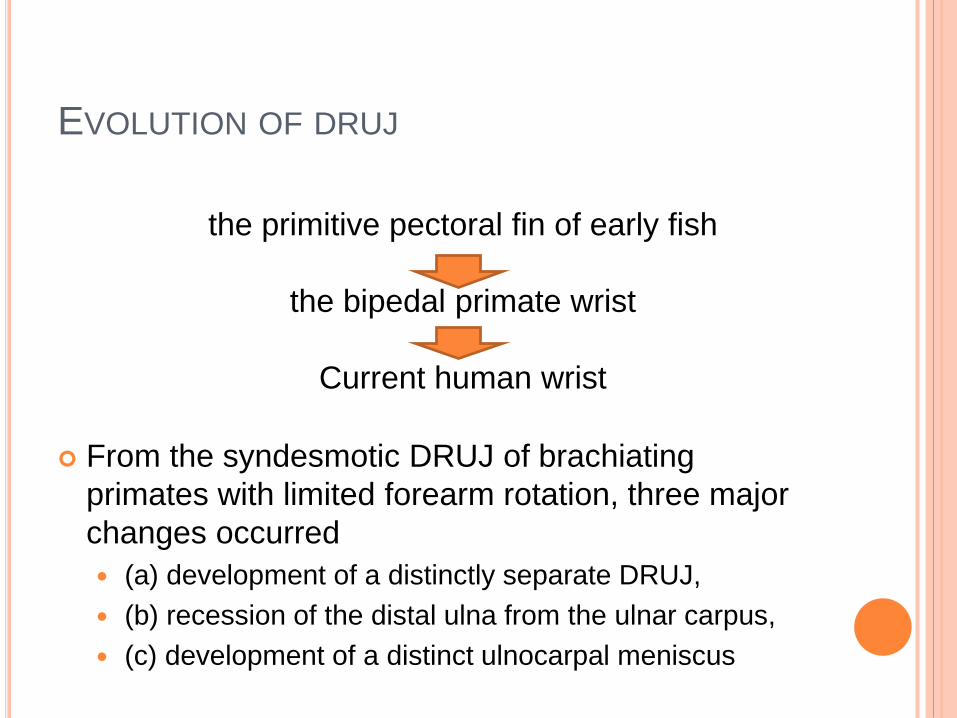
From the syndesmotic DRUJ of brachiating
primates with limited forearm rotation, three major
changes occurred
(a) development of a distinctly separate DRUJ,
(b) recession of the distal ulna from the ulnar carpus,
(c) development of a distinct ulnocarpal meniscus
the primitive pectoral fin of early fish
the bipedal primate wrist
Current human wrist
Distal radioulnar
joint injuries
Acute injury Chronic
instabilty or arthritis
Isolated injuries
along with fractures
ANATOMY
diarthrodial trochoid synovial joint
two parts
the bony radioulnar articulation and
soft tissue stabilizers.
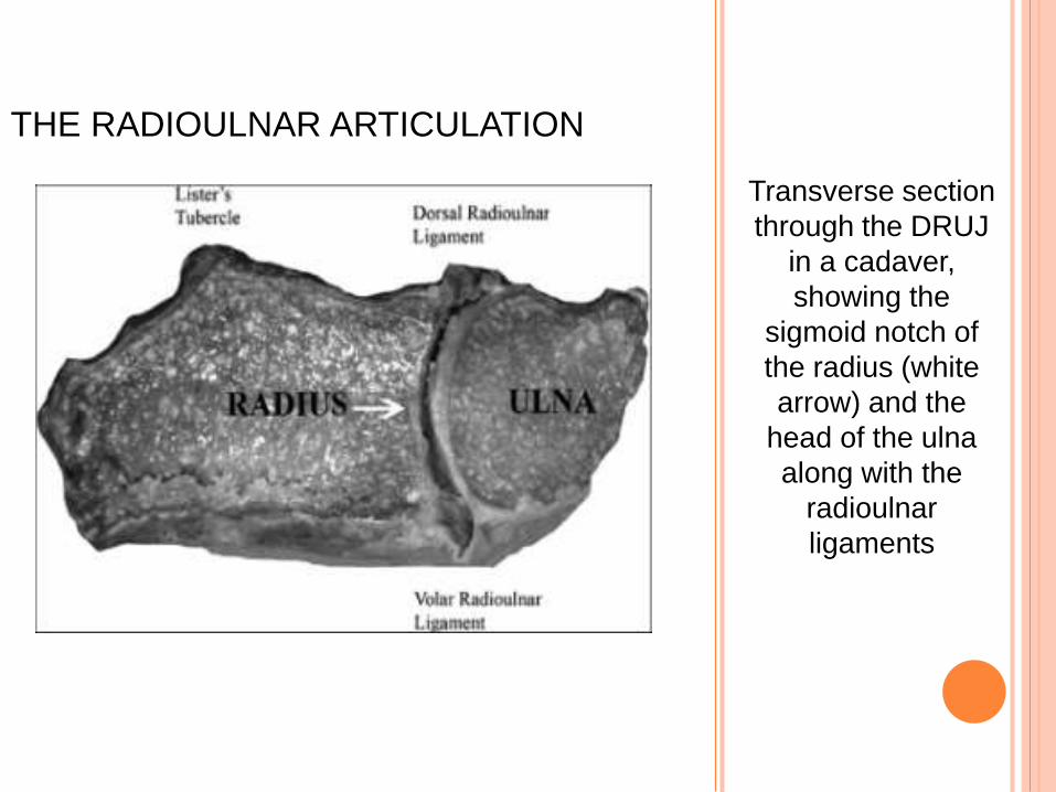
Transverse section
through the DRUJ
in a cadaver,
showing the
sigmoid notch of
the radius (white
arrow) and the
head of the ulna
along with the
radioulnar
ligaments
THE RADIOULNAR ARTICULATION
ANATOMY
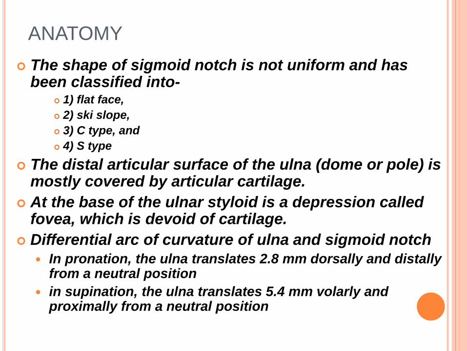
The shape of sigmoid notch is not uniform and has been classified into-
1) flat face,
2) ski slope,
3) C type, and
4) S type
The distal articular surface of the ulna (dome or pole) is mostly covered by articular cartilage.
At the base of the ulnar styloid is a depression called fovea, which is devoid of cartilage.
Differential arc of curvature of ulna and sigmoid notch
In pronation, the ulna translates 2.8 mm dorsally and distally from a neutral position
in supination, the ulna translates 5.4 mm volarly and proximally from a neutral position
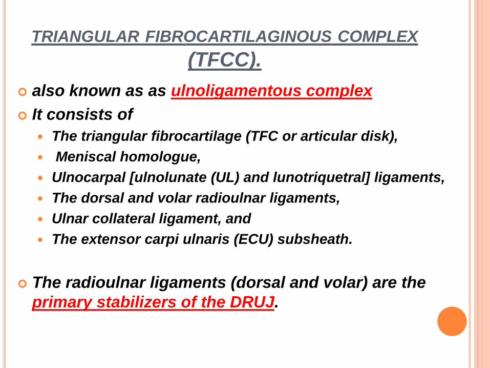
TRIANGULAR FIBROCARTILAGINOUS COMPLEX
(TFCC).
also known as as ulnoligamentous complex
It consists of
The triangular fibrocartilage (TFC or articular disk),
Meniscal homologue,
Ulnocarpal [ulnolunate (UL) and lunotriquetral] ligaments,
The dorsal and volar radioulnar ligaments,
Ulnar collateral ligament, and
The extensor carpi ulnaris (ECU) subsheath.
The radioulnar ligaments (dorsal and volar) are the
primary stabilizers of the DRUJ.
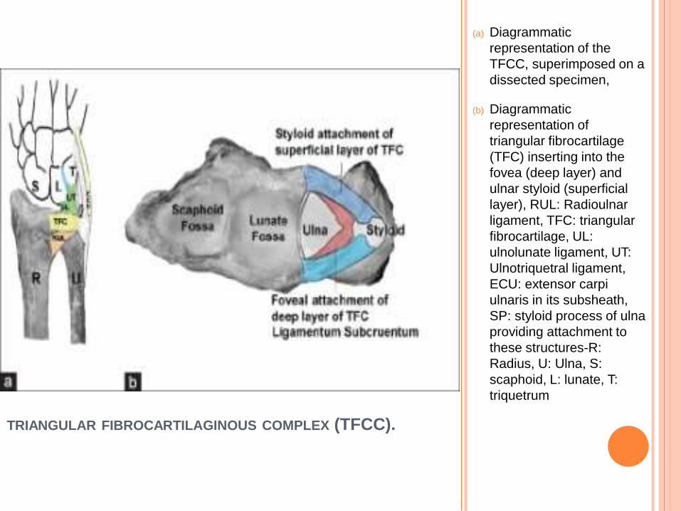
TRIANGULAR FIBROCARTILAGINOUS COMPLEX (TFCC).
(a) Diagrammatic
representation of the
TFCC, superimposed on a
dissected specimen,
(b) Diagrammatic
representation of
triangular fibrocartilage
(TFC) inserting into the
fovea (deep layer) and
ulnar styloid (superficial
layer), RUL: Radioulnar
ligament, TFC: triangular
fibrocartilage, UL:
ulnolunate ligament, UT:
Ulnotriquetral ligament,
ECU: extensor carpi
ulnaris in its subsheath,
SP: styloid process of ulna
providing attachment to
these structures-R:
Radius, U: Ulna, S:
scaphoid, L: lunate, T:
triquetrum
CLINICAL EVALUATION
trauma, eg, a fall on the outstretched hand
(FOOSH).
ulnar-sided wrist pain (USWP), especially on
loading the hand and rotating the forearm,
Persistence of USWP and stiffness following distal
radius fractures (DRF)
Clicking sounds
Obvious instability
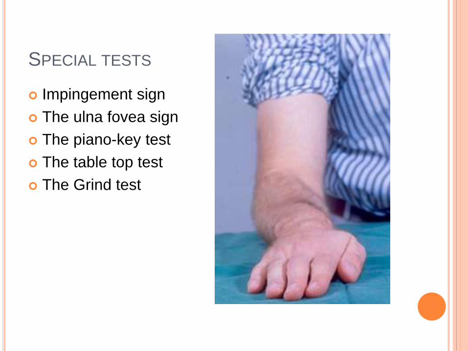
SPECIAL TESTS
Impingement sign
The ulna fovea sign
The piano-key test
The table top test
The Grind test
Ulnar deviation of the wrist with the forearm in neutral produces ulnar wrist pain and occasional clicking
A painful click may be elicited by having the patient clench and ulnarly deviate the wrist and then repeatedly pronate and supinate the wrist
The ulnar impaction test—wrist hyperextension and ulnar deviation with axial compression—also will elicit pain.
The “press test” is another useful provocative test: the seated patient is asked to push the body weight up off a chair using the affected wrist, creating an axial ulnar load. If this reproduces the patient’s pain, the test is considered positive
With the wrist in pronation, an unstable distal ulna may translate dorsally and can be manually reduced with dorsal thumb pressure (“piano key test”).
Tenderness and pain identified when external pressure is applied to the area of the fovea (fovea sign) is indicative of an ulnocarpal ligament lesion.
TFCC instability also is suggested by excessive motion with the “shuck test”—with the radial aspect of the wrist stabilized, anteroposterior stress is applied to the ulnar side of the wrist
RADIOLOGICAL INVESTIGATIONS
Radiographs
Posteroanterior (PA)
True lateral X-ray
Pronation and supination views
A clenched fist PA view in pronation
Weighted lateral stress view in pronation
X-RAY EVALUATION OF DRUJ
a)True PA views should
show the groove for
ECU radial to the ulnar
styloid (red arrow). True
lateral view should show
the palmar edge of
pisiform (red dotted line)
midway between palmar
borders of distal pole of
scaphoid and capitate
(yellow lines);
(b) Scheker-weighted
lateral view with patient
holding 3 lb weight in the
hand showing dorsal
instability of the distal
ulna. Weighted views
provide loading of the
DRUJ, bringing out
instability, which may not
be visible in routine X-
rays
COMPUTED TOMOGRAPHY
Useful to delineate sigmoid notch fractures and
DRUJ injuries
Ligament injuries can be assessed indirectly by
assessing the radioulnar articulation in various
positions and also by loading views
Three-dimensional (3D) reconstructions are helpful
in assessing spatial relationship between the radius
and ulna
MRI HAS 86% SENSITIVITY FOR DETECTION OF TFCC
TEARS.
a) MRI T2-weighted
fat suppression
image, showing a
radial TFCC tear,
fluid seen adjacent to
DRUJ.
b) Proton density-
weighted MRI,
coronal view
suggestive of ulnar
impaction syndrome.
There is articular
cartilage loss with
erosion, marrow
edema, subchondral
cyst, and sclerosis of
triquetrum and lunate
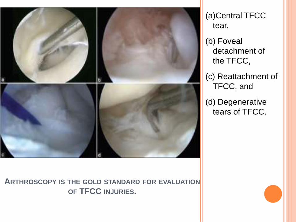
ARTHROSCOPY IS THE GOLD STANDARD FOR EVALUATION
OF TFCC INJURIES.
(a)Central TFCC
tear,
(b) Foveal
detachment of
the TFCC,
(c) Reattachment of
TFCC, and
(d) Degenerative
tears of TFCC.
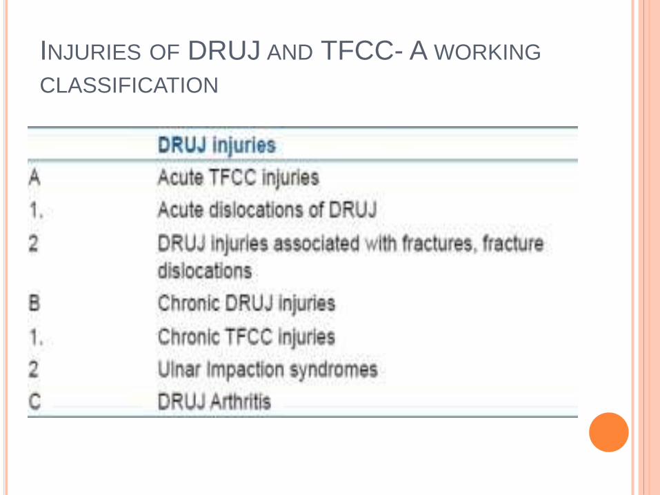
INJURIES OF DRUJ AND TFCC- A WORKING
CLASSIFICATION
TRIANGULAR FIBROCARTILAGINOUS COMPLEX
INJURY
“the traumatic TFCC disruption as a continuum of
injury”– Melone
It was classified into five stages of increasing severity
Stage I: detachment of TFC from ulnar styloid,
stage II: ECU subsheath injury,
stage III: ulnocarpal ligament disruption,
stage IV: lunotriquetral ligament injury, and
stage V: midcarpal ligament injury
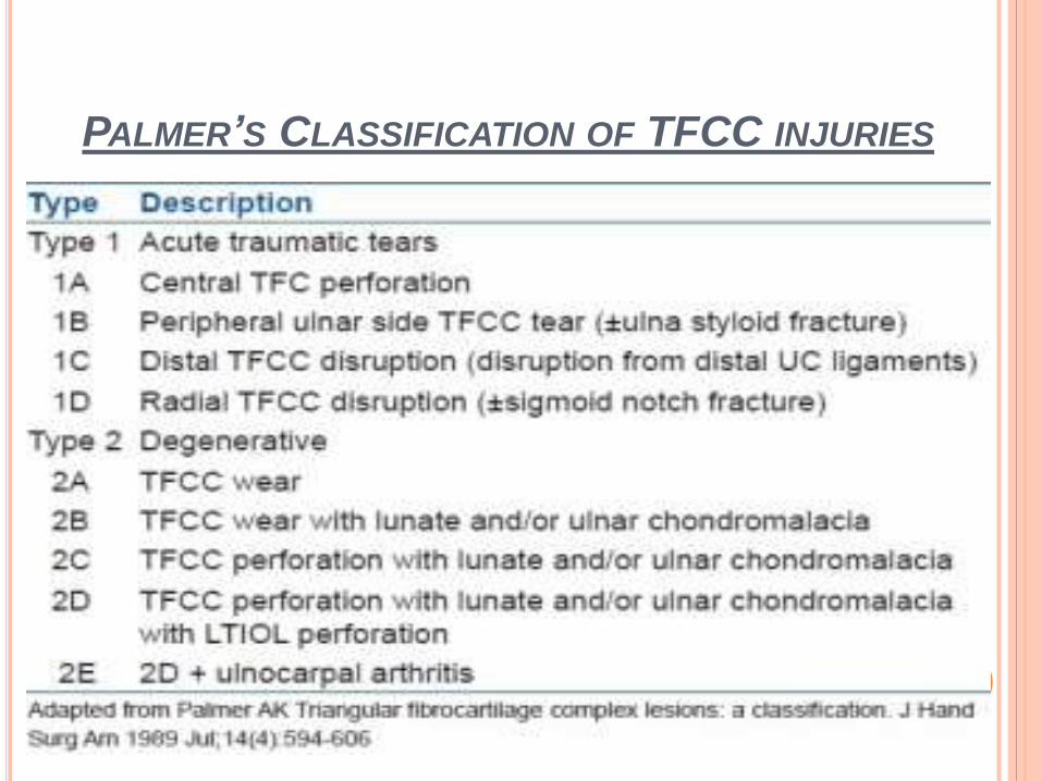
PALMER’S CLASSIFICATION OF TFCC INJURIES
Currently, management of class 1A TFCC (central perforation)
lesions includes nonoperative measures initially. If significant
symptoms persist, arthroscopic débridement may provide relief
For class 1B lesions (avulsion from the ulna, with or without
ulnar styloid fracture), immobilization for 6 weeks followed
by rehabilitation may be sufficient
If symptoms persist, and if there is DRUJ instability,
arthroscopic repair using either an inside-out or an outside-in
technique may produce satisfactory relief of pain and
improvemen
class 1C lesions (distal avulsion of ulnocarpal ligaments), which
result in a volar ulnar “sag” of the carpus, late open or
arthroscopic repair may relieve symptoms
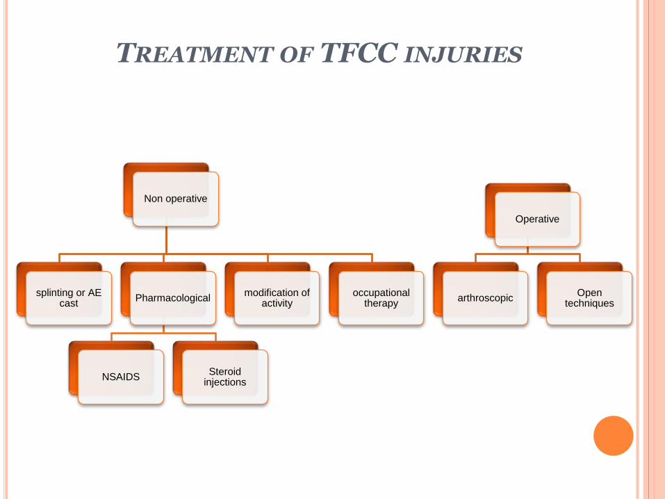
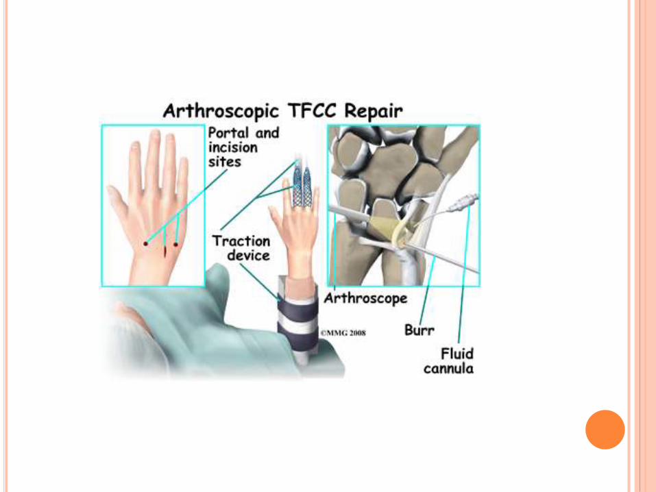
TREATMENT OF TFCC INJURIES
Non operative
splinting or AE cast
Pharmacological
NSAIDSSteroid
injections
modification of activity
occupational therapy
Operative
arthroscopicOpen
techniques
ISOLATED DRUJ DISLOCATIONS
Uncommon injuries
Dorsal or volar
Simple or complex
The dorsal dislocation is more common
closed manipulation and reduction under anesthesia is
usually successful.
Once the joint is reduced, stability must be verified
ISOLATED DRUJ DISLOCATIONS
Immobilize dorsal dislocations in an above elbow plaster
of Paris (POP) cast in supination, and volar dislocations
in pronation for a period of 6 weeks
If instability persists after reduction, radioulnar pinning is
done in reduced position to allow soft tissue healing
TFCC repair, either open or arthroscopic, needs to be
also considered in case of severe disruptions
Soft tissue interposition can result in irreducibility
DRUJ INJURIES ASSOCIATED WITH FRACTURES AND
FRACTURE-DISLOCATIONS
The most common cause of residual wrist disability after
DRF is the DRUJ involvement
Three basic causes that result in radioulnar pain and
limitation of forearm rotation are
instability,
joint incongruence, and
ulnocarpal abutment
it is found that severely displaced DRF result in disruption of
TFCC in the absence of ulna styloid fractures
USF through the base results in DRUJ instability if the
fragment involves the foveal insertion of the TFCC.
DRUJ INJURIES ASSOCIATED WITH FRACTURES
AND FRACTURE-DISLOCATIONS
Fractures through the sigmoid notch produce stiffness and
late onset arthritis of the DRUJ.
Despite the severity of these injuries, with proper diagnosis
and reduction, most patients will have a satisfactory outcome
Assessment of DRUJ stability following DRF are best done
intraoperatively after fixation of the radius fracture by
translation of the ulna in a dorsopalmar direction
DRUJ INJURIES ASSOCIATED WITH FRACTURES
AND FRACTURE-DISLOCATIONS
Careful assessment of the preoperative X-rays can
indicate a possibility of DRUJ instability
1) shortening of radius >5 mm relative to ulna,
2) fracture of the base of ulnar styloid,
3) widening of the DRUJ interval on PA view,
4) dislocation of the DRUJ on lateral view.
Computed tomography scans subluxation and
fractures of the ligamentous margins of radius and
ulna
DRUJ INJURIES ASSOCIATED WITH FRACTURES
AND FRACTURE-DISLOCATIONS
Fragment-specific fixation is helpful
About 61% of DRF are associated with ulna styloid fractures
No significant relationship between functional outcome and
ulnar styloid fractures (USF), which were not fixed following
stable fixation of distal radius fracture
ULNA STYLOID FRACTURES
may also be seen in isolation
While styloid tip fractures are stable, basal fractures of the styloid are associated with DRUJ instability
Fixation of styloid fracture makes the DRUJ stable, provided the TFCC is not otherwise injured
various fixation techniques
closed pinning,
tension band wiring
compression screw fixation,
suture anchor technique
symptomatic nonunions of styloid?
Comminuted, unstable, or displaced distal ulna neck fractures?
GALEAZZI FRACTURE-DISLOCATION
Palmer Type IB TFCC injury is classically seen
80% of these injuries presented with complete
dislocation of DRUJ
operative fixation of the radius is necessary due to
inherent instability.
When the radius fracture is within 7.5 cm of the distal
radius, DRUJ injury is highly likely
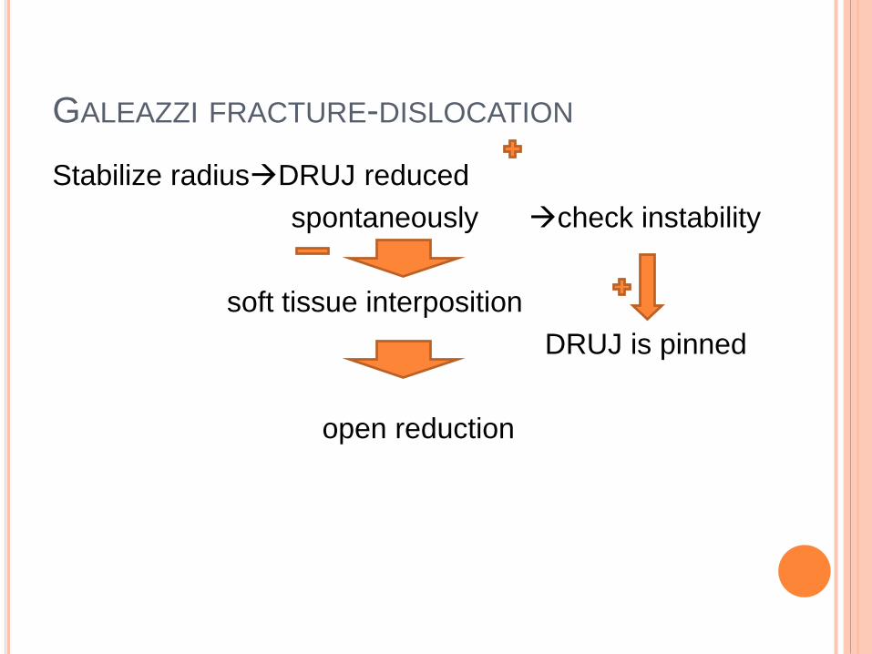
GALEAZZI FRACTURE-DISLOCATION
Stabilize radiusDRUJ reduced
spontaneously check instability
soft tissue interposition
DRUJ is pinned
open reduction
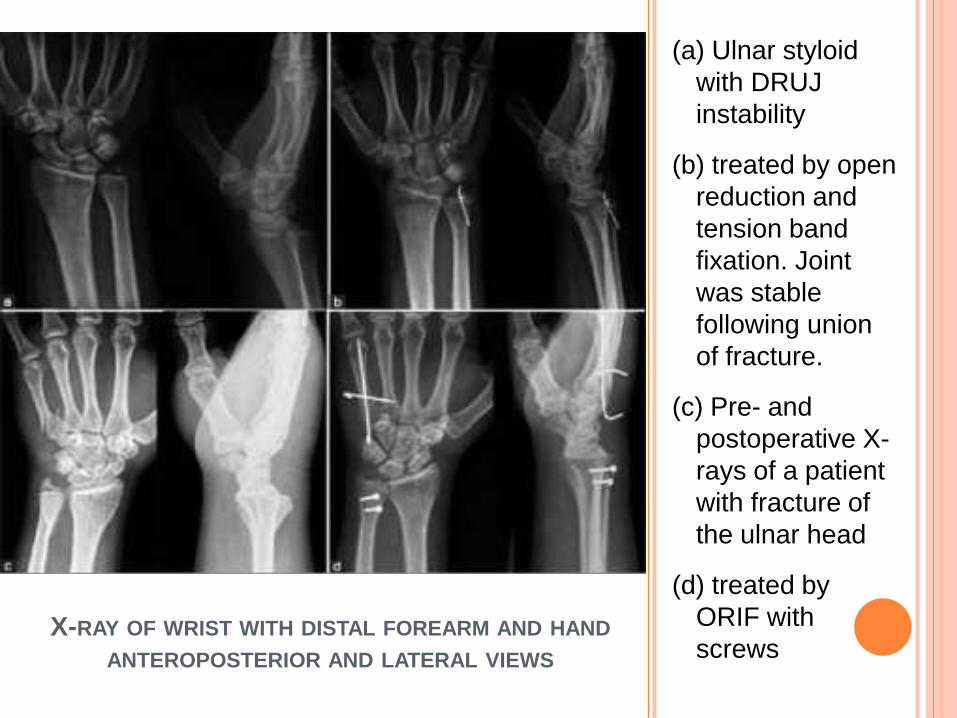
X-RAY OF WRIST WITH DISTAL FOREARM AND HAND
ANTEROPOSTERIOR AND LATERAL VIEWS
(a) Ulnar styloid
with DRUJ
instability
(b) treated by open
reduction and
tension band
fixation. Joint
was stable
following union
of fracture.
(c) Pre- and
postoperative X-
rays of a patient
with fracture of
the ulnar head
(d) treated by
ORIF with
screws
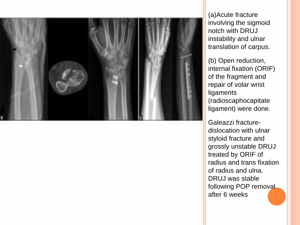
(a)Acute fracture
involving the sigmoid
notch with DRUJ
instability and ulnar
translation of carpus.
(b) Open reduction,
internal fixation (ORIF)
of the fragment and
repair of volar wrist
ligaments
(radioscaphocapitate
ligament) were done.
Galeazzi fracture-
dislocation with ulnar
styloid fracture and
grossly unstable DRUJ
treated by ORIF of
radius and trans fixation
of radius and ulna.
DRUJ was stable
following POP removal
after 6 weeks
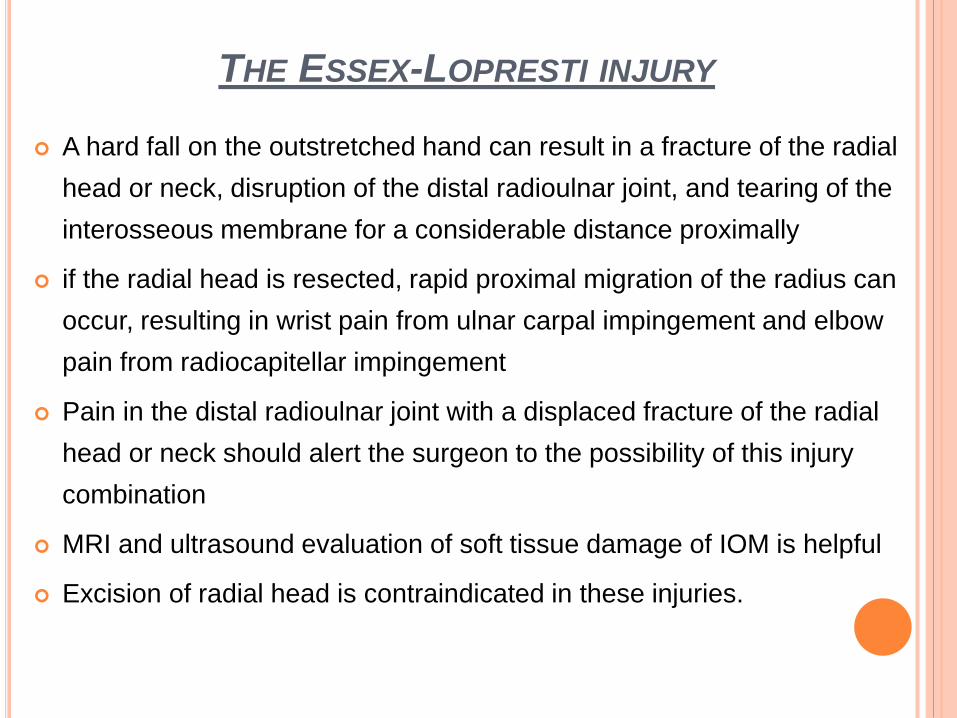
THE ESSEX-LOPRESTI INJURY
A hard fall on the outstretched hand can result in a fracture of the radial
head or neck, disruption of the distal radioulnar joint, and tearing of the
interosseous membrane for a considerable distance proximally
if the radial head is resected, rapid proximal migration of the radius can
occur, resulting in wrist pain from ulnar carpal impingement and elbow
pain from radiocapitellar impingement
Pain in the distal radioulnar joint with a displaced fracture of the radial
head or neck should alert the surgeon to the possibility of this injury
combination
MRI and ultrasound evaluation of soft tissue damage of IOM is helpful
Excision of radial head is contraindicated in these injuries.
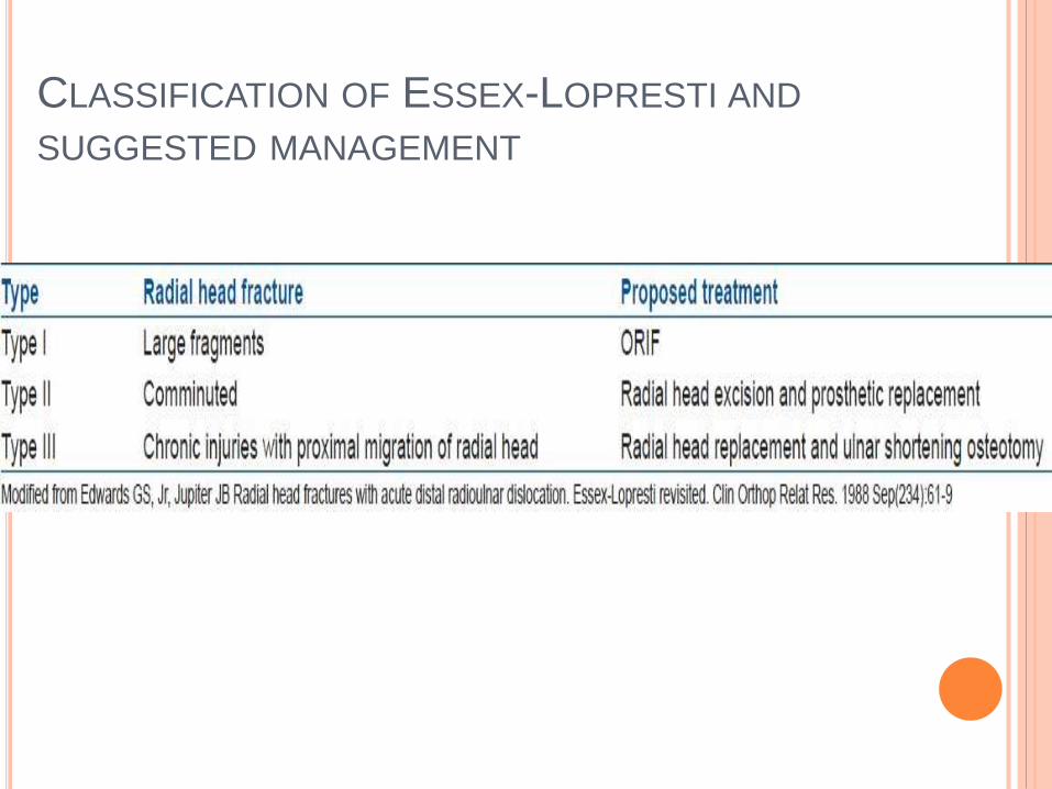
CLASSIFICATION OF ESSEX-LOPRESTI AND
SUGGESTED MANAGEMENT
CHRONIC DRUJ INSTABILITY
Chronic DRUJ instability can result from fractures of the
distal radius and ulna following inadequate treatment or
malunion
If untreated, these lead to chronic pain and disability due to
stiffness, decreased grip strength, and arthritis
There are reports suggesting that anatomical reduction of
DRF is more critical in avoiding persistent DRUJ issues
rather than associated fixing or union of ulna styloid
fractures.
MANAGEMENT
Management of chronic DRUJ instability depends
primarily on the underlying cause
Correct malunion, length discrepancies first
Soft tissue reconstruction indicated in symptomatic
patients in whom TFCC is irreparable & sigmoid notch
incompetent
Arthritis of DRUJ requires salvage procedures
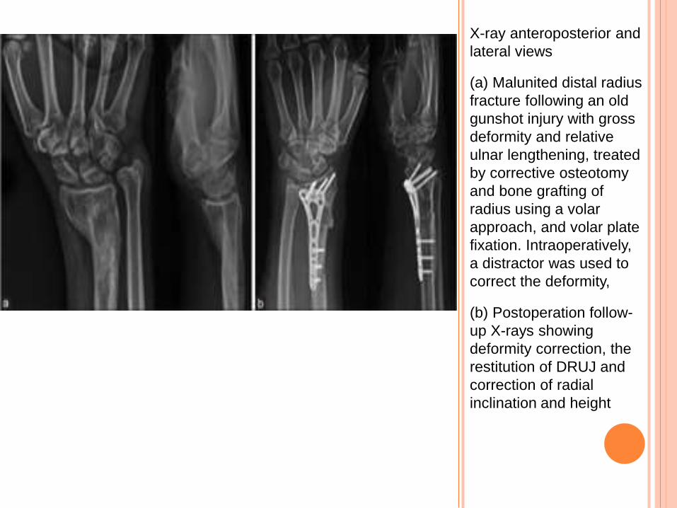
X-ray anteroposterior and
lateral views
(a) Malunited distal radius
fracture following an old
gunshot injury with gross
deformity and relative
ulnar lengthening, treated
by corrective osteotomy
and bone grafting of
radius using a volar
approach, and volar plate
fixation. Intraoperatively,
a distractor was used to
correct the deformity,
(b) Postoperation follow-
up X-rays showing
deformity correction, the
restitution of DRUJ and
correction of radial
inclination and height
MANAGEMENT
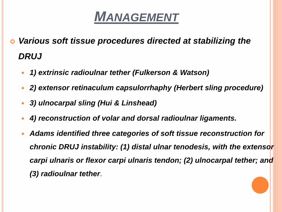
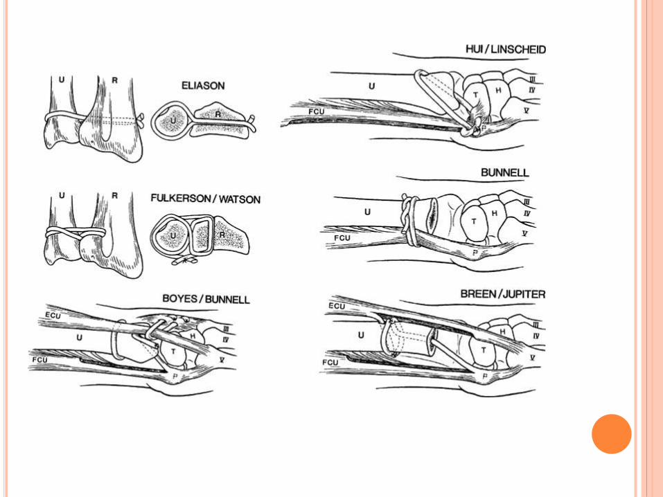
Various soft tissue procedures directed at stabilizing the
4) reconstruction of volar and dorsal radioulnar ligaments.
Adams identified three categories of soft tissue reconstruction for
chronic DRUJ instability: (1) distal ulnar tenodesis, with the extensor
carpi ulnaris or flexor carpi ulnaris tendon; (2) ulnocarpal tether; and
(3) radioulnar tether.
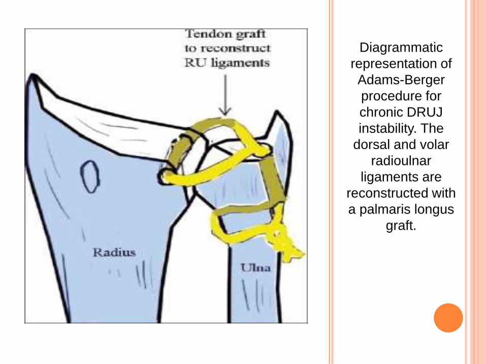
Diagrammatic
representation of
Adams-Berger
procedure for
chronic DRUJ
instability. The
dorsal and volar
radioulnar
ligaments are
reconstructed with
a palmaris longus
graft.
ULNAR IMPACTION SYNDROME
Due to repetitive loading of the ulnocarpal joint, especially in
the presence of ulna plus variance, degenerative changes
occur in the TFC,ulnar head, lunate and triquetral surface,
lunotriquetral articulation and is referred to as ulnar
impaction or ulnocarpal abutment syndrome
progressive wear of TFCC perforation ulnocarpal
arthritis
the most common cause acquired ulna plus variance and
dorsal tilt caused by malunited distal radius fracture
ulna impingement
syndrome??
ULNAR IMPACTION SYNDROME
Typical clinical features are ulnar-sided wrist pain,
especially on loading and rotation movement
Investigations
The PA view demonstrates the ulna plus.
MRI is useful for observing changes in the lunate and
triquetrum
Arthroscopy demonstrates the classical stages described by
Palmer.
TREATMENT
Splinting
NSAIDs
Modification of
activities
wafer resection of the
distal ulna as described
by Feldon
ulna shortening
osteotomy
conservative Surgical
Author prefers an ulna shortening osteotomy
and compression plate fixation
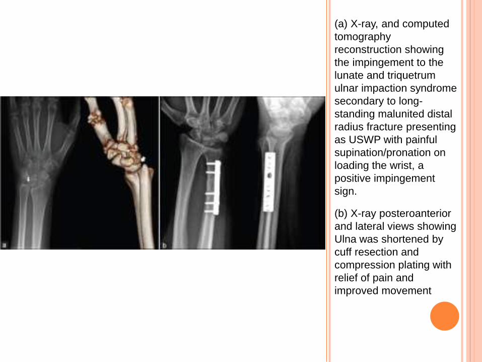
(a) X-ray, and computed
tomography
reconstruction showing
the impingement to the
lunate and triquetrum
ulnar impaction syndrome
secondary to long-
standing malunited distal
radius fracture presenting
as USWP with painful
supination/pronation on
loading the wrist, a
positive impingement
sign.
(b) X-ray posteroanterior
and lateral views showing
Ulna was shortened by
cuff resection and
compression plating with
relief of pain and
improved movement
DRUJ ARTHRITIS
Causes
DRF through the sigmoid notch or the distal ulna
Malunions
chronic instability of DRUJ
failed reconstruction of the DRUJ
Various options are available
Resection of distal ulna (Darrach procedure)
Sauve-Kapandji procedure
Hemiresection-interposition arthroplasty
DRUJ implant arthroplasty
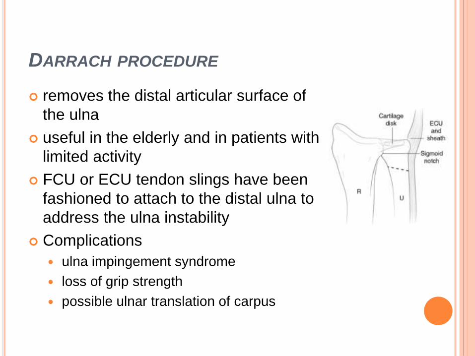
DARRACH PROCEDURE
removes the distal articular surface of
the ulna
useful in the elderly and in patients with
limited activity
FCU or ECU tendon slings have been
fashioned to attach to the distal ulna to
address the ulna instability
Complications
ulna impingement syndrome
loss of grip strength
possible ulnar translation of carpus
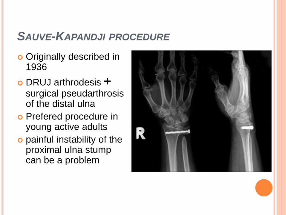
SAUVE-KAPANDJI PROCEDURE
Originally described in 1936
DRUJ arthrodesis + surgical pseudarthrosisof the distal ulna
Prefered procedure in young active adults
painful instability of the proximal ulna stump can be a problem
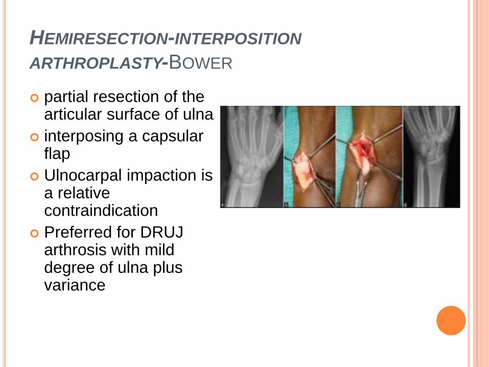
HEMIRESECTION-INTERPOSITION
ARTHROPLASTY-BOWER
partial resection of the articular surface of ulna
interposing a capsular flap
Ulnocarpal impaction is a relative contraindication
Preferred for DRUJ arthrosis with mild degree of ulna plus variance
DRUJ IMPLANT ARTHROPLASTY
Indications
primary DRUJ arthrosis
failed DRUJ surgery
Prosthesis commonly used
Swanson and Herbert prosthesis for distal ulna replacement.
Scheker’s semiconstrained modular implant for total
replacement of the DRUJ (APTIS DRUJ prosthesis)
Though long term results are still awaited, the implant
shows great promise
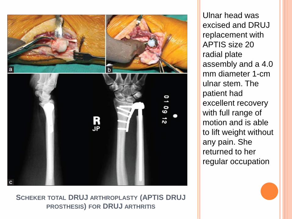
SCHEKER TOTAL DRUJ ARTHROPLASTY (APTIS DRUJ
PROSTHESIS) FOR DRUJ ARTHRITIS
(a) Peroperative
photograph
showing incision
mark.
(b) X-rays lateral
and posteroanterior
views showing
degenerative
changes in the
DRUJ.
(c) Peroperative
photograph
showing ulnar head
devoid of cartilage
with sigmoid notch
osteophytes
SCHEKER TOTAL DRUJ ARTHROPLASTY (APTIS DRUJ
PROSTHESIS) FOR DRUJ ARTHRITIS
Ulnar head was
excised and DRUJ
replacement with
APTIS size 20
radial plate
assembly and a 4.0
mm diameter 1-cm
ulnar stem. The
patient had
excellent recovery
with full range of
motion and is able
to lift weight without
any pain. She
returned to her
regular occupation
CONCLUSION
The DRUJ injuries presents as ulna sided wrist pain
resulting most commonly from traumatic episodes
Clinical examination provide information regarding
the anatomical structures injured
Arthroscopy is considered the gold standard in
diagnosis
Treatment include splinting, ORIF of fractures and
repair of torn ligaments and TFCC by arthroscopy
or open methods
DRUJ arthroplasty is emerging as a treatment in
cases of arthrosis of the joint.
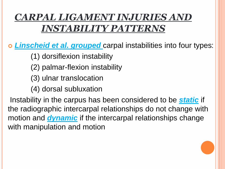
CARPAL LIGAMENT INJURIES AND INSTABILITY PATTERNS
Linscheid et al. grouped carpal instabilities into four types:
(1) dorsiflexion instability
(2) palmar-flexion instability
(3) ulnar translocation
(4) dorsal subluxation
Instability in the carpus has been considered to be static if
the radiographic intercarpal relationships do not change with
motion and dynamic if the intercarpal relationships change
with manipulation and motion
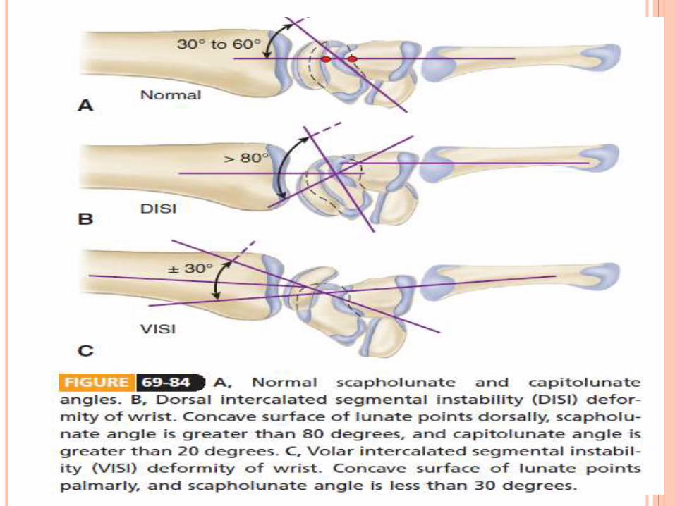
Radiographic evaluation of the proximal carpal row in the lateral projection in which the radius, lunate, capitate, and third metacarpal should have collinear axes within an approximately 15-degree tolerance.
On this projection, the wrist-collapse patterns include (1) patterns in which the distal articular surface of the lunate is tilted to face dorsally, known as dorsal intercalated segment instability (2) patterns in which the distal articular surface of the lunate faces toward the palm, known as volar intercalated segment instability.
Linscheid et al. advocated the concept of dissociative and nondissociative instabilities in the wrist. Dissociative carpal instabilities are those in which there is disruption of the intrinsic interosseous ligaments between the bones of the proximal carpal row. Nondissociative instabilities are those in which the extrinsic radiocarpal ligaments may be disrupted, with intact intrinsic ligaments between the carpal bones.
PROGRESSIVE PERILUNAR INSTABILITY Mayfield, Johnson, and Kilcoyne described four stages of
progressive disruption of ligament attachments and anatomical relationships to the lunate resulting from forced wrist hyperextension
Stage I represents scapholunate failure;
stage II, capitolunate failure
III, triquetrolunate failure
IV, dorsal radiocarpal ligament failure, allowing lunate
dislocation
ROTARY SUBLUXATION OF THE SCAPHOID Injuries to the dorsal and volar portions of the scapholunate
interosseous ligament, the long radiolunate ligament, and the radioscaphocapitate ligament allow the proximal pole of the scaphoid to rotate dorsally. The scaphoid assumes a more vertical orientation, and eventually the scaphoid separates from the lunate .
Watson and Black observed that rotary subluxation of the scaphoid may manifest in four types: (1) dynamic, (2) static, (3) with degenerative arthritis, and (4) secondary to a condition such as Kienböck osteochondrosis.
a fall on the extended wrist is the usual cause.
On examination, pain and tenderness are present along the dorsal radiocarpal articulation at the scapholunate area.
Edema may be present with limitation of motion, particularly in flexion.
The following maneuvers are considered to be helpful in evaluating rotary instability of the scaphoid
“scaphoid test,” in which the examiner places four fingers on the dorsum of the radius with the thumb on the scaphoid tuberosity, using the right hand for the right wrist and the left hand for the left wrist. Ulnar deviation of the wrist aligns the scaphoid with the long axis of the forearm. Applying thumb pressure to the scaphoid tuberosity, the wrist is returned to radial deviation, maintaining the thumb pressure on the scaphoid tuberosity. If the scaphoid is sufficiently unstable, the proximal pole is driven dorsally, and pain results
As the wrist under load progresses from radial deviation to ulnar deviation, the scaphoid normally moves smoothly into extension, aligning with the forearm axis. If scaphoid rotary subluxation is present, the lunate remains in a volar-flexed and dorsal position until sufficient pressure is applied, so that it suddenly shifts from the volar-flexed position and “catches up” with the scaphoid with a “clunking” sensation
the diagnosis of static rotary subluxation of the scaphoid can be made on an anteroposterior radiographic view when a gap of more than 2 mm is noted between the scaphoid and the lunate bones. This gap is seen to increase with an anteroposterior view taken with the fist clenched. Other findings on the anteroposterior view include apparent shortening of the scaphoid and the so-called cortical ring appearance of the axial projection of the scaphoid.
MANAGEMENT
Closed treatment of acute rotary subluxation of the scaphoid consists of attempting reduction by placing the wrist in neutral flexion and a few degrees of ulnar deviation.
Percutaneous pinning can be done with one 0.045-inch (1.16-mm) Kirschner wire placed through the scaphoid into the capitate and a second placed through the scaphoid into the lunate.
If closed reduction is unsuccessful, arthroscopic reduction and percutaneous pin fixation can be attempted
open reduction through a dorsal approach with closure of the scapholunate gap, Kirschner wire internal fixation of the lunate to the scaphoid, and ligament repair usually are indicated.
Management of an old rotary subluxation of the scaphoid may require reconstruction of the scapholunate interosseous ligament with a segment of the extensor carpi radialis brevis tendon plus Kirschner wire fixation
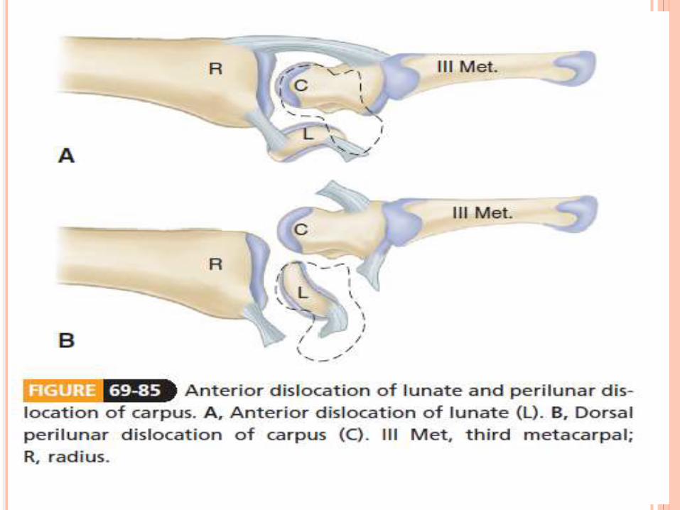
ANTERIOR DISLOCATION OF THE LUNATE
The most common carpal dislocation is anterior dislocation of the lunate
On a lateral radiographic view of the normal wrist, the half-moon–shaped profile of the lunate articulates with the cup of the distal radius proximally and with the rounded proximal capitate distally
AP view, the normal rectangular profile of the lunate when dislocated becomes triangular because of its tilt.
An anteriorly dislocated lunate can cause acute compression of the median nerve
When the injury is treated early, manipulative reduction usually is possible and immobilization for 3 weeks with the wrist in slight flexion is required.
When treated after 3 weeks, the injury can be difficult to reduce by manipulation, and open reduction may be necessary. A dorsal approach has been recommended
TREATMENT OPTIONS FOR WRIST LIGAMENT
INJURIES AND INSTABILITY
For acute injuries, options include closed or arthroscopically
controlled manipulation and percutaneous pinning
If closed methods are unsuccessful, open repair or
reconstruction of ligaments may be required
For late diagnosed problem – limited arthrodesis
Dorsal capsulodesis can be added to limit scaphoid flexion


![[18'] Carpal](https://static.documents.pub/doc/80x56/577d20351a28ab4e1e924083/18-carpal.jpg)


