AD/A-006 016 ATLAS OF GOAT ANATOMY. PART IV. INTERNAL ORGANS Clarence E. Hopkins, Sr. Edgewood Arsenal Aberdeen Proving Ground, Maryland February 19715 DISTRIBUTED BY: National Technical Information Service U.S. DEPARTMENT OF COMMERCE
Transcript
AD/A-006 016
ATLAS OF GOAT ANATOMY. PART IV.INTERNAL ORGANS
Clarence E. Hopkins, Sr.
Edgewood Arsenal
Aberdeen Proving Ground, Maryland
February 19715
DISTRIBUTED BY:
National Technical Information Service
U. S. DEPARTMENT OF COMMERCE
* ~~~SECURITY CLASSIFICATION OF THIS PAGE ("O~n Date entered) _________________
Approved for public relcase, distribution unlimfied.
17. DISTRIBUTION STATEMENT (of the abstract entered In Block 20, If different from Report)
III. SUPPLEMENTARY NOTES L .D CR.flodAced by 3r'-r:r( ~NATIONAL TECHNICAL I
In-house laboratory independent research INFORMATION SERVICE MUS Department of Comme~rce
5ptin~field, VA. 22151
I.KEY WORDS (Continue on reverse side if necesarwy and identify by block number) f~T hInternal organs Dissecting U JL U mGoat atlas DrawingsBPhotographs LabelingPRCS UBE _TIH .
20. ABSTRACT (Continue en reverse aide If neceary and Identify by block nuember)
The Angora goat (Capra /iircus) has been tile minII experimental animal ti.,ed by the Biophysics Division ofthe Biomedical Laboratory for many years. Knowledge tst goat anatomy is imipoitant during both the planningand experimental stages of projects. Because no readily usable information on goat anatomy is available, a sariesof reports on the subject is being prepared. The first iii thle series described thle skeletal anatomy; the seconddescribed the cross-section anatomy; the third described tile muscles on superficial and deep dissection of thehead, neck, and pectoral and pelvic limbs. This report concerns the internal organs and describes the structuresof each organ.
I J173 1473 EDITION OF I NOV $5IS ONSOLETE UNCLASSIFIEDSECURITY CLASSIFICATION OF THIS PAGE (When Data Entered)
PREFACE
The work described in this report was authorized under Task IT061I1A91AIS,In-Hoise Laboratory Independent Research; Atlas of the Goat. This work was started in July1973 and completed in July 1974.
In conducting the research described in this report, the investigator adhered to the"Guide for the Care and Use of Laboratory Animals:' as promulgated by the Committee on theRevision of the Guide for Laboratory Animal Facilities and Care of the Institute of LaboratoryAnimal Resources-National Research Council.
Reproduction of this document in whole or in part is prohibited except withpermission of the Commander, Edgewood Arsenal, Attn: SAREA-TS-R, Aberdeen ProvingGround, Maryland 21010; however, DDC and the National Technical Information Service areauthorized to reproduce the document for US Government purposes.
Acknowledgments
The author wishes to acknowledge the technical assistance of John J. Holter, JosephB. Scott, and Garnet E. Afflck, Jr., Biophysics Divsion, who prepared the photographs.
31 Right Kidney of Goat .......... .......................... .36
4
* n - r - -
A HI AS 01: GOAT ANATOMY
lAR IV: INTFRNAL ORGANS
1. INTRODUCTION.
Thi: Angora goat ((iqpre Wrens) has been the inain experimental animal used hylte Biophysics Division~ ol, tile Biomedical Laboratory For many years. Knowledge ot' goalanatomny is important during both tile planning and experimentaml stages of' projects. Bvccuuiseno readily usable information ()on goalt anatomy is available, a series ol reports on the subject
ais bveing prepared. The first in lte serk-0~ described the skeletal anatomy: lte second2 describediii' cross-sectional anatomy: anid thie third-' described [lie mnuscles onl superficial and deepdissetion of' the head. neck. and pectoral and pelvic Aieis
This report concerns the internal organs and describes the structures of' eachorganl.
11. MATERIALS AND ME-THODS.
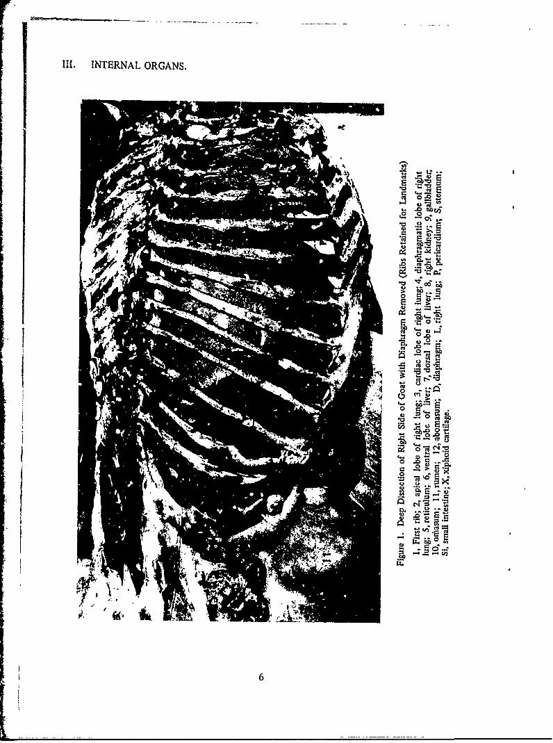
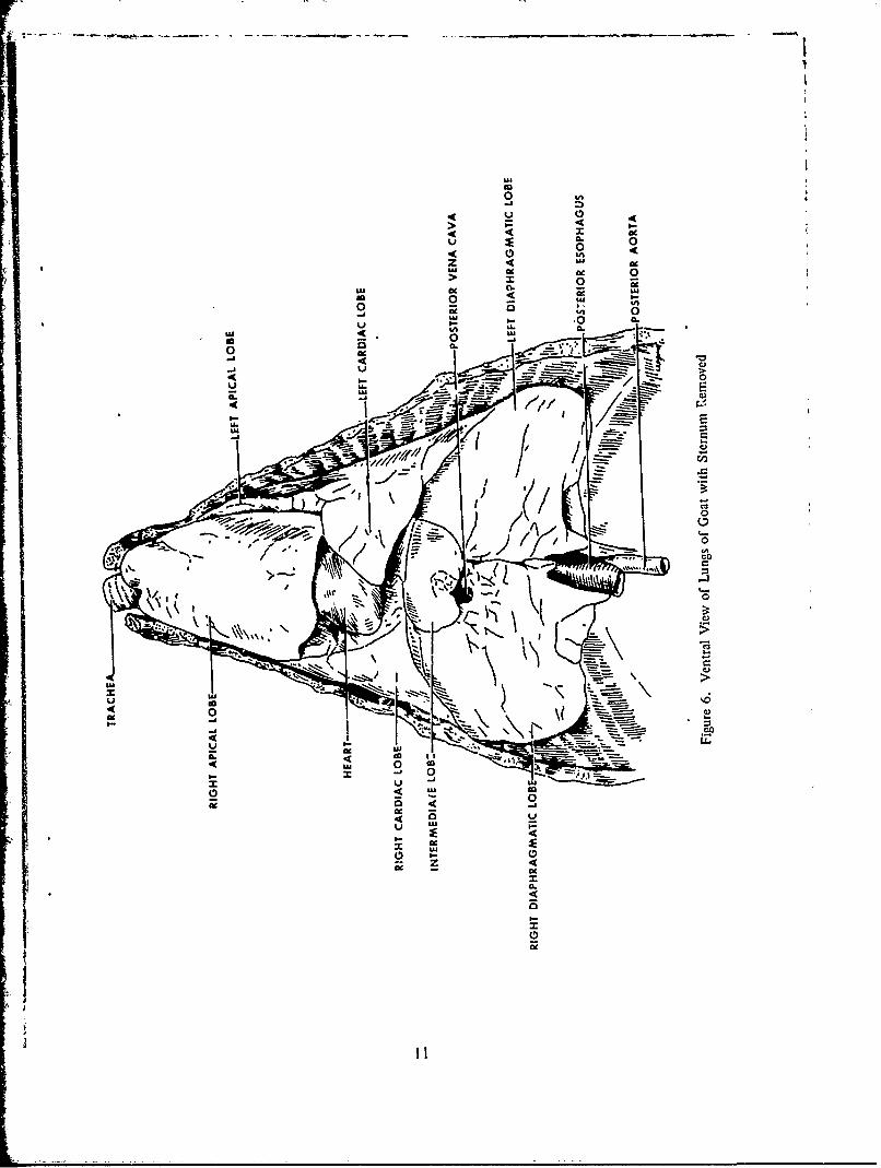
Eight castrated Angorai goats were used. Each goat was euthanized withphenobarbital sodium. The toals were of average size, weighing 40 to 50 kg. Deep dissectionwas done on fo.ur goats showing the right and left lateral side with tile internal organs intact(figures I through 4). Another goat -was dissected to show the ventral view of the qbdonlinalviscera (figvie 5) and the ventral view of' the lungs (figures 6 and 7). Three goats weredissected te obtain the internal organs. The organs were either drawn or photographed andlabeled accv-rdingly. The drawings are placed with some of the photographs with a centimeterscale shc'wing the exact size of the organ.
A total of 14 drawings and 28 photogiaphs was made while the dissection of theorgans was taking place. The organs dissected were the 'brain, tongue, !ungs, heart, liver.kidneys, adrenal, spleen, pancreas, and the stomach (figures I through 31).
The drawing of the digestive system in the domestic goat shows the passagewaythrough the system of thi, four stomachs (abomasum, retictlmini. onmasum. and rumnen)(figure 29).
The anatomy books of Sissoni and Grossman" andl Nlwi" anmd the excellent bookby Miller et al.' were, used as references during the preparation om thi, atlas.
I5
IlINTERNAL ORGANS.
S0
*Z3
6
o
.2 0
Ui
0)
Cq
It, co
-co O
0.
C3
o 'ca
CS W
*00
14
-r 4o,
)
-2 E
. o0. .
IJ
Figure 5. Ventral View of Aboomindi Viscera of Goat with Onientumn Removed
1, Reticulum; 2, abomasurai; 3, rumen-. 4, omasun,; 5. 7, and 8, colon;,6, small intestine; C, caccum; D, diaphragm; L, liver; S, sternum; V, ventralsac of rumen.
10
L.0z JA
> 0go 0 0U
U 0~"A 0 C
0000
4K
-K
-l.
teU,
C-
0
00
-K 4U 0I- 0
S UA
z UJ
Figure 7. Ventral View of Lungs of Goat with Sternum Removed
1, Trachea; 2, right apical lobe; 3, heart; 4, right cardiac lobe; 5, left cardiac lobe;6, left diaphragmatic lobe; 7, right diaphragmatic lobe; 8, posterior esophagus; 9, aorta.
12
co
od
I-
U 4>
U.
t n
0.>
0
us~
UAJ
LAII
LL. co
LAuJ
K >0>
z<b 0
00.
00.
UL
LU 13
Ilk-
14
* LONGITUDINAL FISSURE
_________OLFACTORY BULB
SULCUS RHINALIS ........ ~ - OLFACTORY TRACT
'' .' LATERAL OLFACTORY STRIA
CHIASMA OPTICUMOPINEV
HYPOPHYSIS PIRIFORM LOBE
OCULOMOTOR NERVE-kCEREBRAL PEDUNCLE
-TRIGEMINAL NERVEABDUCENT NERVE
-CEREBELLUMMEDULLA OBLONGATA -CHORIOID PLEXUS
ACCESSORY NERV
SPINAL CORD
VHYPOGLOSSAL NERVE
Figure 10. Base of Goat Brain
15
Figlure 11. Base of Goat Brain
16
I3E
0
-4
L
0v 0U. v
0
'IA
o4A
-I
AA.
0 !2
IL 0
w ,,.
3E
m U
oIAo
A 'A
Z <
0 a
o 0
us
uj LUI I
CLI(V< 2 '
LL.
4A L"
44
II
17Z
a I-
-n A
a:0
-
18V
IFRONTAL POLE -OLFACTORY BULd
LONGITUDINAL FISSURE
TRANSVERSE FISSURE .- OCCIPITAL POLE
MEDULLA OBLONGATA
HYPOGLOSSAL NERVE-SPINAL CORD
4 Figure 14. Dorsal View of Goat Brain
19
Figure 15. Dorsal View of Goat Brain
20
IA~
mha
0 06
ab
UAU
CdC
xIae0
21
- - -- ~ ~ - - - - . - . - - - - - - - - - - C's
0
0
P, UA
22
00
UL
02
04
LL 4ALu tW
4 523
-~ -...~- ~ 'WI1
Cl0
C.,I-0
C)Ca
Cl
U,
Cl
0
00\
C)
24
CI0l
0
00
25
z
mm
>
0 0o
K v4 0
z Cz
0 00
=..-
z z,
Z '
, . .--. I-
LU
"" 1 - "* -
zo o. o ;>
v v 0
> >
4 - 0
z R -
a. 26
26
JA-
iz
27
z 0
2 0 z
wA IA
00
F UU
LUU
2 4
>
0 >
00
0
UL
28
4-a
I..U
IIc~IUI-
29
4N4 Ii o0
l0
30I-
-. 5
3OU
I'0
J -
C.
LW0
I-
0
I-
ULW
(-4
cCLI.
Ik I,
f31
400
wl"MWT , W
(IU
06
C14
0 40
cnu
.J:3.
000
33
w --
CfC)
LL
ow->
Cz U)
4)
z
> W) C) AJ
'-4 0
j (n w 2
34W
Iu
0d ww
0
4 a,
0 0
. Z .00 w 0 u 0
oo
0
aa.
35.a.
4 -€
- o
354
- -,' - - ------------------- -- -- ~ -,-.-
z4
w -
z -4'A* U
4
-aI- 4 0U, U
z0
/~ m**'1~ U)
I-.
Uen -- 0
0
4)
-a .-"a -
4zU
4)
U-.
zU
-a "a
U 4"' Z U- "a
~Ii Iji CINI,'atl(,,
4'
-J
'
~LcI
36
LITERATURE CITED
1. Hopkins, C. E., Sr., Hamm, T. E., Jr., and Leppurt, G. L. EATR 4431. Atlas of Goat Anatomy.Part 1: Osteology. September 1970.
2. Hopkins, C. E., Sr., Ham, T. E., Jr., and Leppart, G. L. EATR 4626. Atlas of Goat Anatomy.Part 11: Serial Cross Section. April 1972.
3. Hopkins, C. E., Sr., Hamm, T. E., Jr., and Leppart, G. L. EATR 4706. Atlas of Goat Anatomy.Part III: Myo!ogy. April 1973.
4. Siuon, S., and Grossman, J. D. Anatomy of Domestic Animals. 4th Edition. W. B. SaundersCompany, Philadelphia, Pennsylvania. 1953.
5. May, N. Anatomy of Sheep. 2d Edition. University of Queensland Press, Wilke Company, Australia.1964.
6. Miller, M. E., Christensen, G. C., and Evans, H. E. Anatomy of Dog. W. B. Saunders Company.Philadelphia, Pennsylvania. 1964.