DivL Performs Critical Cell Cycle Functions in Caulobacter crescentusIndependent of Kinase Activity�
Sarah J. Reisinger,1 Sarah Huntwork,2† Patrick H. Viollier,3 and Kathleen R. Ryan1*Department of Plant and Microbial Biology, University of California, Berkeley, Berkeley, California 947201; Department of
Developmental Biology, Stanford University School of Medicine, Stanford, California 943052; and Department ofMolecular Biology and Microbiology, Case Western Reserve University School of Medicine, Cleveland, Ohio 441063
Received 4 June 2007/Accepted 28 August 2007
The Caulobacter cell cycle is regulated by a network of two-component signal transduction proteins. Phos-phorylation and stability of the master transcriptional regulator CtrA are controlled by the CckA-ChpTphosphorelay, and CckA activity is modulated by another response regulator, DivK. In a screen to identifysuppressors of the cold-sensitive divK341 mutant, we found point mutations in the essential gene divL. DivL issimilar to histidine kinases but has a tyrosine instead of a histidine at the conserved phosphorylation site(Y550). Surprisingly, we found that the ATPase domain of DivL is not essential for Caulobacter viability. Weshow that DivL selectively affects CtrA phosphorylation but not CtrA proteolysis, indicating that DivL acts ina pathway independent of the CckA-ChpT phosphorelay. divL can be deleted in a strain overproducing thephosphomimetic protein CtrAD51E, but unlike �ctrA cells expressing CtrAD51E, this strain is profoundlyimpaired in the control of chromosome replication and cell division. Thus, DivL performs a second functionin addition to promoting CtrA phosphorylation. DivL is required for bipolar DivK localization and positivelyregulates DivK phosphorylation. Our results show that DivL controls two key cell cycle regulators, CtrA andDivK, and that phosphoryl transfer is not DivL’s essential cellular activity.
Several two-component signal transduction proteins havebeen identified in genetic screens for Caulobacter crescentusmutants with defects in cell cycle progression, cell division, andpolar morphogenesis (reviewed in reference 38). The essentialresponse regulator CtrA directly controls the expression of atleast 95 genes (24) and promotes cell division, DNA methyl-ation, and the biogenesis of polar organelles (22). However,CtrA also binds to five sites within the replication origin toinhibit the initiation of chromosome replication (33), and cellswith unrestricted CtrA activity arrest in the G1 phase of the cellcycle (6). CtrA activity must therefore be regulated tightly toallow the orderly progression of DNA replication, morphogen-esis, and cell division.
The following three mechanisms control CtrA activity as afunction of the cell cycle: (i) ctrA expression peaks after DNAreplication has begun, due to the transcriptional effects ofDnaA and GcrA (14); (ii) CtrA is activated by phosphorylationin swarmer cells, where DNA replication has not yet occurred(G1), and in predivisional cells, where replication has alreadybeen initiated (17); and (iii) the CtrA protein is cleared fromcells preparing to initiate DNA replication by the ATP-depen-dent protease ClpXP (6). Combined, these three mechanismsensure that phosphorylated CtrA (CtrA�P) is present onlyearly and late in the cell cycle, precisely when CtrA-dependentgenes are activated or repressed. They also restrict CtrA phos-
phorylation and eliminate the protein itself just before S phaseto free the origin for the initiation of DNA replication.
CtrA phosphorylation and proteolysis are coordinated by aphosphorelay containing the hybrid histidine kinase CckA andthe histidine phosphotransferase ChpT (16). CckA autophos-phorylates on a conserved histidine residue. The phosphorylgroup is passed to a conserved aspartate in the receiver domainat the C terminus of CckA and from there to a histidineresidue in ChpT. ChpT can pass the phosphoryl group to oneof two response regulators, either CtrA or CpdR (3). Phos-phorylation activates CtrA to regulate transcription and DNAreplication, and phosphorylation of CpdR prevents it frompromoting CtrA proteolysis (16). Thus, when the CckA-ChpTphosphorelay is active, CtrA is stable and activated, whereaswhen the phosphorelay is inactive, CtrA becomes dephosphor-ylated and proteolyzed.
Fluctuations in CtrA activity during the cell cycle are linkedto changes in CckA activity, and CckA phosphorylation itself iscell cycle dependent (17). The only factor known to modulateCckA activity is the essential single-domain response regulatorDivK. When phosphorylated, DivK downregulates the activityof the CckA-ChpT pathway. In the cold-sensitive divK341 mu-tant (41) grown at the nonpermissive temperature, excessCckA�P and CtrA�P are present (3), CtrA is no longer pro-teolyzed, and the net result is G1 cell cycle arrest (15). Con-versely, DivK overexpression causes a sharp decrease in thephosphorylation of CckA and an accumulation of chromo-somal DNA (3). The mechanism by which DivK affects theactivity of CckA is currently unknown.
DivK is implicated not only in cell cycle regulation but alsoin the generation of cellular asymmetry. Each Caulobacter celldivision produces two distinct cell types with different mor-phologies, protein contents, and cell fates (reviewed in refer-
* Corresponding author. Mailing address: University of California,Berkeley, Department of Plant and Microbial Biology, 251 KoshlandHall, Berkeley, CA 94720. Phone: (510) 642-5559. Fax: (510) 642-4995.E-mail: [email protected].
† Present address: Department of Biology, Massachusetts Instituteof Technology, Cambridge, MA 02139.
ence 7). The swarmer progeny is motile, contains CtrA�P, andcannot initiate DNA replication, while the stalked progeny issessile, lacks CtrA, and can immediately begin a new round ofDNA replication. DivJ, a histidine kinase that phosphorylatesDivK, resides at the stalked pole of the predivisional cell, whilePleC, a histidine kinase that dephosphorylates DivK, is posi-tioned at the swarmer or flagellated pole (48). Once a cyto-plasmic barrier is created in the late predivisional cell (21),phosphorylated DivK (DivK�P) is thought to accumulate inthe stalked compartment due to the action of DivJ, triggeringthe downregulation of CckA and the degradation of CtrA.In the swarmer compartment, DivK is dephosphorylated byPleC, which maintains the activity of the CckA-ChpT phos-phorelay and preserves CtrA function (27).
To identify components that act in concert with or in parallelto DivK, we performed a screen for temperature-sensitive sup-pressors of the cold-sensitive divK341 mutant. Since divK341cells at the nonpermissive temperature fail to eliminate CtrAactivity, we expected to find mutations that compromise CtrA activityin ctrA itself and in genes that positively regulate CtrA. Threeindependent suppressor mutations were identified in divL,which encodes an essential kinase that regulates the Cau-lobacter cell cycle (51). Depletion of the DivL protein fromCaulobacter cells results in cell filamentation and accumulationof additional copies of the chromosome (36). DivL is unusualin that its dimerization and histidine phosphotransfer (DHp)domain contains a tyrosine residue (Y550) at the conservedphosphorylation site. DivL was shown to autophosphorylate onY550 in vitro and to pass the phosphoryl group to CtrA (51).Conditional divL mutants also contain reduced levels of phos-phorylated CtrA (31), indicating that DivL plays a role in CtrAactivation in vivo.
Where does DivL fit into the network of two-componentsignaling proteins that regulate the Caulobacter cell cycle? Onepossibility is that DivL acts in a pathway parallel to CckA andChpT to phosphorylate CtrA. However, cckA mutants lackdetectable CtrA�P (17, 18), so DivL and CckA may act in thesame signaling pathway. Some results indicate that DivL couldinteract with DivK and thereby modulate the CckA pathway.First, divL mutations can suppress phenotypic defects in pleCand divJ strains, which contain altered levels of active DivK(31, 41). Second, fragments of DivL were identified along withthose of DivJ and PleC in a yeast two-hybrid screen for pep-tides that physically interact with DivK (30).
Here we show that DivL positively regulates CtrA phosphor-ylation without affecting CpdR phosphorylation or the rate ofCtrA proteolysis, suggesting that DivL acts selectively on CtrArather than modulating the CckA-ChpT phosphorelay. DivLalso promotes DivK phosphorylation and localization of DivKto the cell poles. Surprisingly, divL alleles that produce pro-teins incapable of autophosphorylation or phosphoryl transfercan substitute for the wild-type divL gene, causing only mod-erate defects in cell division. DivL is therefore one of the fewhistidine kinase homologs known to perform a key cellularfunction distinct from phosphoryl transfer to a response regu-lator.
MATERIALS AND METHODS
Bacterial strains, plasmids, and culture conditions. The strains and plasmidsused for this study are listed in Table 1. All experiments were performed using
derivatives of Caulobacter crescentus strain CB15N grown to mid-logarithmicphase. CB15N strains were grown in peptone-yeast extract (PYE; complex me-dium), M2G (minimal medium) (9), or M5G [10 mM piperazine-N,N�-bis(2-ethanesulfonic acid) (PIPES), pH 7, 1 mM NaCl, 1 mM KCl, 0.05% NH4Cl, 0.01mM Fe-EDTA, 0.2% glucose, 0.5 mM MgSO4, 0.5 mM CaCl2, and 0.1 mMphosphate] at the indicated temperatures. Solid and liquid media were supple-mented with 3% sucrose, kanamycin (25 �g/ml and 5 �g/ml for solid and liquidmedia, respectively), chloramphenicol (1 �g/ml), naladixic acid (20 �g/ml),oxytetracyline (2 �g/ml and 1 �g/ml, respectively), or spectinomycin (100 �g/mland 25 �g/ml, respectively), as required. Escherichia coli strains were grown inLuria broth at 37°C, and solid and liquid media were supplemented with car-benicillin (100 �g/ml and 50 �g/ml, respectively), chloramphenicol (30 �g/ml and20 �g/ml, respectively), kanamycin (50 �g/ml and 30 �g/ml, respectively), tetra-cycline (12 �g/ml), or spectinomycin (50 �g/ml), as required.
Site-directed mutagenesis of divL to create divL(Y550F) was carried out usingthe QuikChange protocol (Stratagene), with pMR20-divL as the template. Trun-cated divL alleles were generated by PCR, using pKR170 as the template. The3�FLAG epitope in pSK127 and pSK137 was constructed using an oligonucle-otide linker and added the amino acid residues DYKDHDGDYKDHDIDYKDDDDK to the C termini of CpdR and DivL539. For the DivL-enhanced yellowfluorescent protein (DivL-EYFP) fusion, we used PCR to generate an NcoI siteat the 3� end of divL and cloned an �880-bp BamHI-NcoI fragment of divL intopEYFP (Clontech). We integrated the divL-eyfp gene into the chromosomal divLlocus using the suicide vector pNPTS138. The plasmid carrying divL510::eyfp wasgenerated by site-directed mutagenesis of the divL-eyfp fusion gene on pKR196to introduce the L740P mutation, creating integration plasmid pSK170. For theDivJ-YFP fusion, we used PCR to amplify divJ from genomic DNA and thenperformed a three-piece ligation of PCR-amplified divJ digested with SacI andXhoI, pNPTS-cc1063-YFP digested with XhoI and SphI, and pUC19 digestedwith SacI and SphI. The resulting plasmid, with full-length DivJ fused to YFP,was digested with SacI and HindIII, and the fragment was moved into pMR20 togenerate pSK158. For the integration plasmid carrying divK::egfp, pMR20divK-EGFP was digested with SpeI, and the insert was ligated into pNPT228 togenerate pSK171. All plasmids were mobilized from E. coli to C. crescentus byconjugation using E. coli strain S17-1 (9). Sequences of all primers used foramplification or mutagenesis are available upon request.
Isolation of suppressors of divK341. A CB15N strain containing a cold-sensi-tive mutation in the divK gene (41) was mutagenized with UV radiation andplated in PYE containing 0.3% agar at 20°C. Large swarms were selected fromthese plates, and the double mutants were screened for the inability to swarm in0.3% agar at 37°C to identify divK suppressors that conferred a temperature-sensitive phenotype. We complemented the 37°C swarm defect in each suppres-sor strain using a cosmid library (2). Complementing cosmids were isolated, andthe insert ends were sequenced. The complementing cosmid for suppressorstrain KR510 included CC3484, encoding the essential tyrosine kinase DivL. Alow-copy-number plasmid containing only the wild-type divL gene (pMR20-divL) also complemented KR510. A kanamycin resistance gene was integrated at3.75 Mb in the genome of KR510, near divL (29, 47). The divL allele in strainKR510 (divL510) was moved into CB15N by cotransduction with the kanamycinresistance gene, using phage �Cr30 (10).
Two-step gene replacement. Arms of homology flanking divL were generatedby PCR amplification of the regions approximately 1,000 bp upstream anddownstream of divL. The left arm included the first three codons of divL, and theright arm included the last three codons of divL. A streptomycin/spectinomycinresistance gene flanked by transcriptional and translational stop signals (32) frompHP45� was digested with BamHI and ligated between the two PCR-generatedhomology arms in pNPTS138 to generate plasmid pSK149. After pSK149 wasmobilized into strains carrying various divL alleles on pMR20, first integrantswere selected by plating on PYE containing streptomycin, oxytetracycline, andnalidixic acid. Two colonies from each conjugation were inoculated into separatecultures containing PYE with oxytetracycline and streptomycin. After overnightgrowth, the two cultures for each strain were combined, and serial dilutions wereplated for counterselection on PYE containing 3% sucrose, oxytetracycline, andstreptomycin. Fifty to 100 of the sucrose-resistant colonies were screened forkanamycin sensitivity and resistance to streptomycin and oxytetracycline to iden-tify colonies in which the wild-type divL allele had been replaced by the �divLmutation. Gene replacements were verified by two PCR tests. First, to verifyinsertion at the divL locus, we used a forward primer in the streptomycinresistance cassette and a reverse primer placed downstream of the sequenceincluded in the divL integration construct. A second PCR was performed usingprimers within the sacB gene of pNPTS138 to verify that sucrose resistanceresulted from the second recombination event rather than sacB inactivation.
To generate the strain �divL �ctrA/pCtrAD51E, we amplified the Tetr cassette
VOL. 189, 2007 FUNCTIONS OF DivL IN THE CAULOBACTER CELL CYCLE 8309
Strain or plasmid Description or construction Source or reference
StrainsC. crescentus strains
KR684 CB15N, from which all Caulobacter strains were derived 11CJ396 �cckA �ctrA/pCtrAD51E 17LS2716 �ctrA/pCtrAD51E 17LS3196 �divJ 48LS3570 divK341 15NR664 divL::Tn5 (inserted in codon 636) This studyUJ506 �pleC 1KR510 divK341 divL510 This studyKR635 divL510 This studyKR648 divL510/pMR20-divL This studyKR649 divL510/pMR20 This studyKR674 divL::eyfp This studyKR748 �pleC/pMR20divK-EGFP This studyKR1609 CB15N/pMR20-cpdR::3�FLAG This studyKR1612 divL510/pMR20-cpdR::3�FLAG This studyKR1773 �divL/pMR20-divL This studyKR1775 �divL/pMR20-divL(Y550F) This studyKR1776 �divL/pMR20-divL657 This studyKR1777 �divL/pMR20-divL635 This studyKR1808 divL510/pMR20-divJ::yfp This studyKR1809 CB15N/pMR20-divJ::yfp This studyKR1841 divK341 divL::eyfp This studyKR1843 divL::eyfp Pxyl-divK This studyKR1846 CB15N/pMR20divK-EGFP 19KR1848 divL510/pMR20divK-EGFP This studyKR1985 �-pleC/pMR20-divJ::yfp This studyKR2044 divL510::eyfp This studyKR2045 �divL �ctrA/pCtrAD51E This studyKR2046 divL::Tn5 (NR664) divK::egfp This studyKR2047 divK::egfp This study
PlasmidspMR20 Broad-host-range, low-copy-number vector 34pNPTS138 Kanr; sacB-containing integration vector 42pNPT228 Kanr integration vector M. R. K. Alley,
unpublished datapJC52 Vector for fusions to the N terminus of ECFP 4pHPV465 Spec/Strepr suicide vector 45pHP45� Plasmid containing � spectinomycin/streptomycin resistance
cassette32
pEYFP Vector for fusions to the N terminus of EYFP ClontechpCtrA pJS14-ctrA, pSAL14 6pCtrAD51E pJS14-ctrAD51E, pCTD14 6pMR20divK-EGFP 19pHXM-divK Pxyl-divK in high-copy-number vector pJS71 3pNPTS-cc1063-YFP pNPTS138-divJ::yfp M. Laub,
unpublished datapKOC3 Contains frt-flanked Tetr cassette 40pLW126 Vector for inserting Kmr cassette at 3.75 Mb 47pKR174 pMR20-divL This studypKR196 pNPTS138-divL::eyfp This studypKR320 pNPTS138-divL::tet This studypSK54 pMR20-divL(Y550F) This studypSK118 pMR20-divL657 This studypSK123 pMR20-divL635 This studypSK128 pMR20-cpdR::3�FLAG This studypSK137 pMR20-divL540::3�FLAG This studypSK149 pNPTS138-divL::spec This studypSK158 pMR20-divJ::yfp This studypSK170 pNPTS-divL510::eyfp This studypSK171 pNPT228-divK::egfp This study
from pKOC3 (39), using primers that added a BamHI site to each end. Wedigested this fragment with BamHI and ligated it into the BglII site between theleft and right divL homology arms in pNPTS138 to create pKR320. After mo-bilizing pKR320 into �ctrA/pCtrAD51E (17), we selected first integrants onPYE-oxytetracycline-nalidixic acid plates. Individual colonies were selected andgrown overnight in liquid PYE-oxytetracycline, after which they were plated ontoPYE-oxytetracycline containing 3% sucrose for counterselection against thepNPTS138 vector backbone. Sucrose-resistant colonies were screened for kana-mycin sensitivity, oxytetracycline resistance, and spectinomycin resistance toidentify colonies in which the wild-type divL gene had been deleted. We per-formed a PCR using primers within the sacB gene of pNPTS138 to verify thatsucrose resistance resulted from the second recombination event rather thanfrom sacB inactivation.
Isolation of pleC suppressors. divL::Tn5 insertions emerged from a geneticscreen devised to isolate suppressors of the pleC mutant phenotype that will bepublished elsewhere (S. K. Rhadakrishnan, S. Pritchard, and P. H. Viollier, inpreparation). Briefly, a promoterless nptII gene conferring resistance to kana-mycin was transcriptionally fused to the promoter of the pilA gene (PpilA), apromoter that is dependent on PleC for optimal activity (43). The PpilA-nptIItranscriptional reporter was integrated at the chromosomal pilA locus in wild-type CB15N and the �pleC mutant derivative (5). Of the two resulting reporterstrains, the former was resistant to kanamycin (20 �g/ml), while the latter wassensitive. The �pleC PpilA-nptII strain was subsequently mutagenized with a Tn5derivative conferring resistance to tetracycline (20) to isolate mutants that wereable to grow in the presence of kanamycin and tetracycline. The six Tn5 inser-tions that were mapped to divL all occurred downstream of the codon for Y550.The Tn5 elements in strains NR525, NR664, NR1190, NR1193, NR1244, andNR1245 were found to be inserted in the codons for D653, G636, A658, G636,I572, and D604, respectively.
Flow cytometry. Samples for flow cytometry were fixed at a final concentrationof 70% ethanol, stored at 4°C for 1 day to 2 weeks, stained with SYTOX greennucleic acid stain as described previously (35), and analyzed using an EpicsXL-MCL analyzer (Beckman-Coulter).
In vivo phosphorylation, pulse-chase, and immunoblot assays. In vivo phos-phorylation experiments were performed as previously described (6), with thefollowing modifications. Cells to be labeled were grown in M2G overnight,diluted the next day in M5G, diluted after 8 hours of growth in M5G, and grownovernight to an optical density at 660 nm of 0.3. One milliliter of culture washarvested for Western analysis, and another milliliter of culture was labeled for4 min at 28°C, using 30 �Ci [-32P]ATP. Each sample was immunoprecipitatedwith 2.5 �l of anti-CtrA serum (6) along with either 2.5 �l of anti-FLAG serum(Sigma) or 2.5 �l anti-DivK serum (19). Radiolabeled proteins were resolved insodium dodecyl sulfate (SDS)-polyacrylamide gels and quantified using a Ty-phoon phosphorimager (Molecular Dynamics). For pulse-chase experiments,CB15N strains grown at 28°C in M2G were shifted to 37°C for 4 h and then pulselabeled with 10 �Ci/ml [35S]methionine per ml of culture for 5 min, followed bya chase with 1 mM unlabeled methionine and 0.3% Casamino Acids. Samples (1ml) were withdrawn from cultures every 15 min for 2 hours and centrifuged topellet the cells. Cells were lysed using 50 �l SDS buffer (10 mM Tris-Cl, pH 8,1% SDS, 1 mM EDTA) and diluted with 800 �l chilled wash buffer (50 mMTris-Cl, pH 8, 150 mM NaCl, 0.5% Triton X-100). After preclearing of thesample with protein A-agarose, anti-CtrA serum (1.5 �l) and protein A-agarose(Roche) were added to each sample and incubated for 1 h at 4°C with rocking.Immunoprecipitates were washed three times with wash buffer and eluted fromprotein A-agarose using 15 �l 2� Laemmli sample buffer. Samples were sepa-rated in 12% SDS-polyacrylamide gels and analyzed using a Typhoon phosphor-imager (Molecular Dynamics). Immunoblotting was performed using anti-CtrAserum at 1:10,000 and anti-FLAG serum at 1:5,000.
Differential interference contrast (DIC) and fluorescence microscopy. Log-phase cells were immobilized on 1% (wt/vol) agarose pads. Images were acquiredusing a Nikon Eclipse 80i microscope with a PlanApo 100�, 1.40-numerical-aperture objective and a Cascade 512B camera (Roper Scientific). Enhancedgreen fluorescent protein (EGFP) and EYFP were imaged using Chroma filtersets 41001 and 41028, respectively. Images were acquired using Metavue soft-ware (Universal Imaging).
RESULTS
A conditional divL mutation suppresses the phenotype ofthe divK341 mutant. To identify proteins that participate inCaulobacter cell cycle regulation, we screened for mutationsthat restored swarming to the cold-sensitive divK341 mutant
(41) at 20°C. Suppressors were then screened for those thatconferred a new growth or motility defect in swarm agar at37°C. Some temperature-sensitive mutants were comple-mented by providing the ctrA gene on a low-copy-numberplasmid, which was expected because one ctrA mutant previ-ously suppressed the phenotype of the divK341 mutant (50).Other mutants had temperature-sensitive phenotypes not com-plemented by ctrA, so we attempted to complement each oneusing a cosmid library containing Caulobacter genomic DNA(2). Three strains were complemented by cosmids containingdivL and also by the divL gene alone on a low-copy-numberplasmid (pMR20-divL). We integrated a kanamycin resistancegene at a chromosomal locus near divL (3.75 Mb) (29, 47) andtransduced one temperature-sensitive allele (divL510) into thewild-type strain CB15N to examine its phenotype in isolation.In contrast to wild-type cells (Fig. 1A), divL510 cells had arange of lengths at 28°C (Fig. 1B). At 37°C, divL510 cellsbecame more filamentous and accumulated DNA in excess ofone or two chromosomes (Fig. 1E and H). These mutant cellsresembled cells depleted of the DivL protein (36), and theirphenotype was complemented by pMR20-divL (Fig. 1C, F, andI), indicating that divL510 is a recessive loss-of-function allele.The divL510 allele contains two point mutations, leading to theamino acid changes L740P and A140V, but the L740P muta-tion alone is sufficient to cause the observed phenotypes (datanot shown).
DivL promotes CtrA phosphorylation without affecting theCckA-ChpT phosphorelay. The amount of CtrA�P is reducedin a temperature-sensitive divL mutant (31), and the C-termi-nal kinase domain of DivL was shown to phosphorylate CtrAin vitro (51). However, the cckATS1 strain contains undetect-able levels of CtrA�P (18), suggesting that no other kinase cangenerate a significant amount of cellular CtrA�P independentof CckA. To account for these data, DivL could act betweenDivK and CckA, blocking DivK-mediated downregulation ofthe CckA-ChpT phosphorelay. In this model, DivL and CckAboth affect CtrA, but each is essential for viability because theyact in the same signaling pathway.
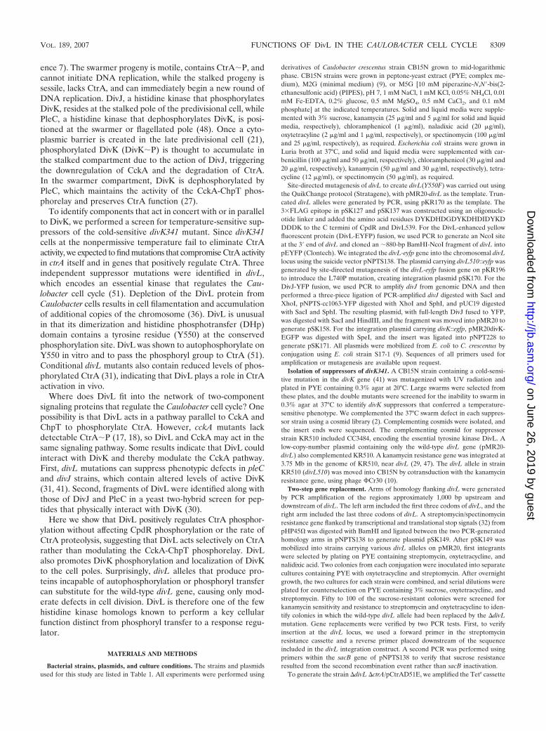
To determine if DivL acts upstream of CckA, we measuredthe phosphorylation of CtrA and CpdR simultaneously instrain CB15N and in cckATS1 (18) and divL510 mutants. Welabeled cultures of each strain with [-32P]ATP and immuno-precipitated CtrA and CpdR-3�FLAG, using anti-CtrA (6)and anti-FLAG (Sigma) antibodies, respectively. In agreementwith previous results (16, 18), cckATS1 cells contained unde-tectable levels of CtrA�P and CpdR�P, although both pro-teins were present (data not shown). CtrA�P was reduced indivL510 cells to �40% of the wild-type level (Fig. 2A, B, andD). Surprisingly, however, divL510 cells contained a wild-typelevel of phosphorylated CpdR (Fig. 2A, C, and D). These datasuggest that DivL specifically affects the amount of phosphor-ylated CtrA rather than modulating the activity of the entireCckA-ChpT phosphorelay.
To confirm that normal CpdR�P levels are maintained indivL510 cells, we measured the half-life of the CtrA protein inwild-type and divL510 cultures grown at the nonpermissivetemperature (37°C). By phosphorylating CpdR, the CckA-ChpT phosphorelay regulates CtrA proteolysis, limiting it tospecific times in the cell cycle. In previous studies, the half-lifeof CtrA was reduced 2.3-fold in a mutant lacking CckA (17)
VOL. 189, 2007 FUNCTIONS OF DivL IN THE CAULOBACTER CELL CYCLE 8311
and 4-fold in a cpdR(D51A) mutant, in which CpdR is presentbut cannot be phosphorylated (16). In the divL510 strain, thehalf-life of CtrA was 25 3 min, compared with 18 3 min forthe wild-type strain CB15N (Fig. 2E); thus, the rate of CtrAproteolysis is not increased in divL510 cells. Because CpdRphosphorylation is not reduced and CtrA proteolysis is notincreased in the divL510 mutant, we infer that DivL selectivelyaffects CtrA phosphorylation.
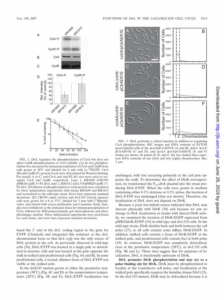
DivL performs a critical function other than regulating thelevel of CtrA�P. DivL clearly affects the cellular level ofCtrA�P, regardless of the molecular mechanism. Is DivL’seffect upon CtrA phosphorylation its only function in Caulo-bacter? To answer this question, we examined the phenotypesof three mutant strains, the �ctrA, �divL �ctrA, and �cckA�ctrA mutants, with each carrying the high-copy-number plas-mid pCtrAD51E (6). When overexpressed, the phosphomi-metic protein CtrAD51E can replace wild-type CtrA, yieldinga mild defect in cell division (17). If the only role of DivL is toregulate CtrA phosphorylation, then we would expect the�divL �ctrA/pCtrAD51E strain to have a phenotype similar tothat of the �ctrA/pCtrAD51E strain.
The divL gene could be deleted in a strain overexpressingCtrAD51E but not in a strain harboring pCtrA (6), whichoverexpresses the wild-type CtrA protein (data not shown).
We deleted the wild-type divL gene in the �ctrA/pCtrAD51Estrain to generate the �divL �ctrA/pCtrAD51E strain, but theresulting cells were filamentous and stalkless and containedexcess chromosomal DNA (Fig. 3C and D). This phenotype ismuch more severe than that of the �ctrA/pCtrAD51E strain(Fig. 3A and B), indicating that the phosphomimetic proteinCtrAD51E can rescue the lethality of the �divL mutant butcannot fully compensate for the absence of the DivL protein. Asimilar result was observed for the histidine kinase CckA (17),where the �cckA �ctrA/pCtrAD51E strain (Fig. 3E and F) wasmuch more impaired in morphology and DNA content thanthe �ctrA/pCtrAD51E strain (Fig. 3A and B). CckA was sub-sequently found to regulate CtrA stability via the responseregulator CpdR, in addition to phosphorylating CtrA via ChpT(17). In the case of DivL, however, the half-life of CtrA was notreduced in the divL510 mutant, and CpdR was phosphorylatednormally (Fig. 2). We therefore propose that DivL performs asecond function critical for Caulobacter cell cycle progressionin addition to promoting CtrA phosphorylation.
DivL is required for DivK localization. Although DivLseems not to regulate CckA, the isolation of divK suppressorsin divL mutants suggests that DivL could still function down-stream of DivK in a different signal transduction pathway. Inthis case, DivL localization may be altered in divK mutants. We
FIG. 1. The divL510 mutant is impaired in cell division and regulation of chromosome replication. DIC images of CB15N (A and D), divL510(B and E), and divL510/pMR20-divL (C and F) cells grown at 28°C or 37°C for 4 h are shown. Bar, 2 �m. The DNA contents of CB15N (G), thedivL510 mutant (H), and the divL510/pMR20-divL mutant (I) grown at 37°C for 4 h were measured by flow cytometry of SYTOX green-stainedcells. The two dashed lines represent DNA contents of one (left) and two (right) chromosomes.
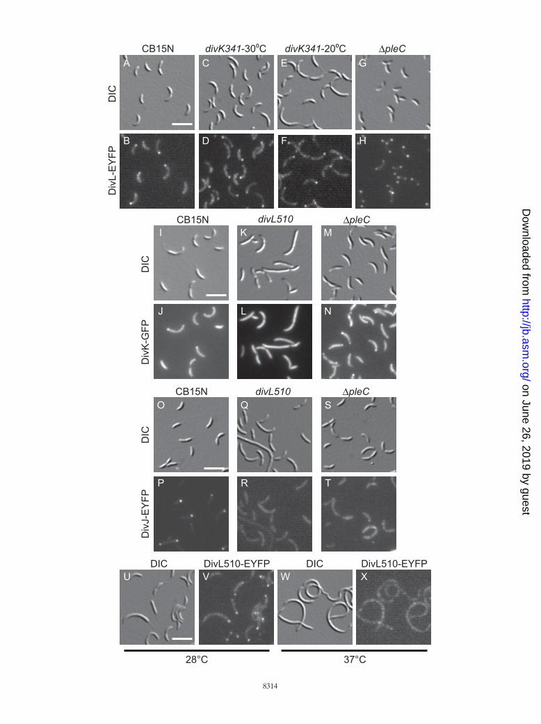
fused the 3� end of the divL coding region to the gene forEYFP (Clontech) and integrated this construct at the divLchromosomal locus so that divL::eyfp was the only source ofDivL protein in the cell. As previously observed in wild-typecells (36), DivL-EYFP was located at a single pole or delocal-ized in swarmer cells and was located at the pole opposite thestalk in stalked and predivisional cells (Fig. 4A and B). In somepredivisional cells, a second, dimmer focus of DivL-EYFP wasvisible at the stalked pole.
In the divK341 mutant grown at either the permissive tem-perature (30°C) (Fig. 4C and D) or the nonpermissive temper-ature (20°C) (Fig. 4E and F), DivL-EYFP localization was
unchanged, with foci occurring primarily at the cell pole op-posite the stalk. To determine the effect of DivK overexpres-sion, we transformed the Pxyl-divK plasmid into the strain pro-ducing DivL-EYFP. When the cells were grown in mediumcontaining either 0.1% dextrose or 0.3% xylose, the location ofDivL-EYFP was unchanged (data not shown). Therefore, thelocalization of DivL does not depend on DivK.
Because a yeast two-hybrid screen indicated that DivL mayinteract physically with DivK (30) and because we saw nochange in DivL localization in strains with altered DivK activ-ity, we examined the location of DivK-EGFP expressed frompMR20divK-EGFP (19) in wild-type and divL510 cells. In thewild-type strain, DivK shuttles back and forth between the cellpoles (27), so all cells contain some diffuse DivK-EGFP. Inaddition, stalked cells contain a focus of DivK-EGFP at thestalked pole, and predivisional cells contain foci at both poles(19). In contrast, DivK-EGFP was completely delocalized,even at the permissive temperature (28°C), in divL510 cells(Fig. 4K and L). These data suggest that for purposes of lo-calization, DivL is functionally upstream of DivK.
DivL promotes DivK phosphorylation and may act as apolar binding site for DivK. DivK must be phosphorylated tolocalize at the Caulobacter cell poles, and localization at thestalked pole specifically requires the histidine kinase DivJ (23).In the divL510 mutant, DivK may be delocalized because it is
FIG. 2. DivL regulates the phosphorylation of CtrA but does notaffect CpdR phosphorylation or CtrA stability. (A) In vivo phosphor-ylation was measured by immunoprecipitation of CtrA and CpdR fromcells grown at 28°C and labeled for 4 min with [-32P]ATP. CtrA(B) and CpdR (C) protein levels were determined by Western blotting.For panels A to C, anti-CtrA and anti-FLAG sera were used to rec-ognize CtrA and CpdR, respectively. Lane 1, KR1609 (CB15N/pMR20-cpdR::3�FLAG); lane 2, KR1612 (divL510/pMR20-cpdR::3�FLAG). (D) Ratios of phosphorylated to total protein were calculatedfor three independent experiments with strains KR1609 and KR1612and normalized to the wild-type strain. Error bars represent standarddeviations. (E) CB15N (open circles) and divL510 (closed squares)cells were grown for 4 h at 37°C, labeled for 5 min with [35S]methi-onine, and chased with excess methionine and Casamino Acids. Sam-ples were withdrawn at the indicated times for immunoprecipitation ofCtrA, followed by SDS-polyacrylamide gel electrophoresis and phos-phorimager analysis. Three independent experiments were performedfor each strain, and error bars represent standard deviations.
Num
ber o
f Cel
lsN
umbe
r of C
ells
Num
ber o
f Cel
ls
A
E0
1000
2000
3000
4000
5000
B
D
F
C
FIG. 3. DivL performs a critical function in addition to regulatingCtrA phosphorylation. DIC images and DNA contents of SYTOXgreen-labeled cells of the �ctrA/pCtrAD51E (A and B), �divL �ctrA/pCtrAD51E (C and D), and �cckA �ctrA/pCtrAD51E (E and F)strains are shown. In panels B, D, and F, the two dashed lines repre-sent DNA contents of one (left) and two (right) chromosomes. Bar,5 �m.
VOL. 189, 2007 FUNCTIONS OF DivL IN THE CAULOBACTER CELL CYCLE 8313
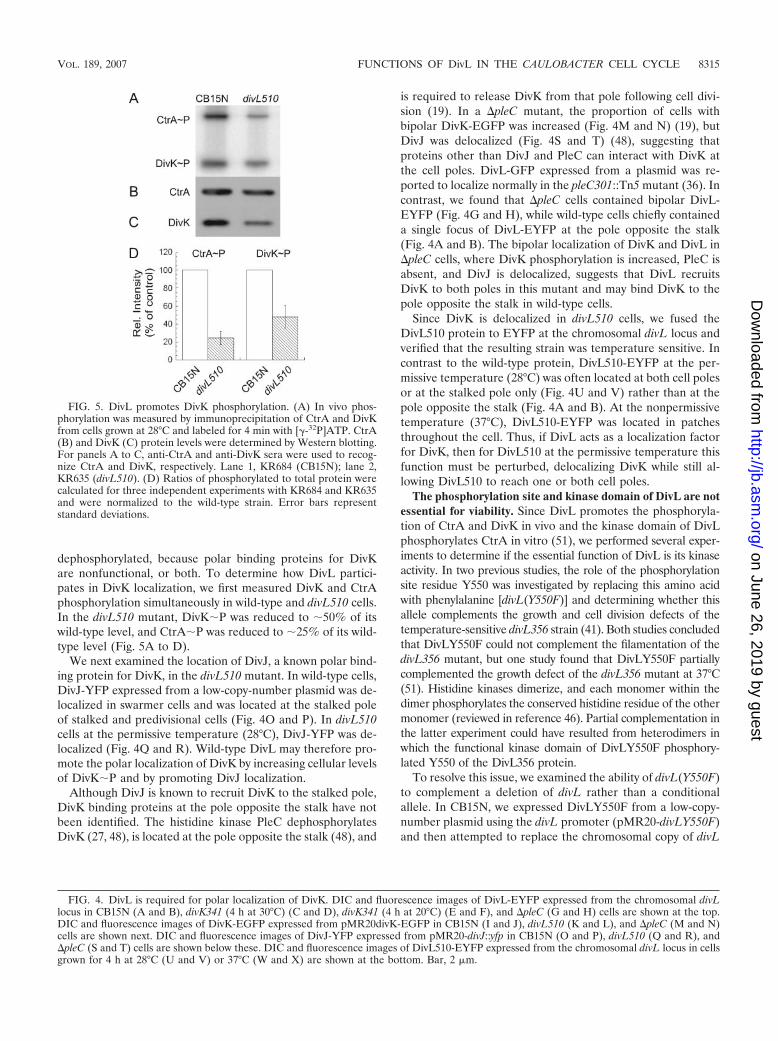
dephosphorylated, because polar binding proteins for DivKare nonfunctional, or both. To determine how DivL partici-pates in DivK localization, we first measured DivK and CtrAphosphorylation simultaneously in wild-type and divL510 cells.In the divL510 mutant, DivK�P was reduced to �50% of itswild-type level, and CtrA�P was reduced to �25% of its wild-type level (Fig. 5A to D).
We next examined the location of DivJ, a known polar bind-ing protein for DivK, in the divL510 mutant. In wild-type cells,DivJ-YFP expressed from a low-copy-number plasmid was de-localized in swarmer cells and was located at the stalked poleof stalked and predivisional cells (Fig. 4O and P). In divL510cells at the permissive temperature (28°C), DivJ-YFP was de-localized (Fig. 4Q and R). Wild-type DivL may therefore pro-mote the polar localization of DivK by increasing cellular levelsof DivK�P and by promoting DivJ localization.
Although DivJ is known to recruit DivK to the stalked pole,DivK binding proteins at the pole opposite the stalk have notbeen identified. The histidine kinase PleC dephosphorylatesDivK (27, 48), is located at the pole opposite the stalk (48), and
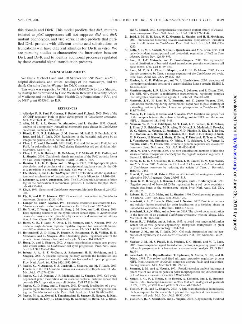
is required to release DivK from that pole following cell divi-sion (19). In a �pleC mutant, the proportion of cells withbipolar DivK-EGFP was increased (Fig. 4M and N) (19), butDivJ was delocalized (Fig. 4S and T) (48), suggesting thatproteins other than DivJ and PleC can interact with DivK atthe cell poles. DivL-GFP expressed from a plasmid was re-ported to localize normally in the pleC301::Tn5 mutant (36). Incontrast, we found that �pleC cells contained bipolar DivL-EYFP (Fig. 4G and H), while wild-type cells chiefly containeda single focus of DivL-EYFP at the pole opposite the stalk(Fig. 4A and B). The bipolar localization of DivK and DivL in�pleC cells, where DivK phosphorylation is increased, PleC isabsent, and DivJ is delocalized, suggests that DivL recruitsDivK to both poles in this mutant and may bind DivK to thepole opposite the stalk in wild-type cells.
Since DivK is delocalized in divL510 cells, we fused theDivL510 protein to EYFP at the chromosomal divL locus andverified that the resulting strain was temperature sensitive. Incontrast to the wild-type protein, DivL510-EYFP at the per-missive temperature (28°C) was often located at both cell polesor at the stalked pole only (Fig. 4U and V) rather than at thepole opposite the stalk (Fig. 4A and B). At the nonpermissivetemperature (37°C), DivL510-EYFP was located in patchesthroughout the cell. Thus, if DivL acts as a localization factorfor DivK, then for DivL510 at the permissive temperature thisfunction must be perturbed, delocalizing DivK while still al-lowing DivL510 to reach one or both cell poles.
The phosphorylation site and kinase domain of DivL are notessential for viability. Since DivL promotes the phosphoryla-tion of CtrA and DivK in vivo and the kinase domain of DivLphosphorylates CtrA in vitro (51), we performed several exper-iments to determine if the essential function of DivL is its kinaseactivity. In two previous studies, the role of the phosphorylationsite residue Y550 was investigated by replacing this amino acidwith phenylalanine [divL(Y550F)] and determining whether thisallele complements the growth and cell division defects of thetemperature-sensitive divL356 strain (41). Both studies concludedthat DivLY550F could not complement the filamentation of thedivL356 mutant, but one study found that DivLY550F partiallycomplemented the growth defect of the divL356 mutant at 37°C(51). Histidine kinases dimerize, and each monomer within thedimer phosphorylates the conserved histidine residue of the othermonomer (reviewed in reference 46). Partial complementation inthe latter experiment could have resulted from heterodimers inwhich the functional kinase domain of DivLY550F phosphory-lated Y550 of the DivL356 protein.
To resolve this issue, we examined the ability of divL(Y550F)to complement a deletion of divL rather than a conditionalallele. In CB15N, we expressed DivLY550F from a low-copy-number plasmid using the divL promoter (pMR20-divLY550F)and then attempted to replace the chromosomal copy of divL
FIG. 4. DivL is required for polar localization of DivK. DIC and fluorescence images of DivL-EYFP expressed from the chromosomal divLlocus in CB15N (A and B), divK341 (4 h at 30°C) (C and D), divK341 (4 h at 20°C) (E and F), and �pleC (G and H) cells are shown at the top.DIC and fluorescence images of DivK-EGFP expressed from pMR20divK-EGFP in CB15N (I and J), divL510 (K and L), and �pleC (M and N)cells are shown next. DIC and fluorescence images of DivJ-YFP expressed from pMR20-divJ::yfp in CB15N (O and P), divL510 (Q and R), and�pleC (S and T) cells are shown below these. DIC and fluorescence images of DivL510-EYFP expressed from the chromosomal divL locus in cellsgrown for 4 h at 28°C (U and V) or 37°C (W and X) are shown at the bottom. Bar, 2 �m.
FIG. 5. DivL promotes DivK phosphorylation. (A) In vivo phos-phorylation was measured by immunoprecipitation of CtrA and DivKfrom cells grown at 28°C and labeled for 4 min with [-32P]ATP. CtrA(B) and DivK (C) protein levels were determined by Western blotting.For panels A to C, anti-CtrA and anti-DivK sera were used to recog-nize CtrA and DivK, respectively. Lane 1, KR684 (CB15N); lane 2,KR635 (divL510). (D) Ratios of phosphorylated to total protein werecalculated for three independent experiments with KR684 and KR635and were normalized to the wild-type strain. Error bars representstandard deviations.
VOL. 189, 2007 FUNCTIONS OF DivL IN THE CAULOBACTER CELL CYCLE 8315
with a spectinomycin/streptomycin resistance cassette (�divL),using a two-step recombination protocol. In a control straincontaining the empty vector pMR20, we were unable to re-place the wild-type divL allele with a �divL mutation, in agree-
ment with previous reports showing that divL is essential forCaulobacter viability (51). However, we successfully replacedthe chromosomal divL gene in a strain containing pMR20-divLY550F (Fig. 6B). The resulting cells were impaired in cell
FIG. 6. DivL variants impaired in kinase activity or phosphoryl transfer support Caulobacter viability and DivK localization. DIC images (A toD) and DNA contents of SYTOX green-stained cells (E to H) are shown for �divL strains expressing the following proteins from pMR20: DivL(KR1773) (A and E), DivLY550F (KR1775) (B and F), DivL657 (KR1776) (C and G), and DivL635 (KR1777) (D and H). (I) Growth of �divLstrains expressing the indicated proteins from pMR20. Serial dilutions of exponential-growth-phase cultures at an optical density at 660 nm of 0.25were made, and 5 microliters of each indicated dilution was spotted onto a PYE plate and incubated for 2 days at 30°C. Three independentexperiments were performed, and a representative plate is shown. (J) Diagram of the DivL protein, including the predicted transmembrane (TM),Per-ARNT-Sim (PAS), DHp, and catalytic (ATPase) domains. Y550 represents the site of phosphorylation, and numbers indicate amino acidresidues. Mutated and truncated DivL proteins used in this study are depicted, and the DivL635 protein is truncated at the site of the Tn5 insertionin strain NR664. (K to N) DIC and fluorescence images of DivK-EGFP expressed from the chromosomal divK locus in CB15N (KR2047) (K andL) and NR664 (KR2046) (M and N). Bar, 5 �m (A to D) or 2 �m (K to N).
division, with an average length two to three times greater thanthat for a control population containing pMR20-divL (Fig.6A), and were often stalkless. Flow cytometry experimentsmeasuring chromosomal DNA content revealed that cells ofboth strains contained primarily one or two chromosomes (Fig.6E and F). In contrast, when cells are depleted of the DivLprotein by making its transcription dependent upon a xylose-inducible promoter, the cells become filamentous and accumu-late additional DNA (36). Thus, a tyrosine residue at position550 is necessary for normal cell division and stalk biogenesisbut is not strictly required for Caulobacter viability.
Conditional alleles of divL have been isolated in screens forpoint mutations that suppress the motility defect of pleC mu-tants (41). Since divL is essential for viability, however, wewere surprised to obtain motile pleC suppressors containingtransposon insertions in the divL open reading frame in aseparate screen (see Materials and Methods). In all cases, thetransposon insertions interrupted the catalytic domain ofDivL downstream of the Y550 phosphorylation site. Spurredby these results, we constructed three divL truncations, ex-pressed each truncated protein from pMR20, and attemptedto delete the chromosomal divL gene in these strains. Twoof the truncations, divL635 and divL657, corresponded totransposon insertion sites, while the third truncation,divL539, was designed to remove both the DHp and ATPasedomains of DivL (Fig. 6J).
We isolated viable strains containing the �divL mutationand expressing the truncated proteins DivL657 and DivL635,but not DivL539. DivL539 contains a C-terminal 3�FLAG tagwhich can be detected using an anti-FLAG antibody on West-ern blots (8). CB15N cells containing pMR20-divL539 pro-duced a FLAG-tagged protein of the correct size (data notshown), suggesting that this protein is produced in Caulobacterbut is unable to support life in the absence of the full-lengthdivL gene. The divL657 and divL635 mutants supported via-bility, but each truncation yielded slower-growing cells (Fig. 6I)with a mixture of lengths (Fig. 6C and D). While �divL/pMR20-divLY550F cells were stalkless (Fig. 6B), �divL/pMR20-divL635 cells often had elongated stalks (Fig. 6D).Again, despite the morphology defects, cells expressing eachtruncated DivL protein had normal DNA contents, containingprimarily one or two chromosomes (Fig. 6G and H).
These findings are at odds with a previous study (36) inwhich a DivL-GFP fusion protein lacking the C-terminal 23amino acids of DivL was incapable of complementing the tem-perature-sensitive lethality of the divL346 mutant. Our exper-iment is different in that the truncated DivL proteins testedwere not GFP fusions, and we assayed complementation of adivL deletion rather than a temperature-sensitive mutation.We are confident that the ATPase domain of DivL is dispens-able for viability, however, because two separate techniques,transposon mutagenesis and deletion analysis, gave similar re-sults. The phenotypes of our divL alleles indicate that DivLcatalytic activity and phosphorylation at Y550 are not requiredfor viability. Thus, DivL performs an essential function otherthan autophosphorylation or phosphoryl transfer to a responseregulator.
Since DivL participates in localizing DivK to the cell poles,we assayed DivK-EGFP localization in the mutant NR664,which contains a Tn5 element inserted in divL after codon 635
and which was the basis for the DivL635 truncation. In thewild-type background, DivK-EGFP expressed from the chro-mosomal divK locus was delocalized in swarmer cells andpresent at the stalked pole (31%) or at both poles (65%) ofcells with a polar stalk (n � 250) (Fig. 6K and L), in agreementwith previous results using DivK-EGFP expressed from a low-copy-number plasmid (19). In the divL::Tn5 mutant NR664,DivK-EGFP was located at the stalked pole (54%) or bothpoles (34%) of cells that possessed a polar stalk (n � 160) (Fig.6M and N). Since localization of DivK was shifted toward thestalked pole in NR664 but was not abolished, we concludedthat the catalytic domain of DivL, and therefore DivL kinaseactivity, is not strictly required for polar localization of DivK.
DISCUSSION
Our studies have revealed unexpected phenotypes of divLmutants that prompt reconsideration of the biochemical func-tion of DivL and indicate that DivL performs more than onerole in Caulobacter cell cycle regulation. Although divL is es-sential for viability (51) and depletion of DivL causes chromo-some accumulation (36), removal of the DivL catalytic domainor replacement of its phosphorylation site tyrosine residue withphenylalanine (Y550F) caused moderate effects on cell mor-phology and had no effect on chromosome content. In contrast,replacement of the phosphorylation site histidine residue inPleC or DivJ with alanine mimics the effect of deleting pleC ordivJ, respectively (23, 44). DivL is therefore unusual in that itperforms a critical function other than phosphoryl transfer toor from a response regulator. The DivL variants that cannotautophosphorylate or participate in phosphoryl transfer arenot perfect substitutes for wild-type DivL. At present, we can-not distinguish between two models to explain these results.DivL may phosphorylate CtrA or another response regulatorat a low level required for precise control of cell cycle progres-sion, or the catalytic domain and Y550 residue may be requiredto create an optimal conformation for interaction with otherproteins.
Two other systems provide examples of histidine kinasesthat function solely or partly via protein-protein interactions.First, the NifL protein regulates the expression of genes fornitrogen fixation in response to redox and fixed nitrogen status(reviewed in reference 26). NifL contains the conserved DHpand catalytic domains of histidine kinases and binds adeninenucleotides (40), but amino acid substitutions at the conservedphosphorylation site, H304, do not disable NifL (49). Ratherthan phosphorylating its partner protein, the transcriptionalregulator NifA, NifL blocks the transcription of genes for ni-trogen fixation under the appropriate conditions by sequester-ing NifA in a complex (28). In the second example, the hybridhistidine kinase RpfC of Xanthomonas campestris performstwo distinct functions, namely, regulating virulence factor pro-duction via phosphoryl transfer to the cognate response regu-lator RpfG and regulating synthesis of the cell-cell communi-cation signal DSF by a protein-protein interaction between theRpfC receiver domain and the enzyme RpfF (12).
We show that the divL510 mutation reduces CtrA phosphor-ylation but not CtrA stability, suggesting that DivL affects CtrAby a pathway independent of CckA-ChpT (Fig. 7). The last 299amino acids of DivL, comprising the DHp and catalytic do-
VOL. 189, 2007 FUNCTIONS OF DivL IN THE CAULOBACTER CELL CYCLE 8317
mains, can phosphorylate CtrA in vitro (51), but the cckATS1(18) and ctrA�3M2 �cckA/pCtrAD51E (17) strains contain noresidual phosphorylation on the CtrA and CtrA�3M2 proteins,respectively. Furthermore, the �cckA/pCtrAD51E and �ctrA�cckA/pCtrAD51E strains are equally impaired in cell divi-sion, chromosome content, and gene expression, suggestingthat in the absence of CckA, the wild-type CtrA protein in theformer strain is not phosphorylated to provide additional CtrAfunction (17). Thus, DivL may provide a low level of CtrAphosphorylation that is below the limit of detection in vivo, butwe favor a model in which DivL regulates CtrA phosphoryla-tion indirectly, perhaps by protecting CtrA�P from dephos-phorylation. Regardless of the mechanism by which DivL pro-motes CtrA phosphorylation, this activity is not DivL’s onlyfunction in Caulobacter, since the phosphomimetic proteinCtrAD51E does not complement the �divL �ctrA double mu-tant as well as it complements the �ctrA mutant alone.
Our protein localization and in vivo phosphorylation resultsindicate that DivK function is also compromised in the divL510mutant. Even at the permissive temperature, DivK phosphor-ylation is reduced and DivK-EGFP is delocalized in divL510cells. DivL could affect DivK by a combination of mechanisms.First, it is formally possible that DivL phosphorylates DivKdirectly. In this scenario, the relative dephosphorylation ofDivK in divL510 cells causes its delocalization. However, in aprevious in vitro study, the DivL kinase domain preferentiallyphosphorylated CtrA over DivK in reaction mixtures contain-ing all three proteins (51). Furthermore, since the ATPasedomain of DivL is not absolutely required for DivK localiza-tion but phosphorylation of DivK is itself needed for localiza-
tion (23), it is unlikely that DivL is a significant source ofDivK�P in the cell. Second, DivL could modulate DivK func-tion by promoting the polar localization and activity of theDivJ histidine kinase (Fig. 7, blue arrow 1). DivJ is normallylocated at the stalked pole but is delocalized in divL510 cells.Since DivJ phosphorylates DivK and recruits DivK to thestalked pole (23, 48), its impaired function could account forthe observed effects on DivK phosphorylation and, thus, local-ization. At present, however, we cannot determine if phos-phoryl transfer from DivJ to DivK is impaired in divL510 cells.Finally, DivL may function as a binding site for DivK at thepole opposite the stalk (Fig. 7, blue arrow 2). In wild-typepredivisional cells, foci of DivK are present at both poles. DivJis thought to bind DivK at the stalked pole (24), but a com-parable DivK binding protein at the flagellated pole has notbeen identified. DivL could perform this function, since it islocated at the flagellated pole. In our model, DivK does notconcentrate at the flagellated pole of divL510 cells because itsinteraction with DivL510 is impaired, nor is DivK sequesteredat the stalked pole because DivJ is also delocalized. Consistentwith this idea, in �pleC cells, where DivK�P is elevated andDivJ is delocalized (48), bipolar DivK localization may bemediated by DivL, whose location is shifted from the flagel-lated pole to both poles.
How does the divL510 mutation suppress the temperature-sensitive growth and motility defects of divK341 cells? We inferthat divK341 is a loss-of-function mutation because it is reces-sive (13) and its G1 cell cycle arrest at the nonpermissivetemperature (15) is distinct from the chromosome accumula-tion caused by divK overexpression (3). Since DivK activity isalso reduced in divL510 cells, the divL510 mutation is notlikely to suppress divK341 by a direct action on DivK. Instead,the divL510 mutation could suppress divK341 by reducing theamount of CtrA�P in the cell, acting parallel to DivK (Fig. 7,red arrow).
It is paradoxical that DivL promotes DivK phosphorylationand localization yet does not appear to modulate the CckA-ChpT phosphorelay. How can DivL alter DivK activity withoutaffecting downstream events? We propose a branch point inthe signal transduction network, at which DivK can eitherdownregulate the CckA-ChpT pathway (Fig. 7, black bar) oract in concert with DivL to affect other cellular processes (Fig.7, green arrows). Alternatively, it is possible that the reductionin DivK�P in divL510 cells is too modest to affect CckA.
Finally, it is unusual that mutations in divL have been iso-lated as suppressors of both divJ and pleC mutants (31, 41; thiswork), when DivJ and PleC have opposing effects on DivKphosphorylation. One model to explain these results is thatdistinct divL mutations increase or decrease DivL’s propensityto interact with DivK�P. Histidine kinase dimers are believedto cycle between two states to accommodate dual activities, i.e.,autophosphorylation and phosphoryl transfer (25). During au-tophosphorylation, the DHp domain (including the phosphor-ylation site histidine residue) of one monomer interacts withthe ATPase domain of the other monomer. During phosphoryltransfer, the ATPase domain must move to allow interactionbetween the phosphorylated histidine and a response regulatorreceiver domain. Since the DHp domain of DivL specificallyinteracted with DivK in a yeast two-hybrid screen (30), variousdivL mutations could promote or block an interaction between
FIG. 7. Model of DivL and DivK activities in Caulobacter cell cycleregulation. Black connectors represent interactions shown in otherstudies. The dotted arrow connecting CckA and CtrA represents thebranched phosphorelay including ChpT and CpdR. Colored connec-tors represent interactions inferred from this work. DivJ phosphoryl-ates DivK and binds to DivK at the stalked pole of the cell. PleCresides at the flagellated pole of the cell and dephosphorylates DivK.When phosphorylated, DivK decreases cellular CtrA activity by down-regulating the CckA-ChpT phosphorelay, leading to CtrA dephos-phorylation and proteolysis. DivL positively regulates CtrA phosphor-ylation by a mechanism independent of CckA and ChpT (red arrow).DivL promotes DivK localization by regulating DivJ localization andactivity (blue arrow 1) and/or by binding directly to DivK at the flag-ellated pole (blue arrow 2). In concert with DivK, DivL regulates cellcycle processes other than CtrA activity (green arrows).
this domain and DivK. This model predicts that divL mutantsisolated as pleC suppressors will not suppress divJ and divKmutant phenotypes, and vice versa. It also predicts that puri-fied DivL proteins with different amino acid substitutions ortruncations will have different affinities for DivK in vitro. Weare pursuing studies to characterize the interaction betweenDivL and DivK and to identify additional processes regulatedby these essential signal transduction proteins.
ACKNOWLEDGMENTS
We thank Michael Laub and Jeff Skerker for pNPTS-cc1063-YFP,helpful discussions, and critical readings of the manuscript, and wethank Christine Jacobs-Wagner for DivK antiserum.
This work was supported by NIH grant GM032506 to Lucy Shapiro,by startup funds provided by Case Western Reserve University Schoolof Medicine and the Mount Sinai Health Care Foundation to P.V., andby NSF grant 0543801 to K.R.
REFERENCES
1. Aldridge, P., R. Paul, P. Goymer, P. Rainey, and U. Jenal. 2003. Role of theGGDEF regulator PleD in polar development of Caulobacter crescentus.Mol. Microbiol. 47:1695–1708.
2. Alley, M. R., S. L. Gomes, W. Alexander, and L. Shapiro. 1991. Geneticanalysis of a temporally transcribed chemotaxis gene cluster in Caulobactercrescentus. Genetics 129:333–341.
3. Biondi, E. G., S. J. Reisinger, J. M. Skerker, M. Arif, B. S. Perchuk, K. R.Ryan, and M. T. Laub. 2006. Regulation of the bacterial cell cycle by anintegrated genetic circuit. Nature 444:899–904.
4. Chen, J. C., and J. Beckwith. 2001. FtsQ, FtsL and FtsI require FtsK, but notFtsN, for colocalization with FtsZ during Escherichia coli cell division. Mol.Microbiol. 42:395–413.
5. Chen, J. C., A. K. Hottes, H. H. McAdams, P. T. McGrath, P. H. Viollier, andL. Shapiro. 2006. Cytokinesis signals truncation of the PodJ polarity factorby a cell cycle-regulated protease. EMBO J. 25:377–386.
6. Domian, I. J., K. C. Quon, and L. Shapiro. 1997. Cell type-specific phos-phorylation and proteolysis of a transcriptional regulator controls the G1-to-S transition in a bacterial cell cycle. Cell 90:415–424.
7. Ebersbach, G., and C. Jacobs-Wagner. 2007. Exploration into the spatial andtemporal mechanisms of bacterial polarity. Trends Microbiol. 15:101–108.
8. Einhauer, A., and A. Jungbauer. 2001. The FLAG peptide, a versatile fusiontag for the purification of recombinant proteins. J. Biochem. Biophys. Meth-ods 49:455–465.
9. Ely, B. 1991. Genetics of Caulobacter crescentus. Methods Enzymol. 204:372–384.
10. Ely, B., and R. C. Johnson. 1977. Generalized transduction in Caulobactercrescentus. Genetics 87:391–399.
11. Evinger, M., and N. Agabian. 1977. Envelope-associated nucleoid from Cau-lobacter crescentus stalked and swarmer cells. J. Bacteriol. 132:294–301.
12. He, Y.-W., C. Wang, L. Zhou, H. Song, J. M. Dow, and L.-H. Zhang. 2006.Dual signaling functions of the hybrid sensor kinase RpfC of Xanthomonascampestris involve either phosphorelay or receiver domain-protein interac-tion. J. Biol. Chem. 281:33414–33421.
13. Hecht, G. B., T. Lane, N. Ohta, J. M. Sommer, and A. Newton. 1995. Anessential single domain response regulator required for normal cell divisionand differentiation in Caulobacter crescentus. EMBO J. 14:3915–3924.
14. Holtzendorff, J., D. Hung, P. Brende, A. Reisenauer, P. H. Viollier, H. H.McAdams, and L. Shapiro. 2004. Oscillating global regulators control thegenetic circuit driving a bacterial cell cycle. Science 304:983–987.
15. Hung, D., and L. Shapiro. 2002. A signal transduction protein cues proteo-lytic events critical to Caulobacter cell cycle progression. Proc. Natl. Acad.Sci. USA 99:13160–13165.
16. Iniesta, A. A., P. T. McGrath, A. Reisenauer, H. H. McAdams, and L.Shapiro. 2006. A phospho-signaling pathway controls the localization andactivity of a protease complex critical for bacterial cell cycle progression.Proc. Natl. Acad. Sci. USA 103:10935–10940.
17. Jacobs, C., N. Ausmees, S. J. Cordwell, L. Shapiro, and M. T. Laub. 2003.Functions of the CckA histidine kinase in Caulobacter cell cycle control. Mol.Microbiol. 47:1279–1290.
18. Jacobs, C., I. J. Domian, J. R. Maddock, and L. Shapiro. 1999. Cell cycle-dependent polar localization of an essential bacterial histidine kinase thatcontrols DNA replication and cell division. Cell 97:111–120.
19. Jacobs, C., D. Hung, and L. Shapiro. 2001. Dynamic localization of a cyto-plasmic signal transduction response regulator controls morphogenesis dur-ing the Caulobacter cell cycle. Proc. Natl. Acad. Sci. USA 98:4095–4100.
20. Jacobs, M. A., A. Alwood, I. Thaipisuttikul, D. Spencer, E. Haugen, R. Kaul,C. Raymond, R. Levy, L. Chun-Rong, D. Guenther, D. Bovee, M. V. Olson,
and C. Manoil. 2003. Comprehensive transposon mutant library of Pseudo-monas aeruginosa. Proc. Natl. Acad. Sci. USA 100:14339–14344.
21. Judd, E. M., K. R. Ryan, W. E. Moerner, L. Shapiro, and H. H. McAdams.2003. Fluorescence bleaching reveals asymmetric compartment formationprior to cell division in Caulobacter. Proc. Natl. Acad. Sci. USA 100:8235–8240.
22. Kelly, A. J., M. J. Sackett, N. Din, E. Quardokus, and Y. V. Brun. 1998. Cellcycle-dependent transcriptional and proteolytic regulation of FtsZ in Cau-lobacter. Genes Dev. 12:880–893.
23. Lam, H., J.-Y. Matroule, and C. Jacobs-Wagner. 2003. The asymmetricspatial distribution of bacterial signal transduction proteins coordinates cellcycle events. Dev. Cell 5:149–159.
24. Laub, M. T., S. L. Chen, L. Shapiro, and H. H. McAdams. 2002. Genesdirectly controlled by CtrA, a master regulator of the Caulobacter cell cycle.Proc. Natl. Acad. Sci. USA 99:4632–4637.
25. Marina, A., C. D. Waldburger, and W. A. Hendrickson. 2005. Structure ofthe entire cytoplasmic portion of a sensor histidine-kinase protein. EMBO J.24:4247–4259.
26. Martinez-Argudo, I., R. Little, N. Shearer, P. Johnson, and R. Dixon. 2004.The NifL-NifA system: a multidomain transcriptional regulatory complexthat integrates environmental signals. J. Bacteriol. 186:601–610.
27. Matroule, J.-Y., H. Lam, D. T. Burnette, and C. Jacobs-Wagner. 2004.Cytokinesis monitoring during development: rapid pole-to-pole shuttling ofa signaling protein by localized kinase and phosphatase in Caulobacter. Cell118:579–590.
28. Money, T., T. Jones, R. Dixon, and S. Austin. 1999. Isolation and propertiesof the complex between the enhancer binding protein NIFA and the sensorNIFL. J. Bacteriol. 181:4461–4468.
29. Nierman, W. C., T. V. Feldblyum, M. T. Laub, I. T. Paulsen, K. E. Nelson,J. Eisen, J. F. Heidelberg, M. R. Alley, N. Ohta, J. R. Maddock, I. Potocka,W. C. Nelson, A. Newton, C. Stephens, N. D. Phadke, B. Ely, R. T. DeBoy,R. J. Dodson, A. S. Durkin, M. L. Gwinn, D. H. Haft, J. F. Kolonay, J. Smit,M. B. Craven, H. Khouri, J. Shetty, K. Berry, T. Utterback, K. Tran, A. Wolf,J. Vamathevan, M. Ermolaeva, O. White, S. L. Salzberg, J. C. Venter, L.Shapiro, and C. M. Fraser. 2001. Complete genome sequence of Caulobactercrescentus. Proc. Natl. Acad. Sci. USA 98:4136–4141.
30. Ohta, N., and A. Newton. 2003. The core dimerization domains of histidinekinases contain recognition specificity for the cognate response regulator. J.Bacteriol. 185:4424–4431.
31. Pierce, D. L., D. S. O’Donnol, R. C. Allen, J. W. Javens, E. M. Quardokus,and Y. V. Brun. 2006. Mutations in DivL and CckA rescue a divJ null mutantof Caulobacter crescentus by reducing the activity of CtrA. J. Bacteriol.188:2473–2482.
32. Prentki, P., and H. M. Krisch. 1984. In vitro insertional mutagenesis with aselectable DNA fragment. Gene 29:303–313.
33. Quon, K. C., B. Yang, I. J. Domian, L. Shapiro, and G. T. Marczynski. 1998.Negative control of bacterial DNA replication by a cell cycle regulatoryprotein that binds at the chromosome origin. Proc. Natl. Acad. Sci. USA95:120–125.
34. Roberts, R. C., C. D. Mohr, and L. Shapiro. 1996. Developmental programsin bacteria. Curr. Top. Dev. Biol. 34:207–257.
35. Sciochetti, S. A., T. Lane, N. Ohta, and A. Newton. 2002. Protein sequencesand cellular factors required for polar localization of a histidine kinase inCaulobacter crescentus. J. Bacteriol. 184:6037–6049.
36. Sciochetti, S. A., N. Ohta, and A. Newton. 2005. The role of polar localizationin the function of an essential Caulobacter crescentus tyrosine kinase. Mol.Microbiol. 56:1467–1480.
37. Simon, R., U. Prieffer, and A. Puhler. 1983. A broad host range mobilizationsystem for in vivo genetic engineering: transposon mutagenesis in gramnegative bacteria. Biotechnology 1:784–790.
38. Skerker, J. M., and M. T. Laub. 2004. Cell-cycle progression and the gen-eration of asymmetry in Caulobacter crescentus. Nat. Rev. Microbiol. 2:325–337.
39. Skerker, J. M., M. S. Prasol, B. S. Perchuk, E. G. Biondi, and M. T. Laub.2005. Two-component signal transduction pathways regulating growth andcell cycle progression in a bacterium: a systems-level analysis. PLoS Biol.3:e334.
40. Soderback, E., F. Reyes-Ramirez, T. Eydmann, S. Austin, S. Hill, and R.Dixon. 1998. The redox- and fixed nitrogen-responsive regulatory proteinNIFL from Azotobacter vinelandii comprises discrete flavin and nucleotide-binding domains. Mol. Microbiol. 28:179–192.
41. Sommer, J. M., and A. Newton. 1991. Pseudoreversion analysis indicates adirect role of cell division genes in polar morphogenesis and differentiationin Caulobacter crescentus. Genetics 129:623–630.
42. Spratt, B. G., P. J. Hedge, S. te Heesen, A. Edelman, and J. K. Broome-Smith. 1986. Kanamycin-resistant vectors that are analogues of plasmidspUC8, pUC9, pEMBL8 and pEMBL9. Gene 41:337–342.
43. Viollier, P. H., and L. Shapiro. 2003. A lytic transglycosylase homologue,PleA, is required for the assembly of pili and the flagellum at the Caulobactercrescentus cell pole. Mol. Microbiol. 49:331–345.
44. Viollier, P. H., N. Sternheim, and L. Shapiro. 2002. A dynamically localized
VOL. 189, 2007 FUNCTIONS OF DivL IN THE CAULOBACTER CELL CYCLE 8319
histidine kinase controls the asymmetric distribution of polar pili proteins.EMBO J. 21:4420–4428.
45. Viollier, P. H., M. Thanbichler, P. T. McGrath, L. West, M. Meewan, H. H.McAdams, and L. Shapiro. 2004. Rapid and sequential movement of indi-vidual chromosomal loci to specific subcellular locations during bacterialDNA replication. Proc. Natl. Acad. Sci. USA 101:9257–9262.
46. West, A. H., and A. M. Stock. 2001. Histidine kinases and response regulatorproteins in two-component signaling systems. Trends Biochem. Sci. 26:369–376.
47. West, L., D. Yang, and C. Stephens. 2002. Use of the Caulobacter crescentusgenome sequence to develop a method for systematic genetic mapping. J.Bacteriol. 184:2155–2166.
48. Wheeler, R. T., and L. Shapiro. 1999. Differential localization of two histi-dine kinases controlling bacterial cell differentiation. Mol. Cell 4:683–694.
49. Woodley, P., and M. Drummond. 1994. Redundancy of the conserved Hisresidue in Azotobacter vinelandii NifL, a histidine autoinase homologuewhich regulates transcription of nitrogen fixation genes. Mol. Microbiol.13:619–626.
50. Wu, J., N. Ohta, and A. Newton. 1998. An essential, multicomponent signaltransduction pathway required for cell cycle regulation in Caulobacter. Proc.Natl. Acad. Sci. USA 95:1443–1448.
51. Wu, J., N. Ohta, J.-L. Zhao, and A. Newton. 1999. A novel bacterial tyrosinekinase essential for cell division and differentiation. Proc. Natl. Acad. Sci.USA 96:13068–13073.