DIY FLUORESCENCE EFFECT PART ONE. Alejandro Ariel García Arriaga, Coacalco de Berriozábal, Estado de México, México. INTRODUCTION: For people who love microscopy having a microscope of every kind would be a dream, and if one of those microscopes is an electron one the dream would come true, since it would be possible to magnify things up to about a million times. Imagine that power at hand, but most of the times it is impossible because the majority of us cannot afford such things, not even for cheaper applications such as confocal, ultraviolet or fluorescence microscopy, they are expensive. But one of the features that characterizes the enthusiast microscopist is that we create things that resemble at least partially those applications that we lack. We love observation, since we love science, and we try to give application of what we observe to this beautiful activity of microscopy. DEVELOPMENT: Several digital cameras or the ones from smartphones have a marvelous function that is called “negative effect” which is a marvelous application that allows you to change the image into a kind of darkfield in which colors also change and some of them change in a beautiful way that resembles that they are emitting light. That an object emits or seems to emit light against a dark background takes our mind to fluorescence, which is a kind of illumination form that is used within the macroscopic world but also the microscopic one. In the latter case there are two microscopes that do that function as both fluorescence and ultraviolet microscopes. For that purpose they use ultraviolet light or also called black light and fluorescent stains are used most of the times as biomarkers. Today I am going to present an observation that I did with my smartphone’s camera and with my microscope camera and its application to microscopy which I have called DIY-FLUORESCENCE. I am going to divide this topic into several parts that I am going to expose in several articles because there are several ways of doing DIY- FLUORESCENCE with the camera effects, not only with the negative one and also by using some stains that are easy to find everywhere. NEGATIVE EFFECT OF CAMERAS. I start by presenting what are the changes with the negative effect if a camera takes one photo of my office, and is turned into the negative effect using the smartphone’s features, so that an appreciation of what the
Transcript
DIY FLUORESCENCE EFFECT PART ONE.
Alejandro Ariel García Arriaga, Coacalco de Berriozábal, Estado de México, México.
INTRODUCTION:
For people who love microscopy having a microscope of every kind would be a dream, and if one of those microscopes is an electron one the dream would come true, since it would be possible to magnify things up to about a million times. Imagine that power at hand, but most of the times it is impossible because the majority of us cannot afford such things, not even for cheaper applications such as confocal, ultraviolet or fluorescence microscopy, they are expensive.
But one of the features that characterizes the enthusiast microscopist is that we create things that resemble at least partially those applications that we lack. We love observation, since we love science, and we try to give application of what we observe to this beautiful activity of microscopy.
DEVELOPMENT:
Several digital cameras or the ones from smartphones have a marvelous function that is called “negative effect” which is a marvelous application that allows you to change the image into a kind of darkfield in which colors also change and some of them change in a beautiful way that resembles that they are emitting light.
That an object emits or seems to emit light against a dark background takes our mind to fluorescence, which is a kind of illumination form that is used within the macroscopic world but also the microscopic one. In the latter case there are two microscopes that do that function as both fluorescence and ultraviolet microscopes. For that purpose they use ultraviolet light or also called black light and fluorescent stains are used most of the times as biomarkers.
Today I am going to present an observation that I did with my smartphone’s camera and with my microscope camera and its application to microscopy which I have called DIY-FLUORESCENCE. I am going to divide this topic into several parts that I am going to expose in several articles because there are several ways of doing DIY-FLUORESCENCE with the camera effects, not only with the negative one and also by using some stains that are easy to find everywhere.
NEGATIVE EFFECT OF CAMERAS.
I start by presenting what are the changes with the negative effect if a camera takes one photo of my office, and is turned into the negative effect using the smartphone’s features, so that an appreciation of what the
negative effect of a camera does can be seen.
As it is appreciated in the photos above some objects seem to emit light against a darker background so
colors turn to a negative look like this:
↔ , ↔ , ↔ , ↔ , ↔ , ↔
As it is evident, purple, blue, red, violet are the most important colors that gives this effect for that reason I consider they are the most important to create DIY-FLUORESCENCE.
CREATING THE DIY –FLUORESCENCE
The simplest way of creating this effect is by placing a sample on the stage of the microscope and
changing the illumination with the negative effect of the camera, obviously for the ones that change with such an effect. In case the camera does feature a negative effect, just take the photo with the brightfield condenser without filters and by using the computer program Paint you can select the image and then turn into a negative for doing DIY- FLUORESCENCE.
RESULTS:
Here are some examples of pictures both taken directly with the negative effect of my microscope camera and
others converted into negative effect by using Paint. As you can see there is no difference with the fluorescent effect.
MOSQUITO 4x 20% zoom 10mp
Mosquito 10X zoom 5mp
Mosquito´s wing 10x 50% zoom
MOSQUITO´S ANTENNA 10x 50% zoom
SPIDER 4x 10% ZOOM
erythrocytes 40x 33% zoom
Poinsettia 40x 33% zoom
Pollen of a yellow lily upon a petal 10x 20%zoom
Spiderweb and dust found on a wall 10x 25%
Spider legs 4x 20% zoom
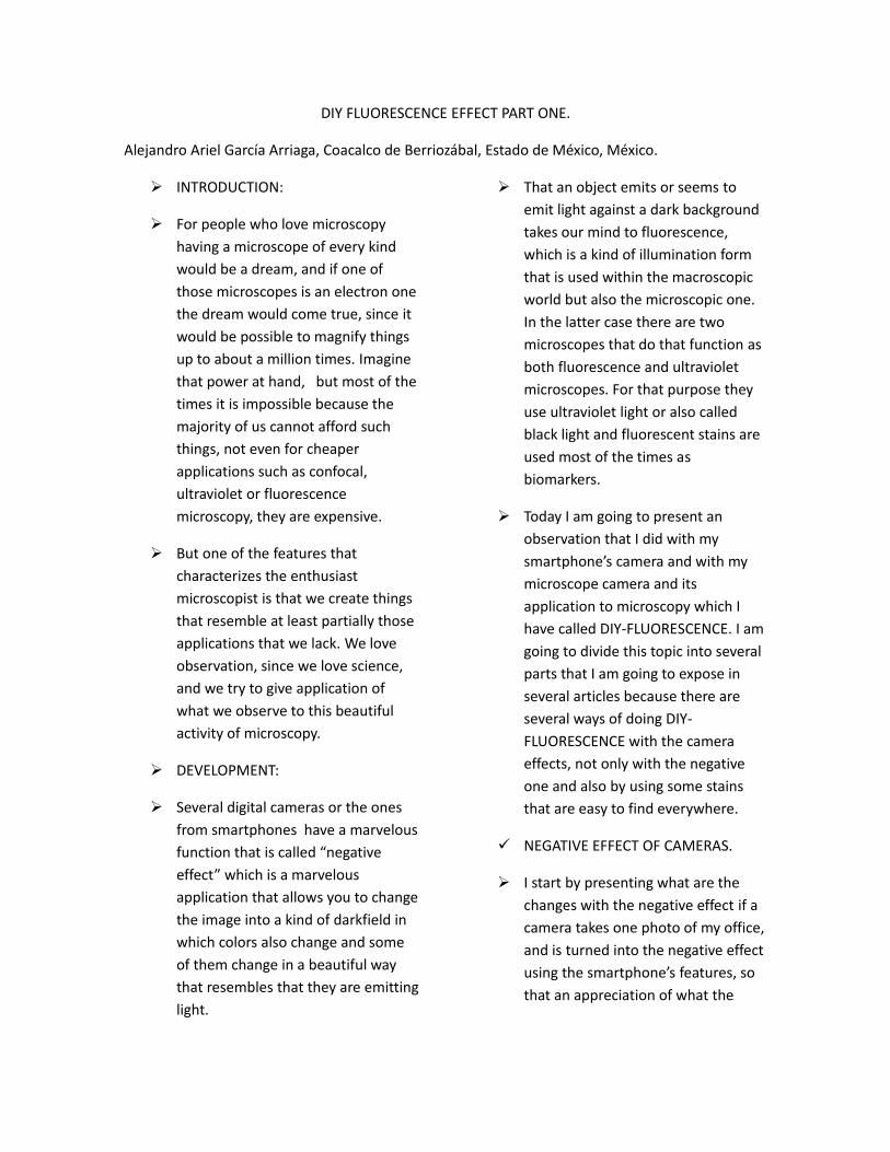
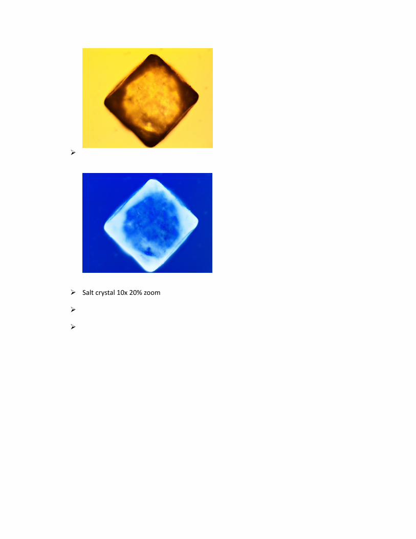
Salt crystal 10x 20% zoom
Sugar crystals 10x 20% zoom
NOTE :
The pictures presented below were taken in exactly the same way as the previous ones, the only difference was that in order to get more contrast and the DIY-FLUORESCENCE effect could be seen better, these
photos were processed with the auto correction of Microsoft Office Picture Manager. When applied it seem as if you “turn on the light” in the picture and it seemed to me that these photos needed that kind of help to get better.
Diatoms and green algae 40x 100% zoom
Mouth epithelial cells 40x 33% zoom
Streptococcus of yogurt 40x 50% zoom
Dry cranberry 40x 50%
Red tomato 40x 25% zoom
Analysis:
Most of the negative effect pictures are objects bright against a blue background. This is because the microscope that I use has a halogen bulb and a halogen light is warm, some times very warm at low intensity. Nevertheless with some examples it was possible because of the sample to get a black back ground. You can achieve this by varying the height of the condenser. As it is seen in the picture, for mosquitoes or parts of them is the nicest for this DIY-fluorescence. Other subjects like the ones that naturally are red like the poinsettia, the cranberry and examples that easily can be transformed with the negative effect. Another factor that helps is when it is used with low magnification powers, although with certain help using the program mentioned above, it is possible to transform a photo with high power magnification into DIY- fluorescence.
CONCLUSION:
Microscope digital cameras are an amazing tool which we can take the maximum advantage of. In addition, we have at hand programs such as Paint, Photoshop, Microsoft Picture Manager and others that can help us improve our observations in the miniature world.
What you can place under the objective and turn it into negative? Whatever you want, remember that some samples should give you amazing results, whilst others probably not, because not all samples are suitable for certain techniques. This is a principle that as microscopists we never have to forget. But by trying them all and learning what are the best for this or that illumination technique, is a great experience that we never would like to miss.
Email author: doctor2408 AT yahoo DOT com DOT mx
(Above in anti-spam format. Copy string to email software, remove spaces and manually insert the capitalised characters.)
Published in the April 2015 issue of Micscape Magazine.