J. Med. Chem. 1990,33,93-97 93 aqueous hydrochloric acid containing 5 g/L carbowax (mol wt 6000) and 0.45 mL of a 0.5 M tris(hydroxymethy1)aminomethane buffer (pH 8.0) containing 0.12 M sodium chloride and 0.05 mL of 1 % dimethyl sulfoxide/water, with or without an inhibitor, was maintained at 37 "C. To this solution Dvalyl-L-leucyl-L-lysine p-nitroanilide (0.1 mL of 3.35 mM in water) was added. The mixture was shaken and incubated at 37 OC for 1 min. The change in absorbance at 405 nm was then measured over 10 min. Inhibition of Trypsin Activity. To a 37 "C mixture of 0.1 mL of 0.01 g/L trypsin in 0.001 N aqueous hydrochloric acid, 2.6 mL of 0.046 M tris(hydroxymethy1)aminomethane buffer con- taining 0.0115 M CaCl, (pH 8.1) and 0.1 mL 1% dimethyl sulf- oxide/water with or without an inhibitor was added 0.2 mL of tosyl-L-arginine methyl ester (0.01 M). The mixture was shaken and incubated at 37 "C for 2 min. The change in absorbance was measured at 247 nm over 10 min. Acknowledgment. We thank the National Institutes of Health for their generous support of this research. Registry No. 2a.HC1,83039-48-9; 2a (free base), 114376-85-1; 2a (semicarbazone), 122314-25-4; 2b-HC1, 83134-12-7; 2b (free base), 122405-43-0; 2b (semicarbazone), 122314-26-5; 2csHC1, 122406-12-6; 2c (free base), 83039-66-1; 2c (semicarbazone, 122314-27-6;2d.HC1, 122314-38-9; 2d (free base), 122405-44-1;2d (semicarbazone), 122314-28-7;2eHC1, 122314-35-6; 2e (free base), 122405-45-2; 2f.HC1, 122314-36-7; 2f (free base), 122405-46-3; 2gHC1, 122314-37-8; 2g (free base), 122405-47-4; 2h.HC1, 122406-13-7; 2h (free base), 122314-43-6; 2bHC1, 122314-39-0; 2i (free base), 122405-48-5; 2j.HC1, 122314-40-3; 2j (free base), 122405-49-6;2k-HC1, 122314-41-4;2k (free base), 122405-50-9; 21-HC1, 122314-42-5;21 (free base), 122405-51-0;3a, 2389-49-3; 3b, 32393-52-5; 3c, 112208-06-7; 4a, 122314-18-5; 4b, 122314-19-6; 4c, 122314-20-9; 5a, 122314-16-3;5b, 122314-17-4;6a, 122332-80-3; 6b, 122314-29-8;6c, 122314-30-1; 6d, 122314-31-2; 6e, 122332-79-0; 6f, 122314-32-3;6g, 122314-33-4;6h, 122314-34-5;7,122314-15-2; 8a, 122314-21-0;8b, 122314-22-1;8c, 122314-23-2; 8d, 122314-24-3; Z-Lys(B0C)-H, 122314-13-0;Z-Orn(B0C)-H, 122314-14-1; BOC- Arg(N02)-H, 71413-14-4; Z-Leu-Leu-OH, 7801-71-0; Z-Leu-Phe- OH, 6401-63-4; Z-Phe-Leu-OH, 4313-73-9; Z-Leu-Val-OH, 7801- 70-9; trypsin, 9002-07-7; plasmin, 9001-90-5; kallikrein, 9001-01-8. of 6a in the deblocking reactions. The title compound exhibited the following: HPLC R, = 3.66 mL (methanol/water, 7/3 v/v, Cle, 5 pm); mp = 139-141 "C; CY]"^^ = -18.2" (c 1.0, methanol); R, = 0.53 (cyclized), 0.46 (uncyclized) (1-butanol/water/acetic acid, 7/2/1, v/v/v); 'H NMR (CD,OD, 200 MHz) 6 0.9 (m, 12 H), 1.4 (m, 8 H), 3.0 (t, J = 7 Hz, 2 H), 3.6 (m, 2 H), 4.3 (m, 3 H), 4.7 (m, 2 H), 5.1 (s, 2 H), 6.0 (m, 1 H, CHO-cyclized), 7.3 (s, 5 H). 13C NMR (CD,OD, 200 MHz) 6 19.65, 19.89, 21.65, 21.86, 27.20, 31.28, 31.82, 33.52, 35.05, 44.51, 45.37, 60.60, 60.90, 67.65, 67.73, 128.61, 128.84, 129.01, 129.50,138.30 (carbinolamine), 157.60 (C=O, Cbz), 162.80 (C=N, iminium), 173.55, 175.59 (amide bonds). Anal. Calcd for CUH&O5C1: C, 57.76; H, 7.88; N, 11.23. Found: C, 57.38; H, 7.98; N, 10.89. Inhibitor Activity Measurements. Rate measurements were performed with the aid of a Beckman Model DU UV-visible spectrophotometer in 1-cm quartz cuvettes thermostated at 37 "C. The inhibition of the enzyme activity was measured four times at five or more inhibitor concentrations. The average change in absorbance at each inhibitor concentration was utilized in the calculation of percent inhibition. All values were within 0.002 standard deviations from the mean. The percent inhibition of the reactions were calculated as follows: % inhibition = (A - B)/A X 100 where A = the change in absorbance without inhibitor and B = the change in absorbance with inhibitor. The concentration inducing a 50% inhibition was obtained by plotting the percent inhibition versus the log of the inhibitor concentration. The standard error for the linear-regression plots was calculated and in each case is less than 5%. Inhibition of Kallikrein Activity. To a 37 "C mixture of 0.02 mL of a 0.3 units/mL human urinary kallikrein solution was added 0.45 mL of 0.46 M tris(hydroxymethy1)aminomethane buffer (pH 8.0) containing 0.0115 M sodium chloride and 0.05 mL of 1% dimethyl sulfoxide/water with or without an inhibitor, 0.1 mL of the substrate, benzoylarginine ethyl ester (0.5 mM). The mixture was shaken and incubated at 37 "C for 2 min. The change in absorbance was then measured at 253 nm over 10 min. Inhibition of Plasmin Activity. A solution of 0.02 mL of a 0.25 casein units/mL plasmin solution in 50% glycerol in 2 mM DNA Intercalating Properties of Tetrahydro-9-aminoacridines. Synthesis and 23Na NMR Spin-Lattice Relaxation Time Measurements Jerrgen Dinesen, Jens Peter Jacobsen,* Frede P. Hansen, Erik B. Pedersen, and Hanne Eggert* Department of Chemistry, Odense University, Campusvej 55, DK-5230 Odense M, Denmark, and Chemical Laboratory II, The H.C. Orsted Institute, University of Copenhagen, Universitetsparken 5, DK-2100 Copenhagen 0, Denmark. Received January 23, 1989 A series of 9-(arylamino)-1,2,3,4-tetrahydroacridines, including the tetrahydro m-AMSA [N-[4-(acridin-g-y1- amino)-3-methoxyphenyl]methanesulfonamide J derivative, has been synthesized. 23Na NMR spin-lattice relaxation rate (l/Tl) measurements have been used to study whether these hydrogenated acridines were capable of intercalative binding to calf thymus DNA. The results have been compared to corresponding measurements for 9-aminoacridine, m-AMSA, and MgCl2 All compounds studied were capable of intercalative binding to DNA. However, it was found that the interaction was strongly influenced by substituents on the 9-arylamino group. Thus, tetrahydro m-AMSA was found to intercalate much more weakly with DNA than m-AMSA. Removal of the 3'-methoxy substituent of the 9-arylamino group resulted in intercalation in DNA that was almost as strong as that for m-AMSA. A large variety of drugs are known to interact strongly with nucleic acids. Many of them bind to double-stranded DNA through intercalation as first described by Lerman.' A great deal of effort has been devoted to the synthesis of several classes of compounds with specific intercalating properties to obtain agents of clinical importance. 9-Aminoacridines constitute a class of compounds that are now recognized for their ability to interact with DNA by intercalation. These compounds show great biological activity and several of them are of clinical importance. More than 500 fully aromatized 9-(ary1amino)acridine derivatives have been found to be antitumor active com- pounds. In particular, the N-[4-(9-acridinylamino)- phenyl]methanesulfonamide (AMSA) has shown a broad spectrum of activity against animal tumors.2 N-[4-(9- (1) Lerman, L. S. J. Mol. Biol. 1961, 3, 18. 0022-2623/90/ 1833-0093$02.50/0 (2) Denny, W. A.; Cain, B. F.; Atwell, G. J.; Hansch, C.; Pan- thananickal, A.; Leo, A. J. Med. Chem. 1982, 25, 276. 0 1989 American Chemical Society
Transcript
J. Med. Chem. 1990,33,93-97 93
aqueous hydrochloric acid containing 5 g/L carbowax (mol wt 6000) and 0.45 mL of a 0.5 M tris(hydroxymethy1)aminomethane buffer (pH 8.0) containing 0.12 M sodium chloride and 0.05 mL of 1 % dimethyl sulfoxide/water, with or without an inhibitor, was maintained at 37 "C. To this solution Dvalyl-L-leucyl-L-lysine p-nitroanilide (0.1 mL of 3.35 mM in water) was added. The mixture was shaken and incubated at 37 OC for 1 min. The change in absorbance a t 405 nm was then measured over 10 min.
Inhibit ion of Trypsin Activity. To a 37 "C mixture of 0.1 mL of 0.01 g/L trypsin in 0.001 N aqueous hydrochloric acid, 2.6 mL of 0.046 M tris(hydroxymethy1)aminomethane buffer con- taining 0.0115 M CaCl, (pH 8.1) and 0.1 mL 1% dimethyl sulf- oxide/water with or without an inhibitor was added 0.2 mL of tosyl-L-arginine methyl ester (0.01 M). The mixture was shaken and incubated at 37 "C for 2 min. The change in absorbance was measured a t 247 nm over 10 min.
Acknowledgment. We thank the National Inst i tutes of Health for their generous suppor t of this research.
Inhibitor Activity Measurements. Rate measurements were performed with the aid of a Beckman Model DU UV-visible spectrophotometer in 1-cm quartz cuvettes thermostated at 37 "C. The inhibition of the enzyme activity was measured four times a t five or more inhibitor concentrations. The average change in absorbance a t each inhibitor concentration was utilized in the calculation of percent inhibition. All values were within 0.002 standard deviations from the mean. The percent inhibition of the reactions were calculated as follows:
% inhibition = (A - B)/A X 100
where A = the change in absorbance without inhibitor and B = the change in absorbance with inhibitor.
The concentration inducing a 50% inhibition was obtained by plotting the percent inhibition versus the log of the inhibitor concentration. The standard error for the linear-regression plots was calculated and in each case is less than 5%.
Inhibit ion of Kallikrein Activity. To a 37 "C mixture of 0.02 mL of a 0.3 units/mL human urinary kallikrein solution was added 0.45 mL of 0.46 M tris(hydroxymethy1)aminomethane buffer (pH 8.0) containing 0.0115 M sodium chloride and 0.05 mL of 1% dimethyl sulfoxide/water with or without an inhibitor, 0.1 mL of the substrate, benzoylarginine ethyl ester (0.5 mM). The mixture was shaken and incubated at 37 "C for 2 min. The change in absorbance was then measured a t 253 nm over 10 min.
Inhibit ion of Plasmin Activity. A solution of 0.02 mL of a 0.25 casein units/mL plasmin solution in 50% glycerol in 2 mM
DNA Intercalating Properties of Tetrahydro-9-aminoacridines. Synthesis and 23Na NMR Spin-Lattice Relaxation Time Measurements
Jerrgen Dinesen, Jens Peter Jacobsen,* Frede P. Hansen, Er ik B. Pedersen, and Hanne Eggert*
Department of Chemistry, Odense University, Campusvej 55, DK-5230 Odense M, Denmark, and Chemical Laboratory I I , The H.C. Orsted Institute, University of Copenhagen, Universitetsparken 5, DK-2100 Copenhagen 0, Denmark. Received January 23, 1989
A series of 9-(arylamino)-1,2,3,4-tetrahydroacridines, including the tetrahydro m-AMSA [N-[4-(acridin-g-y1- amino)-3-methoxyphenyl]methanesulfonamide J derivative, has been synthesized. 23Na NMR spin-lattice relaxation rate (l/Tl) measurements have been used to study whether these hydrogenated acridines were capable of intercalative binding to calf thymus DNA. The results have been compared to corresponding measurements for 9-aminoacridine, m-AMSA, and MgCl2 All compounds studied were capable of intercalative binding to DNA. However, it was found that the interaction was strongly influenced by substituents on the 9-arylamino group. Thus, tetrahydro m-AMSA was found to intercalate much more weakly with DNA than m-AMSA. Removal of the 3'-methoxy substituent of the 9-arylamino group resulted in intercalation in DNA that was almost as strong as that for m-AMSA.
A large variety of drugs are known to interact strongly with nucleic acids. Many of them bind to double-stranded D N A through intercalation as first described by Lerman.' A great deal of effort has been devoted t o the synthesis of several classes of compounds with specific intercalating properties t o obtain agents of clinical importance.
9-Aminoacridines consti tute a class of compounds that are now recognized for their ability to interact with DNA
by intercalation. These compounds show great biological activity and several of them are of clinical importance. More than 500 fully aromatized 9-(ary1amino)acridine derivatives have been found t o be ant i tumor active com- pounds. In particular, the N-[4-(9-acridinylamino)- phenyl]methanesulfonamide (AMSA) has shown a broad spectrum of activity against animal tumors.2 N-[4-(9-
(1) Lerman, L. S. J . Mol. Biol. 1961, 3, 18.
0022-2623/90/ 1833-0093$02.50/0
(2) Denny, W. A.; Cain, B. F.; Atwell, G. J.; Hansch, C.; Pan- thananickal, A.; Leo, A. J . Med. Chem. 1982, 25, 276.
0 1989 American Chemical Society
94 Journal of Medicinal Chemistry, 1990, Vol. 33, No. 1 Dinesen et al.
Scheme I
R,, 4
2c OCH, h
Acridinylamino)-3-methoxyphenyl]methanesulfonamide (m-AMSA) is used clinically in the treatment of acute l e~kemia ,~ and it has been reported to have antiviral ac- tivity, particularly against vaccina virus.4 Recently, it has been found that some 9-(arylamino)acridine derivatives possess marked inhibitory action against mammalian malignant turn or^.^ However, biological activity is not restricted to derivatives of the fully aromatized acridine skeleton.
In pesticide screening of 9-(arylamino)-1,2,3,4-tetra- hydroacridines, there was found fungicide activity against mildew (Erysiphe gramines) on barley, insecticide activity against Aedes larvae in water, and anthelmintic activity in 9-(Substituted-amino)-1,2,3,4-tetrahydro- acridines have been found to exhibit a wide spectrum of pharmacological properties, which include respiratory stimulant activities: anticholinesterase”” activity, hallu- cinogen-countering activities,12 and morphine antago-
These findings have urged us to synthesize the basic skeleton 9-amino-1,2,3,4-tetrahydroacridine (1) together with a series of 9- (arylamino) - 1,2,3,4- tetrahydroacridines (2a-d) (Scheme I), including the tetrahydro derivative of m-AMSA (2d), in order to examine the intercalation ability of these molecules in DNA in comparison with those of the corresponding acridine compounds.
The intercalation of drugs to nucleic acids has been studied by several methods among which analytical ul- tracentrifugation, viscometry, and circular dichroism have been important.16 NMR spectroscopy has been used in several cases focusing mainly on the ’H and 31P nuclei.
nism.8913-15
(3) Arlinh, Z. A.; Sklaroff, R. B.; Gee, T. S.; Kempin, S. J.; How- ard, J.; Clarkson, B. D.; Young, C. W. Cancer Res. 1980, 40, 3304.
(4) Byrd, D. M. Ann. N.Y. Acad. Sci. 1977, 284, 463. (5) Kavadias, G.; William, T.; Janik, E.; Partyka, R. A. Eur. Pat.
Appl. EP. 165, 592; Chem. Abstr. 1986, 105, 6412u. (6) Girgis, N. S.; Pedersen, E. B. Synthesis 1985, 547. (7) Finlander, P.; Fischer, H. P.; Pedersen, E. B. Heterocycles
(8) Shaw, F. H.; Bently, G. A. Aust. J . Expt . Biol. Med. Sei. 1955, 33, 143.
(9) Shaw, F. H.; Bentley, G. A. Aust. J. Expt . Biol. Med. Sei. 1953, 31, 573.
(IO) De La Lande, I. S.; Bentley, G. A. Aust. J . Expt . Biol. M e d . Sci. 1955, 33, 555.
(11) Heilbrom, E. Acta. Chem. Scand. 1961, 15, 1386. (12) Gerehon, S. Nature 1960, 186, 1072. (13) Shaw, F. H.; Bentley, G. A. Med. J. Aust. 1949, 2, 868. (14) Shaw, F. H.; Bentley, G. A. Nature 1952, 169, 712. (15) Shaw, F. H., Gershon, S.; Bentley, G. A. J . Pharm. Pharmacol.
1957, 9, 666. (16) Wilson, D.; Jones, J. L. Intercalation Chemistry; Academic
Press: New York, 1982: Chapter 14.
iga5,23,1437.
’IT,
70
60
50
LO
30
20 R,
1 I I t
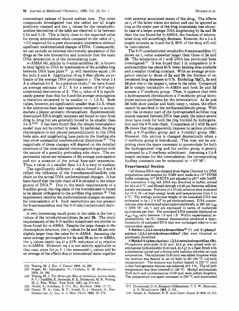
002 OOL 006 008 010 012 r Figure 1. 23Na spin-lattice relaxation rate, l/Tl, obtained a t 5.9 T, versus the ratio of the intercalator 9-amino-1,2,3,4-tetra- hydroacridine (1) to DNA phosphate, r. [PI = 8.07 mM, [Nal/ [PI = 1.2. The temperature was 27 “C.
Recently, some of us have introduced 23Na spin-lattice relaxation time (T,) measurement in studies of intercala- tion of several types of 9-aminoacridines in calf thymus DNA.17 It was shown that 23Na relaxation rate, R1 = l /Tl, is an extremely sensitive parameter for investigation of intercalation. 23Na NMR R1 measurements have therefore been used to investigate the interaction of the 1,2,3,4- tetrahydroacridines with DNA. Met hod
23Na spin-lattice relaxation rate measurements of a Na DNA solution can be interpreted on the basis of a “two- state model”.17 The sodium ions can interact with the charged double-stranded polynucleotide and thereby be in a perturbed region (denoted bound sodium ions, [Na+Ib) or they can reside “free” in the solvent (denoted free so- dium ions, [Na+If). The two states are characterized by different intrinsic relaxation rates, a large rate for the bound state (Rb) and a much smaller rate for the free ions (RJ. The measured R1 of a Na DNA solution are average values of Rf and R,,.
The intercalation process results in a helix extension of the DNA. This increases the average phosphate to phosphate distance and thereby decreases the DNA charge density. A further charge-density reduction results from positive charge on the intercalator, neutralizing some an- ionic charge of DNA. The reduction of the DNA charge density causes a release of bound sodium ions together with a decrease in Rb for the remaining bound sodium ions. Both effects reduce the measured value of R1, but it has been shown17 that the major part of the effect is due to a sizeable decrease in Rb. Only a minor part of the re- duction is caused by release of bound sodium ions.
For addition of small amounts of intercalator a linear dependence of R1 upon P = [I]/[P] (intercalator/DNA phosphate concentration) exists (see Figure 1). The in- tersection point ( r = rJ of this line with R1 = Rf occurs at
(1)
where a is a measure of the reduction of the relaxation rate of the bound sodium ions. a. is the initial concentration of bound sodium ions per DNA base pair before addition of the intercalator, determined to be 0.76.18 n is the ratio
a0
aoa + nPI rs =
(17) Eggert, H.; Dinesen, J.; Jacobsen, J. P. Biochemistry. In press.
Tetrahydro-9-aminoacridines
of mole of bound sodium ions released per mole or added intercalator, and pI is the fractions of drug intercalated relative to the total amount added.
According to the ion-condensation theory, it can be es- timated that"
D O
Journal of Medicinal Chemistry, 1990, Vol. 33, No. 1 95
Scheme I1
CH30 .Q CH30 6 & -
CH30 4 CH,O 4 "03 & - Sn. HCI
2c 3
NHSObCH3
NH "
(2.3q + 1.4xm)p1 l L b cy=- Rb0 - Rf
where q is the charge of the intercalating part of the drug, x the increase of the average distance between the base pair upon intercalation with m = 1 for monointercalation and m = 2 for bifunctional intercalation. Rb0 is the re- laxation rate of the bound sodium ions before addition of the intercalation. R$/(Rbo - Rf) is close to 1 since Rb0 >> Rf RP. In our DNA solutions, we have previously" estimated that Rbo/(Rbo - Rf) = 1.24 a t 5.9 T.
For strong intercalators (K > lo4 M-l), pI approaches unity and can be neglected as was done in the previous study1' of acridine intercalators. However, the present compounds may be weak intercalators and pI has to be included.
The number of released sodium ions per intercalator can be estimated by the simple ion-condensation theory to be given by the following expression"
n = - xm + Q 7.1 (3)
where Q is the total charge of the drug.
relation Equations 1-3 can be combined to give the following
pIx = 0.52/r, - 2.17 (4)
in which Q = q = m = 1 has been used as this applies for the drugs in this study. Moreover, the above mentioned values of a. and Rbo/(Rbo - Rf ) have been inserted.
In eq 2-4, we have assumed that all the bound drugs actually intercalate properly and that only a negligible part binds exclusively through electrostatic mechanism to the phosphate groups. If this is not so, the values of the prolongation factor, x , tend to be overestimated. However, such a electrostatic substitution mechanism of the drug has only a minor effect on the relaxation rate of sodium and cannot be estimated to be very important in view of the small values of rs obtained. Synthesis
Different substituted 9-(arylamino)-1,2,3,4-tetrahydro- acridines have been synthesized by the methods given below. 9-(Arylamino)-1,2,3,4-tetrahydroacridines (2) were prepared in one pot from methyl anthranilate and cyclo- hexanones in a mixture of phosphorus pentoxide, tri- ethylamine hydrochloride, and the appropriate arylamine a t 160-220 "C for 5 min-20 h. The molar ratio of the starting materials was 1:1.8:6:6:6. The reaction is believed to proceed by the mechanism described by Girgis and Pedersed and Nielsen and Pedersen.lg
Since 9- [ (2-methoxyaryl)arnino]-l72,3,4-tetrahydro- acridine (2c) can be synthesized easily in one step from commercially available materials, the tetrahydro derivative (2d) of m-AMSA can be prepared by nitration of the 4'- carbon of tetrahydroacridine 2c. The para isomer was purified by recrystallization from ethyl acetate. Reduction of nitro compound 3 was carried out by tin in hydrochloric acid and the amine salt was obtained. The free amine was liberated by addition of sodium hydroxide. Mesylation of
(18) Manning, G. S. Q. Rev. Biophys. 1978,2, 179. (19) Nielsen, S. V., Pedersen, E. B. Chem. Ser. 1986, 26, 331.
Pyridine
4 2d
Table I. Experimental Values of rs Obtained from Plots of l/T1 versus P
amino compound 4 was carried out by the method of Cain et a1.20v21 with mesyl chloride in dry pyridine a t -5 "C (Scheme 11). Measurements
The values of 23Na spin-lattice relaxation rates deter- mined as functions of the added amount of intercalator are shown in Figure 1 with 9-amino-172,3,4-tetrahydro- acridine (1) as the intercalator. A linear dependency is observed for small concentrations of the intercalator. The relaxation rate converges to the value of the free sodium ion (Rf) at increasing concentration of the intercalator. The initial linear dependency can be extrapolated to yield the value of rs a t the intersection point with the line l/T1 = Rf.
In accordance with the simplified two-site model of the relaxation rate, the Rf values used are the relaxation rate of 17.5 Hz found at 27 "C in a 10" M NaCl solution. This value is identical with the limiting value of the relaxation rate in the DNA solution after addition of the excess amount of the intercalator.
The values of r, have been determined for the series of compounds given in Table I. For comparison, the values of 9-aminoacridine (5), m-AMSA (6), and MgC12 taken from ref 17, are also included. Typically, 7-10 TI mea- surements have been used in the region r < 0.05.
The smallest rs value (rs = 0.116) has been obtained for 9-aminoacridine (5). As discussed by Eggert et al.,17 this value can be explained in terms of a strong monointerca- lation. The largest value (rs = 0.230) is due to MgC12. This value is the result of an electrostatic association of the double positively charged magnesium ion with DNA and
(20) Cain, B. F.; Seelye, R. N.; Atwell, G. J. J. Med. Chem. 1974, 17, 922.
(21) Cain, B. F.; Atwell, G. J., Denny, W . A. J . Med. Chem. 1975, 18, 1110.
96 Journal of Medicinal Chemistry, 1990, Vol. 33, No. 1
concomitant release of bound sodium ions. The other compounds investigated (see the table) are all single positively charged and the rs values for the tetrahydro- acridine derivatives of the table are observed to be between 0.14 and 0.19. This is fairly close to the expected value for strong intercalation when compared to the value of r, -0.5 expected for a pure electrostatic interaction without significant conformational changes of DNA. Consequently, we can exclude an external electrostatic association of the drugs as the sole interaction and conclude that the main DNA interaction is of the intercalating type.
m-AMSA (6), similar to 9-aminoacridine (51, is known to bind tightly to DNA by intercalation of the acridine ring between DNA base pairs.22 pI will therefore equal 1.00 for both 5 and 6. Application of eq 4 then allows an es- timate of the average DNA prolongation x . The value 2.4 A is obtained for 5 . In a previous study,I7 we have found an average estimate of 2.7 A for a series of 9-N-alkyl- substituted derivatives of 5. The r, value of 6 is signifi- cantly greater than that for 5 and the average prolongation is therefore estimated to be smaller ( x - 1.8 A). Both values, however, are significantly smaller than 3.4 A, which is the minimum base pair separation necessary to accom- modate a planar aromatic chromophore. Experimentally determined DNA-length increases are found to vary from dru to drug but are generally found to be smaller than
model' may not be correct in detail. In particular, the drug chromophore is not placed perpendicularly to the DNA helix axis, and neighboring base pairs to the binding site may show substantial conformational distortion^.^^ The magnitude of these changes will depend on the detailed structure of the intercalated chromophore together with the nature of a possibly protruding side chain. The ex- perimental values are estimates of the average prolongation and not a measure of the actual base-pair separation. Thus, a value of x smaller than 3.4 A is easy to explain.
The significantly different x values found for 5 and 6 reflect the influence of the 9-methanesulfoanilide side chain on the actual DNA conformational changes. I t has been found that the side chain of 6 is situated in the minor groove of DNA.26 Due to the steric requirements of a 9-anilino group, the ring plane of the 9-substituent is forced to be almost orthogonal to the acridine ring system.% This requirement restricts the conformational changes necessary for intercalation of 6. Such restrictions are not present for 9-aminoacridine and the 9-N-alkyl-substituted deriv- atives.
A very interesting result given in the table is the low r, values of the tetrahydroacridines 2a and 2b. The steric requirements of the 9-N-anilino substituent are similar to those found for m-AMSA. Despite the large change in the chromophore structure, the r, values for 2a and 2b are only slightly larger than the value for n-AMSA. Assuming the same average prolongation for 2a and 2b as for m-AMSA, the r, values imply (eq 4) a 13% reduction of pI relative to m-AMSA. However, eq 4 is not strictly applicable in that case, since for pI > 1 the measured r, values will be an average of the effects due to intercalated states together
3.4 1 .23924 It has been found that the simple intercalation
Dinesen et al.
~~ ~
(22) Waring, M. J. Eur. J. Cancer 1976, 12, 995. (23) Hogan, M.; Dattagupta, N.; Crothers, D. M. Biochemistry
1979, 18, 280. (24) Waring, M. J. The Molecular Bais of Antibiotic Action; Gitle,
E. F.; Cundliffe, E., Reynolds, P. E., Richmond, M. H., Waring, M. J., Eds.; Wiley: New York, 1981; pp 273-441.
(25) Neidle, S.; Abraham, Z. Crit. R e a Bzochem. 1984, 17, 73. (26) Denny, W. A.; Cain, B. F.; Atwell, G. J.; Hansch, G.; Pan-
thananickal, A, ; Leo, A. J . Med. Chem. 1982, 2Ei, 276.
with external associated states of the drug. The effects on rB of the latter states are minor and can be ignored as long as the major part of the drug intercalates (see above). In case of a larger average DNA lengthening by 2a and 2b than the one found for m-AMSA, the fraction of interca- lated drug will accordingly decrease. However, for a 2.4-.& length increase as found for 5, 80% of the drug will still be intercalated.
The 9-N-unsubstituted tetrahydro-9-aminoacridine (1) shows an rs value somewhat larger than those of 2a and 2b. The interaction of 1 with DNA has previously been investigated.27 It was found that 1 in comparison to 9- aminoacridine has about 60% fewer binding sites available and a smaller binding constant. With an average prolon- gation similar to those of 2a and 2b the fraction of in- tercalated drug decreases to 0.76. Excluding MgCl,, 2c and 2d give rise to the largest r, value in the table. Compound 2d is simply tetrahydro m-AMSA and both 2c and 2d possess a 3'-methoxy group. Thus, it appears that with a hydrogenated chromophore a 3'-methoxy group intro- duces serious problems for the intercalation. As 2c and 2d both show similar and fairly large r8 values, the effect cannot be ascribed to the methanesulfamido group. With only the aromatic part of the hydrogenated acridine ring system inserted between DNA base pairs, the minor groove must have room for both the ring buckled by hydrogena- tion and the 9-N-side chain. Comparison of 1 with 2a and 2b shows that this apparently imposes no serious problem with a 9-N-anilino group and a 3-methyl group (2b). However, this picture is changed dramatically when a 3'-methoxy group is introduced. This may not be sur- prising since the space necessary to accomodate for both the hydrogenated ring and the anilino group is greatly increased by a 3'-methoxy substituent. Assuming a 2.4-A length increase for the intercalation, the corresponding binding constants can be estimated to -lo2 M-I. Experimental Section
Calf thymus DNA was obtained from Sigma Chemical Co. DNA preparations and samples for NMR were made in a PIPES buffer containing lo4 M EDTA and adjusted to pH 7 with NaOH. For sonication, 4 mg/mL DNA was dissolved in buffer, stirred for 24 h at 4 "C, and filtered through a 0.45-km Sartorius cellulose acetate membrane. Portions of a 10-mL solution were sonicated for 10 x 10 s a t high energy levels and temperatures from 0 to 4 "C. The average molecular weight of the sonicated DNA was estimated to be 1.2 X lo6 by gel electrophoresis. DNA concen- trations were determined spectrophotometrically at 260 nm (tm
= 6600 M-' cm-') and are expressed in terms of nucleotide equivalents per liter. The sonicated DNA samples displayed an A2,/ATm. ratio between 1.8 and 1.9. Within experimental re- producibility ( i l % ) thermal denaturation produced a hypo- chromicity of sonicated DNA that was unchanged relative to an unsonicated sample. 9-Amino-1,2,3,4-tetrahydroacridineZ8 (1) and 9-( phenyl-
amino)-1,2,3,4-tetrahydr~acridine~ (2a) were obtained as previously described.
%-Methyl-9-(phenylamino)-l~,3,4-tetrahydroacridine (2b). Phosphorus pentoxide (0.30 mol, 42.6 g) was mixed with tri- ethylamine hydrochloride (0.30 mol, 41.3 g) in a flask fitted with a mechanical stirred and a drying tube (calcium chloride) a t room temperature. The arylamine (0.30 mol) was added dropwise while the mixture was heated in an oil bath to 80-100 "C (oil-bath temperature). The mixture was further heated to 220 "C until a clear homogeneous mixture was achieved (0.5-1 h). The oil bath temperature was then lowered to 140 "C. Methyl anthranilate (0.05 mol) and cyclohexanone (0.09 mol) were added dropwise. The temperature was again increased to 220 "C and maintained
(27) Drummond, D. S.; Simpson-Gildemeister, V. F. W.; Deacocke, A. R. Biopolymers 1965, 3, 135.
(28) Osbirk, A.; Pedersen, E. B. Acta Chem. Scand. B 1979,33,313.
J. Med. Chem. 1990, 33,97-101 97
at this temperature for 20 h. (The reaction progress was followed by taking from the mixture a sample which was treated with 2 M sodium hydroxide and extracted with diethyl ether. The extract was subjected to silica TLC with methylene chloride and methanol ( lO:l) . ) The reaction mixture was allowed to cool to 120 "C and 2 M sodium hydroxide (650 mL) was added. The temperature was lowered to room temperature and the mixture stirred about 0.5-1 h and extracted with ether. After evaporation, the residue was distilled at 225 "C/ l Torr. Compound 2b (5 g, 37%) was obtained by recrystallization from ligroin (100-140 "C), mp 186-187 OC (lit.29 mp 186 "C). 9- [ (2-Met hoxyp heny1)amino 1- 1,2,3,4-tetrahydroacridine
(2c). Phosphorus pentoxide (1.8 mol, 256 g) was mixed with triethylamine hydrochloride (1.8 mol, 448 g) in a flask fitted with a mechanical stirrer and a reflux condenser with a drying tube (calcium chloride) a t room temperature. 2-Methoxyaniline (1.8 mol, 222 g) was added dropwise while the mixture was heated in an oil bath to 60 "C (oil-bath temperature). The mixture was further heated to 160 "C until a homogeneous mixture was achieved (0.5 h). The oil-bath temperature was then lowered to 130 "C. Methyl anthranilate (0.3 mol, 45.4 g) and cyclohexanone (0.54 mol, 53.0 g) were added dropwise. The temperature was again increased to 160 "C and maintained a t that temperature for 15 min. The reaction mixture was allowed to cool to 110 OC and 2 M sodium hydroxide (4000 mL) was added. The aqueous suspension was extracted with 2 X 1500 mL of diethyl ether. The solvent was stripped off under reduced pressure. The residue was further evaporated at 0.1 mmHg to remove 2-methoxyaniline. A black solid formed and was recrystallized twice from ligroin (80-100 "C) to yield 2c (32.4 g, 36%) as white-yellow crystals: mp 131-132 "C; 'H NMR (CDC13/TMS) d/ppm 1.91 (m, 4 H), 2.79 (m, 2 H), 3.19 (m, 2 H), 4.02 (s, 3 H), 6.27 (s, NH), 6.10-8.20 (m, 8 H); '% NMR (CDCl,/TMS) b/ppm 25.09 (C-1), 22.75 (C-2), 22.49 (C-3), 33.98 (C-4), 159.77 (C-4a), 128.60 (C-5) 128.41 (C-6), 124.84 (C-7), 123.08 (C-8), 123.67 (C-8a), 142.96 (C-9), 124.45 (C-ga), 147.24 (C-loa); MS m / e 304 (loo), 289 (13), 273 (ll), 197 (ll), 182 (11). Anal. (CzoHzoNzO) C, H, N. 9-[ (4-Nitro-2-methoxyphenyl)amino]-1,2,3,4-tetrahydro-
acridine (3). Compound 2c (20 mmol, 6.1 g) was dissolved in acetic acid (99%, 80 mL). After addition of nitric acid (68%) at 70 "C and stirring for 4 h, it was poured into ice (500 mL). When the ice had melted, 2 M sodium hydroxide (0.5 L) was added. A yellow precipitate 6.5 g (ortho and paraisomer) was washed with water and dried in vacuum. Recrystallization twice from ethyl acetate yielded 3 (2.4 g, 34%), mp 221-223 "C. Anal. (CzoH19- N303) C, H, N.
(29) Endicott, M.; Alden, B. W.; Sherrill, M. L. J. Am. Chem. SOC. 1946, 68, 1303.
9-[ (4-Amino-2-methoxyphenyl)amino]-lf,3,4-tetrahydro- acridine (4). A mixture of 3 (13 mmol, 4.6 g), tin (83 mmol, 9.9 g), and 6 M hydrochloride acid (100 mL) was heated with stirring to 120 "C (oil-bath temperature) by which time the tetrahydro- acridine had dissolved. The mixture was refluxed for 2 h and cooled. The precipitate was dissolved in water and, after addition of 2 M sodium hydroxide (50 mL), extracted into chloroform. The chloroform extract was evaporated and the residue was recrys- tallized from 50% ethanol to yield 4 (2.5 g, 56%), mp 155-156 " C.
N-[4-[ (1,2,3,4-Tetrahydro-9-acridinyl)amino]-3-methoxy- phenyl]methanesulfonamide (2d). Compound 4 (7.5 mmol, 2.4 g) was dissolved in 25 mL of dry pyridine. Mesyl chloride (15 mmol, 1.16 mL) was added slowly at -5 "C and the mixture was stirred for 1 h. The mixture was evaporated under reduced pressure and dissolved in water (200 mL). Hydrochloric acid (4 M, 20 mL) was added to precipitate the hydrochloride, which was taken up in 200 mL of water. 10 mL of sodium hydrogen car- bonate was added and the precipitate was rrystallized from ethanol (96%) to yield 2d (1.5 g, 50%), mp 248-249 "C (lit.% mp 243-245 "C.
NMR Experiments. All DNA samples for NMR were made 5 mM in DNA phosphate by diluting with distilled water. This gave [Na+]/[P] ratios of 1.2. The NMR intercalator titrations were performed by adding successive aliquots (corresponding to r - 0.005) of a drug stock solution directly to the DNA solution in the NMR tube. For titrations up to r - 0.05, the volume increases by 10%. A control experiment, in which water alone was added up to 20% volume increase, showed that T1, within measuring uncertainties, was unchanged by such dilution. pH was measured to be 7 both before and after the addition of in- tercalators.
=Na NMR spectra were recorded at 5.9 T on a Bruker AC 250 and obtained without lock. The inversion-recovery (180'-7- 90"-acq) pulse sequence was used for the T, measurements with 15 different values of 7 for each experiment. The Tl values were obtained by a three-parameter linear least square fitting procedure. Each Tl value is the average of at least two measurements. The temperature for the NMR measurements was 27 "C.
13C and 'H spectral data given were also obtained a t 5.9 T on a Bruker AC 250 NMR instrument. Registry No. 1,321-64-2; 2a, 14807-16-0; 2b, 110245-49-3; 2c,
(30) Yamato, M.; Takeuchi, Y.; Ikeda, Y. Heterocycles 1987, 26, 191.
Cyclization-Activated Prodrugs. Basic Carbamates of 4-Hydroxyanisole
Walfred S. Saari,* J o h n E. Schwering, Paulette A. Lyle, Steven J. Smith, and Edward L. Engelhardt
Merck Sharp & Dohme Research Laboratories, West Point, Pennsylvania 19486. Received January 18, 1989
A series of basic carbamates of 4-hydroxyanisole was prepared and evaluated as progenitors of this melanocytotoxic phenol. All of the carbamates were relatively stable a t low pH but released 4-hydroxyanisole cleanly a t pH 7.4 a t rates that were structure dependent. A detailed study of the N-methyl-N-[2-(methylamino)ethyl]carbamate showed that generation of the parent phenol followed first-order kinetics with t l l2 = 36.3 min a t pH 7.4, 37 "C, and was accompanied by formation of N,N'-dimethylimidazolidinone. These basic carbamates are examples of cycliza- tion-activated prodrugs in which generation of the active drug is not linked to enzymatic cleavage but rather depends solely upon a predictable, intramolecular cyclization-elimination reaction.
Esterification of therapeutically active agents t o provide prodrugs with improved properties has become a familiar strategy for the circumvention of adverse physicochemical limitations. Ester prodrugs of alcohols and phenols are frequently explored t o improve solubility, absorption, and bioavailability a n d t o extend the duration of action of the parent drug.'S2 It is of course essential for the success of
this strategy that the ester progenitor be capable of de- livering the parent d rug at a practical rate in vivo. Gen- erally, ester prodrugs have depended upon chemical or
(1) Stella, V. J.; Charman, W. N. A.; Naringrekar, V. H. Drugs.