Proc. Nati. Acad. Sci. USA Vol. 87, pp. 3831-3835, May 1990 Biochemistry DNA intercalation and cleavage of an antitumor antibiotic dynemicin that contains anthracycline and enediyne cores (hybrid antibiotic/nucleotide specificity/minor groove/action mechanism) YUKIO SUGIURA*t, TAKASHI SHIRAKI*, MASATAKA KONISHIO, AND TOSHIKAZU OKI* *Institute for Chemical Research, Kyoto University, Uji, Kyoto 611, Japan; and tBristol-Myers Research Institute, Tokyo Research Center, 2-9-3 Shimo-Megro, Meguro-ku, Tokyo 153, Japan Communicated by Samuel J. Danishefsky, February 26, 1990 (received for review October 1, 1989) ABSTRACT Dynemicin is a hybrid containing anthraqui- none and enediyne cores, which contribute to binding and cleavage of DNA, respectively. DNA strand scission by the antitumor antibiotic is significantly enhanced by the addition of NADPH or thiol compounds. The preferential cutting site of dynemicin is on the 3' side of purine bases (i.e., 5'-GC, -GT, and -AG) and is clearly different from the cutting sites of esperamicin and calicheamicin. The double-stranded and the stem regions of single-stranded DNAs are preferentially cleaved by dynemicin. Therefore, dynemicin may be a useful reagent for probing secondary structures of DNA. Pretreat- ment of DNA with Adriamycin and actinomycin D alters the cutting mode of dynemicin. Dynemicin-mediated DNA break- age is strongly inhibited by pretreatment of the DNA with distamycin A and anthramycin, suggestng that dynemicin interacts with the minor groove of the DNA helix. Intercalation of the anthraquinone core into the DNA followed by the attack of the phenyl diradical formed from the enediyne core is considered as a possible mechanism of action of dynemicin. Dynemicin, isolated from the fermentation broth of Mi- cromonospora chersina, possesses potent cytotoxicity and in vivo antitumor activity (1). This antibiotic is a hybrid mole- cule of two typical chemotypes of antitumor agent, enediyne and anthraquinone (Fig. 1). Esperamicin, calicheamicin, and neocarzinostatin, which produce DNA strand breaks, belong to the family of enediyne antitumor antibiotics (2-5). DNA cleavage by esperamicin and calicheamicin appears to in- volve rearrangement of the enediyne unit, a phenylene dirad- ical that can abstract hydrogen atoms from the sugar phos- phate backbone of DNA. On the other hand, daunomycin, Adriamycin, and aclacinomycin belong to the class of an- thracycline antitumor antibiotics and can intercalate into DNA through the planar aromatic rings (6, 7). Indeed, an x-ray diffraction study (8) demonstrated that two daunomy- cin molecules intercalate between the GC base pairs in the self-complementary hexanucleotide CGTACG. Evidently, dynemicin possesses these two structural features in one molecule and hence we were interested in clarifying the mechanism through which dynemicin cleaves DNA. MATERIALS AND METHODS Drugs and Chemicals. Dynemicin was isolated from the fermentation broth of Micromonospora chersin and purified as described (1). Esperamicin A1 and anthramycin were a generous gift of T. W. Doyle (Bristol-Myers) and L. H. Hurley (University of Texas), respectively. Adriamycin and distamycin A were offered by F. Arcamone (Farmitalia). Plasmid pBR322 DNA was isolated from Escherichia coli C600, and restriction enzymes, such as EcoRI, Sal I, and Dra HH3 HC H3 OH 0 OH HHII OH 0 OH FIG. 1. Chemical structures of dynemicin (Left) and its aroma- tized derivative (Right). This aromatic compound was obtained by treating dynemicin with HCl and its structure was identified by NMR and x-ray crystallographic methods. II, were obtained from Takara Shuzo (Kyoto, Japan). Stem- and-loop structure G4 DNA obtained from phage R199/G4ori replicative form DNA was a kind gift of T. Komano (Kyoto University, Japan). Ethidium bromide, actinomycin D, and NADPH were purchased from Sigma. All other chemicals used were of commercial reagent grade. Assay for DNA Cleavage Activity. Analysis of drug (5 ,uM)-induced damage to supercoiled, covalently closed, cir- cular (form I) pBR322 DNA was performed in the presence of NADPH, dithiothreitol, 4-hydroxythiophenol, or NaBH4 (compound used was added at 5 AM-100 mM) for 30 min-24 hr at 37TC, followed by agarose gel electrophoresis to sepa- rate the various DNA products-namely, nicked relaxed circular DNA (form II) and linearized DNA (form III). DNA bands were visualized by using ethidium bromide binding and UV illumination. Nucleotide Sequence Analysis. The reaction samples (total volume, 50 Al) contained a 5'-end-labeled 70-base-pair (bp) (EcoRI-Dde I) fragment or a 128-bp (Sal I-Dra II) pBR322 DNA fragment, sonicated calf thymus carrier DNA (20 ,ug/ml), and 10 mM Tris-HCl buffer (pH 7.5). Nucleotide sequence cleavage was initiated by addition of dynemicin (5-50 AM) and NADPH (5 mM) or dithiothreitol (5 mM), and then the samples were incubated at 370C for 5 hr. Cleavage experiments for the complementary strand (3'-32P-labeled DNA) were carried out by using the Sal I-Dra II pBR322 DNA fragment. Ice-cold ethanol was added to the samples to stop the reaction. After preincubation of the DNA fragment with distamycin A and some intercalators such as ethidium bromide and Adriamycin at 370C for 30 min, the dynemicin- induced DNA cleavage was investigated and compared with the nucleotide sequence cleavage of intact DNA with dyne- micin. DNAs modified with aflatoxin B1 (9), cis-diammine- dichloroplatinum (10), and anthramycin (11) were prepared. Electrophoresis was performed on a 10% polyacrylamide/7 M urea slab gel, and DNA sequencing was carried out by the Maxam-Gilbert method (12). The autoradiograms were scanned with a laser densitometer (LKB model 2222 Ultro- Scan XL). tTo whom reprint requests should be addressed. 3831 The publication costs of this article were defrayed in part by page charge payment. This article must therefore be hereby marked "advertisement" in accordance with 18 U.S.C. §1734 solely to indicate this fact.
Transcript
Proc. Nati. Acad. Sci. USAVol. 87, pp. 3831-3835, May 1990Biochemistry
DNA intercalation and cleavage of an antitumor antibioticdynemicin that contains anthracycline and enediyne cores
YUKIO SUGIURA*t, TAKASHI SHIRAKI*, MASATAKA KONISHIO, AND TOSHIKAZU OKI**Institute for Chemical Research, Kyoto University, Uji, Kyoto 611, Japan; and tBristol-Myers Research Institute, Tokyo Research Center, 2-9-3Shimo-Megro, Meguro-ku, Tokyo 153, Japan
Communicated by Samuel J. Danishefsky, February 26, 1990 (received for review October 1, 1989)
ABSTRACT Dynemicin is a hybrid containing anthraqui-none and enediyne cores, which contribute to binding andcleavage of DNA, respectively. DNA strand scission by theantitumor antibiotic is significantly enhanced by the addition ofNADPH or thiol compounds. The preferential cutting site ofdynemicin is on the 3' side of purine bases (i.e., 5'-GC, -GT,and -AG) and is clearly different from the cutting sites ofesperamicin and calicheamicin. The double-stranded and thestem regions of single-stranded DNAs are preferentiallycleaved by dynemicin. Therefore, dynemicin may be a usefulreagent for probing secondary structures of DNA. Pretreat-ment of DNA with Adriamycin and actinomycin D alters thecutting mode of dynemicin. Dynemicin-mediated DNA break-age is strongly inhibited by pretreatment of the DNA withdistamycin A and anthramycin, suggestng that dynemicininteracts with the minor groove of the DNA helix. Intercalationof the anthraquinone core into the DNA followed by the attackof the phenyl diradical formed from the enediyne core isconsidered as a possible mechanism of action of dynemicin.
Dynemicin, isolated from the fermentation broth of Mi-cromonospora chersina, possesses potent cytotoxicity and invivo antitumor activity (1). This antibiotic is a hybrid mole-cule of two typical chemotypes of antitumor agent, enediyneand anthraquinone (Fig. 1). Esperamicin, calicheamicin, andneocarzinostatin, which produce DNA strand breaks, belongto the family of enediyne antitumor antibiotics (2-5). DNAcleavage by esperamicin and calicheamicin appears to in-volve rearrangement of the enediyne unit, a phenylene dirad-ical that can abstract hydrogen atoms from the sugar phos-phate backbone of DNA. On the other hand, daunomycin,Adriamycin, and aclacinomycin belong to the class of an-thracycline antitumor antibiotics and can intercalate intoDNA through the planar aromatic rings (6, 7). Indeed, anx-ray diffraction study (8) demonstrated that two daunomy-cin molecules intercalate between the GC base pairs in theself-complementary hexanucleotide CGTACG. Evidently,dynemicin possesses these two structural features in onemolecule and hence we were interested in clarifying themechanism through which dynemicin cleaves DNA.
MATERIALS AND METHODSDrugs and Chemicals. Dynemicin was isolated from the
fermentation broth of Micromonospora chersin and purifiedas described (1). Esperamicin A1 and anthramycin were agenerous gift of T. W. Doyle (Bristol-Myers) and L. H.Hurley (University of Texas), respectively. Adriamycin anddistamycin A were offered by F. Arcamone (Farmitalia).Plasmid pBR322 DNA was isolated from Escherichia coliC600, and restriction enzymes, such as EcoRI, Sal I, and Dra
HH3
HC H3
OH 0 OH
HHII
OH 0 OH
FIG. 1. Chemical structures of dynemicin (Left) and its aroma-tized derivative (Right). This aromatic compound was obtained bytreating dynemicin with HCl and its structure was identified by NMRand x-ray crystallographic methods.
II, were obtained from Takara Shuzo (Kyoto, Japan). Stem-and-loop structure G4DNA obtained from phage R199/G4orireplicative form DNA was a kind gift of T. Komano (KyotoUniversity, Japan). Ethidium bromide, actinomycin D, andNADPH were purchased from Sigma. All other chemicalsused were of commercial reagent grade.
Assay for DNA Cleavage Activity. Analysis of drug (5,uM)-induced damage to supercoiled, covalently closed, cir-cular (form I) pBR322 DNA was performed in the presenceof NADPH, dithiothreitol, 4-hydroxythiophenol, or NaBH4(compound used was added at 5 AM-100 mM) for 30 min-24hr at 37TC, followed by agarose gel electrophoresis to sepa-rate the various DNA products-namely, nicked relaxedcircular DNA (form II) and linearized DNA (form III). DNAbands were visualized by using ethidium bromide binding andUV illumination.
Nucleotide Sequence Analysis. The reaction samples (totalvolume, 50 Al) contained a 5'-end-labeled 70-base-pair (bp)(EcoRI-Dde I) fragment or a 128-bp (Sal I-Dra II) pBR322DNA fragment, sonicated calf thymus carrier DNA (20,ug/ml), and 10 mM Tris-HCl buffer (pH 7.5). Nucleotidesequence cleavage was initiated by addition of dynemicin(5-50 AM) and NADPH (5 mM) or dithiothreitol (5 mM), andthen the samples were incubated at 370C for 5 hr. Cleavageexperiments for the complementary strand (3'-32P-labeledDNA) were carried out by using the Sal I-Dra II pBR322DNA fragment. Ice-cold ethanol was added to the samples tostop the reaction. After preincubation of the DNA fragmentwith distamycin A and some intercalators such as ethidiumbromide and Adriamycin at 370C for 30 min, the dynemicin-induced DNA cleavage was investigated and compared withthe nucleotide sequence cleavage of intact DNA with dyne-micin. DNAs modified with aflatoxin B1 (9), cis-diammine-dichloroplatinum (10), and anthramycin (11) were prepared.Electrophoresis was performed on a 10% polyacrylamide/7M urea slab gel, and DNA sequencing was carried out by theMaxam-Gilbert method (12). The autoradiograms werescanned with a laser densitometer (LKB model 2222 Ultro-Scan XL).
tTo whom reprint requests should be addressed.
3831
The publication costs of this article were defrayed in part by page chargepayment. This article must therefore be hereby marked "advertisement"in accordance with 18 U.S.C. §1734 solely to indicate this fact.
Proc. Natl. Acad. Sci. USA 87 (1990)
RESULTSSequence-Specific DNA Cleavage by Dynemicin. Agarose
gel electrophoretic results for dynemicin-mediated strandscission demonstrated that NADPH, 4-hydroxythiophenol,or dithiothreitol stimulated DNA cleavage by dynemicin.Whereas ascorbic acid, sodium dithionite, or sodium boro-hydride had only a weak effect on dynemicin-mediated DNAbreakage. With 4-hydroxythiophenol or dithiothreitol, form IDNA was converted to form II and form III DNAs within 30min. Although the cleavage reaction was slow in the NADPHsystem, extensive fragmentation of DNA was clearly ob-served after 5 hr. Cleavage data for both strands (DNAfragments 32P-labeled at both ends) in the dynemicin-NADPH system are presented in Fig. 2. In this experiment,the Sal I-Dra II fragment (128 bp) was used as DNAsubstrate. As shown in the histogram (Fig. 3), (i) dynemicinpreferentially attacks the 3' side of purine bases such as5'-AG, 5'-GC, and 5'-AI and (ii) among the four basesguanine is a relatively favorable cutting site for dynemicin. Inaddition, the drug appears to cause typical double-strand cutsat the sequence
3'-GATGATGACC5'-CTACTACTGG.
II 111.11 Ill. .,,GCTCAACGGCCTCAACCTACCGAGTTG CCGGAGTTGGAT(I I I' I11 '1
III
IIlllIII II I III I Ii,TACTGGGCTGCTTCCTAATGCAGGA-3'BATGACCCGACGAAGGATTACGTCCT-5'
1 11 I I I
il
I I
11 I
I I 11
AMD
DMC
FIG. 3. Histograms of DNA-cutting sites by dynemicin for intactDNA and actinomycin D (AMD)- or distamycin A (DMC)-pretreatedDNA. Relative DNA cleavage frequencies were obtained fromdensitometric scans of autoradiograms from gels.
Alteration of Dynemicin-Induced DNA Cleavage by Pre-treatment ofDNA with Groove Binders and Intercalators. Fig.4 shows DNA fragments generated by dynemicin after pre-treatment with ethidium bromide, proflavine, Adriamycin,actinomycin D, anthramycin, and distamycin A. These pat-terns were compared with the standard nucleotide-specificcleavage products for intact DNA in the dynemicin-NADPHsystem. Inhibition of the dynemicin-induced DNA cleavage
CGGAG TT 1 2 3 4 5 6 £T ~~~~~~~~A
G ACCGGA CG TT _
A
TG AGAT A~~~~~~~~GcA- TT AGA C .; 8c ~~~~~~~~TCCG c~~~~~~~
GA ~~~~~~~~CA T
C TG CA CT TT AA Ac TG AC T~~~~~~~~
A5tl3t
FIG. 2. Strand scission of 5'-end-labeled (lanes 1-3) and 3'-end-labeled (lanes 4-6) DNA sequences by dynemicin. Lanes 3 and4 show DNA cleavage patterns by dynemicin (50 ,uM) and NADPH(5 mM) at 37°C for 5 hr. Lanes 1 and 6 show intact DNA. Lanes 2 and5 indicate the Maxam-Gilbert sequencing reaction for G+A.
by Adriamycin was considerably greater than by ethidiumbromide or proflavine. Actinomycin D also masked somedynemicin cutting sites such as 5'-GC. In addition, as in lane6, cleavage at certain nucleotides was appreciably enhanced
CCC
___ ~~AG
T
G
A_ r~~~~~~s:S: ko~~~~
TXB E -__ A~~
FIG. 4. DNA-cutting modes by dynemicin after pretreatmentwith ethidium bromide (lane 3), proflavine (lane 4), Adriamycin (lane5), actinomycin D (lane 6), anthramycin (lane 8), or distamycin A(lane 9). After the pretreatment of the DNA fragment with thesecompounds (50 ALM) at 37°C for 30 min, the DNA cleavage reactionswere carried out by dynemicin (50 ,uM) and NADPH (5 mM) at 37°Cfor 5 hr. Lane 7 shows dynemicin-induced DNA cleavage for intactDNA. Lanes 1 and 2 show intact DNA and the Maxam-Gilbertsequencing reaction for G+A, respectively.
3832 Biochemistry: Sugiura et al.
Proc. Natl. Acad. Sci. USA 87 (1990) 3833
Ah
c
tGCTCTGAGCACACT GTCA
r-AVGCCTACGACA94GACTCAATCATGACCTCGTMCGCAAC
TA
.orTAT A
TC A G
AA
T4 Q-4 . AGT
AAAGCCAAAAGGACTAACATATGTTCC
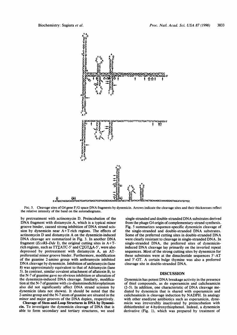
FIG. 5. Cleavage sites ofG4 gene F/G space DNA fragments by dynemicin. Arrows indicate the cleavage sites and their thicknesses reflectthe relative intensity of the band on the autoradiograms.
by pretreatment with actinomycin D. Preincubation of theDNA fragment with distamycin A, which is a typical minorgroove binder, caused strong inhibition ofDNA strand scis-sion by dynemicin near A+T-rich regions. The effects ofactinomycin D and distamycin A on the dynemicin-inducedDNA cleavage are summarized in Fig. 3. In another DNAfragment (EcoRI-Dde I), the original cutting sites in A+T-rich regions, such as TTIATC-5' and CIGTAA-5', were alsodepressed by pretreatment with distamycin A, an AT-preferential minor groove binder. Furthermore, modificationof the guanine 2-amino group with anthramycin inhibitedDNA cleavage by dynemicin. Inhibition ofanthramycin (lane8) was approximately equivalent to that of Adriamycin (lane5). In contrast, similar covalent attachment of aflatoxin B1 tothe N-7 ofguanine gave no obvious inhibition or alteration ofthe dynemicin-induced DNA cleavage. Similarly, modifica-tion at the N-7 ofguanine with cis-diamminedichloroplatinumalso 'did not significantly affect DNA strand scission bydynemicin (data not shown). It should be noted that the2-amino group and the N-7 atom ofguanine are situated in theminor and major grooves of the DNA duplex, respectively.
Cleavage of Stem-and-Loop Structures in DNA by Dynemi-cin. To investigate the dynemicin cleavage of DNA that isable to form secondary and tertiary structures, we used
single-stranded and double-stranded DNA substrates derivedfrom the phage G4 origin ofcomplementary-strand synthesis.Fig. 5 summarizes sequence-specific dynemicin cleavage ofthe single-stranded and double-stranded DNA substrates.Some of the preferred cutting sites in double-stranded DNAwere clearly resistant to cleavage in single-stranded DNA. Insingle-stranded DNA, the preferred sites of dynemicin-induced DNA cleavage lay primarily on the inverted repeatsequences. Most of the strong cutting sites by dynemicin forthese substrates were at the dinucleotide sequences 5'-ATand 5'-GT. A certain bulge thymine was also a preferredcleavage site in double-stranded DNA.
DISCUSSIONDynemicin has potent DNA breakage activity in the presenceof thiol compounds, as do esperamicin and calicheamicin(2-5). In addition, one characteristic of DNA cleavage me-diated by dynemicin that is shared with esperamicin andcalicheamicin is cleavage induction by NADPH. In analogywith other enediyne antibiotics such as esperamicin, dyne-micin was irreversibly inactivated by preincubation withdithiothreitol or 4-hydroxythiophenol. Indeed, a dynemicinderivative (Fig. 1), which was prepared by treatment of
Biochemistry: Sugiura et al.
Proc. Natl. Acad. Sci. USA 87 (1990)
dynemicin with HCI causing aromatization of the enediynestructure, had no DNA cleavage activity under the experi-mental conditions used with dynemicin. This result stronglyindicates that the enediyne chromophore significantly con-tributes to the present DNA strand scission. Presumably, aphenylene diradical produced by cyclization of the diyne-eneis the active form of dynemicin, similar to the mechanismproposed for compounds in this class of antitumor agentsresponsible for potent DNA breakage (2, 4, 13, 14). Fordynemicin, opening the epoxide appears to be the key inactivation to the diaryl radical form of dynemicin-namely,the DNA-cleaving intermediate. A more reasonable mecha-nism for the epoxide ring opening reaction requires theconversion of dynemicin by a one-electron reduction or twosequential one-electron reductions to the hydroquinone. Thequinone methide formed can undergo nucleophilic attack bywater or act as nucleophile and become protonated giving thehydroquinonediol or the quinone alcohol, respectively (15,16). The compounds can then undergo the Bergman reactionand aromatize (Fig. 6). The blocking ofDNA damage by highlevels of thiols and the detection of free radical signals in apreliminary ESR study suggest a radical mechanism for theaction of dynemicin. However, dynemicin-mediated strandscission frequently occurs at guanines and adenines. Thebases adenine and guanine are particularly susceptible toalkylation that could result in cleavage at these sites withoutinvoking the presence of radicals. Perhaps dynemicin takesadvantage of both alkylation and hydrogen atom abstractionto effect DNA cleavage. In the presence of thiol cofactors,dynemicin at concentrations as low as 0.5 ,uM causes DNAcleavage in supercoiled pBR322 DNA. Calicheamicin andesperamicin appear to be more efficient at generating DNAstrand breaks.Although the DNA damage by dynemicin is not particu-
larly base-specific, dynemicin does preferentially attackbases adjacent to the 3' side of purines such as 5'-GC, 5'-GT,5'-AT, and 5'-AG. Among the four bases, guanine is afavored cutting site for dynemicin. It is of interest that thisbase is the most resistant to cleavage by esperamicin (3),calicheamicin (4), and neocarzinostatin (5). The cleavagemode ofdynemicin clearly differs from that ofesperamicin (3)and calicheamicin (4), which show preferential cytosine and
thymine cutting in oligopyrimidine regions. As shown in theautoradiograms (Figs. 2 and 4), the radioactive bands pro-duced by dynemicin were found to be electrophoreticallyidentical to the Maxam-Gilbert products (12), suggesting thepresence of 3'-phosphates at the present DNA breaks. Whenthe cleavage products were treated with base, the productscoincidently migrated with the sequencing markers in thisDNA fragment. It is also observed that dynemicin occasion-ally caused strong strand breaks 3 bp apart in the two DNAstrands (for example, the 5'-CTACTACTGG and its comple-ment 3'-_jATjATjACC). This cleavage pattern is well-known for DNA damage by calicheamicin (4) and espera-micin (3). This cutting mode is characteristic of double-stranded DNA breakage. Furthermore, the asymmetriccleavage pattern on the 3' side of opposite strands suggestsinteraction of dynemicin with the minor groove of the DNAhelix (17). Indeed, the nucleotide cleavage pattern generatedby dynemicin is significantly affected by pretreatment of theDNA with distamycin A and anthramycin, strongly support-ing an interaction between the minor groove of DNA anddynemicin. Distamycin A is a well-known minor groovebinder and anthramycin is a typical modifier of the guanine2-amino group. In contrast, the covalent attachment of afla-toxin B1 or cis-diamminedichloroplatinum to the guanine N-7atom, which is situated in the major groove ofDNA, does notgenerate changes in the dynemicin cleavage pattern. On theother hand, Adriamycin and actinomycin D strongly inhibiteddynemicin-induced DNA cleavage. This suggests that thesedrugs disturb sites of dynemicin-DNA interaction by inter-calating between purine bases. In the EcoRI-Dde I and SalI-Dra II pBR322 DNA fragments, repeated DNase I cleavinginhibition analyses did not show a large number of distinctfootprints for DNA complexed to dynemicin but did showcertain weak footprints of approximately 2 bp, such as 5'-GCand 5'-AT. The sequence-preferred DNA binding sites ofseveral anthraquinone-base intercalating drugs, includingmitoxantrone, which were revealed by DNase I footprintinghave been reported to be 5'-purine-pyrimidine sites, such as5'-GC, -AC, and -AT (18). On the basis of the present resultsand a similar anthraquinone core in dynemicin, therefore, thisdrug is likely to bind to DNA by intercalating or stacking withat least one purine base.
1~H3
NAD(P)H OH OH HN OOH
H(or RSH) O
OH OH OH
OH OH
OO H3
OH 0 OH
OH OHHN
OH OH OH
H3
OH OH HNHOOO
OHHOH OH OH
I1H3
OH 0 HNOOH
OH 0 OH
FIG. 6. Model for the mechanism of DNA breakage by dynemicin.
3834 Biochemistry: Sugiura et al.
Proc. Natl. Acad. Sci. USA 87 (1990) 3835
Fig. 5 shows possible secondary structures near the G4origin of complementary-strand synthesis (19) and someinteresting features of dynemicin cutting, although the sec-ondary structure ofDNA is not unequivocally clarified. Mostof the preferred cleavage sites are in stem regions of thesingle-stranded DNA, indicating that dynemicin does notbind efficiently to single-stranded DNA. The loop regions inthe double-stranded and single-stranded DNAs were alsoresistant to the dynemicin-induced cleavage, although thesingle-stranded DNA was cleaved at certain bases that lieoutside the inverted repeat sequences. This suggests that thesite preferred by dynemicin may form a stem-and-loop struc-ture. Therefore, dynemicin appears to be a probe for stem-and-loop structures. It should be noted that dynemicin pref-erentially cleaved DNA at the predicted stem regions [in-verted repeat DNA sequences that potentially form stem-and-loop structure have often been found at regulatory sitessuch as operator and transcription termination regions andDNA replication origin (20-22)].
In conclusion, dynemicin is a unique hybrid of an an-thraquinone and a 1,5-diyn-3-ene that shows potent DNAcleavage activity in the presence of NADPH and dithiothrei-tol. Preferred cleavage sites in dynemicin-mediated DNAdegradation are at bases adjacent to the 3' side of purinessuch as 5'-GC, 5'-GT, 5'-AT, and 5'-AG, and the presentnucleotide sequence cleavage mode clearly differs from thoseof other enediyne antibiotics, such as esperamicin and cali-cheamicin. The strong inhibition of the dynemicin-inducedDNA cutting by distamycin A and anthramycin indicates aminor groove interaction of B-DNA with dynemicin. Inaddition, dynemicin appears to bind intercalatively to DNAthrough its anthraquinone core. Indeed, the double-strandedDNA and the stem regions of single-stranded DNA are goodsubstrates for the dynemicin-induced DNA strand scission.The proposed mechanism of action most likely involvesphenyl diradical formation similar to that involved in DNAstrand cleavage by esperamicin, calicheamicin, and neocarzi-nostatin.
We are grateful to Drs. T. W. Doyle and D. R. Langley forpertinent discussion of DNA cleaving mechanisms. This study wassupported in part by Grant-in-Aid for Scientific Research on PriorityArea from the Ministry of Education, Science, and Culture, Japan.
1. Konishi, M., Ohkuma, H., Matsumoto, K., Tsuno, T., Kamei,H., Miyaki, T., Oki, T. & Kawaguchi, H. (1989) J. Antibiot. 42,1449-1452.
2. Long, B. H., Golik, J., Forenza, S., Ward, B., Rehfuss, R.,Dabrowiak, J. C., Catino, J. J., Musial, S. T., Brookshire,K. W. & Doyle, T. W. (1989) Proc. Natl. Acad. Sci. USA 86,2-6.
3. Sugiura, Y., Uesawa, Y., Takahashi, Y., Kuwahara, J., Golik,J. & Doyle, T. W. (1989) Proc. Natl. Acad. Sci. USA 86,7672-7676.
4. Zein, N., Sinha, A. M., McGahren, W. J. & Ellestad, G. A.(1988) Science 240, 1198-1201.
5. Goldberg, I. H. (1987) Free Radical Biol. Med. 3, 41-54.6. Arcamone, F. (1981) in Doxorubicin Anticancer Antibiotics
(Academic, New York).7. Neidle, S., Pearl, L. H. & Skelly, J. V. (1987) Biochem. J. 243,
1-13.8. Wang, A., Ughetto, G., Quigley, G. & Rich, A. (1987) Bio-
chemistry 26, 1152-1163.9. Suzuki, T., Kuwahara, J. & Sugiura, Y. (1983) Biochem.
Biophys. Res. Commun. 117, 916-922.10. Mascharak, P. K., Sugiura, Y., Kuwahara, J., Suzuki, T. &
Lippard, S. J. (1983) Proc. Nati. Acad. Sci. USA 80, 6795-6798.
11. Kuwahara, J. & Sugiura, Y. (1988) Proc. Natl. Acad. Sci. USA85, 2459-2463.
12. Maxam, A. M. & Gilbert, W. (1980) Methods Enzymol. 65,499-560.
13. Chin, D.-H., Zeng, C., Costello, C. E. & Goldberg, I. H. (1988)Biochemistry 27, 8106-8114.
14. Myers, A. G. & Proteau, P. J. (1989) J. Am. Chem. Soc. 111,1146-1147.
15. Fisher, J., Abdella, R. J. & McLane, K. E. (1985) Biochemistry24, 3562-3571.
16. Boldt, M., Gaudiano, G., Haddadin, M. J. & Koch, T. H.(1989) J. Am. Chem. Soc. 111, 2283-2292.
17. Sluka, J. P., Horvath, S. J., Bruist, M. F., Simon, M. I. &Dervan, P. B. (1987) Science 238, 1129-1132.
18. Fox, K. R., Waring, M. J., Brown, J. R. & Neidle, S. (1986)FEBS Lett. 202, 289-294.
19. Godson, G. N., Barrell, B. G., Standen, R. & Fiddes, J. C.(1978) Nature (London) 276, 236-247.
20. Sugimoto, K., Oka, A., Sugisaki, H., Takanami, M., Nishi-mura, A., Yasuda, S. & Hirota, Y. (1979) Proc. Natl. Acad. Sci.USA 76, 575-579.
21. Tapper, D. P. & Clayton, D. A. (1981) J. Biol. Chem. 256,5109-5115.
22. Tschumper, G. & Carbon, J. (1982) J. Mol. Biol. 156, 293-307.