21
DNA Repair Biochemistry 302 Bob Kelm January 31, 2005

DNA RepairBiochemistry 302
Bob KelmJanuary 31, 2005

Summary of types of DNA damage
• Depurination• Deamination• Alkylation• UV photoproducts• Oxidation (maybe most important)
– ROS, reactive oxygen species (H2O2, hydroxyl radicals, and superoxide radicals)
– ROS generated during irradiation or as byproducts of aerobic metabolism
– Defense systems (e.g. catalase and superoxide dismutase)– Oxidants escaping cellular defense can promote…
• Deoxyribose oxidation • Base oxidation• Strand breaks

Summary of DNA Repair SystemsDNA repair system
Types of Damage
Enzyme/Proteins involved
Direct repair Pyrimidine dimers Alkylated bases Mismatched bases
Photoreactivating enzymes Methyl transferases
Excision Repair (nucleotide or base)
Alkylated bases Deaminated bases Pyrimidine dimers Lesions that disort the DNA helix
DNA glycosylases AP endonuclease UvrABC nuclease (Helicase, DNA Pol I, DNA ligase)
Mismatch Repair Mismatched bases on nascent strands
Mut proteins Helicases Exonucleases DNA Pol III, ligase
Error-Prone Repair Recombinatorial repair
Cross-linked bases Intercalation sites Pyrimidine dimers ds breaks
UmuC, UmuD RecA RecA and other factors
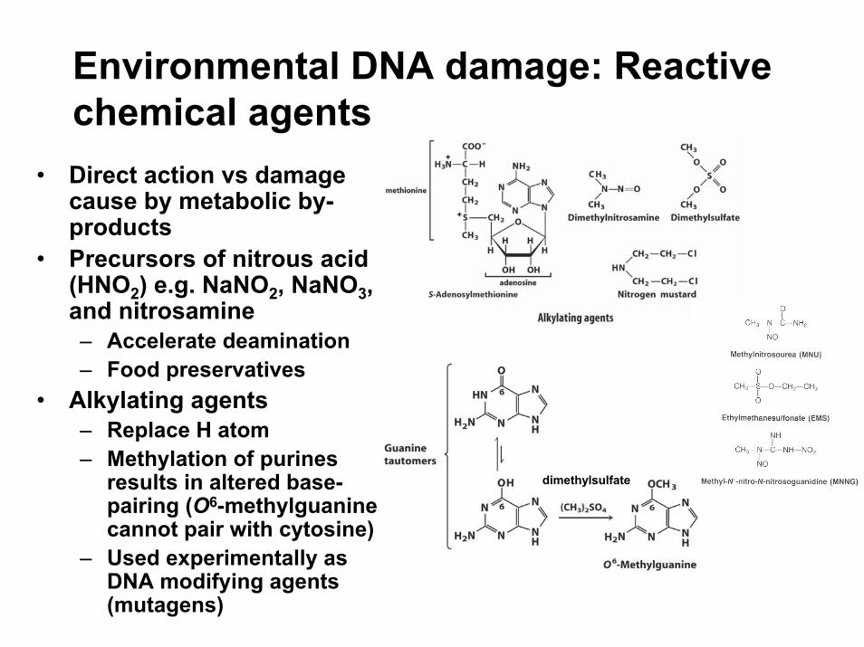
Environmental DNA damage: Reactive chemical agents
• Direct action vs damage cause by metabolic by-products
• Precursors of nitrous acid (HNO2) e.g. NaNO2, NaNO3, and nitrosamine– Accelerate deamination– Food preservatives
• Alkylating agents– Replace H atom– Methylation of purines
results in altered base-pairing (O6-methylguanine cannot pair with cytosine)
– Used experimentally as DNA modifying agents (mutagens)
dimethylsulfate

DNA repair systems I: Direct change of a modified base
• O6-Alkylguanine alkyl-transferase (methyltransferase)– Catalyzes transfer of alkyl
to Cys residue resulting in protein turnover
– Alkylated form self-regulates its own transcription
– Energetically costly- an entire protein consume to fix 1 base
• MutT nucleotide hydrolase– Accumulates in O2-stressed
cells (8-oxo-G can bp w/A)– Cleaves 8-oxo-dGTP prior to
incorporation in DNA• Photoreactivation
8
6
Fig. 25.11

Environmental DNA damage: Radiation and pyrimidine dimers
• One of the first forms of DNA damage discovered
• Irradiation of bacteria w/ 260 nm light → condensation of adjacent ethylene groups
• Human skin cells particularly susceptible
• Ionizing radiation (x-rays and gamma rays)– Ring opening and base
fragmentation– Breaks in DNA backbone
• UV + ionizing radiation exposure accounts ~10% of all DNA damage caused by environmental agents
lethal mutagenic AT→GC
200-400 nm near-UV
Lehninger Principles of Biochemistry, 4th ed., Ch 8

DNA repair systems I: Direct repair of thymine dimers by DNA photolyases
Fig. 25.10
56
• Enzyme (E. coli and yeast) uses a “photosynthesis-like” free-radical-dependent mechanism.
• Enzyme binds to lesion in the dark and breaks C5-C5 and C6-C6 bonds in the light.
• Enzyme contains two cofactor chromophores that absorb light at specific λs (photoantennaMTHFpolyGlu transmits light energy to FADH−) .
• Excited FADH• transfers an electron to the dimer. Electronic rearrangement restores thymine monomers.
• Enzyme is not found in mammalian cells.

DNA repair systems II: Base Excision Repair (BER)
• Damaged bases repaired – Products of deamination
(uracil), depurination, alkylation, and oxidation
– Thymine dimers (phage T4 specific mechanism)
• Four-step mechanism– Removal of damaged base via
specific DNA-N-glycosylase– Nicking of abasic strand by
AP endonuclease– Gap repair synthesis by DNA
polymerase I – Nick repair by DNA ligase
• AP endonucleases types– Cut 5′ to abasic site– Cut 3′ to abasic site
Lehninger Principles of Biochemistry, 4th ed., Ch 25

Base Excision Repair:Removal of 8-oxo-guanine by hOGG1 (glycosylase/β-lyase)
• OGG1 recognizes oxoG opposite C.
• Active site Lys of hOGG1 attacks the C1′ of deoxyribose resulting in the extrusion of oxoG.
• BER apparatus restores correct G/C base-pairing.
Hoogsteen mode
Aminal intermediate Schiff base
ε
rearrangement
DNA backbone cleavage
From Bruner, S. D. et al. Nature 403: 859 (2000)
(Example of a modified base-specific DNA glycosylase)

Molecular basis of oxoG recognition(What’s peculiar about these structures?)
From Bruner, S. D. et al. Nature 403: 859 (2000)
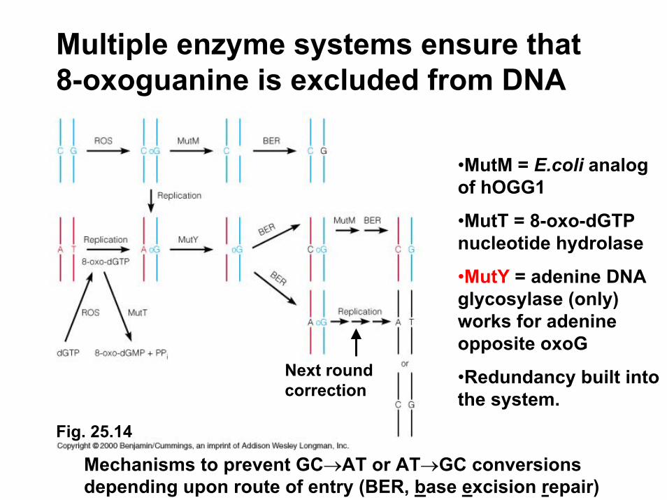
Multiple enzyme systems ensure that 8-oxoguanine is excluded from DNA
Mechanisms to prevent GC→AT or AT→GC conversions depending upon route of entry (BER, base excision repair)
•MutM = E.coli analog of hOGG1
•MutT = 8-oxo-dGTP nucleotide hydrolase
•MutY = adenine DNA glycosylase (only) works for adenine opposite oxoG
•Redundancy built into the system.
Next round correction
Fig. 25.14

DNA repair systems III: Nucleotide Excision Repair (NER)
• Typically occurs with bulky lesions that distort the DNA helix– pyrimidine dimers not
removed by BER– Intra-strand G crosslinks
(cisplatin-induced)– alkylation
• E. coli machinery (similar in yeast and mammals)– UvrA, B, C excinuclease
(catalyzes two specific endonucleolytic cleavages)
– UvrD helicase II– DNA polymerase and DNA
ligase
c) Monofunctional adducts ~ 2%
a) Intra-strand crosslink ~ 90%
b) Inter-strand crosslink ~ 5%
http://www.md.huji.ac.il/courses/bioorganic/cisplatin_3.ppt
Cisplatin = cis-diaminedichloroplatinum (cancer chemotherapeutic agent but highly toxic, only short-term efficacy)

Cisplatin-DNA adducts
Note how guanines become de-stacked.
H-bonding between N7 of G & cisplatin
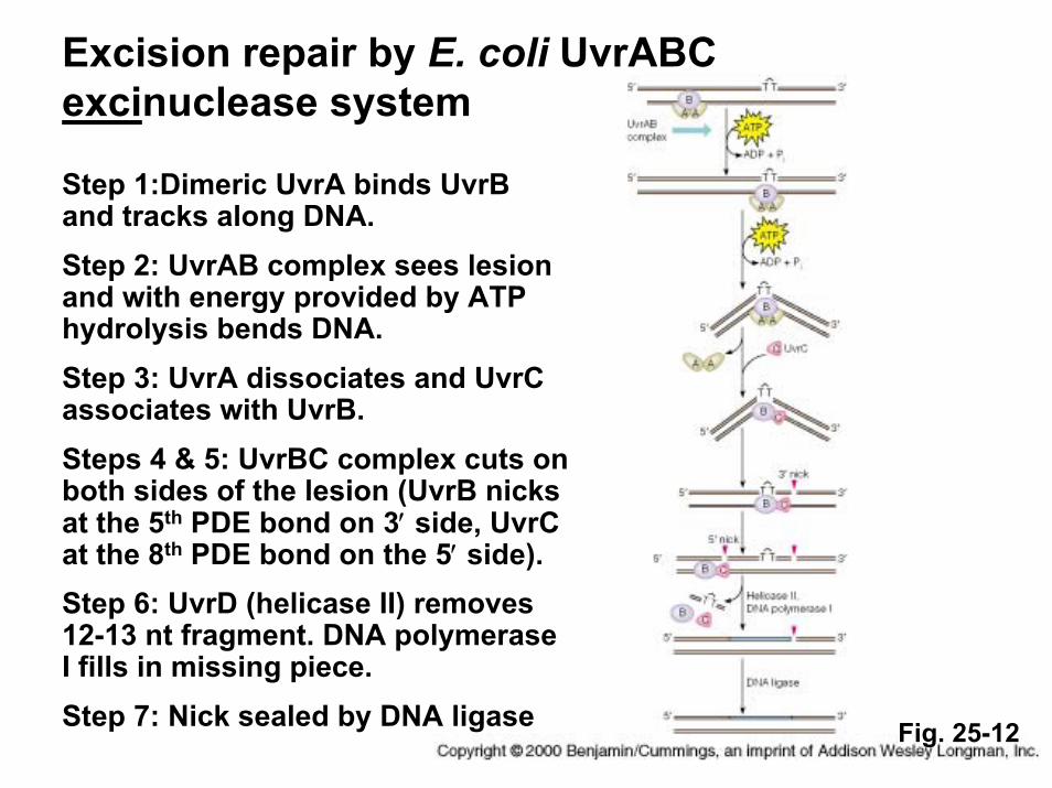
Step 1:Dimeric UvrA binds UvrB and tracks along DNA.Step 2: UvrAB complex sees lesion and with energy provided by ATP hydrolysis bends DNA.Step 3: UvrA dissociates and UvrC associates with UvrB.Steps 4 & 5: UvrBC complex cuts on both sides of the lesion (UvrB nicks at the 5th PDE bond on 3′ side, UvrC at the 8th PDE bond on the 5′ side).Step 6: UvrD (helicase II) removes 12-13 nt fragment. DNA polymerase I fills in missing piece.Step 7: Nick sealed by DNA ligase
Excision repair by E. coli UvrABC excinuclease system
Fig. 25-12

Similarity of NER repair in humans• Human excinuclease
– 16 polypeptides: no homology to UvrABC subunits
– NER sole repair pathway for pyrimidine dimers in humans
• Xeroderma pigmentosum (XP)– Deficiency in one or more XP
NER proteins (XPA→XPG) – Extreme sensitivity to light and
high incidence of skin cancers– Neurological abnormalities:
Due to high rate of oxidative metabolism in neurons.
• Translesional bypass– DNA polymerase η (eta): inserts
two A residues opposite T-T– XP-variant (XP-V) afflicted
people lack Pol η function
Lehninger Principles of Biochemistry, 4th ed., Ch 25

Site-specific DNA methylation….
• Bacteria– Restriction & modification– Mismatch error correction
• Eukaryotes– Tissue-specific inactivation of
genes during development – Suppression of transposon
migration– Formation of Z-DNA
• Occurrence– E. coli: N6 of adenine (major),
N4 of cytosine (minor), GATC restriction sites
– Eukaryotes: 3-5% of cytosines (C5), mainly in CpG islands (animal cells and higher plants, absent in insects)

DNA repair systems IV: Mismatch Repair• Corrects mismatched bases
arising from….– Replication errors– Deamination of 5-mC to T– Nonhomologous recombination – Specificity G-T > others > C-C
• Strand recognition system E. coli-specific– Hemi-methylation – Certain mismatch repair factors
use DNA methylation to identify the newly replicated strand with mismatched nucleotide.
• Enzymatic Components– MutS, MutL (motor proteins)
MutH (endonuclease), – MutU or DNA helicase II (also
known as UrvD)– DNA polymerase III, DNA ligase
Lehninger Principles of Biochemistry, 4th ed., Ch 25
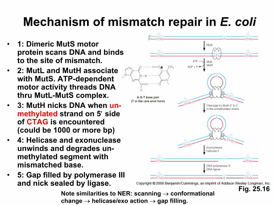
Mechanism of mismatch repair in E. coli• 1: Dimeric MutS motor
protein scans DNA and binds to the site of mismatch.
• 2: MutL and MutH associate with MutS. ATP-dependent motor activity threads DNA thru MutL-MutS complex.
• 3: MutH nicks DNA when un-methylated strand on 5′ side of CTAG is encountered (could be 1000 or more bp)
• 4: Helicase and exonuclease unwinds and degrades un-methylated segment with mismatched base.
• 5: Gap filled by polymerase III and nick sealed by ligase.
Fig. 25.16
4
Note similarities to NER: scanning → conformationalchange → helicase/exo action → gap filling.
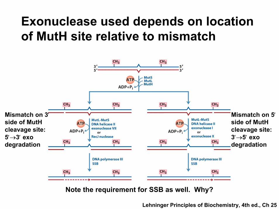
Exonuclease used depends on location of MutH site relative to mismatch
Note the requirement for SSB as well. Why?
Mismatch on 5′side of MutHcleavage site: 3′→5′ exo degradation
Mismatch on 3′side of MutHcleavage site: 5′→3′ exo degradation
Lehninger Principles of Biochemistry, 4th ed., Ch 25

Mismatch repair in eukaryotes
• Recognition mechanism does not seem to involve methylation and/or GATC sequences (no MutH)
• Different MutS homologs (MSH2, MSH3, MSH6)– Heterodimerize with different mismatch specificities– MSH2/MSH6 (single-base mismatches, bind less well to
longer mispaired loops)– MSH2/MSH3 (longer mismatches: 2 to 6 bp)
• MutL homologs – Heterodimer of MLH1 and PMS1 (post meiotic
segregation)• Mutations in mismatch repair genes confer
increased cancer risk– hMLH1 and hMSH2 mutations most prevalent– Early onset HNPCC (hereditary nonpolyposis colon
cancer)

Summary• DNA damage induces base modifications that are
mutagenic due to effects on base-pairing.• Direct repair: Reverses modification w/o excision
and resynthesis of base or nucleotide.• Base excision: Removes & replaces individual
nucleotides: diverse and specific• Nucleotide excision repair: Removes larger and
bulkier lesions.• Mismatch repair: Corrects errors in replication
through MutSLH complex.• Methylation: Required for genome protection
(prokaryotes) and gene regulation (eukaryotes).


