DNA Tetrahedron Delivery EnhancesDoxorubicin-Induced Apoptosis of HT-29Colon Cancer CellsGuiyu Zhang1, Zhiyong Zhang2 and Junen Yang3*
Abstract
As a nano-sized drug carrier with the advantage of modifiability and proper biocompatibility, DNA tetrahedron(DNA tetra) delivery is hopeful to enhance the inhibitory efficiency of nontargeted anticancer drugs. In this investigation,doxorubicin (Dox) was assembled to a folic acid-modified DNA tetra via click chemistry to prepare a targeted antitumoragent. Cellular uptake efficiency was measured via fluorescent imaging. Cytotoxicity, inhibition efficiency, andcorresponding mechanism on colon cancer cell line HT-29 were evaluated by MTT assay, cell proliferationcurve, western blot, and flow cytometry. No cytotoxicity was induced by DNA tetra, but the cellular uptakeratio increased obviously resulting from the DNA tetra-facilitated penetration through cellular membrane.Accordingly, folic acid-DNA tetra-Dox markedly increased the antitumor efficiency with increased apoptosislevels. In details, 100 μM was the effective concentration and a 6-h incubation period was needed forapoptosis induction. In conclusion, nano-sized DNA tetrahedron was a safe and effective delivery system forDox and correspondingly enhanced the anticancer efficiency.
Keywords: DNA tetrahedron, Doxorubicin, Folic acid, Apoptosis, Colon cancer
BackgroundDoxorubicin (Dox) is one of the most widely used anti-neoplastic agent, and numerous clinical studies demon-strated that Dox can strikingly hinder the growth oftumor cells in various cellular growth cycles by inhibit-ing the synthesis of RNA and DNA [1, 2]. Previous stud-ies suggested that the proliferation of tumor cells wereeffectively blocked in G1 phase, and the metastasis wasalso inhibited by Dox at a certain concentration [3]. Inaddition, effective inhibition can be achieved in a rela-tively smaller dose when compared with other antican-cer drugs [4]. However, Dox usually induced side effectsresulted from the lack of specific targeting for tumorcells and nonselective inhibition of DNA and RNA,which seriously limited the clinical applications [5, 6].Meanwhile, the low cellular intake capacity reduces theaccumulation of Dox in tumor cells [7]. Therefore, an ef-ficient delivery system for Dox should be developed to
make Dox more specific and effective targeting, moreeasily to be encapsulated, and of an excellent intake cap-acity and bio-compatibility.Nano-sized drug carriers, such as liposome and inorganic
nanoparticles, may facilitate Dox penetrating the tumor cellmembrane, improving the targeting efficiency [8]. Nonethe-less, liposome-based delivery cannot reduce the side effectsto normal cells because of the relatively poor targeting;meanwhile, inorganic carriers, such as mesoporous silicananoparticles, cannot be completely biodegraded in vivo,hampering the process of further drug uptake and bringingpotential bio-toxicity. For these functional delivery systems,the complexity of preparation, inhomogenization of nano-particles structures, and low encapsulation efficiencyobstacle the clinical expansions [9–11]. As a nano-sizeddrug carrier with the excellent performance on drug deliv-ery, DNA-based structures, such as DNA tetrahedron(DNA tetra), may penetrate the membrane via avoiding theincompatibility between electro-negative DNA and plasmamembrane [12–17]. Drugs and targeting molecules can beboth covalently attached to DNA tetrahedron. Furthermore,easy absorption and biodegradation of DNA tetrahedral
* Correspondence: [email protected] of Cardiology, Tangshan Gongren Hospital, Tangshan, Hebei063000, ChinaFull list of author information is available at the end of the article
sequences avoid long-time retention. Guanine-rich aptamerdrugs, such as AS1411, have been successfully deliveredinto A549 tumor cells and performed as targeting agentsand inhibitors [18]. Immune regulatory factors, such asCpG and siRNA, can also be delivered to tumor cells viaDNA tetra carrier to regulate immune responses [19]. Withthe advantages of low-toxicity, proper biocompatibility, andadjustable targeting, DNA tetra showed bio-safety and po-tentials for Dox delivery.In this study, functional particles of DNA tetrahedron
assembled with Dox as anticancer drug, and folic acid asspecific recognition molecules [20], were designed, syn-thesized, and characterized. The anticancer efficiencywas evaluated on colon cancer cells considering signifi-cantly upregulated folate receptors on the surface of cellmembrane. Specifically, the level of cellular uptake, thedegree of Dox-induced apoptosis, and the inhibition oncellular proliferation were measured on HT-29 cell lines.
MethodsRegents and EquipmentDNA oligonucleotide chains were purchased fromTAKARA in Dalian, China. Dulbecco’s modified Eagle’smedium (DMEM) and fetal bovine serum were pur-chased from Gibco in NY, USA. Penicillin and strepto-mycin were purchased from Beyotime biotechnology inShanghai, China. Folic acid, doxorubicin, 3-(4,5-di-methyl-2-thiazolyl)-2,5-diphenyl-2-H-tetrazolium brom-ide (MTT), and agarose were purchased from Sigma-Aldrich in MO, USA. All the antibodies were purchasedfrom Abcam Company in Shanghai, China. Other re-agents were purchased from Sinopharm Chemical Re-gent Co., Ltd., in Shanghai, China.UV-Vis spectrophotometer (Thermo Evolution 201)
and constant temperature incubator were purchasedfrom Thermo Fisher in the USA; centrifugal machine(GT10-1) was purchased from Beijing Era BeiliCentrifuge Co., Ltd.; fluorescence spectrophotometer(UV-1800) was purchased from Shimadzu Corpor-ation. Confocal laser scanning microscopy (Visitech)was purchased from Leica companies; polymerasechain reaction instrument (PCR, T100), protein elec-trophoresis, and nucleic acid electrophoresis apparatuswere purchased from Bio-Rad Company; dynamiclight particle size analyzer was purchased from Beck-man Company; cell culture plates with 96-well or 24-well were purchased from Dow Corning Corporation.High-performance liquid chromatography (HPLC,Agilent 1200) with C18 column was purchased fromAgilent Technologies.The human colon cancer cell line HT-29 was main-
tained in DMEM supplemented with 10% fetal bovineserum, 100 U/mL penicillin, and 100 μg/mL streptomycin
in a humidified atmosphere containing 95% air and 5%carbon dioxide at 37 °C.
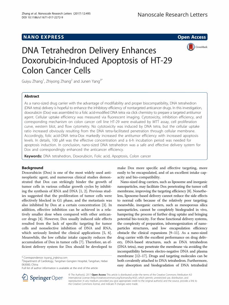
Synthesis and Purification of DNA TetrahedronIn this investigation, synthesis followed the schematicprocedures in Fig. 1, and the single-strand DNA(ssDNA) sequences of DNA tetrahedron are provided inFig. 1 as well. In details, each ssDNA was dissolved in0.5× TE buffer, and the corresponding optical density(OD) value of DNA was determined by UV spectropho-tometer at 260 nm. Additional TE buffer was supple-mented to make four chains at the same concentration.The mixing ratio of four ssDNAs was 1:1:1:1 at 1 μM in100 μL. The reaction was performed in a polymerasechain reaction (PCR) machine with the cycling condi-tions: 95 °C, 10 min, naturally cooled to 4 °C. All of thesingle-strand DNA were purified by HPLC with 260 nmas characteristic absorption peak. In HPLC spectrum,the peak time of DNA tetra was faster than that of singlestrand, and product was collected at the correspondingtime point.
Synthesis and Characterization of Folic Acid-DNA tetra-DoxFree hydrogen groups of Dox and folic acid were modi-fied with azide group and then coupled with 3′-OH ofssDNA via click chemistry reaction [21]. When adding adifferent amount of functional group tagged ssDNA, theratio of functional groups could be stoichiometricallycontrolled via specific hybridization of side chains. Forsynthesis of folic acid-DNA tetra, the molar ratio of folicacid and DNA tetrahedron was set as 1:1. For synthesisof DNA tetra-Dox, the molar ratio of Dox and DNAtetrahedron was set as 4:1. For synthesis of folic acid-DNA tetra-Dox, the molar ratio of folic acid, DNA tetra-hedron, and Dox was respectively set as 1:1:3. All thesynthesis were carried out at micromolar level at 37 °Cand then stored at 4 °C [22].In this study, the series of DNA tetra complexes was
characterized by polyacrylamide gel electrophoresis(PAGE) with 8% separation gel (39% Acr-Bis) to checkthe purity and relative molecular sizes. The samples con-sisted of the different DNA tetra structures and 6× load-ing buffer with mixing ratio of 2:1. The samples werestained and analyzed after gel electrophoresis for 90 minat 110 V. To figure out the differences on particle sizes,DNA tetra samples were also scanned by dynamic lightscattering (DLS) instruments with dynamic light.
DNA tetra-Facilitated Cellular UptakeFor the different coupling structures designed in thisresearch, the uptake rates by HT-29 cells were comparedto figure out the drug delivery efficiency. Cellular uptakeefficiency was evaluated and quantified utilizing thecharacteristic fluorescence spectrum of Dox, i.e., excitation
Zhang et al. Nanoscale Research Letters (2017) 12:495 Page 2 of 7
light at 470 nm and emission light at 590 nm. The HT-29cells at 2 × 10⁵/mL were seeded in 24-well plates andcultured for 24 h. Cells were incubated with various DNAtetra structures at 10 μM for another 24 h. The mediumwas then discarded, and the cells were rinsed with PBSthree times. For cell fixation, 4% paraformaldehyde wasimmediately added at room temperature for a 30-minco-incubation, and the cells were rinsed with PBS threetimes again. At last, the 24-well plates were observed bylaser confocal microscope to compare the cellular uptakeefficiency based on the light intensity of emission light.
Cytotoxicity and Anticancer EfficiencyCytotoxicity and anticancer efficiency were evaluatedusing MTT assay, where a redox reaction occursbetween the MTT in DMSO and intracellular succinatedehydrogenase. HT-29 cells were seeded in 96-wellculture plates and cultured for 24 h.For the cytotoxicity of DNA tetra as drug carrier,
medium containing DNA tetra structures at the concen-tration of 0–100 μM was added for another 24- or 48-hincubation. Then, 100 μL of MTT solution (5 mg/mL)was added to each well, and the mixture was incubatedat 37 °C for 4 h. The liquid was then removed, and thecells were lysed and dissolved with 200 μL DMSO. Theabsorbance of supernatant was measured at 570 nm byMicroreader. The non-treated HT-29 sample was deemedas control group.For the DNA tetrahedron coupled with folic acid or Dox,
on the one side, the research focused on the stability,
bio-safety, and inhibition efficiency; on the other side,whether the Dox of DNA tetra-Dox has comparable antitu-mor properties as before or not was also explored.Therefore, cytotoxicity and antitumor efficiency of DNAtetra complexes were evaluated. Complexes respectively at100 μM were incubated with HT-29 cells. Cell sampleswere collected every 6 h and then detected by MTT assayto evaluate the effect of incubation period. Particularattentions were paid for the difference on anticancerefficiency resulted from the variations of structures.Furthermore, concentration effects on inhibition of HT-29after being treated with folic acid-DNA tetra-Dox wereevaluated at 0–200 μM via MTTassays.
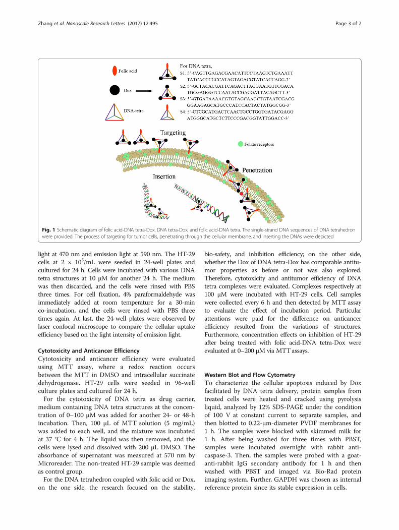
Western Blot and Flow CytometryTo characterize the cellular apoptosis induced by Doxfacilitated by DNA tetra delivery, protein samples fromtreated cells were heated and cracked using pyrolysisliquid, analyzed by 12% SDS-PAGE under the conditionof 100 V at constant current to separate samples, andthen blotted to 0.22-μm-diameter PVDF membranes for1 h. The samples were blocked with skimmed milk for1 h. After being washed for three times with PBST,samples were incubated overnight with rabbit anti-caspase-3. Then, the samples were probed with a goat-anti-rabbit IgG secondary antibody for 1 h and thenwashed with PBST and imaged via Bio-Rad proteinimaging system. Further, GAPDH was chosen as internalreference protein since its stable expression in cells.
Fig. 1 Schematic diagram of folic acid-DNA tetra-Dox, DNA tetra-Dox, and folic acid-DNA tetra. The single-strand DNA sequences of DNA tetrahedronwere provided. The process of targeting for tumor cells, penetrating through the cellular membrane, and inserting the DNAs were depicted
Zhang et al. Nanoscale Research Letters (2017) 12:495 Page 3 of 7
Flow cytometry was further utilized to quantify thelevels of apoptosis at the selected concentration andtime point via MTT assays. HT-29 cells were incubatedwith inhibitors for a period of time and then measuredvia quantitative flow cytometry using Annexin V-PIdouble staining method.
Cell Proliferation QuantifyingCell counting method was used to quantify the cell pro-liferation with time. In details, after being treated withfolic acid-DNA tetra-Dox for a period of time at100 μM, cells were digested by 0.25% trypsin for 30 s,and then, the equal volume of complete medium wasadded to terminate reaction. Supernatant was discardedafter centrifugation in 1000 rpm for 3 min, and cellswere re-suspended with complete medium. The numberof cells in each detection point was recorded underoptical microscope using cell count plates, so as to drawthe cellular proliferation curve.
Statistics AnalysisIn this research, significances of differences were deter-mined using Student’s t test (two-tailed; two-sampleequal variance). P < 0.05 means significant differencesbetween different groups.
ResultsPreparation of Folic Acid-DNA tetra-DoxIn specific synthesis process, Dox was mixed with DNAtetrahedron in different proportions to complete loadingand assembling of Dox (wm = 543.52) and folic acid(wm = 441.4). As shown in Fig. 2a, due to that Dox andfolic acid are monomers with relative low and similarmolecular weight, DNA in a tetrahedron and the conju-gates with one functional molecule had roughly equiva-lent molecular weights. However, Dox or folic acidassembling with all the four DNA vertices of the tetrahe-dron made molecular size of DNA tetra increased obvi-ously. As shown in Fig. 2b, most of DNA tetrahedral
monomer size was less than 15 nm. With the increase ofcoupling groups, the symmetrical structure of DNA tetrawas destroyed and the medium of particle sizes ofcompounds increased significantly. In particular, sizeexpansion of folic acid-DNA tetra-Dox extended themedium of diameter to 20 nm.
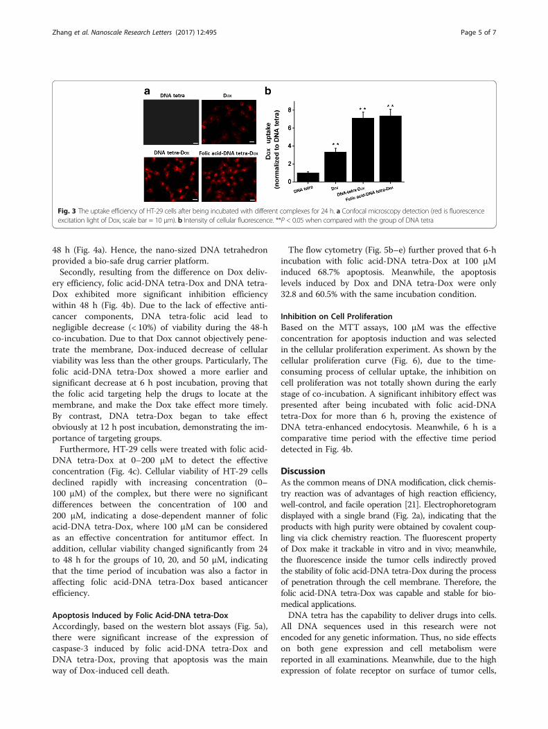
Drug Delivery EfficiencyAfter being incubated with different DNA tetra struc-tures for 24 h, the intracellular efficiency of Dox wasevaluated with fluorescence imaging, where the intracel-lular red fluorescence is the characteristic fluorescenceof Dox; hence, cells incubated with DNA tetra did notexhibit any fluorescent signal as shown in Fig. 3a, andthe red signals of cells incubated with folic acid-DNAtetra-Dox and DNA tetra-Dox complexes were obviouslyhigher than that with Dox, meaning the significantlyenhanced cellular uptake efficiency of Dox resultingfrom the DNA tetra facilitated penetration through themembrane, as well as the intracellular stability of theconjugates between Dox and DNA tetra.The further quantitative analysis of fluorescence inten-
sity (Fig. 3b) evidenced the visual findings, and therewas no significant difference between folic acid-DNAtetra-Dox and DNA tetra-Dox complexes (P < 0.05).The methodology of co-incubation of drugs and cellsobstacles the complete exhibition of targeting ability offolic acid. The relative high concentration confirmed thespecific recognition of folate receptors, which is theoret-ically more obvious in the in vivo applications with thecomplex circulating system. In brief, the DNA tetradelivery guaranteed the anticancer efficiency of Dox.
Cytotoxicity and Anticancer EfficiencyFirst, the viability of HT-29 cells co-incubated with dif-ferent concentrations of DNA tetra were examined usingMTT assay. There were no obvious cytotoxicity of DNAtetra treatment in HT-29 cells at 0–100 μM for 24 and
Fig. 2 Characterization of DNA tetra with different constituents. a Left to right: DNA ladder, DNA tetra, folic acid-DNA tetra, folic acid-DNA tetra-Dox, andDNA tetra-Dox imaged in 8% SDS-PAGE. b The size distribution of DNA tetra, folic acid-DNA tetra, folic acid-DNA tetra-Dox, and DNA tetra-Dox detectedvia dynamic light scattering
Zhang et al. Nanoscale Research Letters (2017) 12:495 Page 4 of 7
48 h (Fig. 4a). Hence, the nano-sized DNA tetrahedronprovided a bio-safe drug carrier platform.Secondly, resulting from the difference on Dox deliv-
ery efficiency, folic acid-DNA tetra-Dox and DNA tetra-Dox exhibited more significant inhibition efficiencywithin 48 h (Fig. 4b). Due to the lack of effective anti-cancer components, DNA tetra-folic acid lead tonegligible decrease (< 10%) of viability during the 48-hco-incubation. Due to that Dox cannot objectively pene-trate the membrane, Dox-induced decrease of cellularviability was less than the other groups. Particularly, Thefolic acid-DNA tetra-Dox showed a more earlier andsignificant decrease at 6 h post incubation, proving thatthe folic acid targeting help the drugs to locate at themembrane, and make the Dox take effect more timely.By contrast, DNA tetra-Dox began to take effectobviously at 12 h post incubation, demonstrating the im-portance of targeting groups.Furthermore, HT-29 cells were treated with folic acid-
DNA tetra-Dox at 0–200 μM to detect the effectiveconcentration (Fig. 4c). Cellular viability of HT-29 cellsdeclined rapidly with increasing concentration (0–100 μM) of the complex, but there were no significantdifferences between the concentration of 100 and200 μM, indicating a dose-dependent manner of folicacid-DNA tetra-Dox, where 100 μM can be consideredas an effective concentration for antitumor effect. Inaddition, cellular viability changed significantly from 24to 48 h for the groups of 10, 20, and 50 μM, indicatingthat the time period of incubation was also a factor inaffecting folic acid-DNA tetra-Dox based anticancerefficiency.
Apoptosis Induced by Folic Acid-DNA tetra-DoxAccordingly, based on the western blot assays (Fig. 5a),there were significant increase of the expression ofcaspase-3 induced by folic acid-DNA tetra-Dox andDNA tetra-Dox, proving that apoptosis was the mainway of Dox-induced cell death.
The flow cytometry (Fig. 5b–e) further proved that 6-hincubation with folic acid-DNA tetra-Dox at 100 μMinduced 68.7% apoptosis. Meanwhile, the apoptosislevels induced by Dox and DNA tetra-Dox were only32.8 and 60.5% with the same incubation condition.
Inhibition on Cell ProliferationBased on the MTT assays, 100 μM was the effectiveconcentration for apoptosis induction and was selectedin the cellular proliferation experiment. As shown by thecellular proliferation curve (Fig. 6), due to the time-consuming process of cellular uptake, the inhibition oncell proliferation was not totally shown during the earlystage of co-incubation. A significant inhibitory effect waspresented after being incubated with folic acid-DNAtetra-Dox for more than 6 h, proving the existence ofDNA tetra-enhanced endocytosis. Meanwhile, 6 h is acomparative time period with the effective time perioddetected in Fig. 4b.
DiscussionAs the common means of DNA modification, click chemis-try reaction was of advantages of high reaction efficiency,well-control, and facile operation [21]. Electrophoretogramdisplayed with a single brand (Fig. 2a), indicating that theproducts with high purity were obtained by covalent coup-ling via click chemistry reaction. The fluorescent propertyof Dox make it trackable in vitro and in vivo; meanwhile,the fluorescence inside the tumor cells indirectly provedthe stability of folic acid-DNA tetra-Dox during the processof penetration through the cell membrane. Therefore, thefolic acid-DNA tetra-Dox was capable and stable for bio-medical applications.DNA tetra has the capability to deliver drugs into cells.
All DNA sequences used in this research were notencoded for any genetic information. Thus, no side effectson both gene expression and cell metabolism werereported in all examinations. Meanwhile, due to the highexpression of folate receptor on surface of tumor cells,
Fig. 3 The uptake efficiency of HT-29 cells after being incubated with different complexes for 24 h. a Confocal microscopy detection (red is fluorescenceexcitation light of Dox, scale bar = 10 μm). b Intensity of cellular fluorescence. **P < 0.05 when compared with the group of DNA tetra
Zhang et al. Nanoscale Research Letters (2017) 12:495 Page 5 of 7
folic acid is selected as specific targeting molecules of thedrug delivery system to enhance the uptake efficiency ofDNA tetra complexes. However, with the circumstancesof DNA tetra at relatively high concentration, the advan-tage of folate receptor targeting was not fully reflected
Fig. 4 The cytotoxicity and antitumor efficiency of Dox complexes andDNA tetra for HT-29 cells. a The viability of HT-29 cells incubated withDNA tetra at different concentrations for 24 or 48 h. b The antitumorefficiency of Dox complexes at 100 μM. c The viability of HT-29 cellsincubated with folic acid-DNA tetra-Dox at different concentrations for24 or 48 h
Fig. 5 The cellular expression of apoptosis-related caspase-3 after beingtreated with different complexes (a) and the flow cytometry of HT-29cells after being incubated with DNA tetra, Dox, DNA tetra-Dox, and folicacid-DNA tetra-Dox (b–e)
Fig. 6 Inhibition of cell proliferation induced by the incubation withfolic acid-DNA tetra-Dox at 100 μM for different time periods
Zhang et al. Nanoscale Research Letters (2017) 12:495 Page 6 of 7
in vitro. Accordingly, for in vivo application, the folicacid was potential to enhance the targeting efficiency inthe complex circulating system.Currently, anticancer drugs commonly bring unex-
pected side effects, so the research on enhancing thetumor cells targeting ability and delivery efficiency hasbeen a hot topic [23–25]. Previous studies showed thatcapacity of DNA tetrahedron for carrying drugs wasbased on its good compatibility. DNA tetrahedron is anartificial container of favorable modification and properbiocompatibility. In this study, the successful coupling ofDox and folic acid with DNA tetrahedron achieved satis-fying inhibitory effect and lead to obvious apoptosis ofHT-29 cell. Owing to that DNA tetra can be modifiedand coupled with drugs and targeting molecules, theenrichment of local drug concentration in tumor cellwas realized. The above results proved the good recogni-tion for tumor cells and corresponding enhanced inhibi-tory effect via DNA tetra delivery, and this nano-sizeddrug delivery system can be used more extensively.
ConclusionsAs a newly developed drug delivery strategy, nano-sizedDNA structures are of low cost, high stability, and feasi-bility to synthesize; meantime, it is bio-safe due to thelack of exogenous immune activity. DNA tetrahedroncoupling strategy facilitates the targeted delivery of Dox,enhances the anticancer efficiency of Dox on colon can-cer cells, and provides a promising inspiration and ideafor drug design.
FundingThe authors gratefully acknowledge the financial supports provided by HebeiProvince Science and Technology Support Program (CN) (no. 142777101D).
Authors’ ContributionsGZ and ZZ designed the research. GZ, ZZ, and JY performed theexperiments and statistics. All the authors read and approved themanuscript.
Competing InterestsThe authors declare that they have no competing interests.
Publisher’s NoteSpringer Nature remains neutral with regard to jurisdictional claims inpublished maps and institutional affiliations.
Author details1Department of Infectious Diseases, Tangshan Gongren Hospital, Tangshan,Hebei 063000, China. 2Department of Pathology, Tangshan GongrenHospital, Tangshan, Hebei 063000, China. 3Department of Cardiology,Tangshan Gongren Hospital, Tangshan, Hebei 063000, China.
Received: 3 May 2017 Accepted: 10 August 2017
References1. Litwiniec A, Grzanka A, Helmin-Basa A, Gackowska L, Grzanka D (2010)
Features of senescence and cell death induced by doxorubicin in A549cells: organization and level of selected cytoskeletal proteins. J Cancer ResClin Oncol 136:717–736
2. Wang Y, Wei X, Zhang C, Zhang F, Liang W (2010) Nanoparticle deliverystrategies to target doxorubicin to tumor cells and reduce side effects. TherDeliv 1:273–287
3. Idani H, Matsuoka J, Yasuda T, Kobayashi K, Tanaka N (2000) Intra-tumoralinjection of doxorubicin (adriamycin) encapsulated in liposome inhibitstumor growth, prolongs survival time and is not associated with local orsystemic side effects. Int J Cancer 88:645–651
4. Hardman WE, Moyer MP, Cameron IL (2000) Dietary fish oil sensitizes A549lung xenografts to doxorubicin chemotherapy. Cancer Lett 151:145–151
5. Chen Y, Wan Y, Wang Y, Zhang H, Jiao Z (2011) Anticancer efficacyenhancement and attenuation of side effects of doxorubicin with titaniumdioxide nanoparticles. Int J Nanomedicine 6:2321–2326
6. Filyak Y, FilyakO SS, Stoika R (2008) Doxorubicin inhibits TGF-beta signalingin human lung carcinoma A549 cells. Eur J Pharmacol 590:67–73
7. Akbarzadeh A, Samiei M, Joo SW, Anzaby M, Hanifehpour Y, Nasrabadi HT etal (2012) Synthesis, characterization and in vitro studies of doxorubicin-loaded magnetic nanoparticles grafted to smart copolymers on A549 lungcancer cell line. J Nanobiotechnology 10:46
8. Shuai X, Ai H, Nasongkla N, Kim S, Gao J (2004) Micellar carriers based onblock copolymers of poly(epsilon-caprolactone) and poly(ethylene glycol)for doxorubicin delivery. J Control Release 98:415–426
9. Chen AM, Zhang M, Wei D, Stueber D, Taratula O, Minko T et al (2009) Co-delivery of doxorubicin and Bcl-2 siRNA by mesoporous silica nanoparticlesenhances the efficacy of chemotherapy in multidrug-resistant cancer cells.Small 23:2673–2677
10. Goren D, Horowitz AT, Tzemach D, Tarshish M, Zalipsky S, Gabizon A (2000)Nuclear delivery of doxorubicin via folate-targeted liposomes with bypass ofmultidrug-resistance efflux pump. Clin Cancer Res 6:1949–1957
11. Janes KA, Fresneau MP, Marazuela A, Fabra A, Alonso MJ (2001) Chitosannanoparticles as delivery systems for doxorubicin. J Control Release 73:255–267
12. Ke Y, Sharma J, Liu M, Jahn K, Liu Y, Yan H (2009) Scaffolded DNA origamiof a DNA tetrahedron molecular container. Nano Lett 9:2445–2447
13. Ozhalici-Unal H, Armitage BA (2009) Fluorescent DNA nanotags based on aself-assembled DNA tetrahedron. ACS Nano 3:425–433
14. Xia ZW, Wang P, Liu XW, Liu T, Yan YN, Yan J et al (2016) Tumor-penetrating peptide-modified DNA tetrahedron for targeting drug delivery.Biochemistry 55:1326–1331
15. Fu J, Yan H (2012) Controlled drug release by a nanorobot. Nat Nanotech30:407–408
16. Jiang Q, Song C, Nangreave J, Liu X, Lin L, Qiu D et al (2012) DNA origamias a carrier for circumvention of drug resistance. J Am Chem Soc 134:13396–13403
17. Ouyang X, Li J, Liu H, Zhao B, Yan J, Ma Y et al (2013) Rolling circleamplification-based DNA origami nanostructrures for intracellular delivery ofimmunostimulatory drugs. Small 9:3082–3087
18. Xu X, Zhao Y, Lu H, Fu C, Li X, Jiang L et al (2016) G4-tetra DNA duplexinduce lung cancer cell apoptosis in A549 cells. Nanoscale Res Lett 11:437
19. Kim H, Akagi T, Akashi M (2010) Preparation of CpG ODN-encapsulated anionicpoly (amino acid) nanoparticles for gene delivery. Chem Lett 39:278–279
20. Zheng B, Yang S, Wang M, Yang X, Teng L, Xie J et al (2015) Non-covalentnanocomplexes of folic acid and reducible polyethylenimine for survivinsiRNA delivery. Anticancer Res 35:5433–5441
21. AH EI-s, Brown T (2010) Click chemistry with DNA. Chem Soc Rev 39:1388–140522. Li J, Fan C, Pei H, Shi J, Huang Q (2013) Smart drug delivery nanocarriers
with self-assembled DNA nanostructures. Adv Mater 25:4386–439623. Maxwell PH, Wiesener MS, Chang GW, Clifford SC, Vaux EC, Cockman ME et
al (1999) The tumour suppressor protein VHL targets hypoxia-induciblefactors for oxygen-dependent proteolysis. Nature 399:271–275
24. Volinia S, Calin GA, Liu CG, Ambs S, Cimmino A, Petrocca F et al (2006) AmicroRNA expression signature of human solid tumors defines cancer genetargets. Proc Natl Acad Sci U S A 103:2257–2261
25. Wilhelm SM, Carter C, Tang L, Wilkie D, McNabola A, Rong H et al (2004)BAY 43-9006 exhibits broad spectrum oral antitumor activity and targets theRAF/MEK/ERK pathway and receptor tyrosine kinases involved in tumorprogression and angiogenesis. Cancer Res 64:7099–7109
Zhang et al. Nanoscale Research Letters (2017) 12:495 Page 7 of 7