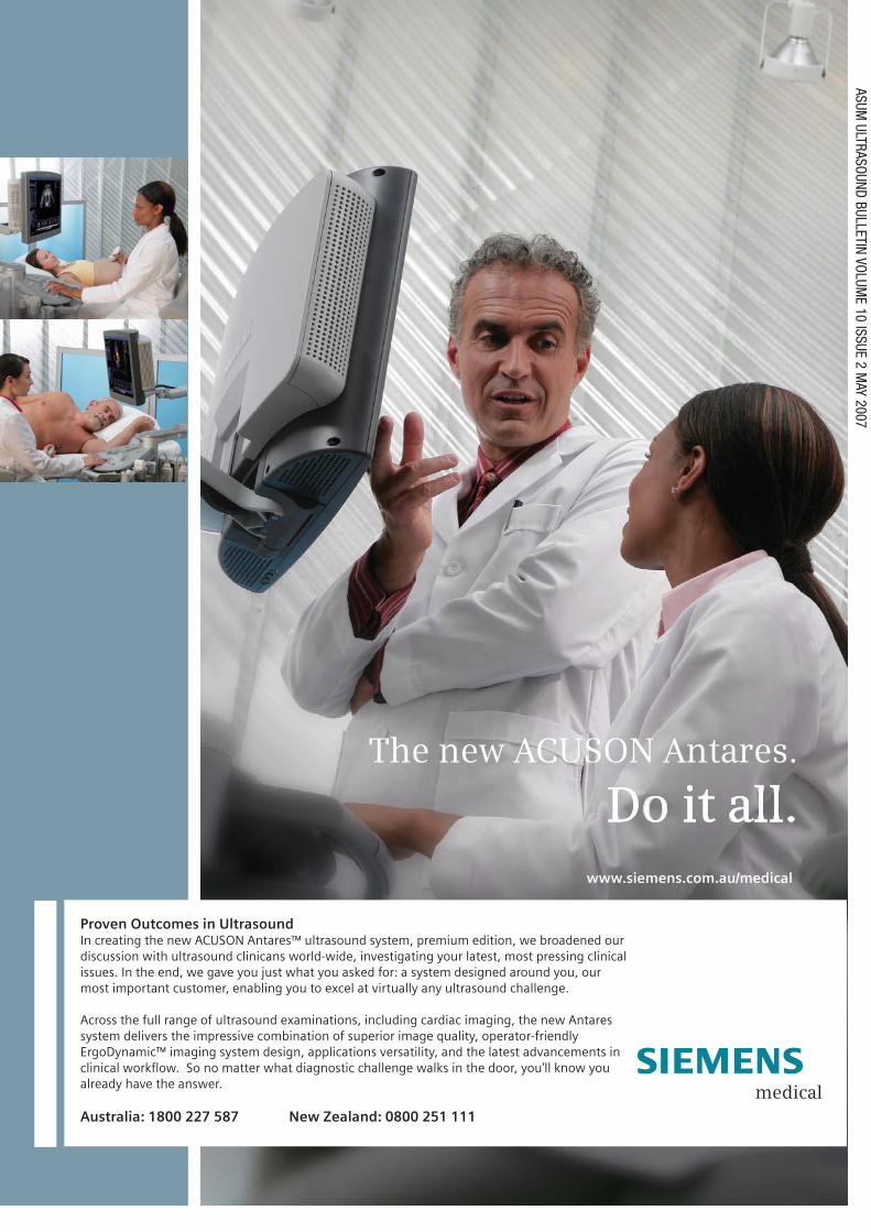
ASUM ULTRASOUND BULLETIN VOLUME 10 ISSUE 2 MAY 2007 The new ACUSON Antares. Do it all. Proven Outcomes in Ultrasound In creating the new ACUSON Antares™ ultrasound system, premium edition, we broadened our discussion with ultrasound clinicans world-wide, investigating your latest, most pressing clinical issues. In the end, we gave you just what you asked for: a system designed around you, our most important customer, enabling you to excel at virtually any ultrasound challenge. Across the full range of ultrasound examinations, including cardiac imaging, the new Antares system delivers the impressive combination of superior image quality, operator-friendly ErgoDynamic™ imaging system design, applications versatility, and the latest advancements in clinical workflow. So no matter what diagnostic challenge walks in the door, you'll know you already have the answer. Australia: 1800 227 587 New Zealand: 0800 251 111 www.siemens.com.au/medical
Transcript
ASUM ULTRASOUND BULLETIN VOLUM
E 10 ISSUE 2 MAY 2007
The new ACUSON Antares.
Do it all.
Proven Outcomes in UltrasoundIn creating the new ACUSON Antares™ ultrasound system, premium edition, we broadened our discussion with ultrasound clinicans world-wide, investigating your latest, most pressing clinical issues. In the end, we gave you just what you asked for: a system designed around you, our most important customer, enabling you to excel at virtually any ultrasound challenge.
Across the full range of ultrasound examinations, including cardiac imaging, the new Antares system delivers the impressive combination of superior image quality, operator-friendly ErgoDynamic™ imaging system design, applications versatility, and the latest advancements in clinical workflow. So no matter what diagnostic challenge walks in the door, you'll know you already have the answer.
Australia: 1800 227 587 New Zealand: 0800 251 111
www.siemens.com.au/medical
ISSN 1441-6891
Ultrasound Bulletin
ISSN 1441-6891Volume 10 Issue 2 May 2007
Journal of the Australasian Society for Ultrasound in Medicine
Every aspect of Aplio is designed to reduce operator fatigue and patient stress during long scanning sessions. From the highly customisable and movable panel to lightweight transducers, the Aplio is ergonomically designed to
adjust to the operator’s needs for comfort and convenience.
• Flexible control panel can be positioned according to patient posture and the examination procedure
• Programmable main panel, screen layout and touch-control screen menu and Quick Scan
• Programmable keys can be customised according to operator and clinical needs
• Highly portable with four-wheel swivel castors for remote studies
To find out more, contact Toshiba on 1300 655 155or email [email protected]
Australasian Society for Ultrasound in Medicine37th Annual Scientifi c Meeting
Proff ered Paper & Poster Abstract Submission DeadlineFriday, 11 May 2007
Proff ered Paper & Poster Abstract Notifi cationFriday, 22 June 2007
Early Bird Registration DeadlineFriday, 13 July 2007
Accommodation DeadlineMonday, 6 August 2007
1ASUM Ultrasound Bulletin 2007 May 10 (2)
President Dr Matthew AndrewsHonorary SecretaryMrs Roslyn Savage
Honorary TreasurerDr Andrew Ngu
Chief Executive OfficerDr Caroline Hong
ULTRASOUND BULLETINOfficial publication of the Australasian Society for Ultrasound in MedicinePublished quarterlyISSN 1441-6891Indexed by the Sociedad Iberoamericana de Informacion Cientifien (SIIC) DatabasesEditorProf Ron BenzieUniversity of Sydney, Division of Women's and Children's Health, Nepean Hospital, Penrith, NSW 2750Co-EditorMr Keith HendersonASUM Education Manager
Editorial CoordinatorMr James HamiltonASUM Education Officer
Assistant EditorsMs Kaye Griffiths AMANZAC Research CRGH Institute NSW
Ms Janine Horton Nanosonics NSW
Ms Louise LeeGold Coast Hospital QldMr Adam LunghiEcho Services WADr Amarendra TrivediFrankston Hospital VicEditorial contributionsOriginal research, case reports, quiz cases, short articles, meeting reports and calen-dar information are invited and should be addressed to The Editor at the address belowMembership and geneal enquiriesto ASUM at the address belowPublished on behalf of ASUMby Minnis CommunicationsBill Minnis Director4/16 Maple GroveToorak Melbourne Victoria 3142 Australiatel +61 3 9824 5241 fax +61 3 9824 5247email [email protected] specifically indicated, opinions expressed should not be taken as those of the Australasian Society for Ultrasound in Medicine or of Minnis CommunicationsAUSTRALASIAN SOCIETY FOR ULTRASOUND IN MEDICINEABN 64 001 679 161
A new method for ultrasound 24 evaluation of the biceps brachii tendon
A prenatal diagnosis of 30 holoprosencephaly using 2D and 3D ultrasound
A twin pregnancy with abnormal 33 fetus and complete hydatidiform mole: an evolving diagnosis
Cornelia de Lange syndrome: the 37 value of 3D and 4D ultrasound
BOOK REVIEWS
General Ultrasound in the Clinically Ill 39
Textbook of diagnostic Ultrasound
Handbook of Early Pregnancy Care
REPORTS
CADUCEUS exchange recipient 42 Morten Boesen's Australian experience
EDUCATION
Stephen Bird tours WA for the 44 Giulia Franco Teaching Fellowship
CCPU report 45
NOTICES
Corporate members 46
New members 46
Calendar 47
Guidelines for authors 48
Ultrasound Bulletin
Dr Matthew Andrews on the first CCPU recipient and recent ASUM activities
On the shoulders of giants
CEO reports on the move to new premises and ASUM's growing stature overseas
Opinion: George Condous questions whether ultrasound is properly used in the diagnosis of ectopic pregnancy
Peter Dietz reviews pelvic floor ultrasound and urges more general acceptance of its use as a standard diagnostic option in pelvic floor medicine
Tear of the distal biceps brachii tendon is a trau-matic event, this article investigates current diag-nostic imaging and suggests a better approach
A discussion of ultrasound in the diagnosis of holoprosencephaly
This article investigates the rare occurrence of a complete hydatidiform mole coexisting with a live fetus and discusses the combined use of ultrasound and b-hCG testing in its detection
The prize winning poster at the 2006 ASUM ASM in Melbourne investigates the value of 3D and 4D ultrasound in the identification of limb abnormalities in the fetus
Ultrasound professionals review the latest books on the subject
The CADUCEUS exchange program continues with the latest Danish visitor to Australia
WA is treated to the Giulia Franco Teaching Fellow's ultrasound expertise
Details of the certificate in clinician performed ultrasound courses both basic and advanced for 2007
Thursday, 13 September 2007
Skills Development Workshop
GE Healthcare Room Toshiba Room Philips Room Siemens Room
9.00 am–9.50 am Mrs Lynette Hassall – Breast Ultrasound
Mrs Shirley Comninos – 18 Week Scan
Dr Joseph Polak – Vascular Scanning, The Challenges
10.00 am–10.40 am Dr Tom Stavros – Breast Implants Dr Alison Lee Tannock – Fetal Heart Ultrasound
Ms Deb Coghlan – CVI Scanning
10.40 am–11.10 am Morning break
11.10 am–12.00 pm Dr Susane Fraser – Breast Cytology and Basic Breast Pathology
Mrs Yvonne Butcher – Unusual Carotid Pathology
Dr David Nyberg – Tertiary Scanning
12.00 pm–1.00 pm Lunch
1.00 pm–1.50 pm Dr Tom Stavros – Ultrasound of the Groin
Ms Deb Coghlan – Arm Arteries Dr David Nyberg – 11–12 Week Scan
2.00 pm–2.40 pm Dr Alison Lee Tannock – Fetal Heart Ultrasound
Ms Deb Coghlan – Arm Veins Dr Carlo Martinoli – Wrist and Hands
2.40 pm–3.10 pm Afternoon break
3.10 pm–3.50 pm Dr Carlo Martinoli – Post Operative Shoulder
Dr David Nyberg – Tertiary Scanning
Mrs Yvonne Butcher – Tips for Difficult DVT Scans
4.00 pm–5.00 pm Mr Richard Allan – Appendix Mrs Yvonne Butcher – Renal Artery Doppler Ultrasound, The Challenges
Mrs Shirley Comninos – 18 Week Scan
Draft Program Cairns
Friday, 14 September 2007
Plenary Session
9.00 am–10.00 am Dr Julie Campbell – Tissue Engineered Vascular and Urogenital Grafts
10.00 am–10.30 am Professor Torben Lorentzen – Liver Abscesses, Imaging and Treatment Guided By Ultrasound
10.30 am–11.00 am Morning Tea – Exhibition
Concurrent Sessions Vascular Breast and Small Parts Musculoskeletal
11.00 am–11.30 am Ms Deb Coghlan – Peripheral Vascular Disease – Lower Extremity Imaging including Stents and Grafts
Dr Tom Stavros – Ultrasound of Aggressive Malignant Breast Nodules
Dr Eugene McNally – Ultrasound Intervention in the Upper Limb
11.30 am–12.00 pm Professor David Evans – Doppler Ultrasound in the Functional Assessment of the Cerebral Circulation
Dr Susane Fraser – Rural and Remote Breast Diagnosis
Dr Carlo Martinoli – Brachial Plexus Ultrasound
12.00 pm–12.30 pm Dr Roxanne Wu – Duplex and the Surgeon:Great Expectations
Mr Stephen Bird – Scrotum Dr Shane Brun – Sports Medicine in Real Life
12.30 pm–1.30 pm Lunch – Exhibition
1.30 pm–2.00 pm Symposia by Sponsors
Concurrent Sessions Vascular Small Parts Musculoskeletal
2.00 pm–2.20 pm Dr Joseph Polak – Carotid Intima/Media Thickness
Dr Tom Stavros – What Thyroid Nodules we Biopsy According to SRU Panel
Dr Eugene McNally – Ultrasound Intervention in the Lower Limb
2.20 pm–2.40 pm Ms Deb Coghlan – Upper Extremity Vascular Disorders/Imaging
Mr Stephen Bird – Salivary Glands Dr Carlo Martinoli – Ultrasound of Ankle Tendons
2.40 pm–3.00 pm Professor David Evans – Ultrasonic Detection of Cerebral Emboli
Dr Tom Stavros – Breast / Mammogram Correlation
Dr Eugene McNally – Ultrasound in Arthritis
3.00 pm–3.30 pm Afternoon Tea – Exhibition
Plenary Session
3.30 pm–4.00 pm Professor David Evans – Cerebral Embolism Research in Leicester
Concurrent Sessions Vascular Urology and Gynaecology Musculoskeletal
4.00 pm–4.20 pm Dr Christina Steffen – Salvaging the Diabetic Foot – a Short Talk about Short Bypasses
Associate Professor Hans Peter Dietz – Ultrasound of the Pelvic Floor – The Basics
Dr Eugene McNally – Rotator Cuff Tears: A Dynamic Approach
4.20 pm–4.40 pm Professor David Evans – Recent Developments in Doppler Ultrasound
Professor Ajay Rane – Ultrasound Vaginal Grafts Dr Carlo Martinoli – Ultrasound of Entrapment Neuropathies of the Upper Extremity
4.40 pm–5.00 pm Dr Joseph Polak – AAA Screening Associate Professor Hans Peter Dietz – Pelvic Floor Trauma – Myth or Reality?
Dr Eugene McNally – Ultrasound of the Hindfoot: Technique and Pathology
5.00 pm–7.00 pm Welcome Reception
Sunday, 16 September 2007
Concurrent Sessions Gynaecology General Obstetrics
9.00 am–9.20 am Associate Professor Hans Peter Dietz – Ultrasound of Implants in Pelvic Reconstructive Surgery
Professor Torben Lorentzen – Percutaneous Gastrostomy Guided by Ultrasound and Fluroscopy
Dr David Nyberg – Fetal Syndromes
9.20 am–9.40 am Professor David Ellwood – How Useful is Ultrasound in the Management of Secondary PPH?
Dr Richard Allan – Ultrasound on Chronic Liver Disease
Dr Jon Hyett – Managing Monochorionic Twin Pregnancies
9.40 am–10.00 am Associate Professor Hans Peter Dietz – The Prediction of Delivery Mode and Intrapartum Maternal Trauma
Mrs MIchelle Pedretti – Peyronies Disease Dr Robert Cincotta – 3D Ultrasound in the 1st Trimester
10.00 am–10.30 am Professor David Ellwood – Saline Hysterosonography – a useful adjunct to the Gynaecological Scan
Dr Matthew Andrews – Interventional Ultrasound: Review of the Basics
Professor Yves Ville – Fetal Brain
10.30am–11.00 am Morning Tea – Exhibition
Plenary Session
11.00 am–11.30 am Mr Richard Allan – Liver Doppler
11.30 am–12.00 noon Dr Robert MIller – The Pivotal Role of Ultrasound in IVF – Before, During and After
12.00 noon–12.30 pm Professor Yves Villes – An Update on Fetal Therapy
12.30 pm Finish*This provisional program is correct at time of printing but the organisers reserve the right to alter the program if and as is deemed necessary
Annual Scientific Meeting 2007Saturday, 15 September 2007
Plenary Session
9.00 am–9.30 am Dr Jon Hyett – Novel First Trimester Markers for Down Syndrome
9.30 am–10.00 am Professor David Ellwood – Labour Ward Ultrasound – What, When and Why?
10.00 am–10.30 am Professor Yves Ville – Increased Nuchal Translucency with Normal Karyotype
12.00 pm–12.30 pm Dr Jon Hyett – Prenatal Diagnosis of Genetic Syndromes
Dr Shane Brun – Sports Medicine Beyond Real Life
Dr Joseph Polak – Carotid IMT: Protocols and Approaches
12.30 pm–1.30 pm Lunch – Exhibition
Concurrent Sessions Obstetrics Musculoskeletal Gynaecology and Renal
1.30 pm–2.00 pm Dr David Nyberg – The Gravid Cervix Dr Neil Simmons – Sonography of Lower Limb Nerve Entrapments
Dr Kerry McMahon – Mistakes made in Obstetrics and Gynaecology Imaging Reporting
2.00 pm–2.30 pm Professor Yves Ville – Fetal Infections Dr Carlo Martinoli – Shoulder Ultrasound Beyond the Rotator Cuff
Associate Professor Hans Peter Dietz – Ultrasound for the Pelvic Floor Surgeon
2.30 pm–3.00 pm Dr David Nyberg – The 2nd Trimester Sonogram
Dr Neil Simmons – Sonography of Bursae Professor Torben Lorentzen – Ultrasound in Nephrology
3.00 pm–3.30 pm Afternoon Tea – Exhibition
Concurrent Sessions Gynaecology and Early Pregnancy Obstetrics Proffered Papers
3.30 pm–3.50 pm Professor David Ellwood – Early Pregnancy Failure – The Role of Assessment Units
Dr David Nyberg – Use of 3D Ultrasound Proffered Papers
3.50 pm–4.20 pm Dr Kerry McMahon – Imaging the Uterus from Congenital Anomalies to Post Menopause
Dr Rob Cincotta – Fetal Therapy: what the Sonographer Needs to Know
Proffered Papers
4.20 pm–5.30 pm Poster Defence Session
7.00 pm–12.00 midnight
ASUM ASM Gala Dinner
4 ASUM Ultrasound Bulletin 2007 May 10 (2)
Critical Dates
Friday, 11 May 2007
Friday, 22 June 2007
Friday, 13 July 2007
Monday, 6 August 2007
ASUM extends a warm welcome to you at upcoming ASUM meetings
www.asum.com.au
Upcoming ASUM Meetings
ASUM WA Ultrasound CPD Meeting 2007Perth, Australia June 30 - 1 July 2007
Go to www.asum.com.au for more details
ASUM and RANZCR 3rd Combined ASM 2007 Wellington, New Zealand July 20 - 22 July 2007 Go to www.asum.com.au for more details
ASUM 37th Annual Scientifi c Meeting 2007Cairns, Australia September 13 - 16 September 2007
Go to www.asum.com.au for more details
ASUM Multidisciplinary Workshop 2008 Sydney, Australia 5 March - 9 March 2008
Go to www.asum.com.au for more details
World Federation for Ultrasound in Medicine and Biology 2009 Sydney, Australia 30 August- 3 September 2009 Go to www.asum.com.au for more details
TBA
5ASUM Ultrasound Bulletin 2007 May 10 (2)
President’s message
THE EXECUTIVE
Dr Matthew Andrews
First Certificate of Clinician Performed UltrasoundA milestone in ASUM's history occurred recently with the awarding of the first CCPU to Dr Anthony Joseph. Tony was awarded the CCPU (Emergency) in Acute Pelvic Pathology by ASUM Council and I would like to congratulate him on his achievement and also all of the ASUM members who have contributed to the develop-ment of the CCPU, culminating in presentation of this certificate. In par-ticular, ASUM is extremely grateful to Dr Glenn McNally, chair of the ASUM CCPU Certification Board and driver of the whole CCPU concept.
Multidisciplinary Workshop Yet another successful ASUM Multidisciplinary Workshop (MDW) was held in March at Jupiters on the Gold Coast. This year the meet-ing was held in conjunction with the International Society of Ultrasound in Obstetrics and Gynaecology (ISUOG), ensuring a high standard of lectures, instruction and a well attended meet-ing. On behalf of ASUM, I would like to thank and congratulate the organ-isers, speakers, instructors and the trade, who provided equipment for the workshop components of the meeting. The MDW and the Annual Scientific Meeting are now attracting similar numbers of registrants and are clearly the two major scientific events organ-ised by ASUM. The MDW format of holding a larger meeting with con-current sessions, rather than a series of smaller specific topic meeting as ASUM organised previously, seems to be popular with our members. ASUM
is continually monitoring its scientific and instructional program and wel-comes any comments, suggestions or feedback from members.
Combined New Zealand ASUM and RANZCR Branch Scientific Meetings The policy of holding a combined scientific meeting of the NZ branches of the two organisations on a bian-nual basis was decided several years ago. This year’s meeting will be held in Wellington on 19th–22nd July and promises to deliver a first class pro-gram. While there are some organi-sational challenges in holding a com-bined meeting, I believe the principle, from an ASUM perspective, is sound and should be encouraged and contin-ued. Ultrasound is rarely practised in isolation from other imaging modali-ties. The opportunity for ultrasound practitioners to view ultrasound in the context of other diagnostic imaging techniques is not available at ultra-sound-only meetings. The radiology component of the meeting provides an overall perspective of patient imaging, allowing sonographers and sonologists to see where their work fits in over-all patient management. It also pro-vides an opportunity to interact with
the providers of imaging other than ultrasound in a non-workplace envi-ronment. I particularly encourage NZ members to attend the meeting, and suggest Australian members also give it consideration.
WFUMB World Congress Sydney 2009Organisation of this meeting is well underway under the leadership of the congress convenor, Dr Stan Barnett. The scientific program is coming together and there will be many inno-vative features at this World Congress, building on the success of previ-ous meetings. Many members have responded to the call for involvement in the congress and as the program develops, their contributions will mate-rialise. I reiterate that this meeting will provide a once in a professional life-time opportunity for ASUM and indi-vidual members, thus I urge members to participate in any manner they feel they can contribute. Apart from the personal satisfaction, participation will be impressive on the CV.
ASUMI would like to acknowledge the many people who contribute to the suc-cess of ASUM as a society. As with
The first CCPU recipient Dr Andrew Joseph (Centre) is awarded his certificate by Dr Caroline Hong and Dr Glenn McNally.
6 ASUM Ultrasound Bulletin 2007 May 10 (2)
7ASUM Ultrasound Bulletin 2007 May 10 (2)
THE EXECUTIVE
any professional organisation, its strength is dependent upon the mem-bership, the members’ honorary con-tributions and the support provided by the secretariat. ASUM is fortunate to have a very active and highly moti-vated membership, many of whom volunteer their services all with the objective of providing ‘Excellence in Ultrasound’ to the community. The highly skilled secretariat is an invalu-able resource to the profession. ASUM provides a unique cross-section of membership from a range of medi-cal craft groups, sonographers, scien-tists and the ultrasound trade, brought together in a spirit of professional camaraderie and cooperation. While I encourage members to participate beyond their membership – ASUM has plentiful resources to assist – I believe it is important to acknowledge that the membership itself contributes signifi-cantly to the Society’s success.
Our front cover this month has an ultrasound image of a likely pentalogy of Cantrell sent by email a t t a c h m e n t from Vietnam. Thanks to Drs Ha and Roberts
for sharing this interesting case with us. In the past, cover pictures have been taken from articles inside the journal, but now the precedent has been set, let’s see if we can rise to the challenge. Perhaps it will encourage others both here and overseas to get on the cover.
The lead article in this issue is an invited one from Peter Dietz, an internationally recognised expert in the new subspecialty of pelvic floor ultrasound. He illustrates for us how much important information can be derived from imaging, including 3D and 4D ultrasound. With increasing concerns being raised about the safety of VBAC and the search for methods of predicting who might safely deliver vaginally after C-Section, we will hear more about ultrasound of the pelvic floor.
Our invited Opinion, which will be a regular feature, is contributed by another gynaecologic sonologist with a reputation beyond our shores. George Condous, whose work in pregnancies of unknown location will be familiar to many of you, raises important questions about the diagnosis and management of ectopic pregnancy and argues the need for early pregnancy units led by appropriately trained specialists. His new book Early Pregnancy is reviewed
Prof Ron Benzie
in this issue. In his article, Stephen Bird describes
a new method for ultrasound evaluation of the distal biceps tendon. This work won a Bronze Award at WFUMB in Seoul in 2006 and we are grateful to be able to publish it here.
Micallef, whose contribution won Best Poster Award in Melbourne at the 2006 ASUM Annual Scientific Meeting, demonstrates how 3D ultrasound can help in the management of Cornelia de Lange syndrome.
Webb reviews holoprosencephaly and also emphasises the place of 3D ultrasound in its prenatal diagnosis.
For those of you who are interested in eponymic syndromes and their origins, Cantrell first described his pentalogy in 1958. In 1933 Cornelia de Lange published the splendidly titled article ‘Sur un type nouveau de degeneration (Typus Amstelodamensis)’ in Arch Med Enf 1933; 36: 713.
Truly we stand on the shoulders of giants. And if you want to know more about the great ones who gave their names to syndromes, www.whonamedit.com is a fascinating resource. Although at this time Cantrell is not yet listed.
I will end with a second plea (the first one in the last issue having spectacularly failed to elicit even negative responses). Please consider this journal as a vehicle for publishing work in progress, case histories, opinions, critiques – anything you feel will appeal to our varied readership.
ASUM Beresford Buttery Teaching FellowshipProudly sponsored by
GE Healthcare
The Beresford Buttery Teaching Fellowship replaces the Beresford
Buttery Overseas Traineeship,
which was established in 1996,
in conjunction with GE Healthcare
in memory of Beresford Buttery
FRACOG, DDU, COGUS who passed
away in China in 1995 while serving
as ASUM’s representative on WFUMB.
Beresford enthusiastically
promoted ultrasound education and
worked tirelessly for ASUM throughout
most of his professional career. The
Beresford Buttery Teaching Fellowship
focuses on major city centres in
Australia and New Zealand.
We are very excited about these
new arrangements as more of our
members will be able to benefit
by attending these workshops and
meetings.
Front cover image: Pentalogy of CantrellThe patient, aged 30 years, had a normal pregnancy three years earlier. She attended the Tu Du Hospital Saigon, for Down syndrome screening and a CRL length of 62 cm was consistent with a gestation of 12 weeks and 5 days. The nuchal fold was prominent and measured 5.2 mm. There was a large ventral hernia and colour flow Doppler confirmed that the heart was within the ventral hernia. A heart rate of 170 / min was recorded.
The association of a ventral hernia with defects of the sternum and diaphragm is consistent with a diagnosis of Pentalogy of Cantrell. It is not possible to comment on possible structural abnormalities of the heart but it is unusual to make this diagnosis at such an early gestational age. The pregnancy was terminated. Cantrell et al.1 described a syndrome with omphalocele in association with a ventral diaphragmatic hernia. The Pentalogy of Cantrell consists of (1) A deficiency of the anterior diaphragm; (2) A midline supra umbilical abdominal wall defect; (3) A defect in the diaphragmatic pericardium; (4) Congenital cardiac abnormailities and; (5) A defect of the lower sternum.
Ectopia cordis is a rare malformation at the severe end of the spectrum of anterior body wall defects.Reference: 1 Cantrell JR, Holler JA, Ravitch MM. A syndrome of congenital defects involving the abdominal wall, sternum, diaphragm, pericardium and heart. Surg Gynaecol Obstet 1958; 107: 602–14.
Expand your vision and visualise in dimensions.Today’s Philips ultrasound advances make it possible to get more with less. This is
especially true for volumetric imaging. When you integrate advanced volume imaging
into your exams, you can decrease the acquisition time of the exam because you are
acquiring a few volume sets instead of 30 or 40 images to complete the study. More
information, in less time. It just makes sense.
To learn more, simply contact Philips 1800 251 400 (Aust) 0800 251 400 (NZ) or
ASUM Head OfficeThe staff has settled nicely into the new office at Crows Nest / St Leonards. Many members have been to the office and most have remarked favourably on the positive change and in the improved professional layout. The design and open plan fit out is modern, bright and vibrant. The lease with the old office at Willoughby expires in May 2007 and the timing of the purchase of these new premises and ASUM’s relocation could not have been better. The office is staffed by eight core employees and we work with volunteer members in serving the society. If any of you are in Sydney, please feel welcome to visit us.
First CCPU Congratulations go to Dr Anthony Joseph for being the first to be awarded the Certificate in Clinician Performed Ultrasound (CCPU). This is indeed a major achievement and a historical milestone for ASUM. We certainly anticipate that many more will be pursuing the CCPU as a natural pro-gression of their education and train-ing as ultrasound continues to grow in its applications in all aspects of medicine.
The world market for medical ultrasound systems, in accordance to the research reports, is expected to undergo rapid growth to $4.5 billion by 2010, which means there will be growth in sales of ultrasound equip-ment as well as a demand for ongoing education and training worldwide.
The CCPU was developed in
response to the demand for credential-ling, certification and established stan-dards of practice for clinicians who use ultrasound at the point of care.
The ASUM Council feels it is important that education and training be provided for this particular form of ultrasound examination. Limited diagnostic point of care imaging has undergone marked proliferation in recent years and is different in depth and scope when compared to referral diagnostic ultrasound examinations. ASUM, together with a number of colleges and professional associations, has introduced the CCPU to provide credentials by which medical practi-tioners can demonstrate that they are appropriately trained in the protocols and standards of practice relevant to this particular form of ultrasound examination.
A special class of ASUM mem-bership has been created, the Clinical Affiliate. CCPU candidates may join ASUM as a full Medical Member or as a Clinical Affiliate.
The program is currently open to fellows or registrars in the second or subsequent year of their training pro-gram of the:■ Australasian College for Emergency
Medicine (ACEM);■ Royal Australian and New Zealand
College of Obstetricians and Gynaecologists (RANZCOG);
■ Royal Australasian College of Surgeons (RACS); and
■ All clinicians who have completed the FAST / AAA Module.A number of colleges, societies and
associations are currently in discus-sion with the ASUM Council regard-ing proposals to extend the CCPU to other specialty areas. It is planned to extend the CCPU to include rheu-matology, rural and remote general practice, anaesthesia and intensive care medicine, military medicine and several of the surgical subspecialties, including breast and endocrine surgery,
10 ASUM Ultrasound Bulletin 2007 May 10 (2)
Parker material to come
11ASUM Ultrasound Bulletin 2007 May 10 (2)
colorectal surgery and trauma surgery. Fellows and Registrars in other spe-cialist disciplines may be admitted at the discretion of the ASUM CCPU Certification Board.
Education is currently offered through on-line modules and interac-tive courses. Accredited courses are also available from other providers. Full details of the requirements of the CCPU are available at: www.asum.com.au/ccpu.htm.
A brochure is being sent to Fellows and Registrars of the ACEM and RANZCOG that outlines all the ASUM CCPU courses on offer between July 2007 and June 2008.
CCPU basic coursesIn 2007, it is planned to run six basic courses in Sydney or Melbourne. Course numbers are strictly limited due to the practical nature and places are subject to spaces available and sufficient registrations being received. In the first half of 2008, eight basic courses are planned.
CCPU advanced coursesFour advanced courses are planned for 2007 and eight for 2008. In addi-tion, the three George Condous courses in Melbourne (20th and 21st July), Brisbane (22nd and 23rd July) and Sydney (28th and 29th July) on Early Pregnancy and Gynaecological Scanning have been approved by the CCPU Certification Board as satisfy-ing all the requirements of the CCPU (O&G) and CCPU (Emergency) Advanced Acute Pelvic Modules.
For further information refer to the ASUM Website http://www.asum.com.au or contact [email protected].
Diploma of Diagnostic UltrasoundAccess to the on-line DDU Tutorial is available to current, registered can-didates only. Your application must be approved and processed before admission to this site is gained. Please remember to complete each section in its entirety before moving onto the next.
The number of DDU candidates continues to rise with the DDU Part I attracting a total of 44 candidates and the DDU Part II a record number of 25 candidates to sit this year. The written portions for both the Part I and the Part II will be held on Monday 14th May 2007 in Auckland, Melbourne, Perth and Sydney. The Part II Viva for all specialties, excluding cardiology, will be held in Melbourne on Saturday 16th June 2007. Part II cardiology candi-dates will sit their Viva examination in Melbourne on Thursday 14th June 2007.
Remember, to apply to sit the DDU examination, you must be a current ASUM Medical Member in a special-ist practice or currently enrolled in a specialist training program, and an Australian or New Zealand resident.
The 2008 DDU examination dates will be published in the DDU Handbook in August this year, a copy of which may be downloaded from our website. With the exception of cardiol-ogy candidates, we will be alternating the sitting of the viva examination between Melbourne and Sydney.
Diploma of Medical UltrsonographyThe new look DMU website design has been well received. One of the major
benefits is the easy access and user- friendliness of different sections of the handbooks and forms.
In previous years, DMU candidates had to sit all of the Part II Examination together but from this year, candidates have the choice of sitting only the Part II Written Examination and sitting the other two examinations, namely the Oral and the Practical Examination, the following year. This greater flexibility allows candidates longer preparation time and opportunity for better profes-sional development.
A DMU information brochure is currently being prepared and this will be placed on the website when it is completed.
This year, a total of 156 candidates have applied to sit the DMU examina-tions. Ninety-five candidates are sitting Part I and 61 candidates have applied for the Part II Examinations. These numbers are comparable to those of previous years. The written examina-tions for all Part I and Part II candidates will be offered in eight major loca-tions throughout Australia and New Zealand on Saturday 28th July 2007. The Part II Practical Examinations will be held at individual practices through-out New Zealand and Australia during August. The oral examinations will be conducted on 1st and 2nd September 2007 in Sydney, Brisbane, Auckland, Melbourne and Perth.
Eligibility to sit for the DMU includes, being a current member of ASUM, being an Australian or New Zealand resident and satisfying the entry criteria as outlined in the DMU Handbook. Candidates who wish to sit for the DMU Part II Practical Examination must have at least two
Left: ASUM has settled into its new home at Crows Nest. Right: Caroline Hong and David Rogers meet ECR President Christian Herold at the European Congress of Radiology's Vienna meeting.
THE EXECUTIVE
Musculoskeletal Ultrasound 2007 18-19 August 2007
Crowne Plaza Hunter Valley Resort Hunter Valley, New South Wales
You are invited to participate in ‘MSK Ultrasound 2007’ being held at the Crowne Plaza Hunter Valley Resort, from 18-19 August 2007. The intensive program will be workshop-focused and delivered by specialist radiologists and sonographers who are recognised for their extensive knowledge and teaching skills. Interactive workshops and open teaching forums are included in the registration fee.
Workshop Facilitators: Bill Breidahl, Stephen Bird, Frank Burke, Greg Cowderoy, Phil Lucas, Jenny Noakes, Paul O’Connell, John Read, Neil Simmons, Amanda Woodward
Program themes: Hip, Groin, Foot, Ankle, Shoulder, Elbow, Wrist & Hand
Social program: Join us for a night of fine wine and entertainment at the Gala Dinner. Award winning wines from local winery Meerea Park have been matched to a sumptuous 5 course dinner. Tempt your taste buds with the menu on the Phoenix Conferencing website
For program updates and registration information visit the Phoenix Conferencing website
This year, the ASUM DMU Board of Examiners is implementing a grad-ing system for every candidate sitting the two MCQ Part I Examinations and the Part II Written Examination (Distinction, Credit, Pass and Fail). This is intended to give candidates feedback on their knowledge content and encourages the enhancement of their future study. For the oral and the practical examinations, pass and fail grades will continue to be awarded.
ASUM always welcomes any feed-back from candidates in relation to the DMU website, the Handbook, exami-nation information and the examina-tion process, itself, as well as our customer service.
Membership – Early bird renewals are due on 30 June 2007Membership renewals for the year 1st July 2007 to 30th June 2008 are being processed now and will be mailed out to current members in May. The early bird specials apply until 30th June 2007 and we urge you to renew early so that you will continue to enjoy your membership benefits. Once you receive your subscription renewal, pay-ments may be made on-line, via post or by fax to +61 (02) 9438 3686.
Please remember that access to your Members Only area of our web-site at www.asum.com.au is linked to the email address you originally provided. If you have had a change of email, let us know by sending an email to [email protected] requesting we reset your password.
No society other than ASUM
can provide you with so many ben-efits and special privileges, in a truly unique professional multidisciplinary environment. The strength of ASUM comes from the quality and diversity of the expert contribution of its medi-cal specialists, medical professionals, scientists, sonographers and corporate members, all of whom are interested in the common goal of promoting excel-lence in ultrasound.
ECR 2007 and WFUMB 2009 Sydney CongressI was privileged to be part of the ASUM team at the European Congress of Radiology (ECR) 2007 Congress that was held in Vienna in March this year, to promote the WFUMB 2009 Sydney Congress. The ECR represents the medical specialty of radiology throughout Europe and the Mediterranean and its annual event is attended by more than 16 000 del-egates. Dr David Rogers, Mrs Sue Rogers, Dr Glenn McNally and I were charged with hosting the ASUM soci-ety booth that was generously offered by ECR at no cost to ASUM. There were more than 60 society booths at this congress, from all over the world; they were all of similar sizes, with displays in the society booth section, as part of the large exhibition and meeting.
At this meeting, we were privi-leged also to meet with the European Federation of Societies for Ultrasound in Medicine and Biology (EFSUMB) President Elect, Dr Norbert Gritzmann and Dr Gerhard Mosbeck, the President of the Austria Society for Ultrasound, in exploring ways to work together
collaboratively and also in promot-ing World Federation of Societies for Ultrasound in Medicine and Biology (WFUMB) 2009 Sydney and WFUMB 2010 Austria congresses. The ASUM team was also warmly received by the ECR 2007 President, Dr Christian Herold, at the President’s international welcome reception.
ASUM has already made progress in the planning for the WFUMB 2009 Congress that will be held from 30th August to 3rd September 2009, at the Sydney Convention and Exhibition Centre (SCEC).
The SCEC has demonstrated its commitment to considering the envi-ronment in all business activities and the important role it plays in its posi-tion on the Sydney Harbour foreshore. Recently, the SCEC achieved Green Globe Benchmarked Convention Centre status. The only other con-vention centre to have this rating is Kuala Lumpur. This Green Globe is the global benchmarking, certification and improvement system for sustain-able travel and tourism. This bench-mark accreditation reflects the Centre’s commitment to the environment and the achievements of its environmental program. ASUM is proud to be host-ing the WFUMB 2009 Sydney World Congress at the SCEC in Darling Harbour Sydney from 30th August to 3rd September 2007.
ASUM Education and MeetingsThere has been a flurry of activity at ASUM.
The Giulia Franco Teaching Fellowship, sponsored by Toshiba, was awarded to Stephen Bird who
Left: The WFUMB booth at ECR in Vienna, Right: Julia and Boris Tuscheck, Dr Caroline Hong, Prof Paladini, Dr Matthew Andrews and Dr Andrew Ngu on the Gold Coast.
THE EXECUTIVE
14 ASUM Ultrasound Bulletin 2007 May 10 (2)
presented in Western Australia. He travelled to Perth, Bunbury, Albany and Kalgoorlie in March this year.
ASUM in the Hunter 2007 was also presented on 25th May 2007 by Assoc Prof Jon Hyett.
We also hope to see as many mem-bers attend our ASUM upcoming meet-ings listed below:30th June and 1st July 2007 – ASUM WA Ultrasound CPD Meeting 2007, Royal Perth Hospital.19th–22nd July 2007 – ASUM NZ Branch and RANZCR NZ Branch Joint Annual Scientific Meeting 2007. Wellington New Zealand.20th–21st July 2007 – Melbourne – ASUM presents: The Early Pregnancy and Gynaecological Scanning Foundation Theoretical Courses (two-day course).22nd–23rd July 2007 – Brisbane – ASUM presents: The Early Pregnancy and Gynaecological Scanning Foundation Theoretical Courses (two-day course).28th–29th July 2007 – Sydney – ASUM presents: The Early Pregnancy and Gynaecological Scanning Foundation Theoretical Courses (two-day course).13th–16th September 2007 – ASUM Annual Scientific Meeting 2007. Cairns Australia.Feb/March 2008 (Dates TBA) – ASUM MDW Sydney Australia.19th–22nd September 2008 – ASUM Annual Scientific Meeting 2008 Auckland New Zealand. 30th August to 3rd September 2009 – WFUMB 2009 Sydney World Congress to be hosted by ASUM. Sydney Australia
For a full listing of ASUM meet-ings and to register please see the ASUM Website Calendar at http://www.asum.com.au
Tanzania – call for helpJohn Morshead is a radiographer who has worked in Melbourne for the past 10 years. Prior to that he spent six years working in Tanzania, where he was involved in radiographer train-ing. While in Tanzania, with some Australian Government funding, John established an ultrasound facility and training program, before returning to Australia.
John plans to return to Tanzania in January 2008. The Society of Radiography in Tanzania plans to
upgrade its course to degree level and John has been invited to assist with their training. He will be based at Uhumbiib Medical Centre, the university training hospital in Dar Es Salaam.
The training school is in need of basic medical imaging textbooks and John is seeking donations of any used ultrasound, radiography or radiology texts. Old and non-current editions would be more than welcome.Mr John Morshead can be contacted at:
Radiology DepartmentJohn Fawkner Hospital 275 Moreland RoadMoreland Victoria 3058
Or18 Clarendon Street Coburg Vic 3058tel 03 9383 6949email [email protected] tel +61 3 9383 1633
CongratulationsASUM also congratulates John Buhler and Ultrasonix Medical Corporation for being named the 2007 Medical Device Company as part of the 9th annual BC Biotechnology Awards held by Life Sciences British Columbia. The award was presented at a gala cer-emony April 18th at the Hyatt Regency Hotel in Vancouver, Canada.
The BC Biotechnology Awards are presented annually by Life Sciences British Columbia to recognise indi-viduals and organisations that have made outstanding contributions to the development of British Columbia’s biotechnology industry, and to increase public awareness and understanding of life sciences in the province.
Should we see all ectopic pregnancies using ultrasound pre-operatively? I think this is an unrealistic goal and, more importantly, this question is not particularly relevant in the modern management of ectopic pregnancy.
More and more ectopic pregnancies are managed non-surgically, using either medical management in the form of methotrexate or a ‘wait and see’ expectant approach. Such conservative approaches do not require histological confirmation of chorionic villi at the time of surgery and it is this confirmation that allows a unit to accurately assess its ectopic pregnancy pick-up rates. Therefore, will we ever truly know the real rates of ectopic pregnancy diagnosis using ultrasound? I don’t think so. If an early pregnancy unit can boast the ability to determine its true positive pick up rate of ectopic pregnancy using ultrasound, then I believe such a unit is doing its early pregnancy population a disservice by not offering more conservative treatment modalities.
I am still amazed that women who present with lower abdominal pain with or without bleeding in the first trimester undergo both a transabdominal scan followed by a transvaginal scan (TVS). This requires the woman to initially have a full bladder, then she leaves the scan room to empty her bladder before returning for her TVS – the investigation that should have been performed in the first place. This is completely unnecessary, inap-propriate and an archaic approach. Transabdominal ultrasonography is not diagnostic for ectopic pregnancy. The transvaginal and not transabdominal ultrasound should be the first and only approach used in women who present with early pregnancy complications. In a recent study, TVS was not painful and was found to be acceptable by women and 99% said that they would agree to have a similar procedure in the future1. Women with clinical signs of a ruptured ectopic pregnancy who are haemodynamically compromised should not have surgery delayed to have an ultrasound examination performed.
There is no doubt that the management of early pregnancy complications, and in particular ectopic preg-nancy, has been revolutionised by the introduction of rapid immunoassay of human chorionic gonadotrophin (hCG) and high-resolution transvaginal ultrasonography. Currently, over 90% of ectopic pregnancies can be visualised on TVS pre-operatively2,3, and TVS performed by a trained ultrasonographer not only confirms the viability and gestation of a pregnancy but, most importantly, its location. The diagnosis of ectopic pregnancy is not usually based on ultrasound signs alone, however, in a recent large prospective study, the capability of TVS when used as a single test to positively identify an ectopic pregnancy was clearly demonstrated3. The diagnosis of ectopic pregnancy should be based upon the positive visualisation of an adnexal mass using TVS rather than on the absence of an intrauterine gestational sac.
It is still possible to see ultrasound reports that read ‘empty uterus, ectopic pregnancy cannot be excluded’. This is not helpful and may result in unnecessary intervention. In well-trained hands, this situation is not akin to a ‘query ectopic pregnancy’. When the pregnancy cannot be visualised on TVS either inside or outside the uterus, then this is a pregnancy of unknown location (PUL)4–12 and not a ‘query ectopic pregnancy’. If the woman is clinically stable, laparoscopy is not appropriate even if the initial serum hCG is above a particular discriminatory zone8. If an ectopic pregnancy cannot be seen on TVS by an experienced ultrasonographer, then there is every chance that it will not be seen at laparoscopy13.
In conclusion, ‘to see or not to see’ is not the question. It is more important to ask: why are women still undergoing laparotomy instead of laparoscopy if surgery is indicated? Why do we not explore more conserva-tive outpatient approaches to the management of ectopic pregnancy? Why do we persist in performing transab-dominal scans instead of a primary transvaginal scan? Why do we still see ultrasound reports that read ‘empty uterus, ectopic pregnancy cannot be excluded’? Why is early pregnancy management still viewed as ‘scraps’ for unsupported junior O&G residents? These questions are much more relevant to modern early pregnancy practice. It is time to focus our efforts on developing a consensus for management of all first trimester compli-cations. This begins with dedicated early pregnancy units led by appropriately trained specialists.
References1 Dutta RL, Economides DL. Patient acceptance of transvaginal sonography in the early pregnancy unit setting. Ultrasound Obstet
Gynecol. 2003; 22 (5): 503–7.
2 Cacciatore B, Stenman UH, Ylostalo P. Diagnosis of ectopic pregnancy by vaginal ultrasonography in combination with a discrimi-natory serum hCG level of 1000 IU/L (IRP). Br J Obstet Gynaecol 1990; 97 (2): 904–8.
3 Condous G, Okaro E, Khalid A, Lu C, Van Huffel S, Timmerman D, Bourne T. The accuracy of transvaginal ultrasonography for the diagnosis of ectopic pregnancy prior to surgery. Hum Reprod 2005; 20 (5): 1404–9.
Ectopic pregnancy: to see or not to see?George Condous
Nepean Clinical School, University of Sydney, Early Pregnancy and Acute Gynaecology Unit, Nepean Hospital, Penrith, NSW 2750, Australia
Correspondence to Assoc Prof George Condous. Email [email protected]
ASUM Ultrasound Bulletin May 2007; 10 (2): 15–16 OPINION
16 ASUM Ultrasound Bulletin 2007 May 10 (2)
Ectopic pregnancy – to see or not to see?
4 Condous G, Kirk E, Lu C, Van Calster B, Van Huffel S, Timmerman D, Bourne T . There is no role for uterine curettage in the contemporary diagnostic workup of women with a pregnancy of unknown location. Hum Reprod 2006; 21 (10): 2706–10.
5 Condous G, Okaro E, Khalid A, Timmerman D, Lu C, Zhou Y, et al. The use of a new logistic regression model for predicting the outcome of pregnancies of unknown location. Hum Reprod 2004; 19 (8): 1900–10.
6 Condous G, Okaro E, Khalid A, Lu C, Van Huffel S, Timmerman D, Bourne T. A prospective evaluation of a single-visit strategy to manage pregnancies of unknown location. Hum Reprod 2005; 20 (5): 1398–1403.
7 Condous G, Kirk E, Syed A, Van Calster B, Van Huffel S, Timmerman D, Bourne T. Do levels of serum CA 125 and creatine kinase predict the outcome in pregnancies of unknown location? Hum Reprod 2005; 20 (12): 3348–54.
8 Condous G, Kirk E, Lu C, Van Huffel S, Gevaert O, De Moor B, et al. Diagnostic accuracy of varying discriminatory zones for the prediction of ectopic pregnancy in women with a pregnancy of unknown location. Ultrasound Obstet Gynecol 2005; 26 (7): 770–5.
9 Kirk E, Condous G, Haider Z, Lu C, Van Huffel S, Timmerman D, Bourne T. The practical application of a mathematical model to predict the outcome of pregnancies of unknown location. Ultrasound Obstet Gynecol 2006; 27 (3): 311–15.
10 Gevaert O, De Smet F, Kirk E, Van Calster B, Bourne T, Van Huffel S, et al. Predicting the outcome of pregnancies of unknown location: Bayesian networks with expert prior information compared to logistic regression. Hum Reprod 2006; 21 (7): 1824–31.
11 Condous G, Kirk E, Van Calster B, Van Huffel S, Timmerman D, Bourne T. Failing pregnancies of unknown location: A prospec-tive evaluation of the human chorionic gonadotrophin ratio. BJOG 2006; 113 (5): 521–27.
12 Kirk E, Condous G, Van Calster B, Van Huffel S, Timmerman D, Bourne T. Rationalizing the follow up of pregnancies of unknown location. Hum Reprod 2007; in press.
13 Condous G, Okaro E, Bourne T. The conservative management of early pregnancy complications: a review of the literature. Ultrasound Obstet Gynecol 2003; 22 (4): 420–30.
14 Condous G. The management of early pregnancy complications. Best Pract Res Clin Obstet Gynaecol 2004; 18 (1): 37–57.
17ASUM Ultrasound Bulletin 2007 May 10 (2)
IntroductionThe uptake of a new diagnostic method depends on many factors, some of them having absolutely nothing to do with medicine. And even if acceptance is rapid, it takes decades for new developments to be reflected in general clinical practice. Clearly, this is true for pelvic floor ultrasound, the modality under review in this article.
In 2007, many colleagues working in general obstetrics and gynaecology in the developed world would consider an assessment incomplete without diagnostic imaging, whether the patient be gynaecological or obstetric. The situation is still very different in urogynaecology where a cursory clini-cal examination is generally regarded as sufficient.
Paradoxically, imaging methods played a more promi-nent role in what is now urogynaecology in the 1960s than in the 1980s. It was plain and contrast radiological imaging then, which were first used to describe bladder descent1–3 and later central and posterior compartment prolapse4.
Ultrasound was introduced from the mid-1980s onwards, via the transvaginal5 and introital/transperineal/translabial6 routes. Magnetic resonance imaging has been used in a research context7 but, as yet, none of these methods has become a generally accepted part of clinical practice. This is at least partly due to the fact that urogynaecology and female urology are defined by another diagnostic method, urodynamics, which investigates lower urinary tract func-tion and encourages the practitioner to ignore pelvic floor anatomy. It is high time that clinicians realised what modern imaging has to offer to the gynaecologist, urogynaecolgist and urologist working in the field of urinary incontinence and female pelvic organ prolapse. There also is a case to be
made for pelvic floor ultrasound in women with anorectal dysfunction, not just for imaging of the anal sphincter, but also for symptoms of obstructed defecation.
The author holds that clinical examination alone, par-ticularly when the examiner is unaware of its shortcomings, is a woefully inadequate tool to assess pelvic floor function and anatomy. In 1943, Howard Gainey, an obstetrician from Kansas City, first described levator trauma in parous women8. In editorial comments two reviewers stated ‘I am convinced that there is more to the examination of the postnatal patient than I have been practising’ and that ‘None of us has learned to examine the pelvis completely’. This is just as true today as it was in 1943. Our examination skills are poor, focusing on surface anatomy, rather than true structural abnormalities. The results are predictable, and imaging can go a long way towards explaining why so many women have surgery for prolapse, only to come back with recurrence9.
The introduction of prolapse quantification systems such as the POP-Q10 has done little to change the situation. While quantification of prolapse beyond Stages 1–4 of the Baden-Walker classification has to be regarded as progress, it still is only surface anatomy, and surface anatomy with-out appreciation of function. Clinical examination often results in false negative findings due to levator co-activa-tion: we ask the patient to bear down, and she reflexively contracts the pelvic floor, trying to stop loss of urine, flatus or stool, or contracts simply because of the stressfulness and embarrassment of the situation, or in anticipation of a cold metal instrument. Levator activation is particu-larly common in nulliparous women11, is very likely part of a generalised defensive reflex12, and has to be overcome by visual or
Pelvic floor ultrasoundHans Peter Dietz
University of Sydney, Nepean Clinical School, Penrith, New South Wales 2750, AustraliaCorrespondence to Hans Peter Dietz. Email [email protected]
ASUM Ultrasound Bulletin May 2007; 10 (2): 17–23 DIAGNOSTIC ULTRASOUND
Fig. 1: Transducer placement on the perineum (left) and schematic representation of imaging in the midsagittal plane. Left image courtesy of N. Pangilinan, Manila, right image adapted from Ultrasound Obstet Gynaecol 2004; 23: 80–92 with permission.
18 ASUM Ultrasound Bulletin 2007 May 10 (2)
tactile biofeedback. Ultrasound helps you see levator co-activa-tion and impending loss of urine or stool, alerting you to the need for bladder or bowel emptying and/or biofeedback teaching.
While there have been attempts at quantifying levator function13, they have largely been ignored by pelvic floor surgeons, and do not contain any reference to either muscle elasticity or distensibility or morphological integrity, two of the main predictors of pelvic organ descent14,15. While it may well be possible to improve our clinical assessment skills, this won’t happen unless we allow imaging tech-niques to show us what the actual problems are. Avulsion of the anteromedial aspects of the pubovisceral muscle off the pelvic sidewall16,17 – the missing link between vaginal childbirth and prolapse or major levator trauma – is pal-pable, but palpation of levator trauma requires considerable skill and teaching18,19, preferably with imaging confirmation. Certainly, diagnosis by imaging is a lot more reproducible than diagnosis by palpation (kappa 0.83 v. 0.495 in own unpublished data), and much easier to teach. Suspected levator trauma, however, is by no means the only reason to perform pelvic floor imaging (Table 1).
Equipment and examination techniqueBasic requirements for translabial pelvic floor imaging would include a B-mode capable 2D ultrasound system with cine loop function and a 3.5–6 Mhz curved array transducer. A videoprinter is usually the most convenient method of
documentation. A midsagittal view is obtained by placing a transducer (usually a curved array with frequencies between 3.5 and 8 MHz) on the perineum (Fig. 1a), after covering the transducer with a glove, condom or thin plastic wrap for hygiene reasons. Due to reverberations, powdered gloves can markedly impair imaging quality and should be avoided. Imaging can be performed in dorsal lithotomy, with the hips flexed and slightly abducted, or in the standing position. Requiring the supine patient to place her heels close to the buttocks will result in an improved pelvic tilt. Bladder filling should be specified and for some applications prior voiding is preferable. The presence of a full rectum may impair diag-nostic accuracy and sometimes necessitates a repeat assess-ment after bowel emptying. Parting of the labia can improve image quality. Best results are realised in pregnancy and poorest in menopausal women with marked atrophy, most likely due to varying hydration of tissues.
The transducer can usually be placed firmly against the symphysis pubis without causing significant discomfort unless there is marked atrophy. The resulting image includes the symphysis anteriorly, the urethra and bladder neck, the vagina, cervix, rectum and anal canal (Fig. 1b). Posterior to the anorectal junction, a hyperechogenic area indicates the central portion of the levator plate, i.e., the puborectalis/pubococcygeus or pubovisceral muscle. The cul de sac may also be seen, filled with a small amount of fluid, echogenic fat or peristalsing small bowel. Parasagittal or transverse views may yield additional information, e.g. enabling assessment of the pubovisceral muscle and its insertion on the arcus tendineus of the levator ani, and for imaging of transobturator implants.
There has been disagreement regarding image orienta-tion in the midsagittal plane. Some prefer orientation as in the standing patient facing right20 which requires image inversion on the ultrasound system, a facility that was not universally available in the past. Others (including the author) prefer an orientation as on conventional transvaginal ultrasound (cranioventral aspects to the left, dorsocaudal to the right). The latter also seems to be more convenient when using 3D/4D systems – see Fig. 2 for the standard representation of a 3D volume of the pelvic floor. The top left (A) represents the midsagittal plane, with the bottom left
Fig. 2: Standard acquisition screen of 3D pelvic floor ultrasound. The midsagittal plane is shown in (A), the coronal plane in (B), the axial plane in (C) and a rendered axial plane (i.e., a semitransparent representation of all pixels in the box seen in A–C) in (D).
Table 1: Proposed indications for pelvic floor ultrasound:
■ Recurrent urinary tract infections
■ Urgency, frequency, nocturia and/ or urge urinary incontinence
■ Stress urinary incontinence
■ Insensible urine loss
■ Bladder-related pain
■ Persistent dysuria
■ Symptoms of voiding dysfunction
■ Symptoms of prolapse, i.e., the sensation of a lump or a dragging sensation
■ Symptoms of obstructed defecation such as straining at stool, chronic constipation, vaginal or perineal digitation and the sensation of incomplete bowel emptying
■ Faecal incontinence
■ Pelvic or vaginal pain after anti- incontinence or prolapse surgery
■ Vaginal discharge or bleeding after anti- incontinence or prolapse surgery
Fig. 3: Pelvic floor Ultrasound, midsagittal plane. The image on the left is taken at rest, the one on the right is on maximal Valsalva.
Hans Peter Dietz
19ASUM Ultrasound Bulletin 2007 May 10 (2)
(C) being an axial plane slice, and the bottom right repre-senting a rendered volume showing the levator hiatus (D).
Translabial ultrasound of the lower urinary tract, even if limited to B-mode imaging in the midsagittal plane, yields information equivalent or superior to the lateral urethro-cystogram. Comparative studies have mostly shown good correlation between radiological and ultrasound data for the assessment of bladder neck mobility and funnelling of the bladder neck. Fig. 3 shows the standard imaging used to describe bladder neck mobility with a Valsalva manoeuvre. The position of the bladder neck is determined relative to the inferoposterior margin of the symphysis pubis. The one remaining advantage of x-ray fluoroscopy may be the ease with which the voiding phase can be observed although some investigators have used specially constructed equip-ment to document voiding with ultrasound21.
I will try and describe the main uses of the method in the following paragraphs
Anterior compartmentClinical examination is limited to grading anterior compart-ment prolapse, which we call ‘cystocele’. In fact, imaging will identify a number of anatomical situations that are dif-ficult, if not impossible, to tell apart clinically. There are at least two types of cystoceles with very different functional implications (Fig. 4). A cystocele with intact retrovesical angle is generally associated with voiding dysfunction and a low likelihood of stress incontinence, while a cystourethrocele is associ-
ated with above average flow rates and urodynamic stress incontinence. In addition, occasionally a cystocele will turn out to be due to a urethral diverticulum (see Fig. 5 for a 3D representation of an unusual anterior urethral diverticulum), a Gartner duct cyst or an anterior enterocele, all likely to be missed on clinical examination.
Urethral structure and spatial relationships are much better appreciated in the axial plane (Fig. 5) which is par-ticularly useful in the differential diagnosis of Gartner cyst versus urethral diverticulum. Recently, synthetic suburethral slings have become very popular. Ultrasound can confirm the presence of such a sling, distinguish between transob-turator and transretzius slings, especially when examining the axial plane (see Fig. 6), and even allow an educated guess regarding the exact type and material of the sling22. A tight ‘C’-shaped appearance at rest and a gap of less than 1 cm between tape and symphysis pubis makes functional obstruction very likely and suggests that tape division would be beneficial in a patient with worsened symptoms of blad-der irritability or clinically significant voiding dysfunction.
Translabial ultrasound may detect foreign bodies or bladder tumours23,24 and can be used to determine residual urine, using a formula originally developed for transvagi-nal ultrasound25. While detrusor wall thickness (DWT) has probably been overrated as a diagnostic tool in the context of detrusor overactivity26,27, increased DWT is associated with symptoms of the overactive bladder27,28, and may be a predictor of postoperative de novo urge incontinence and/or
Fig. 4: The two main types of cystocele as imaged on maximal Valsalva in the midsagittal plane: Cystourethrocele (Green Type 2) on the left, associ-ated with urinary stress incontinence and good voiding function, and an ‘isolated cystocele’ (Green Type 3) on the right, associated with prolapse and voiding dysfunction rather than stress incontinence.
Fig. 5: Urethral diverticulum on 3D pelvic floor ultrasound. The orthogonal planes A-C clearly illustrate the location and extent of the diverticulum.
Fig. 6: Suburethral slings as seen on translabial ultrasound, axial plane. The TVT (left) is curving ventrally, while the Monarc tracks laterally towards the insertion of the levator ani muscle.
Pelvic floor ultrasound
20 ASUM Ultrasound Bulletin 2007 May 10 (2)
detrusor overactivity after anti-incontinence procedures29. As opposed to the situation in the male, DWT in women is not predictive of voiding dysfunction30.
Central compartmentTranbslabial ultrasound is less useful in the assessment of central compartment prolapse. Generally, uterine prolapse is obvious clinically, as is vault descent. Having said that, translabial ultrasound may graphically show the effect of an anteriorised cervix in women with an enlarged, retroverted uterus, explaining symptoms of voiding dysfunction, and supporting surgical intervention in order to improve voiding in a woman with an incarcerated retroverted fibroid uterus. On the other hand, mild descent of an anteverted uterus may result in compression of the anorectum, explaining symp-toms of obstructed defecation, a situation that is termed a ‘colpocele’ on defecation proctography.
Posterior compartmentAs regards the posterior compartment, clinically we diag-nose ‘rectocele’ without being able to distinguish the several different conditions leading to downwards displacement of the posterior vaginal wall. A second degree rectocele may be due to a true rectocele, i.e., a defect of the rectovaginal septum – the most common condition, associated with symptoms of prolapse and obstructed defecation31 – or due to an abnormally distensible, intact rectovaginal septum (common and associated mainly with prolapse symptoms), a combined recto-enterocele (less common), an isolated enterocele (uncommon) or just a deficient perineum giving the impression of a ‘bulge’32. See Fig. 7 for a comparison of the first two conditions.
In addition, there is scope for functional imaging of the anorectum, with rectal intussusception and prolapse being
visible on Valsalva (Fig. 8). When an asymptomatic or even mildly symptomatic rectal intussusception is picked up on translabial imaging, one may want to provide the patient with visual biofeedback. If we can show that straining at stool is obviously counterproductive (whether due to rec-tocele, colpocele or rectal intussusception) the patient will hopefully be more likely to modify her behaviour. In some, this is all that is needed to cure symptoms of obstructed def-ecation and dyschezia.
Finally, there is sphincter assessment. The anal sphincter is generally imaged by endo-anal ultrasound, using high resolution probes with a field of vision of 360°. This method is firmly established as one of the cornerstones of a colorec-tal diagnostic workup for anal incontinence and is beyond the scope of this review. Due to the limited availability of such probes in gynaecology, obstetricians and gynaeco-logists have taken to using high-frequency curved array or endovaginal probes placed exo-anally, i.e., transperineally, in the coronal rather than the midsagittal plane33–35. There are advantages to this approach- not just from the point of view of the patient. Exo-anal imaging (Fig. 9) reduces distortion of the anal canal and allows dynamic evaluation of the anal sphincter and mucosa at rest and on sphincter contraction, which seems to enhance the definition of muscular defects. However, resolutions may be inferior36 to those obtained by endoanal ultrasound, and we still lack good comparative studies.
The axial planeAt the moment, axial plane imaging is limited to the assess-ment of the levator ani muscle, although technological improvements may soon allow us to evaluate paravaginal and paraurethral tissues as well. Translabial ultrasound has confirmed 60-year-old clinical data8 and magnetic reso-
Fig. 7: The distinction between a ‘true rectocele’, i.e., a defect of the rectovaginal septum (shown by the left pair of images) and perineal hypermobility, i.e., descent of the rectal ampulla without fascial defect (right pair of images). Adapted from Ultrasound Obstet Gynaecol 2005; 26: 73–77 with permission.
Fig. 8: Rectal intussusception as evident in the midsagittal plane. Images are at rest (left), on mild Valsalva (middle) and maximal Valsalva (right). From Pelvic Floor Ultrasound. Springer Verlag, London 2007 with permission.
Hans Peter Dietz
21ASUM Ultrasound Bulletin 2007 May 10 (2)
nance imaging studies37–39 showing that major morphological abnormalities of levator structure and function are common in vaginally parous women15. Very recently, it has been proven that such morphological abnormalities are due to vaginal delivery16, see Fig. 10 for a comparison of magnetic resonance, ultrasound and clinical findings in a patient with unilateral levator avulsion.
Such major delivery-related levator trauma, affecting the inferomedial aspects of the pubovisceral muscle, seems to be part of the missing link between prolapse and childbirth. While clearly there are other factors, which probably include microtrauma or altered biomechanics of otherwise intact muscle, levator trauma seems to enlarge the hiatus40 and results in anterior and central compartment prolapse15. The larger the defect, the higher is the likelihood of prolapse40,
as quantified on multi-slice or tomographic ultrasound (see Fig. 11). Levator defects seem to be associated with cys-tocele recurrence after anterior repair41, probably increase the likelihood of symptomatic prolapse by about 70% (own unpublished data), and are even more strongly associated with symptoms of prolapse than with objective prolapse, espe-cially of the anterior and central compartment (own unpub-lished data). This last observation implies that levator defects are markers for ‘traumatic prolapse’, and it makes perfect sense that congenital prolapse (i.e., prolapse in the absence of overt pelvic floor trauma) is less likely to cause symptoms.
These levator defects are palpable, but palpation requires significant teaching19,42 and is clearly less repeatable (kappa = 0.41) than identification by ultrasound (kappa = 0.83 on analysis of whole volumes and kappa = 0.61 for single
Fig. 9: Findings after repair of 3rd degree tears. The image on the left shows an excellent repair with no discernible scarring of the external anal sphincter (EAS). The central image shows mild scarring in an asymptomatic patient. The right image illustrates a major residual defect which was palpable and associated with flatus incontinence. From Pelvic Floor Ultrasound. Springer Verlag, London 2007 with permission.
Fig. 10: Delivery-related levator trauma as seen on exploration of a large vaginal tear after vaginal delivery (left), as imaged on translabial 4D ultrasound (middle) and on MR (right).
Pelvic floor ultrasound
22 ASUM Ultrasound Bulletin 2007 May 10 (2)
Hans Peter Dietz
slices) as shown by the author (own unpublished data).Another factor only apparent on axial plane imaging
is the degree of hiatal distension on Valsalva. If the hiatus enlarges to over 25 cm2 on Valsalva, we speak of ‘balloon-ing’, and the degree of distension is strongly associated with prolapse43 and symptoms of prolapse (unpublished own data). It seems that ballooning is associated with prolapse recurrence after rectocele repair44, and the same probably holds for other forms of prolapse surgery.
If delivery-related trauma and excessive distensibility of the levator are indeed risk factors for female pelvic organ prolapse and recurrence after reconstructive surgery, then of course we should be aware of it preoperatively and adjust our surgical approach accordingly. In fact, we may even want to develop surgical methods that reduce the size and distensibility of the hiatus or reconnect the detached muscle in an attempt to prevent prolapse recurrence.
New surgical techniques such as anterior vaginal wall mesh repair with transobturator anchoring45, clearly reduce the effective size of the hiatus by providing a hammock-like bar across the anterior aspect of the hiatus (Fig. 12). The same may, to some extent, be true for posterior mesh techniques. First attempts at repairing levator trauma have failed (unpublished own data), and the unusual nature of this trauma will likely require innovative techniques.
ConclusionEven in the late 1980s it was quite apparent that translabial ultrasound was particularly useful for clinical audit. When I worked on my my MD thesis in Heidelberg, we found that vaginal hysterectomy and anterior repair did little to correct cystocele and frequently made women incontinent who had been continent preoperatively. This study was never published – my supervisor made it clear that he didn’t want such embar-rassing data in the public domain. Not much later, ultrasound was pivotal in showing that colposuspension resulted in a more durable elevation of the bladder neck than anterior repair, and, later, that laparoscopic colposuspension did just the same, although with slightly more ‘give’ over time. When bone anchor slings were introduced in the mid 1990s, ultra-sound rapidly showed that those procedures were very poor surrogates for colposuspension, although it took several years for this conclusion to be accepted on clinical grounds.
And then there was the tension-free vaginal tape (TVT) technique. Imaging helped elucidate the mode of action: plain, simple mechanical compression of the urethra, noth-ing fancy like ‘activation of muscular forces’ as claimed by some. By 1998 or 1999 it was clear that results regarding stress continence should be durable, very likely better than for colposuspension procedures. Other implants such as the Sparc and the Monarc were assessed in the same way, show-ing a virtually identical mode of action, but less compressive effect. Procedural modifications were studied, demonstrat-ing that the original TVT technique (using an intraop-erative cough test for sling adjustment) was unnecessarily obstructive. Again and again, imaging helped us to adjust practice several years before clinical data in the literature told us what we already knew. While most clinicians ignored ultrasound data, those who didn’t were several years ahead of their colleagues.
All this was prior to the widespread introduction of 3D / 4D ultrasound. Even in 2001, it was obvious that imag-ing could make a real difference to the way we investigated and treated women with pelvic floor disorders. Now it is difficult to see how anybody could avoid coming to that conclusion. Current trends, i.e., the near universal introduc-tion of 4D ultrasound, new software options and increasing availability of training, will likely lead to more general acceptance of ultrasound as a standard diagnostic option in pelvic floor medicine.
The issue of levator trauma, one of the most significant developments in clinical obstetrics in the last decade, will take pelvic floor ultrasound from a niche application into the mainstream. The crucial issue, as always, is teaching and the provision of up-to-date resources. It may still be another decade or two before this new imaging method truly becomes part of the gynaecological mainstream.
For further information see http://www.medfac.usyd.edu.au/people/academics/profiles/pdietz.php and the recent-ly published textbook ‘Pelvic Floor Ultrasound’, Eds. HP Dietz, AB Steensma and L Hoyte, Springer Verlag London 2007.
References1 Schubert E. Topographie des Uterus und der Harnblase im
2 Jeffcoate TNA, Roberts H. Observations on stress incontinence of urine. Am J Obstet Gynecol 1952; 64: 721–38.
3 Green TH. Urinary stress incontinence: differential diagnosis, patho-physiology, and management. Am J Obstet Gynecol 1975; 122 (3): 378–400.
4 Richter K. Die Bedeutung der radiologischen Beckenviszerographie
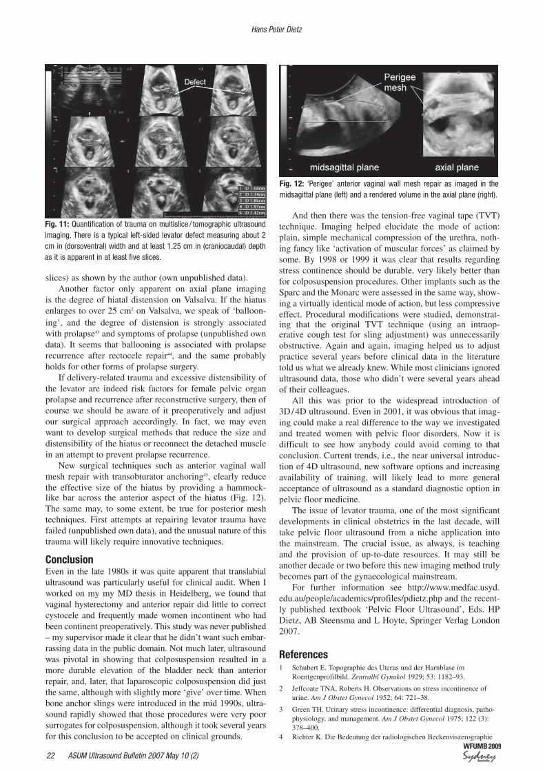
Fig. 11: Quantification of trauma on multislice / tomographic ultrasound imaging. There is a typical left-sided levator defect measuring about 2 cm in (dorsoventral) width and at least 1.25 cm in (craniocaudal) depth as it is apparent in at least five slices.
Fig. 12: ‘Perigee’ anterior vaginal wall mesh repair as imaged in the midsagittal plane (left) and a rendered volume in the axial plane (right).
23ASUM Ultrasound Bulletin 2007 May 10 (2)
fuer eine rationelle Therapie der weiblichen Stressinkontinenz. Geburtshilfe Frauenheilkd 1987; 47: 509–17.
5 Quinn MJ, Beynon J, Mortensen NJ, Smith PJ. Transvaginal endo-sonography: a new method to study the anatomy of the lower urinary tract in urinary stress incontinence. Br J Urol 1988; 62 (5):414–8.
6 Kohorn EI, Scioscia AL, Jeanty P, Hobbins JC. Ultrasound cystoure-thrography by perineal scanning for the assessment of female stress urinary incontinence. Obstet Gynecol 1986; 68 (2): 269–72.
7 Dohke M, Mitchell DG, Vasavada SP. Fast magnetic resonance imaging of pelvic organ prolapse. [Review] [15 refs]. Techniques in Urology 2001; 7 (2): 133–8.
8 Gainey HL. Post-partum observation of pelvic tissue damage. Am J Obstet Gynecol 1943; 46: 457–66.
9 Olsen AL, Smith VJ, Bergstrom JO, Colling JC, Clark AL. Epidemiology of surgically managed pelvic organ prolapse and uri-nary incontinence. Obstet Gynecol 1997; 89 (4): 501–6.
10 Bump RC, Mattiasson A, Bo K, et al. The standardization of termi-nology of female pelvic organ prolapse and pelvic floor dysfunction. Am J Obstet Gynecol 1996; 175 (1): 10–7.
11 Oerno A, Dietz HP. Levator co-activation is a significant confounder of pelvic organ descent on Valsalva. Neurourol Urodyn 2006; 25 (6): 527–8.
12 van der Velde J, Laan E, Everaerd W. Vaginismus, a component of a general defensive reaction. an investigation of. Int Urogynecol J 2001; 12 (5): 328–31.
13 Laycock J. Clinical evaluation of the pelvic floor. In: Schuessler B, ed. Pelvic floor re-education: Principles and practice. London: Springer; 1994: pp 42–8.
14 Dietz H, Shek K, Clarke B. Biometry of the pubovisceral muscle and levator hiatus by three-dimensional pelvic floor ultrasound. Ultrasound Obstet Gynecol 2005; 25 (6): 580–5.
15 Dietz H, Steensma A. The prevalence of major abnormalities of the levator ani in urogynaecological patients. BJOG 2006; 113 (2): 225–30.
16 Dietz H, Lanzarone V. Levator trauma after vaginal delivery. Obstet Gynecol 2005; 106 (4): 707–12.
17 Lien KC, Mooney B, DeLancey JO, Ashton-Miller JA. Levator ani muscle stretch induced by simulated vaginal birth. Obstet Gynecol 2004; 103 (1): 31–40.
18 Dietz HP, Hyland G, Hay-Smith J. The assessment of levator trauma: A comparison between palpation and 4D pelvic floor ultrasound. Neurourol Urodyn 2006; 25 (5): 424–7.
19 Kearney R, Miller JM, Delancey JO. Interrater reliability and physi-cal examination of the pubovisceral portion of the levator ani muscle, validity comparisons using MR imaging. Neurourol Urodynam 2006; 25 (1): 50–4.
20 Schaer GN. Ultrasonography of the lower urinary tract. Curr Opin Obstet Gynecol 1997; 9 (5): 313–6.
21 Schaer GN, Siegwart R, Perucchini D, DeLancey JO. Examination of voiding in seated women using a remote-controlled ultrasound probe. Obstet Gynecol 1998; 91 (2): 297–301.
22 Dietz HP, Barry C, Lim YN, Rane A. Two-dimensional and three-dimensional ultrasound imaging of suburethral slings. Ultrasound Obstet Gynecol 2005; 26 (2): 175–9.
23 Tunn R, Petri E. Introital and transvaginal ultrasound as the main tool in the assessment of urogenital and pelvic floor dysfunction: an imag-ing panel and practical approach. Ultrasound Obstet Gynecol 2003; 22 (2): 205–13.
24 Dietz HP. Ultrasound Imaging of the Pelvic Floor: Part 1: 2D aspects. Ultrasound Obstet Gynecol 2004; 23 (1): 80–92.
25 Haylen BT, Frazer MI, Sutherst JR, West CR. Transvaginal ultrasound in the assessment of bladder volumes in women. Preliminary report. Br J Urol 1989; 63 (2): 149–51.
26 Yang JM, Huang WC. Bladder wall thickness on ultrasonographic cystourethrography: affecting factors and their implications. J Ultrasound Med 2003; 22 (8): 777–82.
27 Lekskulchai O, Dietz HP. Detrusor wall thickness is of limited use as a test for detrusor overactivity. Int Urogynecol J 2006; 17 (S2): S133–134.
28 Robinson D, Anders K, Cardozo L, Bidmead J, Toozs-Hobson P, Khullar V. Can ultrasound replace ambulatory urodynamics when investigating women with irritative urinary symptoms? BJOG 2002; 109 (2):145–8.
29 Robinson D, Khullar V, Cardozo L. Can bladder wall thickness pre-dict postoperative detrusor overactivity? Int Urogynecol J 2005; 16 (S2): S106.
30 Lekskulchai O, Dietz HP. Is detrusor hypertrophy in women associat-ed with symptoms and signs of voiding dysfunction? In: ICS Annual Scientific Meeting; 2006; Christchurch, New Zealand; 2006.
31 Dietz HP, Korda A. Which bowel symptoms are most strongly associ-ated with a true rectocele? Aust NZ J Obstet Gynaecol 2005; 45 (6): 505–8.
32 Dietz HP, Steensma AB. Posterior compartment prolapse on two-dimensional and three-dimensional pelvic floor ultrasound: the distinction between true rectocele, perineal hypermobility and entero-cele. Ultrasound Obstet Gynecol 2005; 26 (1): 73–7.
33 Peschers UM, DeLancey JO, Schaer GN, Schuessler B. Exoanal ultra-sound of the anal sphincter: normal anatomy and sphincter defects. BJOG 1997; 104 (9): 999–1003.
34 Kleinubing H, Jr., Jannini JF, Malafaia O, Brenner S, Pinho TM. Transperineal ultrasonography: new method to image the anorectal region. Dis Col Rect 2000; 43 (11): 1572–4.
35 Yagel S, Valsky DV. Three-dimensional transperineal sonography for evaluation of the anal sphincter complex: another dimension in under-standing peripartum sphincter trauma. Ultrasound Obstet Gynecol 2006; 27 (2): 119–23.
36 Cornelia L, Stephan B, Michel B, Antoine W, Felix K. Trans-perineal versus endo-anal ultrasound in the detection of anal sphincter tears. Eur J Obstet Gynecol Reprod Biol 2002; 103 (1): 79–82.
37 DeLancey JO, Speights SE, Tunn R, Howard D, Ashton Miller JA. Localized levator ani muscle abnormalities seen in MR images: site, size and side of occurrence. Int Urogynecol J 1999;10 (S1): S20–S1.
38 DeLancey JO, Kearney R, Chou Q, Speights S, Binno S. The appear-ance of levator ani muscle abnormalities in magnetic resonance images after vaginal delivery. Obstet Gynecol 2003;101 (1): 46–53.
39 Kearney R, Miller J, Ashton-Miller J, Delancey J. Obstetric factors associated with levator ani muscle injury after vaginal birth. Obstet Gynecol 2006; 107 (1): 144–9.
40 Dietz HP. Quantification of major morphological abnormalities of the levator ani. Ultrasound Obstet Gynecol; in print.
41 Adekanmi OA, Freeman R, Puckett M, Jackson S. Cystocele: Does anterior repair fail because we fail to correct the fascial defects? A clinical and radiological study. Int Urogynecol J 2005; 16 (S2): S73.
42 Dietz H, Steensma A. Dimensions of the levator hiatus in symptom-atic women. Ultrasound Obstet Gynecol 2005; 26 (4): 369–70.
43 Barry CL, Dietz HP, Lim YN, Rane A. A short-term independent audit of mesh repair for the treatment of rectocele in women, using 3-dimensional volume ultrasound: a pilot study. Aust NZ Continence J 2006; 12 (4): 94–9.
44 Shek KL, Dietz HP, Rane A. Transobturator mesh anchoring for the repair of large or recurrent cystocele. Neurourol Urodyn 2006; 25: 554–55.
Pelvic floor ultrasound
24 ASUM Ultrasound Bulletin 2007 May 10 (2)
DIAGNOSTIC ULTRASOUND ASUM Ultrasound Bulletin May 2007; 10 (2): 24–29
IntroductionTear of the distal biceps brachii tendon is an unusual and dramatic event that most commonly occurs as a result of forced extension of the flexed load bearing elbow1. Typically, the patient is a male manual worker with a clinical history of having lifted a heavy object and his elbow giving way and reporting a sensation of something snapping within the joint.
Sonography is often the modality of choice for assess-ment of tendons as it provides high resolution, cost effective, dynamic imaging, free of ionising radiation and enjoys high patient acceptance.
Requests for assessment of most tendons are generally greeted with optimism by sonographers and sonologists, how-ever when ‘distal biceps brachii tendon’ appears written on the referral the smiles disappear, followed by a phone call to the MRI department. Until now, this scenario has been com-mon, however the use of a new acoustic window makes sono-graphic imaging of the distal insertion of the biceps brachii as simple and rewarding as imaging the Achilles tendon.
The conventional approachTraditionally, the same logic has been applied to the biceps brachii tendon as tendons elsewhere in the body:1) Scan straight along the length of the tendon;2) Use the shortest possible tissue path; and3) Attempt to scan perpendicular to the tendon fibres.
The patient is positioned with the elbow fully extended and wrist supinated allowing access to the antecubital fossa. An anterior approach and sagittal scan plane is used with the transducer placed upright in the antecubital fossa. The distal end of the transducer is pressed into the soft tissues using a ‘heel and toe’ manoeuvre to image as perpendicular to the direction of the tendon fibres as is possible (Figs. 1, 2).
A new method for ultrasound evaluation of the distal biceps brachii tendonStephen J. Bird
Benson Radiology, North Adelaide, South Australia 5006, AustraliaCorrespondence to SJ Bird. Email [email protected]
The difficultiesThe conventional approach only occasionally provides sat-isfactory results, in particular the most distal portion of the tendon, as it inserts onto the radial tuberosity and the adja-cent bicipito-radial bursa is often poorly visualised.
The image quality when using this conventional approach is hampered by a variety of physical limitations:1) Anisotropy is impossible to overcome due to the deep
diving course of the tendon toward the radial tuberos-ity. Even real time compound imaging or beam steering technology combined with ‘heel and toe’ pressure can-not, in most cases, produce insonation perpendicular to the direction of the inserting tendon fibres.
2) The tendon insertion is located on the medial aspect of the radial tuberosity making direct sagittal visualisation difficult. Forced supination of the forearm will improve, but not completely overcome the problem (Figs. 3, 4).
3) The tendon insertion is in a deep location, often several centimeters from the skin surface, requiring a lower fre-quency transducer of poorer spatial resolution capabil-ity to achieve adequate penetration. This problem is exacerbated in the acute post-traumatic phase when the antecubital fossa is swollen.
4) When scanning through the mid antecubital fossa, the overlying brachioradialis and pronator teres muscle belly edges lie immediately anterior to the biceps brachii tendon and result in refraction of the sound beam which significantly degrades image quality. This is akin to scanning an ovary or prostate through the midline of the abdominal wall with refraction artifact generated by the rectus abdominus musculature (Figs. 5, 6).The combination of a difficult-to-access tendon insertion,
in a deep location, angled steeply away from the transducer
and overlaid with refraction causing muscle belly edges and fat provides an imaging challenge that requires a new solution.
In 2005 Giuffre BM and Lisle DA described a posterior approach with a pronated forearm which allows visualisa-tion of the biceps brachii insertion onto the radial tuberos-ity2. This approach works well, but has the limitation of visualising only the most distal 5–10 mm of the insertion and does not allow visualisation of the bicipito-radial bursa, more proximal tendon or musculotendinous junction (Figs. 7, 8).
The solution1) Use a high quality acoustic window that does not degrade
image quality at depth;2) Directly access the medial aspect of the radial tuberosity;
and
3) Scan exactly perpendicular to the long length of the tendon fibres to eliminate anisotropy and reveal the tendon fibrillar echotexture from musculotendinous junction to insertion.The key to achieving all of these objectives and produc-
ing high quality images of the biceps brachii insertion is to utilise the pronator teres muscle belly as an acoustic window. The pronator teres muscle runs obliquely across the proximal aspect of the anterior compartment of the forearm.
Pronator teres arises from dual heads on the humerus and ulna that merge to form a single muscle belly which inserts onto the shaft of the radius. It courses directly anterior and medial to the biceps brachii tendon insertion providing an ideal acoustic window free of refraction artifact.
A new method for ultrasound evaluation of the distal biceps brachii tendon
26 ASUM Ultrasound Bulletin 2007 May 10 (2)
The patient is positioned in the traditional manner with elbow extended and wrist supinated. Steeply angling the transducer from medial to lateral across the proximal fore-arm provides an acoustic window directly through the mid belly of the pronator teres muscle. This line of sight leads directly to the biceps brachii insertion on the radial tuber-
osity (Figs. 9, 10). Subtle supination and pronation of the wrist adjusts the position of the radial tuberosity and tension of the tendon insertion, both of which are used to fine-tune the image quality. The full length of the biceps brachii ten-don from insertion to musculotendinous junction may be visualised using this technique (Fig. 11). The periosteum of
Fig. 11: Normal biceps brachii insertion using pronator teres acoustic window: Arrow periosteal fibrocartilage.
Fig. 12: Brachial artery provides an excellent acoustic window.
Fig. 7: Giuffre and Lisle posterior approach. Fig. 8: Giuffre and Lisle technique.
Stephen J. Bird
27ASUM Ultrasound Bulletin 2007 May 10 (2)
Fig. 13: Bicipito-radial bursa fluid filling a partial tear defect in the biceps brachii tendon.
Fig. 14: Transverse image of the bicipito-radial bursa.
Fig. 15: Biceps brachii tendonosis. Fig. 16: Partial tear of the biceps brachii insertion.
the radial tuberosity including the periosteal fibrocartilage (Fig. 11 arrow) and the bicipito-radial bursa are imaged with excellent clarity. Occasionally, the brachial artery will be located immediately superficial to the biceps brachii ten-don, further improving the acoustic window properties and enhancing image quality (Fig. 12).
In cases where a partial tear is suspected, more strenu-ous pronation and supination of the wrist provides a useful provocative manoeuvre.
Supination and pronation adjusts the tendon tension and displaces fluid from the bicipito-radial bursa into partial tears or avulsions (Fig. 13).
Transverse images of the tendon may be obtained by rotating the transducer through 90 degrees while maintain-ing the pronator teres acoustic window. Transverse images are particularly useful for assessing the bicipito-radial bursa which appears as a ‘U’ shaped fluid collection immediately
deep to the tendon (Fig. 14).This technique provides excellent and easily reproduc-
ible visualisation of the biceps brachii insertion with a mini-mum of patient discomfort, even in the acute post traumatic phase. Image quality is only slightly affected by the size of the patient and degree of antecubital fossa swelling due to the nature of the acoustic window that is employed.
PathologyUsing the conventional technique, attention was focused on differentiating ruptured biceps brachii tendons from intact tendons. This new technique provides greater diagnostic con-fidence and allows more subtle pathology to be diagnosed.
Biceps brachii tendonosis is seen as a loss of fibrillar echotexture and swelling of the tendon (Fig. 15).
Partial tears are seen as anechoic fluid clefts running part way through the tendon (Fig. 16).
A new method for ultrasound evaluation of the distal biceps brachii tendon
▼▼
▼
▼
28 ASUM Ultrasound Bulletin 2007 May 10 (2)
Stephen J. Bird
Fig. 19: Median nerve and brachial artery ‘guarding’ the bicipito-radial bursa. Fig. 20: Needle placement into the bicipito-radial bursa.
Fig. 21: Median nerve.
Partial avulsions of the tendon insertion from the radial tuberosity may be seen as fluid tracking beneath the proxi-mal avulsed tendon, while distal fibres remain inserted and intact. Care must be taken not to misdiagnose the nor-mal periosteal fibrocartilage appearance as fluid tracking between the tendon and the radial tuberosity enthesis. In cases of avulsion, fluid is invariably seen in the bicipito-radial bursa deep to the tendon which communicates freely between the avulsed tendon end and the enthesis. Real time assessment during supination and pronation of the forearm is most helpful to observe the fluid communication, as is provocative testing with resisted elbow flexion and supination (Fig. 17).
In the acute phase, complete rupture of the biceps brachii tendon results in a ‘bare’ radial tuberosity and adjacent fluid collection (Fig. 18). Chronic tears are often accompanied by clinical symptoms including pain during physical activity, weakness in flexion of the elbow, supination of the forearm and grip strength3. Sonographically a ‘bare’ radial tuberosity and wasting of the biceps muscle belly is evident.
Bicipito-radial bursa injectionThe bicipito-radial bursa lies deep to the biceps brachii tendon and is in close proximity to the brachial artery and medial nerve. The anatomical position as well as the previously discussed imaging challenges make placement of steroid and local anaesthetic under ultrasound guidance difficult. The pronator teres acoustic window approach may be used to obtain high resolution images of the bicipito-radial bursa and biceps brachii tendon insertion. Rotating the transducer 90 degrees into the axial plane allows the bursa and tendon to be imaged in cross section. In this plane the location of the median nerve and brachial artery in relation to the bicipito-radial bursa are easily appreciated (Fig. 19). A needle path through the forearm musculature in this plane provides excellent needle visualisation and safe access to the bursa (Fig. 20).
Median nerveSlight modification to the described biceps brachii insertion technique, produces excellent images of the median nerve as it passes between the heads of the pronator teres. This is the
Fig. 17: Partial avulsion of biceps brachii insertion. Fig. 18: Bare radial tuberosity following acute biceps brachii rupture.
▼ ▼
29ASUM Ultrasound Bulletin 2007 May 10 (2)
most difficult portion of the median nerve to visualise due to the deep course taken through the antecubital fossa. It is also an important structure to examine as irritation of the median nerve may complicate bicipito-radial bursitis or biceps bra-chii partial tear4. To locate the median nerve, simply scan the biceps brachii tendon in the longitudinal plane and angle the transducer 5–10 degrees posterior. The pronator teres acous-tic window will demonstrate the median nerve coursing deep through the proximal forearm in excellent detail.
Longitudinal and transverse images of the median nerve may be obtained through this acoustic window (Fig. 21).
SummaryThe described angled approach through the pronator teres muscle belly provides high quality images of the biceps bra-chii tendon from the musculotendinous junction to the inser-tion. The bicipito-radial bursa and radial tuberosity perios-teum are well visualised and with a slightly modified tech-nique, the median nerve may be examined coursing through the antecubital fossa. Use of this technique greatly enhances
the diagnostic confidence of sonography for assessment of the biceps brachii insertion and related structures.
AcknowledgementMusculoskeletal Images are from the University of Washington Musculoskeletal Atlas: A Musculoskeletal Atlas of the Human Body by Carol Teitz, M.D. and Dan Graney, Ph.D.
tures: a historical perspective and current concepts. Am J Orthop 2001; 30(3): 193–200.
2 Giuffre BM, Lisle DA. Tear of the distal biceps brachii tendon: A new method of ultrasound evaluation. Australas Radiol 2005; 49(5): 404–6.
3 Fritz RC: Magnetic resonance imaging of the elbow. Semin Roentgenol 1995; 30 (5): 241–64.
4 Morrey BF. Tendon injuries about the elbow: The elbow and its disor-ders. 2nd Ed. Philadelphia: WB Saunders; 1993.
A new method for ultrasound evaluation of the distal biceps brachii tendon
30 ASUM Ultrasound Bulletin 2007 May 10 (2)
IntroductionHoloprosencephaly results from failure of the normal sepa-ration of the embryonic forebrain (prosencephalon) during the fourth to sixth week of gestation. At its most severe this results in the formation of a single ventricle and absent falx, and a spectrum of midline facial defects. The incidence is approximately 1: 10 000 births1.
Causative factors include aneuploidy (most commonly trisomies 13 and 18), as in the case reported here; also teratogens, congenital infection such as toxoplasmosis and maternal diabetes. Chromosomal abnormalities represent one-third of cases2.
Three types of the condition are recognised; alobar, semilobar and lobar holoprosencephaly with alobar being the most severe. Severely affected fetuses commonly die at birth or in the first few months.
Holoprosencephaly – definitionsThe three types of holoprosencephaly are characterised by the degree of division of the prosencephalon.
Fig. 1 demonstrates the normal fetal brain. Fig. 2 Alobar type. There is a single crescent shaped ventricle. Fig. 3 Semilobar type. There is separation of the cerebral hemispheres in the occipital area. The occipital and tem-poral horns of the lateral ventricles are partly formed.Fig. 4 Lobar type. The cerebral hemispheres are connect-ed in the frontal area. There is mild ventricular dilatation. Typical sonographic features of holoprosencephaly:
Alobar and semilobar features 1 Single crescent-shaped ventricle;2 Fused thalami;3 Absent midline brain structures (falx cerebri, cavum
septum pellucidum, corpus callosum);4 Mid-line facial abnormalities – most severe in alobar
type (such as cyclopia); and5 Absent third ventricle.
Lobar features1 Fusion of lateral ventricles anteriorly; 2 Absent cavum septum pellucidum, corpus callosum;3 Incomplete falx; 4 Incomplete interhemispheric fissure; and5 Normal face or less severe anomaly (such as cleft lip)2–4.
Other possible sonographic findings of holoprosencephaly include polyhydramnios, macrocephaly, microcephaly, intra-uterine growth restriction, and reduced intraorbital distance.
Case reviewA 36-year-old gravida 2, para 1 woman attended for her
A prenatal diagnosis of holoprosencephaly using 2D and 3D ultrasoundAlison L. Webb and Ron J. Benzie
Christopher Kohlenberg Department of Perinatal Ultrasound, Nepean Hospital, University of Sydney, Penrith, New South Wales 2075, Australia.Correspondence to Alison Webb. Email [email protected]
ASUM Ultrasound Bulletin May 2007; 10 (2): 30–32DIAGNOSTIC ULTRASOUND
routine fetal anatomy scan at a country centre. When a complex cranio-facial fetal anomaly was diagnosed at this scan, she was referred to the Nepean Hospital’s Perinatal Ultrasound Department where she presented at 19 weeks and 2 days gestation.
Our ultrasound examination (2D and 3D) demonstrated several cranio-facial abnormalities including a single large cerebral ventricle / alobar holoprosencephaly, hypotelorism, prominent forehead and a flattened facial profile with a fore-head proboscis. Other fetal structures appeared normal.
The patient was counselled and underwent an immediate uncomplicated amniocentesis. The fetus died spontaneously in utero five days later at 20 weeks gestation. Karyotyping after amniocentesis revealed trisomy 13.
Figs. 5–9 are scan images demonstrating the main findings.
Discussion – sonographic downfalls and tipsThere are reports of diagnosis of holoprosencephaly in the first trimester from nine weeks gestation using transabdominal and transvaginal ultrasound1.
Diagnosis depends on distinguishing this pathology from other conditions causing profound ventriculomeg-aly. The most significant sonographic finding in severe
Adapted from Romero et al3
Fig. 1: Normal fetal brain Fig. 2: Alobar type
Fig. 3: Semilobar typeFig. 4: Lobar type
31ASUM Ultrasound Bulletin 2007 May 10 (2)
holoprosencephaly is the single crescent-shaped ventricle with the crescent-shaped cortex seen anteriorly, and fused thalami posteriorly without the third ventricle between them. In ventriculomegaly there are always two separate lateral ven-tricles and the midline cranial structures are usually present.
Hydranencephaly may mimic holoprosencephaly. However, the finding of a crescent-shaped rim of cortex and mid-line facial anomalies are indicative of holoprosen-cephaly as opposed to hydranencephaly2.
Antenatal diagnosis of lobar holoprosencephaly is dif-ficult and unreliable as the absent cavum septum pellucidum (CSP) is subtle and is the only real feature1,2.
The incomplete falx may be overlooked and isolated ventriculomegaly may be wrongly diagnosed. However, the absent CSP and flattened frontal horns are the hallmarks for lobar holoprosencephaly.
A large Dandy-Walker cyst can be confused with holo-prosencephaly. However, there will be a normal supratento-rial system seen with a true Dandy-Walker cyst4.
If prenatal sonographic findings include a large crescent-shaped single ventricle and severe midline facial abnormali-ties, look for characteristics of trisomy 13 such as abnormal hands / feet, omphalocele and heart defects4.
Holoprosencephaly may also occur with other syndromes such as Di George syndrome or campomelic dysplasia prompting a thorough search for multi-system anomalies.
Fig. 9: Surface rendered 3D facial profile.
Fig. 5: Single dilated crescent-shaped ventricle and fused thalami. Fig. 6: Normal cerebellum and fused thalami.
Fig. 7: Measurement of proboscis in 2D. Fig. 8: 2D facial profile. The proboscis is seen protruding from the
flattened forehead. The nose appears to be absent.
ConclusionsPrenatal diagnosis of alobar and semilobar holoprosen-cephaly is possible with 2D and 3D ultrasound; it is more difficult to diagnose lobar holoprosencephaly confidently.
Holoprosencephaly may be diagnosed with ultrasound in the first trimester.
In this particular case study, the additional use of 3D ultrasound, specifically when imaging the fetal face, assisted in the explanation of the severity of the baby’s condition.
A prenatal diagnosis of holoprosencephaly using 2D and 3D ultrasound
32 ASUM Ultrasound Bulletin 2007 May 10 (2)
AcknowledgementAn acknowledgement to David Fauchon, Mani Viswasm and Lesa Posa for their technical assistance.
References1 Rumack C, Wilson S, Charboneau J. Diagnostic Ultrasound Vol 2, 3rd
Ed. USA: Elselvier Mosby; 2005.
2 Platypus. Canada: TKI Medcon; 1999.
3 Romero R, Pilu G, Jeanty P, Ghidini A, Hobbins J. Prenatal Diagnosis of Congenital anomalies. USA: Appleton and Lange; 1998.
4 Sanders R. Structural Fetal Anomalies. The Total Picture. USA: Mosby Year Book Inc.; 1996.
Alison L. Webb and Ron J. Benzie
33ASUM Ultrasound Bulletin 2007 May 10 (2)
ASUM Ultrasound Bulletin May 2007; 10 (2): 33–36 DIAGNOSTIC ULTRASOUND
IntroductionHydatidiform mole is the benign form of gestational tro-phoblastic disease characterised by marked trophoblastic proliferation and hydropic degeneration of the chorionic villi. Although rare, complete and partial moles can be found in association with a normal fetus in twin gestations occurring in 1 in 20 000 to 1 in 100 000 pregnancies. A woman with a coexistent mole and fetus has a one-in-four chance of a live birth1. These pregnancies are at high risk of both maternal and fetal complications including preeclampsia, persistent trophoblastic disease, hyperthyroid-ism, theca lutein cysts, spontaneous miscarriage, intrauterine death and preterm labour.
The presenting symptoms of a complete mole and a co-existing fetus (CMCF) are similar to those of a singleton hydatidiform mole.
Patients typically present in the first and second trimester with intermittent vaginal bleeding, hyperemesis gravidarum, rapid uterine enlargement and markedly elevated b-hCG levels.
Sonographically, a complete hydatidiform mole in a twin pregnancy will appear as a normal fetus and placenta next to a complex echogenic intrauterine mass containing multiple cystic areas.
A twin pregnancy with a normal fetus and complete hydatidiform mole: an evolving diagnosisSharon M. Watson and Ron J. Benzie
Christopher Kohlenberg Department of Perinatal Ultrasound, Nepean Hospital, University of Sydney, Penrith, New South Wales 2750, AustraliaCorrespondence to Sharon Watson. Email [email protected]
Fluid collections with irregular contours and thin walls may also be seen in addition to the small cystic spaces. Bilateral ovarian enlargement with theca lutein cysts has also been described2.
The sonographic appearance of a molar pregnancy in the early first trimester may, however, be non-specific as dem-onstrated by this case. The majority of molar pregnancies in the first trimester may simply appear as either a missed abortion or anembryonic pregnancy. While, conversely, a non-molar abortion (miscarriage) may undergo hydropic change which may sonographically mimic the features of a hydatidiform mole3. The progressive sonographic changes in a case of CMCF occurring between initial presentation at 5 weeks 5 days gestation and diagnosis at 12 weeks are presented here for review.
Case historyOver the course of a six-week period a primigravida 21-year-old woman presented with ongoing vaginal bleeding, lower abdominal pain and increasing hyperemesis.
At each subsequent examination the embryo was seen to have normal growth and the ovaries to have a normal sonographic appearance. Figs. 1–7 illustrate the evolution of the diagnosis.
Fig. 1: 1st Presentation 5 weeks, 5 days. Ultrasound findings: Transabdominal and transvaginal scanning identi-fied a single embryo with a CRL of 2.2 mm consistent with a gestation of 5 weeks 5 days. No cause for p.v. bleeding was identified. b-hCG = 28 085 (normal range 500–200 000).
Fig. 2: 2nd Presentation 6 weeks 1 day. Ultrasound findings: A dichorionic-diamniotic twin pregnancy was identified on transvaginal scanning. The second gestational sac contained a lobulated avascular mass of uniformly echogenic material measuring 1.2 x 1.0 x 0.8.cm related to the developing placenta. Fetal elements could not be seen.Diagnosis: An ongoing viable pregnancy with a coexistent degenerating failed gestation was considered the most likely diagnosis. A complete hyda-tidiform mole was not considered as a possible differential diagnosis. b-hCG = 46 325 (normal range 500–200 000).
34 ASUM Ultrasound Bulletin 2007 May 10 (2)
Fig. 5: 5th Presentation 9 weeks 4 days. Ultrasound findings: The area of mixed echogenicity could be seen adjacent to the gestational sac containing the viable fetus. Diagnosis: A resolving haematoma with areas of recent haemorrhage. b-hCG recorded at 9 weeks 2 days 193 894 IU/L Not available at time of the scan (normal range 10 000–100 000).
Fig. 6: 6th Presentation 12 weeks 1 day.Ultrasound Findings: Anteriorly and to the right of the normal fetus and placenta is a mass of hyperechoic tissue interspersed with anechoic spaces of varying sizes. The mass measured 6.9 x 5.2 x 4.6 cm.
Fig. 3: 3rd Presentation 6 weeks 6 days. Ultrasound Findings: The second gestational sac appears irregular in outline. The rounded hyperechoic material had doubled in size and almost occupied the entire sac. Diagnosis: Unchanged from the previous examination.b-hCG 92 844 IU / L (normal range 500–200 000).
Fig. 4: 4th Presentation 8 weeks 4 days. Ultrasound findings: The 2nd gestational sac was poorly visualised. The sac borders were ill defined and it appeared to be filled with material of mixed echogenecity (arrow). b-hCG not recorded.
Sharon Watson and Ron J. Benzie
35ASUM Ultrasound Bulletin 2007 May 10 (2)
A twin pregnancy with a normal fetus and complete hydatidiform mole
Fig. 7: Transverse scans though the complete mole with colour Doppler demonstrating vascularity. Diagnosis: Complete hydatidiform mole with a coexistent fetus. b-hCG 537 668 IU / L (Normal Range 10 000–100 000)The serum bHCG reached a peak of 561 044 units recorded at 12 weeks 3 days gestation. Over the course of 3 weeks the serum bHCG declined to normal. After counselling the patient elected to terminate the pregnancy.
Fig. 8: Histopathology confirmed the ultrasound diagnosis of a complete mole with a coexistent first trimester placenta and fetus.
apparent in the early first trimester. Transabdominal ultra-sound was unable to demonstrate the hydropic villi as cystic spaces until the 12th week of gestation, when the b-hCG was also abnormally elevated.
The initial transvaginal examination at 5 weeks only identified the viable embryo.
The examinations at 6 weeks 1 day demonstrating a uniformly hyperechoic mass projecting into the gestational sac from the placental surface and the subsequent scan at 6 weeks 6 days showing increase in mass size, have features consistent with a failed gestation and associated hemor-rhage / clot.
When correlated with the b-hCG levels the initial diag-nosis of a viable pregnancy with a coexistent degenerating failed gestation would seem to account for the patient’s symptoms. In retrospect the hyperechoic mass is thickened hydropic trophoblast5. The subsequent transabdominal scans at 8–9.5 weeks appeared to confirm the initial diagnosis. It is arguable whether transvaginal scanning at the examina-tions at 8–9.5 weeks may have revealed the characteristic sonographic features of a hydatidiform mole.
Discussion A complete hydatidiform mole with a coexistent live fetus is a rare entity. The majority of these pregnancies result from dizygotic dichorionic twinning and can be differenti-ated from a partial mole by identifying a normal fetus and normal placenta. The clinical management is complex, and prognosis poor. The fetal loss rate is estimated at approxi-mately 60%, while severe maternal complications occur in 10% of cases. The risk of persistent trophoblastic disease is markedly increased with CMCF ranging from 20–50% of cases1. In this setting the diagnosis should be both accurate and timely.
The symptoms of hydatidiform mole are however essen-tially non-specific in the first trimester. Ultrasonography in conjunction with b-hCG levels which exceed those of normal pregnancy are crucial to facilitating an accurate diagnosis. While markedly elevated b-hCG levels may suggest com-plete hydatidiform mole it is not reliable enough in twin pregnancies where b-hCG levels can be significantly but normally elevated4.As demonstrated by this case the typi-cal sonographic features of hydatidiform mole may not be
36 ASUM Ultrasound Bulletin 2007 May 10 (2)
Sharon Watson and Ron J. Benzie
The incidence of theca lutein cysts identified in conjunc-tion with complete moles ranges between 20 and 60%6. Theca lutein cysts are seen infrequently in the first trimester with complete hydatidiform moles. They are more readily seen in the second trimester as the ovaries respond to the markedly elevated levels of b-hCG. Theca lutein cysts were not identified at any time throughout the entire series of scans in this case.
Conclusion Ultrasound can identify complete hydatidiform moles exist-ing with a normal pregnancy. The hydatidiform changes are often less marked in the first trimester, exhibiting features which are indistinguishable from a missed abortion. Studies performed by Lazarus6 and subsequently confirmed by Benson2 reviewed the ability of ultrasound to detect histo-logically confirmed hydatidiform moles. Both concluded that the majority (72–82%) of complete hydatidiform moles demonstrated a typical appearance but that diagnostic accu-racy declined in very early gestations. An empty sac, or one containing complex material should be viewed with a degree of suspicion even when found coexisting with normal fetus and placenta.
As demonstrated in this instance clinical review of the patient symptoms, correlation with b-hCG and serial ultra-sound eventually led to an accurate diagnosis.
AcknowledgementsThe authors gratefully acknowledge the contribution Peter Paton of the Nepean Hospital Pathology Department and Lesa Posa of the Audio Visual Department, Nepean Hospital.
References 1 Wee L, Jauniaux E. Prenatal diagnosis and management of twin preg-
nancies complicated by a coexisting molar pregnancy. Prenat Diag 2006; 25 (9): 772–76.
3 Sebire NJ, Rees H, Paradinas F, Seckl M and Newlands. The diagnos-tic implications of routine ultrasound examination in histiologically confirmed early molar pregnancies. Ultrasound Obstet Gynaecol 2001; 18 (6): 662–65.
4 Vaisbuch E, Ben-Arie A, Dgani R, Perlman S, Sokolovsky N and Hagay Z. Twin pregnancy consisting of a complete hydatidiform mole and co-existent fetus: report of two cases and review of the literarture. Gynaecol Oncol 2005; 98 (1): 19–23.
5 Bovicelli L, Ghi T, Pilu G, Farina A, Savelli L, Simonazzi G, Calzolari E, Ferlini A, Santini D and Valeri B. Prenatal diagnosis of a complete mole coexisting with a dichorionic twin pregnancy: Case report. Hum Reprod 2004; 19 (5): 1231–34.
6 Lazarus E, Hulka CA, Siewart B, and Levine D. Sonographic appear-ance of early complete molar pregnancies. J Ultrasound Med 1999; 18 (9): 589–93.
37ASUM Ultrasound Bulletin 2007 May 10 (2)
IntroductionCornelia de Lange Syndrome (De Lange Syndrome, Brachmann de Lange Syndrome) is a genetic disorder in which most cases occur sporadically. It is characterised by pre- and post-natal symmetrical growth retardation, cra-niofacial abnormalities, limb abnormalities, diaphragmatic hernias, genitourinary features, cardiac anomalies, severe mental retardation and hirsutism. The main craniofacial fea-tures include microbrachycephaly, micrognathia, anteverted nostrils and long philtrum. Flexion contractures of the elbow are present in approximately 80% of cases and 20% of cases demonstrate severe upper limb anomalies (Fig. 1)1–3.
Clinical backgroundA 28-year-old gravida 1 para 0 had a fetal anatomy scan performed at a rural centre that identified limb abnormalities at 18 weeks 4 days of gestation by the last normal menstrual period. She was referred for a tertiary review at Nepean Hospital and was re-scanned at 19 weeks 1 day with a GE Voluson 730 Expert using 2D, 3D and 4D imaging. This scan identified the following:■ Fetal size equivalent to 17 weeks 4 day gestation sugges-
tive of intra-uterine growth restriction;■ Bilateral shortening and absence of a radius or ulna in the
forearms (Figs. 2 and 3);■ Absence of hands bilaterally;■ Micrognathia (Fig. 4);■ Long philtrum (Fig. 4); and■ Flexion contractures of the elbows.
Later that day, she was counselled in the Genetics Department where the diagnosis of Cornelia de Lange syn-drome was suggested and a decision was made to terminate
the pregnancy. An autopsy was agreed to after the procedure in the hope of a more definitive diagnosis for the risk of future recurrence.
The autopsy findings supported the sonographic find-ings. It also revealed a small membranous ventricular septal defect, bicuspid aorta, minor gastrointestinal anomalies, brachycephaly and a left foot containing four metatarsals and hypoplastic phalanges. The left arm was found to have a single shortened forearm bone with the presence of a single metacarpal and digit with a fingernail.
The patient returned seven months later with a subse-quent pregnancy for nuchal translucency screening and later for a fetal anatomy scan. These revealed low risk for trisomy 21 and trisomies 18/13 and a structurally normal fetus. Fig. 5 demonstrates 3D images comparing this fetus with the previ-ous Cornelia de Lange fetus.
DiscussionCornelia de Lange syndrome has proved in the past to be a difficult diagnosis to make by prenatal ultrasound4. However, the frequency in prenatal diagnosis of facial anomalies and skeletal malformations is likely to increase with the rapid
Cornelia de Lange syndrome: the value of 3D and 4D ultrasoundBen Micallef, David Fauchon and Ron J. Benzie
Christopher Kohlenberg Department of Perinatal Ultrasound, Nepean Hospital, University of Sydney, Penrith, NSW 2750, Australia.Correspondence to Ben Micallef. Email [email protected]
ASUM Ultrasound Bulletin May 2007; 10 (2): 37–38 CASE REVIEW
Fig. 1: Examples of various types of malformed upper limbs found in de Lange syndrome. (From JM Berg et al. The de Lange syndrome. Oxford: Pergamon; 1970).
Figs. 2 and 3: Shortening and absence of a radius or ulna in the forearms.
▼
Occiput
▼
Right arm/elbow
38 ASUM Ultrasound Bulletin 2007 May 10 (2)
improvement of 2D ultrasound and the development and increased use of 3D and 4D ultrasound5.
The current literature comparing the diagnostic per-formance of 2D versus 3D ultrasound for the diagnosis of congenital anomalies has not provided conclusive results5. When used with 2D ultrasound, 3D ultrasound improves the detection rate of a wide range of anomalies6. This case is an example of the benefits 3D and 4D ultrasound add, in the sonographic detection of limb and facial abnormalities and the counselling of the expectant parents.
The fetal face is the most frequently documented struc-ture using 3D ultrasound6. When an adequate 3D volume of the face is obtained, the reconstructed image can be rotated and manipulated using the multiplanar function in conjunc-tion with the rendered display to better demonstrate the area of interest5. This application is most commonly used to improve visualisation of clefts of the lip and palate6. In this case, the use of 3D imaging, primarily the rendered display, assisted in the diagnosis of micrognathia and a long phil-trum when demonstrated in conjunction with the 2D profile images as demonstrated in Fig. 4.
The examination of the fetal limbs was also made easier with the use of 3D and 4D ultrasound. As demonstrated in Fig. 6, the 2D images of the limb anomalies are very difficult to appreciate compared to the 3D images. Furthermore, the use of 4D ultrasound in the patient’s following pregnancy, improved the visualisation of the normal free moving elbows and ruled out elbow flexion contractures.
When counselling the parents, the 3D and 4D ultrasound played a valuable part in describing the anomalies encoun-tered. It also provided a reduced level of anxiety in the patient’s subsequent pregnancy when she was shown a 3D image of her normal baby (Fig. 5). Most patients and people with non-sonographic background have great difficulty in comprehending 2D ultrasound images. A better understand-ing of both normal and abnormal surface anatomy is gained when it is illustrated by 3D ultrasound images. This enables more objective and focused counselling, and allows special-ists to explain management more effectively.6
3D ultrasound has been reported as offering a significant diagnostic advantage in detecting fetal anomalies when used
Ben Micallef, David Fauchon and Ron J. Benzie
with traditional 2D ultrasound imaging6. Our department has also found 3D and 4D ultrasound to be a beneficial addition to 2D ultrasound imaging. This case in particular has high-lighted the benefits in both diagnosis and counselling.
AcknowledgementThis was the prize winning poster at the 2006 ASUM Annual Scientific Meeting in Melbourne.We would like to thank Mani Viswasam for assistance in gathering the infor-mation.
References1 Le Vaillant C, Quere MP, David A, Berlivet M, Boog G. Prenatal
diagnosis of a ‘minor’ form of Brachmann-de Lange syndrome by three dimensional sonography and three dimensional computed tomography. Fetal Diag Ther 2004; 19 (2): 155–59.
2 Borck G, Redon R, Sanlaville D, Rio M, Prieur M, Lyonnet S, Vekemans M, et al. NIPBL mutations and genetic heterogeneity in Cornelia de Lange syndrome. Journal of Medical Genetics 2004 [Online] vol. 41, Available online at: http://jmg.bmjjournals.com/cgi/content/full/41/12/e128 [Accessed 19 June 2006].
3 Gorlin R, Cohen M, Levin L. Syndromes of the Head and Neck (3rd ed). New York: Oxford University Press; 1990.
4 Ranzini AC, Day-Salvatore D, Farren-Chavez D, Mclean DA, and Greco R. Prenatal Diagnosis of de Lange syndrome. J Ultrasound Med, 1997; 16 (11): 755–58.
5 Goncalves L, Lee W, Espinoza J and Romero R. Three- and 4-Dimensional Ultrasound in Obstetric Practice. Does it help? J Ultrasound Med 2005; 24 (12): 1599–1624.
6 Tritsch I, Platt L. Three-dimensional ultrasound experience in obstet-rics. Curr Opin Obstet Gynecol 2002; 14 (6): 569–75.
Fig. 4: Micrognathia.
Fig. 5: 3D images comparing structurally normal fetus with the previous Cornelia de Lange syndrome fetus.
Fig. 6: 2D ultrasound (left) and 3D demonstrate superior images of anomalies.
▼
Left forearm
39ASUM Ultrasound Bulletin 2007 May 10 (2)
General Ultrasound in the Critically illAuthors Daniel Lichtenstein, M.R. Pinsky and F. JardinPublisher SpringerYear 2004 ISBN 3540208224Price $A210.00
This 189-page hard cover text makes for interesting and, at times, thought-provoking reading. Daniel Lichtenstein MD, from France, is described in the two forewords to the book, as ‘an intensivist and physician-sonographer’ and ‘one of the leaders in the field’.
Lichtenstein describes the book as having the dual purposes of describ-ing ‘the fullest exploitation possible of general ultrasound in the intensive care unit’ and helping to ‘popular-ise a method that remains obscure to those who have never used it’. The text is therefore aimed at radiologists who may be involved in the emer-gency medicine setting, intensivists and emergency medicine physicians. In countries such as Australia and New Zealand, the book is also applicable to sonographers working in acute care hospitals. Both the clinical applica-tions of ultrasound techniques in the critically ill and the rationale / philos-ophy of ultrasound performed by criti-cal care physicians are well discussed and appear to be supported by the author’s extensive experience and own research.
Three separate sections are includ-ed. Part I Generalities consists of four brief chapters; Basic Notions, which is a very limited description of the basics of ultrasound imaging; The Ultrasound Equipment, which is a brief, some-what quirky in places, description of basic equipment; Specific Notions of Ultrasound in the Critically Ill, is an interesting five pages on the advan-tages and disadvantages of scanning the critically ill patient and provides some insights into specific features of this area of imaging, such as indica-tions for the ultrasound examination; and last a very brief, scantily illustrated chapter General Ultrasound: Normal Patterns. In this last chapter of the section, Lichtenstein indicates that the term ‘general ultrasound’ is usu-ally understood to mean ‘abdominal ultrasound’ and indeed this chapter is an overview of the basic, normal ultrasound appearance of the main
organs of the abdomen. The rest of the book, however, uses the term ‘general ultrasound’ to have a much broader scope than this and includes an almost ‘whole of body’ approach.
At 18 chapters and 127 pages the Part II: Organ by Organ Analysis sec-tion forms the major part of the text and includes some very interesting information on the applications, advan-tages, disadvantages and limitations of ultrasound in a diverse range of situa-tions. The chapters cover all areas of the abdomen, including the peritoneum and retroperitoneal space; the venous system including upper extremity cen-tral veins, IVC, lower limb veins; the mediastinum; general ultrasound of the heart; head and neck; and soft tissues in various areas of the body. Four chapters are devoted to ultrasound techniques in the lung: pleural effusion, pneu-mothorax, diseases of the lung tissue and disorders of the diaphragm. The chapter Lung Ultrasound Applications presents some of the clinical potentials of applying what the author calls ‘the seven principles of lung ultrasound’.
Part III: Clinical Applications of Ultrasound discusses the applica-tion of ultrasound in specific settings or situations. Included are chapters such as: Ultrasound in the Surgical Intensive Care Unit (ICU); Ultrasound in Trauma; Emergency Ultrasound Outside the ICU; Interventional Ultrasound; Emergency Ultrasound and Antibiotic Therapy; Learning and Logistics of Emergency Ultrasound; and Ultrasound, a Tool for Clinical Examination. This section makes for interesting reading and offers a dif-ferent perspective on the use of ultra-sound in many situations to that found in standard ultrasound texts written for sonologists and sonographers. The final chapter Concluding Remarks offers a discussion of the role, value and applicability of ultrasound in the critically ill patient.
Overall, this is a very interesting text, however, a major criticism is the quality of some of the images which have all been obtained using quite old equipment. Lichtenstein justifies this by indicating that he would prefer to ‘keep characteristic figures, as a clinically contributive image is defi-nitely better, in the emergency, than the sophisticated image dear to the imaging specialist’. Indeed there may well be other aspects of the text and its
philosophy that the imaging specialist may object to, but I thought there were many interesting aspects and much to be learned from this book.
I would recommend the book to anyone working in an acute care set-ting with a busy emergency department and all those involved in teaching gen-eral sonologists and sonographers, and emergency medicine physicians.
Margo Gill
Textbook of Diagnostic Ultrasonography 6th Edition, Vols 1 & 2Editor / Author Sandra L Hagen-Ansert Publisher Mosby Elsevier ISBN 13 9780323028035, ISBN 10 3323028039Approximate cost $A425.00 inc GST
This text was first published in 1978 and has since been widely used by those training in ultrasonography. The sixth edition is an extensive text and includes new chapters on physics, con-trast agents in abdominal applications, ultrasound-guided interventional tech-niques, emergency abdominal scan-ning, musculoskeletal system, neonatal hip/spine, female infertility and 3D / 4D evaluation of fetal anomalies.
Sandra Hagen-Ansert and 18 con-tributors have covered all aspects of sonography in a thoroughly compre-hensive manner, explaining that ‘the primary goal of the text is to serve as an in-depth resource for students studying sonography as well as a ref-erence for experienced practitioners’. The chapters cover sonographer tech-nique including practical instructions on patient history taking, patient posi-tioning, protocols and probe manipula-tion and orientation.
Each chapter is laid out with learn-ing objectives, chapter outlines, key terms and definitions. This makes it very easy for the reader to find the information they are looking for. The sonographic examination details fol-low on from the sections on anatomy, physiology, laboratory data, pathology and numerous tables and charts with a distinctive sonographic findings icon. Over 3000 illustrations are included and there are many new full color illustrations, high quality ultrasound images and gross pathology photos.
The chapter on musculo-
BOOK REVIEWS
40 ASUM Ultrasound Bulletin 2007 May 10 (2)
skeletal (MSK) ultrasound is limited, reflecting the use of ultrasound for MSK in the USA. Shoulder, wrist and Achilles regions have been covered with general MSK technique and arte-facts outlined. Although not covering other regions, it has practical instruc-tions for examining the musculoskel-etal system and provides the trainee with a good start in MSK.
This is an excellent text for trainee sonographers and a very good practi-cal addition to any department. The chapters are clearly laid out and a workbook to accompany the text is also available.
Glenda McLeanTutor SonographerMonash Medical Centre
Handbook of Early Pregnancy CareEditors Thomas H. Bourne and George CondousPublisher Taylor & FrancisISBN 1842143239Approx cost $A120
While this book is succinct and infor-mative, editor Condous’ chapter on what to do when you cannot see a pregnancy, which you suspect is there, is worth the book’s price alone.
The concept of pregnancies of unknown location (PUL) is one we
should accept and bear in mind before we assume an empty uterus is due to a complete miscarriage.
The point is made that complete miscarriage is not diagnosable by ultra-sound. A serum hCG must be done to exclude an ectopic pregnancy. The rare case of the patient with an earlier scan in the pregnancy, confirming an intra-uterine gestation, who a week later has an empty uterus, might be the excep-tion to that rule.
Ectopic pregnancy is important enough to claim six out of the 16 chapters. There is an excellent chapter by Kirk on expectant management of ectopic pregnancy, with emphasis on the strict criteria to be fulfilled before embarking on that route. Surgical man-agement is also well reviewed by Istre.
Bottomley’s chapter on caesarean scar pregnancy reminds us that with increased caesarean section incidence, we will see more of these and if sus-pected, referral to a specialist centre may save a life.
I strongly recommend that every ultrasound clinic where pregnant women are seen should have a copy of this book and it should also be required reading for radiologists and obstetri-cians in training.
SONOGRAPHER REGISTRATION WORKING PARTYSonographer registration will happen – Help us to make it happen on our terms
What the current accreditation system meansSince 2001, the Australasian Sonographer Accreditation Registry (ASAR) has been the body for accreditation of sonographers in Australia. This involves:• Assessing and evaluating education and training courses and institutions to uphold standards and consistency of
sonographer education and training; and• Ensuring sonographers meet the required level of expertise and maintain that level via mandatory continuing
professional development (CPD).
The Sonographer Registration Working Party, comprising representatives of all professional associations and stakeholders – ASAR, ASUM, ASA, AIR, and CSANZ – has been formed to work towards developing a national sonographer registration board.
What future national Sonographer Registration would mean for you and the ultrasound profession• Offers protection to patients and the community by assuring the quality and safety of ultrasound service provision
by registered sonographers;• Enhanced accountability of practitioners in ultrasound;• Avoids the multiplicity of state based registration boards and their associated fees, thus facilitating interstate work
opportunities;• Uniform national standards – assessment of character and fitness to practice;• Recognition of sonography as an allied health profession; and• Protection of the title 'sonographer'.
All enquiries may be addressed to the ASUM representative, Ros Savage, via email [email protected]
ASUM Giulia Franco Teaching FellowshipProudly sponsored by
Toshiba Medical
Since the introduction of ultrasound, Toshiba has been at the technological forefront of this diagnostic imaging technology. Throughout the years, Toshiba’s innovations have set new standards and created new applications that have significantly extended ultrasound capabilities.
The Giulia Franco Teaching Fellowship was established by ASUM in association with Toshiba Medical to provide educational opportunities for sonographers in all parts of Australia and New Zealand. It is named to commemorate Giulia Franco whose passion for ultrasound took her to all parts of Australia and New Zealand, and continued as she moved into a business career with Toshiba. It was first awarded in 2004.
The Giulia Franco Teaching Fellowship will focus on major city centres.
BOOK REVIEWS
41ASUM Ultrasound Bulletin 2007 May 10 (2)
ULTRASOUND TRANSMISSION GEL* US 01-50 5 litre SONICPAC® with dispenser, 1 per box, 4 per case*US 01-08 0.25 litre dispencer, 12 per box
• AQUASONIC ® 100
ULTRASOUND TRANSMISSION GEL* US 01-01 20g over wrapped sterilized foil pouches 48 per box
• STERILE AQUASONIC ® 100
ULTRASOUND GEL* US 03-50 5 litre SONICPAC® with dispenser, 1 per box, 4 per case
• AQUASONIC ® CLEAR ®
ULTRASOUND GEL PAD - STANDOF* US 04-02 2cm x 9cm gel pad, 6 pads per box
• AQUAFLEX ®
ULTRASOUND LOTION* US 21-28 1 U.S. gallon with dispenser bottle, 4 per pack
NEThe 3rd Annual Special Interest Group Seminar of the Australian Sonographers Association
2 0 0 7SIGSpecial Interest Group
Registrations now open!
Join us for this highly targeted event, which will examine the role of ultrasound in enhancing quality patient management in the areas of perinatal and musculoskeletal ultrasound.
Early bird closes Friday 15 June 2007 – register now to ensure workshop selection
For further information please contact:ASA National Office, PO Box 709 Moorabbin VIC 3189P: 03 9585 2996 F: 03 9585 2331E: [email protected] W: www.A-S-A.com.au
11-12 August 2007, Hilton on the Park, Melbourne
Incorporating FMF-approved Nuchal Translucency course, Friday 10 August
42 ASUM Ultrasound Bulletin 2007 May 10 (2)
ASUM REPORT
From December 2006 to January 2007, Victoria House Medical Imaging was lucky enough to host the CADUCEUS Exchange Program Scholarship winner, Dr Morten Boesen. The CADUCEUS scholarship is a joint creation of the ASUM and the Danish Society for Diagnostic Ultrasound (DSDU) to share knowledge and promote the worldwide excellence of ultrasound practice.
Morten Boesen is the third person to be awarded this honour. He follows in the footsteps of his Danish col-league, Christoffer Brushoj, who visited us last year and the Australian sonogra-pher, Mary Langdale, who travelled to Denmark in late 2006 to share her ultra-sound knowledge with the Danes.
Morten is medically qualified and on his way to becoming an orthopaedic sports surgeon. He is currently writ-ing his PhD under the guidance of Dr Soren Torp-Pedersen on the diagnosis and treatment of Achilles tendinopa-thies. He is also the team doctor of the largest soccer club in Demark (F.C. Copenhagen).
Morten came to us with consid-erable experience in musculoskeletal ultrasound, but found the practice of ultrasound in Australia very differ-ent to what he was accustomed to in Denmark. In Australia, highly qualified and experienced sonographers perform the initial scan on most patients and produce a series of images that form the basis of review and discussion with the radiologists. In Denmark, there are no sonographers, nor are films taken in
2006/2007 CADUCEUS Scholarship Australian Visit
his practice. There, the doctor does all the scans and there are no pictures to review. Morten immediately appreci-ated the advantages of using sonogra-phers and has taken home the thought of instigating a Danish sonographer training program.
Ultrasound at Victoria House Medical Imaging is a bit of a cornu-copia and Morten loved the variety. He was impressed by the many differ-ent pathologies, the detailed scanning techniques, and how much information was obtained from each scan. He was a quick learner and his cheerful attitude meant our patients were very willing to assist him and wanted to know all about him. And before you ask, no, he has not met Princess Mary.
Because of Morten’s interest in sports medicine, physicians Dr Gary Zimmerman and Dr Peter Larkins made him welcome in the sports medicine clinic. Morten really appreciated sitting in on their consulting sessions and the opportunity to go with Gary, who is the Western Bulldogs team doctor, to one of their training sessions.
Morton was also able to spend a day and naturally some part of the night with David Young, one of the ortho-
Cheryl Bass, Frank Burke, Mary Langdale
and Stephanie Pritchard on the latest Danish
visitor under the CADUCEUS program
Left: Morten Boesen, Cheryl Bass and Gary Zimmerman, scanning at the Tennis Open, Right: Morten Boesen and Cheryl Bass hard at work.
paedic surgery legends in Melbourne.During the Australian Tennis Open,
Morten, having represented Denmark in Badminton, with his brothers, was very keen to be involved in our ultra-sound service for the players in the medical centre underneath the Rod Laver Stadium. Tim Wood, Tennis Australia’s doctor, sponsored Morten to receive a visitor's pass. Morten was very excited and spent a very happy day seeing players with Gary Zimmerman and Dr Peter Larkins. Unfortunately, on that day the Ultrasound machine was not needed, so he only got to scan the ball.
Morten also spent a week in Sydney. Robyn Tantau, sonographer, was very helpful in assisting him in placements for his stay. He visited Dr John Read, musculoskeletal radiolo-gist, Prof George Murrel, orthopaedic surgeon and Nathan Gibbs, physician.
Nathan befriended Morten, orga-nising among other things, a special guided tour inside the Opera House with his brother in law. And just to ensure that Morten didn’t think that Victoria was the only state that played Aussie Rules, he took Morten to a Sydney Swans’ training session.
As well as being the acronym for Australia’s and Denmark’s ultrasound exchange program, CADUCEUS is an ancient astrological symbol, a sym-bol with complex interpretations; the simplest expresses that life is about maintaining a healthy balance.
Morten was certainly able to
43ASUM Ultrasound Bulletin 2007 May 10 (2)
Organise your 2007–2008 Educational DiaryCheck out our full range of programs and have a look at the exciting new ones coming up
Sample of upcoming programs:
n July 23–27 Ultrasound in O&G Workshop
n July 30–August 3 Vascular FastTrack
n August 9–13 Advanced Vascular – NEW 4 day program
n August 8–10 3D Ultrasound Workshop
n October 8–12 MSK Workshop
Come and join us in 2007Check the website or your annual booklet for dates,
or just give us a call
CADUCEUS: Collaborative Australasian Danish Undertaking for Continued Excellence in Ultrasound
Expressions of interest in the CADUCEUS Scholarship should to be directed to the ASUM CEO, Dr Caroline Hong, by email to [email protected]
The CADUCEUS scholarship and exchange program has been possible as a result of a Memorandum of Agreement which was signed on 8th April 2005.
The signatories were Dr David Rogers and Dr Caroline Hong on behalf of the Australasian Society For Ultrasound In Medicine (ASUM) and Dr Christian Nolsøe and Dr Michael Bachmann Nielsen on behalf of the Danish Society For Diagnostic Ultrasound (DSDU).
achieve this. On his visit, Morten's wife, Camilla and gorgeous three-year-old daughter, Ulrikka, accompanied him. In addition to Morten’s heavy work schedule, he and his family made the most of every opportunity to explore Melbourne and Sydney. Melbourne turned on a hot few days for their six-week visit and I think Ulrikka’s favou-rite place was the Prahran Swimming Pool. Morten is a delightful person and we thoroughly enjoyed having him at Victoria House Medical Imaging and wish him well for the future.
Left: Two CADUCEUS fellows, Mary Langdale and Morten Boesen, chat together at Victoria House Medical Imaging, Right: Morten Boesen(CADUCEUS Fellow) with the team from Victoria House Medical Imaging; Stephanie Pritchard, Cheryl Bass, Frank Burke, Mary Langdale, Morten and Ross McKellar (Musculoskeletal Imaging Fellow).
Mr Stephen Bird was the recipient of the 2006 Giulia Franco Teaching Fellowship, kindly supported by Toshiba. Stephen toured Western Australia with his top notch presenta-tions, starting his week of teaching lectures in Perth on 24th March 2007. The flavour of the trip was definitely musculo-skeletal (MSK) ultrasound and Stephen was able to demonstrate his technique for imaging the biceps brachii insertion, among other topics.
The Perth session was well received by all who attended. The workshop was kept small, with the 25 attendees representing all the major practices and teaching hospitals in Perth. The program was didactic in nature, com-bining lectures with two hands-on live scanning sessions. The topics covered included the Elbow, Upper Limb Neuro and Difficult Areas in MSK Ultrasound Demystified.
A Toshiba Aplio, kindly loaned by Royal Perth Hospital, performed beau-tifully and was used in the two scan-ning sessions of the day. Participants were reluctant for the day to end and Stephen was still lecturing an hour after the expected finish.
Following the session, Stephen commenced his 2000 km journey around the lower half of the state.
Bunbury was his second stop and he was worked hard there, providing 12 sonographers with a day of lectures, covering another didactic program of live scanning and lectures that includ-ed Shoulder, Elbow and Basic Arm Neurology, Groin and Hip, Ankle, Calf and Difficult areas in MSK.
The next stop in the tour was fur-ther south in Albany. The venue for the evening lecture was the Albany Regional Hospital where Stephen met twelve sonographers and medical imaging technologists, some of whom had made the trip to Albany from surrounding towns. Then it was on to Esperance. Stephen made his final stop of his trip in Kalgoorlie, 600 km North East of where he had started in Perth six days previously.
Stephen presented some wonder-ful lectures in Kalgoorlie Hospital’s Medical Imaging Department on Thursday evening, covering the elbow, wrist and shoulder. We also welcomed him into the department on Friday, where he demonstrated his skill with
2006 Giulia Franco Teaching Fellow strikes Gold in outback WA
the ultrasound transducer, providing hands on workshops for sonographers, among the bustle of the department.
Stephen was given a day’s reprieve to do a bit of sight seeing in and around Kalgoorlie prior to catching the train back to Adelaide.
During this time, he was caught panning for gold and striking it lucky – it is doubtful that he will be able to retire on the basis of his gold find, but we would welcome him back to try his luck in the Goldfields again.
Stephen may not have had a huge amount of success with the gold pan-ning but for all those who attended his lecture series certainly did strike gold. He has demonstrated an unwavering passion for ultrasound and a great wealth of knowledge in musculo-skel-etal ultrasound, which he has shared among the WA attendees with an infec-tious enthusiasm.
Stephen Bird tours outback WA for the
Giulia Franco Teaching Fellowship
Forging ahead; Stephen Bird takes time out from his whirlwind WA tour with the Giulia Franco Teaching Fellowship.
Important notice
DMU Pass 2007
ASUM Council awarded Merrin Jackson (NSW) a DMU (Vascular) on 3rd
March 2007.
45ASUM Ultrasound Bulletin 2007 May 10 (2)
The Certificate in Clinician Performed Ultrasound (CCPU) was developed in response to the demand for credential-ling, certification and established stan-dards of practice for clinicians who use ultrasound at the point of care.
The ASUM Council feels it is important that education and training be provided for this particular form of ultrasound examination. Limited diagnostic point of care imaging has undergone marked proliferation in recent years and is different in depth and scope when compared to referred diagnostic ultrasound examinations. ASUM, together with a number of colleges and professional associations, has introduced the CCPU to provide credentials by which medical practi-tioners can demonstrate that they are appropriately trained in the protocols and standards of practice relevant to this particular form of ultrasound examination.
A special class of ASUM membership has been created, the Clinical Affiliate.
Candidates may join ASUM as a full Medical Member or as a Clinical Affiliate.
The program is currently open to fellows or registrars in the second or subsequent year of their training
program of the:■ Australasian College for Emergency
Medicine (ACEM);■ Royal Australian and New Zealand
College of Obstetricians and Gynaecologists (RANZCOG); and
■ Royal Australasian College of Surgeons (RACS) and all clinicians who have completed the FAST / AAA Module.A number of colleges, societies and
associations are currently in discus-sion with the ASUM Council regard-ing proposals to extend the CCPU to other specialty areas. It is planned to extend the CCPU to include rheu-matology, rural and remote general practice, anaesthesia and intensive care medicine, military medicine and several of the surgical subspecialties, including breast and endocrine surgery, colorectal surgery, and trauma surgery. Fellows and Registrars in other spe-cialist disciplines may be admitted at the discretion of the ASUM CCPU Certification Board.
Education is currently offered through on-line modules and interac-tive courses. Accredited courses are also available from other providers. Full details of the requirements of the CCPU are available at: www.asum.
CCPU Reportcom.au/ccpu.htm.
A brochure is being sent to Fellows and Registrars of the ACEM and RANZCOG that outlines all the ASUM CCPU courses on offer between July 2007 and June 2008.
Basic coursesIn 2007, it is planned to run six basic courses in Sydney or Melbourne. (Course numbers are strictly limited due to the practical nature and places are subject to spaces available and suf-ficient registrations being received.) In the first half of 2008, eight basic courses are planned.
Advanced CoursesFour advanced courses are planned for 2007 and eight for 2008. In addi-tion the three George Condous courses in Melbourne (20th and 21st July), Brisbane (22nd and 23rd July) and Sydney (28th and 29th July) on Early Pregnancy & Gynaecological Scanning have been approved as satisfying all the requirements of the CCPU (O&G) and CCPU (Emergency) Advanced Acute Pelvic Modules.
For further information refer to the ASUM Website http://www.asum.com.au or contact [email protected].
ASUM Chris Kohlenberg Teaching FellowshipsProudly sponsored by
GE Healthcare
Since its foundation, GE Healthcare has constantly been at the forefront of research and technical innovation, with GE today being recognised as a world leader in the supply of diagnostic imaging systems.
The Chris Kohlenberg Teaching Fellowships were established by ASUM in association with GE Healthcare to increase the opportunity for members outside the main centres to have access to quality education opportunities.
It has been awarded annually since 1998 to commemorate Dr Chris Kohlenberg, who died while travelling to educate sonographers.
MEMBERSHIP RENEWALSJust a reminder to all our valued members, as at the end of this financial year (30th June 2007) all current membership with ASUM will lapse. We have endeavoured to mail out the subscription renewal notices late May. In the meantime, should you have any questions or concerns regarding your membership, please feel free to contact Marie Cawood at our office via email to [email protected]
EARLYBIRD RENEWALSMedical / Scientific / Sonographer members $313.50* if paid by 30th June 2007Associate members $242.00* if paid by 30th June 2007Trainee members $242.00* if paid by 30th June 2007Retired members $104.50* if paid by 30th June 2007Corporate members $1155.00* if paid by 30th June 2007Corresponding ordinary members $203.50* if paid by 30th June 2007Corresponding associate members $154.00* if paid by 30th June 2007*Inclusive of GST
PAY ON-LINE & WIN A PRIZEPay on-line at our website www.asum.com.au to enter the draw for a bottle of wine.
46 ASUM Ultrasound Bulletin 2007 May 10 (2)
Siemens Ltd - Medical Solutions (Acuson)Nick Kapsimallis 02 9491 [email protected] Marketing General Manager: Cameron Marcucciowww.medical.siemens.com
GE HealthcareStephanie Mason 02 9846 [email protected] General Manager: Kevin Potterwww.gemedicalsystems.com
Healthsite Recruitment Australia(Allied Health Recruitment Agency)Ian Stewart 07 5445 4604ian.stewart@healthsiterecruit-ment.comwww.healthsiterecruitment.com
ASUM Corporate Members 2007Meditron Pty Ltd (Acoustic Imaging, Dornier, Kontron, Focus Surgery)Michael Fehrmann 03 9879 [email protected]
Philips Medical Systems Australasia P/L (incorporating formerly ATL, HP, Agilent)Kathryn Davis 02 9947 [email protected] CEO: Harry van Dykwww.medical.philips.com
TRAINEE (8)Shin Cheng NSWAndrew Cheung NSWKim Mae Chia NSWRosalie Grivell SAGeoffrey Lee VicRitu Mogra VicDennis Wang NSWKes Wickremesekera NZ
CORRESPONDING (1)Marion Cairnduff Canada
200725th May ASUM in the HunterRadiology Conference CentreJohn Hunter HospitalContact Christian Abel 0414 495 [email protected]
14th June – Melbourne ASUM DDU Cardiology Oral ExaminationContact Marie Cawood.Ph:+612 9438 2078Fx:+612 9438 3686email: [email protected]: www.asum.com.au
16th June ASUM DDU Oral Examination All except Cardiology – MelbourneContact Marie CawoodPh:+612 9438 2078Fx:+612 9438 3686email: [email protected]: www.asum.com.au
30th June and 1st JulyASUM WA Ultrasound CPD Meeting 2007(2 days) Royal Perth Hospital Contact Michelle Pedrettiemail: [email protected]
19th JulyNuchal Translucency Accreditation One Day Theoretical Course FMF Certification of Competence in 11–14 Week Scan 2007The Ellott Theatre, Wellington Convention Centre, Wakefield Street, WellingtonRegistration will be available online Medical Industry Association of New ZealandPh: (09) 917 3645 Fx: (09) 917 3651email: [email protected]: www.mianz.co.nz
20th–21st July – Melbourne22nd–23rd July – Brisbane28th–29th July – SydneyASUM Presents: (2 days) The Early Pregnancy and Gynaecological Scanning Foundation Theoretical Courses 2007ConvenorAssoc Prof George CondousASUM Head OfficePO Box 943, Crows Nest NSW 1585Sydney, AustraliaPh: 02 9438 2078Fx: 02 9438 3686email: [email protected]
19th–22nd JulyASUM NZ and RANZCR NZ Third Combined Scientific Meeting 2007 New Zealand(4 days) Venue: Wellington Convention Centre Wellington New ZealandContact Jodie Preston-ThomasPh: 0011 649 917 3645Fx: 0011 649 917 3651email: [email protected]
27th July2nd ABDA Educational Programme (1st Annual Indonesian Society of Oncologic Imaging (ISOI) and 12th Annual ISUM Meeting(2 days) Venue: Balihai Resort & SPA, Kuta, BaliContact: Secretariat ISUM/ISOI, Jl. Raya Radio Dalam IB, Kebayoran Baru, Jakarta Selatan 12140, IndonesiaPh: +62-21-7230060, 7258135 Fx: +62-21-7230061email: [email protected] / [email protected]
28th JulyASUM DMU Part I & Part II Written Examinations-ProvisionalVenue as allocated. Candidates receive individual notification. Contact DMU CoordinatorPh: +61 2 9438 2078 Fx: +61 2 9438 3686 email: [email protected]
13th–17th September ASUM 2007 37th Annual Scientific Meeting of the Australasian Society for Ultrasound in Medicine(4 days) Venue Cairns Convention Centre, Cairns North Queensland Australia Contact ASUM PO Box 943, Crows Nest NSW 1585Ph: +61 2 9438 2078 Fx: +61 2 9438 3686 website: http//:www.asum.com.au or online registration
2008TBA ASUM Multi-Disciplinary Workshop(5days) Sydney, Australia
26th July ASUM DMU Part I & Part II Written Examination-ProvisionalVenue: As allocated. Candidates receive individual notification. Contact: DMU CoordinatorPh: +61 2 9438 2078 Fx: +61 2 9438 3686 email: [email protected]
18th–21st September38th Annual Scientific Meeting of the Australasian Society for Ultrasound in medicine 2008(4 days) Venue: Skycity Auckland Convention Centre Auckland, New ZealandContact: Karen WilliamsonPh: 0011 649 917 3645website: www.mianz.co.nz
200930th August–3rd SeptemberASUM hosts WFUMB 2009 World Congress in Sydney Australia (5 days) Venue: Sydney Convention and Exhibition CentreContact: Dr Caroline Hong ASUM CEO Ph: + 61 2 9438 2078Fx: + 61 2 9438 3686email: [email protected] or [email protected]
Corporate newsCorporate members are invited to pub-lish news about the company, including structural changes, staff movements and product developments. Each cor-porate member may submit one article of about 200 words annually. Logos, illustrations and tables cannot be pub-lished in this section.
FormatManuscripts should be submitted in triplicate in print and on PC formatted diskette as MS Word documents.Images must be supplied separately and not embedded. PowerPoint pre-sentations are not accepted.● Font size: maximum 12 pt, mini-mum 10 pt● Double spacing for all pages● Each manuscript should have the following: Title page, abstract, text, references, tables, legends for illustrations.● Title page should include the:
Title of manuscript, the full names of the authors listed in order of their contribution to the work, the depart-ment or practice from which the work originated, and their position.
Corresponding author’s name, con-tact address, contact telephone number and facsimile number (where avail-able) for correspondence.● Abbreviations may be used after being first written in full with abbre-viation in parentheses.● References should be cited using the Vancouver style, numbered according to the sequence of citation in the text, and listed in numerical order in the bibliography. Examples of Vancouver style:
1 In-text citation Superscript. If at the end of a sentence the number(s) should be placed before the full stop or comma.2 Journal article Britten J, Golding RH, Cooperberg PL. Sludge balls to gall stones. J Ultrasound Med
1984; 3: 81–84.3 Book: Strunk W Jr, White EB. The elements of style (3rd ed.). New York: Macmillan, 1979.4. Book section Kriegshauser JS, Carroll BA. The urinary tract. In: Rumack CM, Wilson SR, Charboneau JW, eds. Diagnostic Ultrasound. St Louis, 1991: 209–260.
AbstractManuscripts for feature articles and original research must include an abstract not exceeding 200 words, which describes the scope, major find-ings and principal conclusions. The abstract should be meaningful without reference to the main text.
ImagesImages may be submitted as hard copy (in triplicate) or in digital format. Images sent must have all personal and hospital or practice identifiers removed. Do not embed images in text. Separate images are required for publication purposes.
A figure legend must be provided for each image. Hard copy images should be presented as glossy print or original film. Any labelling should be entered on the front of the glossy print using removable labels. Send one copy of illustrations without labelling as this can be added electronically prior to publication. On the back of the print include the author’s name, figure num-ber and a directional arrow indicating the top of the print.
Digitised graphics should be sup-plied as JPG or TIFF files on PC formatted 3.5” diskette or CD, which must be clearly labelled with the author’s name and the names of the image files.
CopyrightAuthors are required to provide assur-ance that they own all property rights to submitted manuscripts, and to trans-fer to ASUM the right to freely repro-duce and distribute the manuscript.
Authors are invited to submit papers for publication in the categories described below. Final responsibility for accepting material lies with the Editor, and the right is reserved to introduce changes necessary to ensure conformity with the editorial standards of the Ultrasound Bulletin.
Original researchManuscripts will be subject to expert referee prior to acceptance for publi-cation. Manuscripts will be accepted on the understanding that they are contributed solely to the Ultrasound Bulletin.
Quiz casesA case study presented as a quiz, involv-ing no more than three or four images and a paragraph briefly summarising the clinical history as it was known at the time. It will pose two or three ques-tions, and a short explanation.
Case reportsCase reports are more substantial pre-sentations resembling short scientific papers which illustrate new informa-tion, or a new or important aspect of established knowledge.
Review articlesReview articles are original papers, or articles reviewing significant areas in ultrasound and will normally be illus-trated with relevant images and line drawings. Unless specifically com-missioned by the Editor, articles will be subject to expert referee prior to acceptance for publication.
Forum articlesMembers are invited to contribute short articles expressing their observa-tions, opinions and ideas. Forum arti-cles should not normally exceed 1000 words. They will not be refereed but will be subject to editorial approval.
Calendar itemsOrganisers of meetings and educa-tional events relevant to medical ultra-sound are invited to submit details for publication. Each listing must contain: activity title, dates, venue, organising body and contact details including name, address, telephone and facsimile numbers (where available) and email address (where available). Notices will not usually be accepted for courses run by commercial organisations.